Article

Antimicrobial Activity, Genetic Diversity and Safety Assessment of Lactic Acid Bacteria Isolated from European Hakes (Merluccius merluccius, L.) Caught in the Northeast Atlantic Ocean

1
Grupo de Seguridad y Calidad de los Alimentos por Bacterias Lácticas, Bacteriocinas y Probióticos (Grupo SEGABALBP), Sección Departamental de Nutrición y Ciencia de los Alimentos (Nutrición, Bromatología, Higiene y Seguridad Alimentaria) (SD-NUTRyCIAL), Facultad de Veterinaria, Universidad Complutense de Madrid, Avda. Puerta de Hierro, s/n, 28040 Madrid, Spain
2
Sección Departamental de Farmacia Galénica y Tecnología Alimentaria (SD-FARMATEC), Facultad de Veterinaria, Universidad Complutense de Madrid, Avda. Puerta de Hierro, s/n, 28040 Madrid, Spain
*
Authors to whom correspondence should be addressed.
Antibiotics 2025, 14(5), 469; https://doi.org/10.3390/antibiotics14050469
Submission received: 15 April 2025 / Revised: 27 April 2025 / Accepted: 2 May 2025 / Published: 6 May 2025

Abstract

:
Background/Objectives: The overuse and misuse of antibiotics has contributed significatively to the growing problem of the emergence and spread of antibiotic resistance genes among bacteria, posing a serious global challenge to the treatment of bacterial infectious diseases. For these reasons, there is a current and growing interest in the development of effective alternative or complementary strategies to antibiotic therapy for the prevention of fish diseases, which are mainly based on the use of probiotics—in particular, those belonging to the Lactic Acid Bacteria (LAB) group. In this context, the aim of the present study was to characterise, evaluate the genetic diversity and assess the safety of candidate probiotic LAB strains for aquaculture isolated from faeces and intestines of European hakes (Merluccius merluccius, L.) caught in the Northeast Atlantic Ocean (Ireland). Methods: The direct antimicrobial activity of the LAB isolates was tested by the Stab-On-Agar method against key ichthyopathogens. Subsequently, their taxonomic classification and genetic diversity were determined by 16SrDNA sequencing and Enterobacterial Repetitive Intergenic Consensus-PCR (ERIC-PCR), respectively. To ensure the in vitro safety of the LAB isolates, their biofilm-forming ability was assessed by a microtiter plate assay; their sensitivity to major antibiotics used in aquaculture, human and veterinary medicine by a broth microdilution method and their haemolytic and gelatinase activity by microbiological assays. Results: All LAB isolates were biofilm producers and susceptible to chloramphenicol, oxytetracycline, flumequine and amoxicillin. A total of 30 isolates (85.7%) were resistant to at least one of the tested antibiotics. None of the 35 LAB isolates showed haemolytic or proteolytic activity. Conclusions: Among the isolated strains, five LAB strains exhibiting the highest antimicrobial activity against aquaculture-relevant ichthyopathogens, taxonomically identified as Streptococcus salivarius, Enterococcus avium and Latilactobacillus sakei, were selected for further characterisation as potential probiotic candidates to promote sustainable aquaculture. To our knowledge, this is the first study to report that hake intestines and faeces represent viable ecological niches for the isolation of LAB strains with antimicrobial activity.

1. Introduction

Aquaculture is currently emerging as a successful strategy to maintain global food security for fish and other fishery products in both industrialised and developing countries [1]. Since 9.8 billion people are expected to populate the planet by 2050, the aquaculture industry, like other food-producing sectors, must sustainably maintain its production growth [2,3]. Aquaculture produced an estimated 94 million tonnes of aquatic animals, surpassing capture fisheries for the first time [3]. In this regard, one of the main problems facing modern aquaculture is to provide a sufficient and constant supply of products with adequate nutritional, hygienic and sanitary quality and safety, as well as the prevention and control of infectious fish diseases, mainly caused by bacteria, viruses, protozoa, helminths and fungi at the larval stages [4,5,6,7,8,9,10,11].
Traditionally, antibiotics have been used as a therapeutic and prophylactic treatment for bacterial ichthyopathologies. However, there is an increasing reluctance and restriction on their use, not only by health agencies but also by the fish farms and consumers, due to their detrimental effects on animal and human health, food safety and the environment [1,12]. In this respect, the excessive use of antibiotics in aquaculture has contributed to the growing and serious problem of the emergence and spread of transmissible bacterial resistance genes to (multiple) antibiotics, which is a major global problem for the treatment of infectious diseases of bacterial aetiology [13,14,15,16,17]. It should be noted that, although vaccination can be an effective strategy for the prevention of ichthyopathologies, its application in aquaculture is difficult due to the limited number of effective commercial vaccines, the variability of the degree and period of protection they offer, the problems of their application at the larval stages, both because of the immaturity of their immune system and the difficulty of their handling, the growth retardation that they can cause, the stress that their use can cause in fish and the alterations that they can cause in the quality of the product [18,19]. Moreover, there are currently no effective vaccines available for the treatment of many of the bacterial, viral and parasitic diseases affecting fish, so the only way to control them is by preventing infections through biosecurity measures and/or by using antimicrobial compounds (antibiotics, antivirals and antiparasitics) [20,21,22]. For all these reasons, there is currently a great interest in the development of novel and effective strategies for the prevention of bacterial ichthyopathologies as alternative or complementary strategies to antibiotherapy and vaccination, such as the use of probiotics, which are defined as live microorganisms that confer a health benefit to the host [17,23,24,25].
Lactic Acid Bacteria (LAB) are the safest and most frequently proposed bacteria for the development of probiotics for aquaculture due to their Qualified Presumption of Safety (QPS) status recognized by the European Food Safety Authority (EFSA) and because several strains are legally accepted for use as probiotics in humans and as zootechnical additives [26]. However, so far, the only strain authorized in the EU as a probiotic for aquaculture is Pediococcus acidilactici CNCM I-4622, due to its beneficial effect on the growth and functional development of fish and crustaceans. In addition, LAB have been evaluated as probiotics due to their antimicrobial properties, such as the production of organic acids and ribosomally synthetized antimicrobial peptides, referred to as bacteriocins [23,24,27]. The use of bacteriocin-producing LAB as probiotics and biocontrol agents in aquaculture could play a crucial role in improving animal health, productivity and product quality, while reducing the environmental impact [5,28,29,30,31].
The first step in this process is acquiring a collection of bacteriocinogenic LAB with suitable probiotic potential. It is also suggested that isolating bacteria from aquatic environments or host-related bacteria can enhance the adaptive capacity of native microbiota, improving their ability to colonize the gastrointestinal tract, mucous membranes and aquatic ecosystems [28,30,31,32,33,34,35,36,37,38]. According to this, the use of isolated and characterised LAB of fish origin could contribute to the development of a sustainable aquaculture in the One Health context, not only for the prevention and control of the most relevant ichthyopathologies but also to contribute to their health, immune status and to the productivity and economic profitability of aquaculture farms. Additionally, fish and fishery products are among the most traded food products worldwide, because they are considered an important source of proteins and minerals. Specifically, the European hake (Merluccius merluccius, L.) is one of the most popular and appreciated white fish, with a significant commercial impact on the fishing industry. Specifically, 62,500 tonnes of European hakes were consumed in Spain in 2021 [3,39]. In this context, the objective of this work was the characterization, genetic diversity evaluation and safety assessment of candidate probiotic LAB strains for aquaculture isolated from the faeces and intestines of European hakes (Merluccius merluccius L.) captured in the Northeast Atlantic Ocean (Ireland).

2. Results

2.1. Antimicrobial Activity and Taxonomic Identification of Bacteria Isolated from European Hakes

A total of 286 bacterial isolates from De Man, Rogosa and Sharpe (MRS) agar plates, which is an elective medium for the isolation of LAB, were selected, according to their different colony morphologies and including representatives from the eight hake samples for the evaluation of their antimicrobial activity. The pre-selection of isolates was made based on their antimicrobial activity spectrum. This involved selecting isolates that represented all the identified groups of antimicrobial activities. In this sense, both isolates with narrow and broad spectra of activity were selected. Conversely, those strains that did not show activity against any indicator were discarded. A total of 66 isolates were pre-selected for their taxonomic identification. The pre-selected isolates were taxonomically identified as Staphylococcus spp. (41%), Lactococcus spp. (32%), Streptococcus spp. (9%), Enterococcus spp. (9%), Macrococcus spp. (1.5%), Bacillus spp. (1.5%), Lactobacillus spp. (1.5%), Leuconostoc spp. (1.5%), Aerococcus spp. (1.5%) and Rothia spp. (1.5%). Bacterial isolates identified as LAB (n = 35) were selected for further safety assessment.
The results of the direct antimicrobial activity of the 35 LAB isolates are shown in Table S1. In brief, all isolates exhibited a broad antimicrobial spectrum, inhibiting the growth of at least three indicator strains and 57% inhibited the growth of at least half of the indicator microorganisms (Figure 1). Specifically, 23% of the LAB isolates inhibited five to six indicators, 37% inhibited seven to eight and 20% inhibited nine or more (Figure 1). The most sensitive indicators were the Gram-negative fish pathogens Aeromona salmonicida CLFP-23 and Aeromona salmonicida CECT4237, followed by Streptococcus parauberis LMG22252 and Tenacibacullum maritimum NCIMB2154, all of them reported as important fish pathogens [40,41,42]. In the case of Lactococcus garvieae, 71% inhibited at least half of the tested indicators, 52% inhibited at least three Gram-positive indicators and 57% inhibited at least five Gram-negative indicators. Moreover, 67% of Streptococcus salivarius inhibited at least five indicators, 50% inhibited three Gram-positive indicators and 67% inhibited more than five Gram-negative indicators. In addition, 34% of Enterococcus avium inhibited half of the indicators—specifically, 83% inhibited at least one Gram-positive indicator, and all of them (100%) inhibited at least two Gram-negative indicators (Figure 1).

2.2. Genetic Diversity Analysis by ERIC-PCR

Phylogenetic relatedness of the 35 LAB was determined by ERIC-PCR (Figure 2). Lc. garvieae isolates presented a high phylogenetic diversity, showing six different clusters (I-VI). Lc. garvieae isolates were identified in three different hakes (hakes A, B and F). Specifically, isolates from hake A were grouped into two different clusters (IV and VI), and isolates from hake B into four clusters (I, III, V and VI). In the case of St. salivarius isolated from hake D, they were highly similar (76%), detecting two ERIC-PCR patterns: cluster I, divided into Ia and Ib, and cluster II. On the other hand, E. avium isolates were found in three different hakes (hakes E, G and H) and presented a high phylogenetic relationship (83%). These isolates were grouped in cluster I and II, the latter divided into IIa and IIb. Interestingly, two isolates (E. avium MH19 and E. avium MH18) belonging to hake H were grouped into two different clusters, namely I and IIa, respectively. In addition, cluster IIb included the E. avium isolates from hakes E and G.

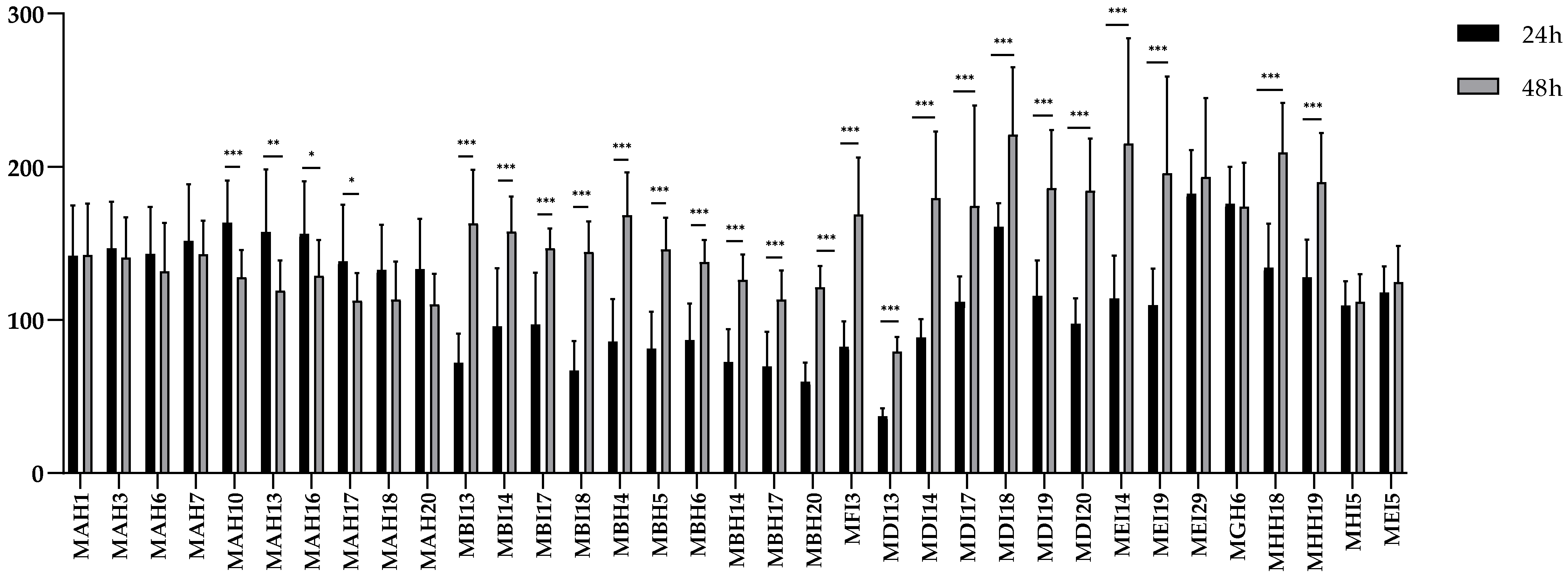
2.3. Biofilm Formation

All LAB isolates from hakes were biofilm producers (Figure 3), with St. salivarius MDI13 being the isolate that showed the lowest biofilm production. A comparison was made between biofilm formation at 24 and 48 h for the different isolates. Interestingly, 25 isolates (71.4%) showed significant differences, with 81% (n = 21) of them being higher biofilm producers at 48 h compared to 24 h. Specifically, 71.4% (n = 15) of the Lc. garvieae isolates showed significant differences, with 52.4% (n = 11) showing a significant increase in biofilm production at 48 h and only 19% (n = 4) showing a significant decrease at that time. Lb. sakei (n = 1) showed no significant differences in biofilm formation at both incubation times. With respect to St. salivarius, 100% (n = 6) showed a significant increase in biofilm formation at 48 h, and 66% of the E. avium isolates (n = 6) also showed a significant increase in biofilm formation at 48 h. However, Leuconostoc carnosum (n = 1) and Lb. sakei (n = 1) showed no significant differences at both incubation times.

2.4. LAB Susceptibility to Antibiotics

The susceptibility of the 35 LAB isolates to antibiotics recommended by the EFSA (i.e., ampicillin, vancomycin, gentamicin, kanamycin, streptomycin, erythromycin, clindamycin, tetracycline and chloramphenicol) [43] and to others used in aquaculture (i.e., florfenicol, oxytetracycline, flumequine, amoxicillin and trimethoprim-sulfamethoxazole) is shown in Table 1.
All the LAB isolates were susceptible to chloramphenicol, oxytetracycline, flumequine and amoxicillin. A total of 30 isolates (85.7%) were resistant to at least one of the tested antibiotics. The most frequently found resistances were to ampicillin (68.6%), kanamycin (62.8%), trimethoprim-sulfamethoxazole (62.8%) and clindamycin (62.8%), followed by streptomycin (48.6%), florfenicol (31.4%), gentamicin (11.4%), erythromycin (5.7%) and tetracycline (5.7%).

2.5. Haemolytic and Gelatinase Activity

None of the 35 LAB isolates showed haemolytic or proteolytic activity.

3. Discussion

In this work, 35 LAB isolates from faeces and intestines of European hakes, which showed antimicrobial activity against relevant Gram-positive and Gram-negative ichthyopathogens, were selected to assess their in vitro safety. LAB is the group of bacteria most frequently proposed as probiotics for aquaculture [23,24]. Previous studies have already described the great diversity of bacterial species, including LAB, found in fish or fishery products [14,38,46,47,48,49,50,51,52,53]. Specifically, some species and genera of LAB isolated in our study have also been identified in fish and fish products, such as E. avium isolated from the gut of rohu (Labeo rohita) and catla (Catla catla) [54], Lc. garvieae from Nile tilapia (Oreochromis niloticus), Japanese threadfin bream (Nemipterus japonicus) [55] and rainbow trout (Oncorhynchus mykiss, Walbaum) [56] and also Streptococcus spp. from fermented fish products [57]. LAB are the most studied group of microorganisms as probiotics, and there have been numerous reviews evaluating their application and results in aquaculture productions [31,58,59,60,61]. Microbial antagonism by LAB may be due to competition for nutrients and the production of organic acids and other antimicrobial metabolites, including ethanol, hydrogen peroxide and ribosomally synthesised peptides [24,62,63].
Previous studies have shown that food-producing animals are frequently colonised by biofilm-forming bacteria [64,65]. The increased biofilm formation at 48 h by LAB isolated from hakes has also been observed in LAB strains of other origins [66,67]. The formation of the three-dimensional biofilm architecture is a multi-step process involving adsorption, adhesion, microcolony formation, maturation and dispersion. In this respect, biofilm formation is considered a mechanism employed by probiotic bacteria to resist stress conditions, such as pH changes and lack of nutrients [68,69,70]. Interestingly, biofilm formation can induce a distinct physiological state in bacteria, often characterised by reduced growth and metabolic rates. However, several studies have shown that LAB within biofilms can not only sustain but sometimes enhance their production of antimicrobial compounds. For example, Lactobacillus casei and Lactobacillus reuteri have exhibited significant antimicrobial and antibiofilm activities under biofilm-forming conditions, particularly against antibiotic-resistant pathogens such as Proteus mirabilis. Biofilm formation may increase the tolerance of LAB to environmental stressors such as nutrient deprivation, improving their persistence and functional performance within the host. Therefore, rather than limiting the broad-spectrum antimicrobial potential of LAB, biofilm formation may actually enhance their resilience and effectiveness, making them even more suitable for probiotic applications [71,72].
In this work, a total of 30 LAB isolates (85.7%) were resistant to at least one of the antibiotics tested. It should be noted that Lc. garvieae is the species that showed more resistances to antibiotics (ampicillin, gentamicin, kanamycin, streptomycin, clindamycin, florfenicol and trimethoprim-sulfamethoxazole). The most common resistances found in Lc. garvieae were to clindamycin, trimethoprim-sulfamethoxazole and streptomycin, as similarly previously described for isolates of this species from other niches such as cultured rainbow trout and a rearing environment [73]. All Lc. garvieae tested in this study showed resistance to clindamycin, which is considered as an intrinsic trait of this species [44]. Indeed, intrinsic resistance to clindamycin was initially proposed as a selective criterion to distinguish between Lc. garvieae and Lc. lactis species [74]. However, the fact that Lc. lactis may possess the Macrolide–Lincosamide–Streptogramin B (MLSB) resistance gene erm(B) makes the resistance to this antibiotic not suitable for distinguishing between these two species [75]. Moreover, 100% and 80.9% of the Lc. garvieae isolates from hakes were resistant to trimethoprim-sulfamethoxazole and streptomycin, respectively, a finding also observed in previous studies [73,76]. These results suggest that streptomycin and trimethoprim-sulfamethoxazole resistances could be intrinsic characteristics of this species. Additionally, 90.4% of the Lc. garvieae isolates showed resistance to kanamycin, which has been previously reported [77]. Furthermore, 71.4% of the Lc. garvieae isolates showed resistance to ampicillin, which has been previously reported for Lc. garvieae isolated from both rainbow trout and cows [78,79]. Finally, 52.3% showed resistance to florfenicol, which agrees with recent studies [80]. Lc. garvieae is considered an emerging pathogen worldwide and of great importance not only for animal (fish and livestock) health but also for human health due to its high zoonotic potential and pathogenicity. Therefore, finding Lc. garvieae strains resistant to several antibiotics could hamper future treatments of this zoonosis with important implications for public health [81,82].
Regarding the St. salivarius isolates from hakes, two of them (33.3%) exhibited resistance to erythromycin, which is consistent with previous studies in which resistance to this antibiotic has been reported as a common trait of this species [83,84]. In addition, 33.3% and 16.6% of St. salivarius strains showed resistance to tetracycline and gentamicin, respectively. Resistance to these antibiotics among streptococcal species (e.g., Streptococcus agalactiae, Streptococcus pneumoniae, Streptococcus pyogenes and Streptococcus dysgalactiae subsp. equisimilis) is usually observed at high rates [85,86,87,88]. Previous studies have also detected resistance to other antibiotics, such as kanamycin and ampicillin, in streptococci [85,89,90,91]. In particular, in our study, half of the St. salivarius isolates (50%) were resistant to kanamycin and 66.6% to ampicillin.
With respect to the E. avium isolates from hakes, 66.6% showed resistance to ampicillin, which has been frequently reported for enterococci [92,93]. Resistance to β-lactam antibiotics is of particular concern, as these drugs, alone or in combination, are frequently used in the treatment of enterococcal infections. This type of resistance significantly limits therapeutic options and represents a challenge in the management of these infections [94,95,96,97]. In addition, one isolate of E. avium (16.6%) showed resistance to clindamycin, which has been previously described for enterococci isolated from shellfish and fish [98].
On the other hand, Lt. carnosum MHI5, the only isolate within this species found in hakes, showed resistance to trimethoprim-sulfamethoxazole, which has previously been suggested as an intrinsic antibiotic resistance in this species [99]. Leuconostoc are generally susceptible to β-lactam antibiotics, such as ampicillin [100]. However, Lt. carnosum species can show resistance to this antibiotic, as it was found in this study for Lt. carnosum MHI5, reaching a MIC of 4 µg/mL. This unusual behaviour highlights the need for constant monitoring of antimicrobial resistance [101].
In this study, 21 LAB isolates from hakes (60%) showed multidrug resistance (MDR), which is defined as the acquired resistance of a microorganism to at least one antibiotic in three or more antimicrobial categories [102]. Regarding this, 85,7% of the Lc. garvieae isolates were resistant to antibiotics belonging to the categories of β-lactams (ampicillin); aminoglycosides (gentamicin, kanamycin and/or streptomycin); amphenicols (florfenicol) and/or folate pathway antagonists (trimethoprim-sulfamethoxazole), and 50% of St. salivarius were resistant to β-lactams (ampicillin), aminoglycosides (kanamycin), macrolides (erythromycin) and/or tetracycline. The occurrence of MDR may be due to the acquisition of mobile genetic elements (MGEs), such as bacteriophages, plasmids and integrative and conjugative elements [103]. In recent years, the identification of antibiotic resistance in LAB has attracted considerable interest, as they may act as reservoirs of antibiotic resistance genes that could subsequently be transferred to commensal or pathogenic bacteria in animals and even humans [104,105,106,107]. The present findings highlight a concerning trend, with 60% of the isolates exhibiting MDR, underscoring the risk of horizontal gene transfer in aquatic environments and contributing to what has been described as a silent pandemic. The widespread presence of antibiotics in these ecosystems not only disrupts the native microbial communities but also promotes the dissemination of resistance genes. Although various strategies have been developed to remove antibiotics from aquatic environments, many are limited by high operational costs and the potential generation of secondary pollutants [108,109].
Only five strains (14,28%)—namely, St. salivarius MDI13, St. salivarius MDI20, E. avium MEI4, E. avium MGH6 and Lb. sakei MEI5—did not show resistance to any of the 15 antibiotics tested in this work. Regarding the latter, Lb. sakei species has previously been described as a possible probiotic for fish due to an enhanced humoral and cellular immune response [110].
The absence of haemolytic activity is considered as a safety trait for potential probiotic candidates. The ability to produce haemolysins is a characteristic of some pathogenic bacteria that can degrade local tissues, converting them into nutrients for the bacteria, increasing the severity of infections [111]. Moreover, the absence of gelatinase production makes these isolates safer for the host. Gelatinase is an extracellular metalloendopeptidase capable of degrading substrates in host tissues, thereby enhancing bacterial migration and diffusion through damaged tissues [112]. Based on these findings, five strains (Streptococcus salivarius MDI13 and MDI20, Enterococcus avium MEI4 and MGH6 and Lactobacillus sakei MEI5) were selected primarily based on safety criteria. These were the only strains that did not exhibit antibiotic resistance, haemolytic activity or gelatinase production. In addition to fulfilling these essential safety requirements, they also demonstrated strong antimicrobial activity and biofilm-forming capacity. This combination of safety and functional properties supports their selection as the most promising probiotic candidates. Future studies will aim to further assess their safety by analysing the virulence factors, antibiotic resistance genes and mobile genetic elements, as well as through complementary in vivo testing.

4. Materials and Methods

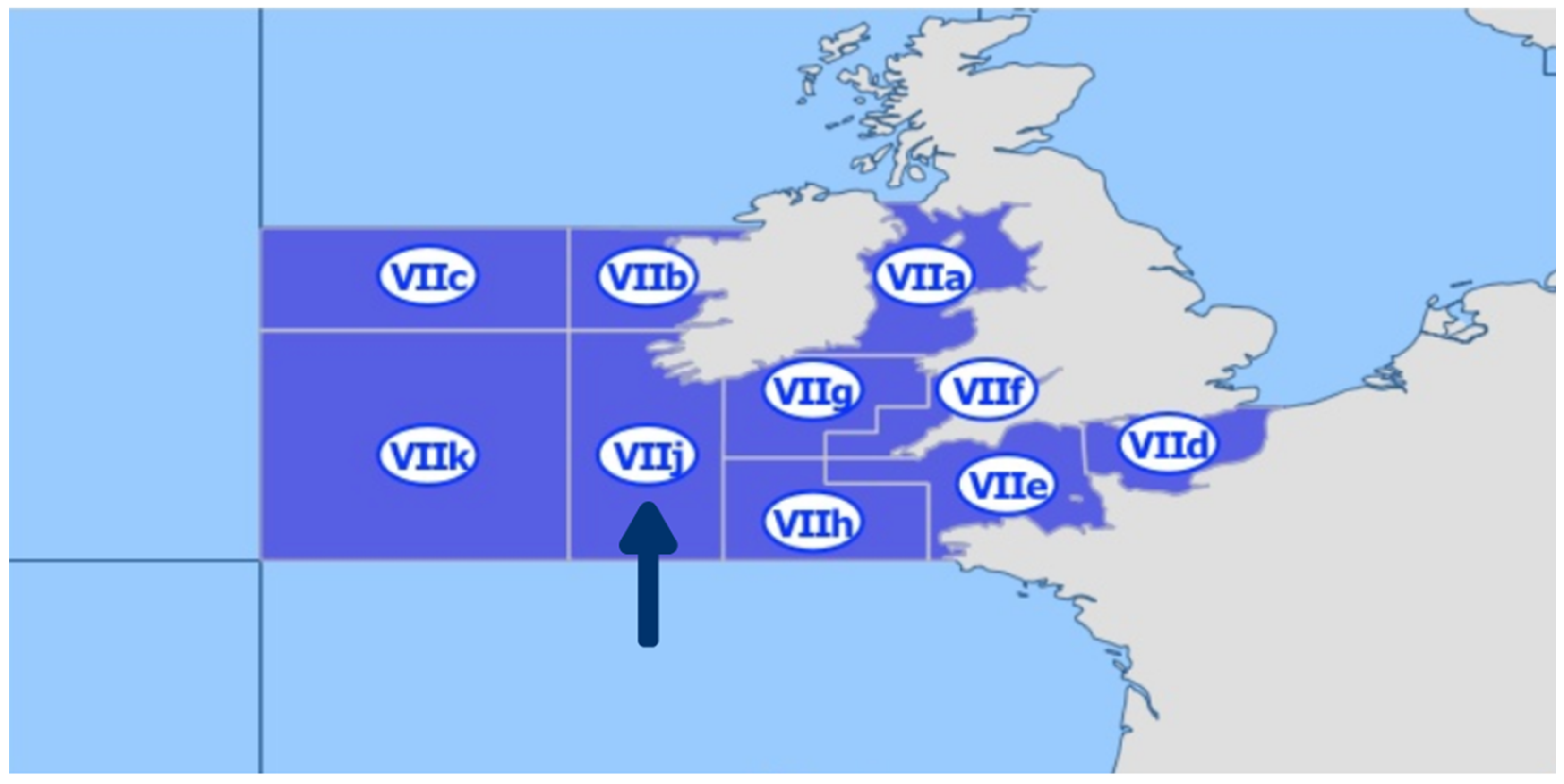
4.1. Study Area, Sample Collection and Bacterial Isolation

Four European hake specimens from the Northeast Atlantic Ocean (Southwest of Ireland), specifically from the sub-area 27.VIIj (Figure 4), obtained during two consecutive years (June 2021 and June 2022) were sampled for this work [113].
For each sampling (June 2021 and 2022), four non-eviscerated commercial European hakes, provided by a Galician skipper dedicated to professional fishing, were used for bacteria isolation, making up a total of eight hake samples. For each hake, one gram of faeces was extracted, serially diluted in sterile peptone water (Oxoid Ltd., Basingstoke, UK) and spread over De Man, Rogosa and Sharpe (MRS, Oxoid, Basingstoke, UK) agar (1.5%, w/v, Scharlau, Barcelona, Spain) plates. In the case of the hake intestines, each sample was washed with phosphate-buffered saline (PBS; Oxoid Ltd., Basingstoke, UK) and homogenised in a stomacher (Seward, NY, USA) with sterile peptone water and then serially diluted and poured onto MRS agar plates (1.5%, w/v). The plates were incubated at 30 °C for 24–72 h in aerobiosis and anaerobiosis. A total of 286 isolates with different morphology, including representatives from the 8 hake samples, were selected to evaluate their antimicrobial activity. The schematic view of the screening used in this study is illustrated in Figure 5.

4.2. Screening of Bacteria with Antimicrobial Activity Against Ichthyopathogens

To assess the direct antimicrobial activity of the isolates and select those of great interest, a Stab On Agar Test (SOAT) was carried out against different ichthyopathogens of relevance to aquaculture (e.g., A. salmonicida CLFP-23, A. salmonicida CECT4237, Aeromonas hydrophila CECT839, A. hydrophila CECT5734, Edwarsiella tarda CECT886, Lactococcus garvieae CF00021, Lc. garvieae CLG4, Listonella anguillarum CECT4344, St. parauberis LMG22252, T. maritimum NCIMB2154, T. maritimum CECT1161 and Yersinia ruckeri LMG3279) and against other microorganisms of importance for humans (Listeria monocytogenes CECT911 and Listeria ivanovii CECT913) [110]. For this purpose, bacteria were stabbed into MRS agar (1.5%, w/v) using sterile sticks, and after incubation at 30 °C for 5 h, the corresponding semisolid medium (0.8% agar, w/v) inoculated with the indicator microorganism (1 × 10⁶ cfu/mL) was poured over the plates. After incubation at 30 °C for 16–24 h, the inhibition halos formed around the isolates under study were measured (mm) [114]. A total of 66 isolates were pre-selected based on their antimicrobial activity.

4.3. Taxonomic Identification of Bacterial Isolates

The identification of the isolates was achieved through comprehensive sequencing of the 16S rDNA. To accomplish this, total DNA was extracted from the isolates that had been pre-selected based on their antimicrobial properties, utilising the InstaGene Matrix (Bio-Rad Laboratories, Inc., Hercules, CA, USA) according to the manufacturer’s guidelines. The amplification of the 16S rDNA gene was performed using PCR, incorporating 25 µL of DreamTaq Hot Start PCR Master Mix 2× (Thermo Scientific, Waltham, MA, USA), 0.5 µM of the forward primer fD1 (5′-AGAGAGAGTTTGGATCCTGGCTCAG-3′), 0.5 µM of the reverse primer rD1 (5′-TAAGGAGGAGGAGGAGGTGATCCAGCC-3′), 50–100 ng of purified DNA and 19 µL of molecular biology-grade water (Thermo Scientific, Waltham, MA, USA) [115]. The PCR mixtures underwent a series of amplification cycles, beginning with an initial denaturation at 95 °C for 3 min, followed by 35 cycles consisting of denaturation at 95 °C for 30 s, hybridisation at 60 °C for 30 s, elongation at 72 °C for 1 min and concluding with a final elongation at 72 °C for 5 min using a thermal cycler (Eppendorf, Hamburg, Germany). The resulting amplicons were then purified with the NucleoSpin® Gel and PCR Clean-up kit (Macherey-Nagel™, Düren, Germany) and forwarded to Eurofins Genomics (Ebersberg, Germany) for DNA sequencing. The nucleotide sequences obtained were analysed using the NCBI BLAST nucleotide server (https://blast.ncbi.nlm.nih.gov/, accessed on 15 April 2024) for taxonomic identification. Out of the 66 identified isolates, only LAB (n = 35) were selected for further characterization.

4.4. Genetic Diversity Analysis by Enterobacterial Repetitive Intergenic Consensus-PCR (ERIC-PCR)

To examine the genetic diversity among the various LAB isolated from European hake, ERIC-PCR was conducted using the primers ERIC-1R (5′-ATGTAAGCTCCTGGGGGGGGGGGGATTCAC-3′) and ERIC-2 (5′-AAGTAAGTG ACTGGGGGGGGGTGAGCG-3′), as previously detailed by Araújo et al. (2015) [15]. PCR reactions of 50 µL were prepared with 25 µL of MyTaq Mix (Bioline Reagents, Ltd., London, UK), 0.7 µM of each primer, 50–100 ng of purified DNA, 3 µM of MgCl2 and 19 µL of molecular biology-grade water. The PCR cycles included an initial denaturation step at 95 °C for 1 min, followed by 35 cycles consisting of denaturation, annealing and elongation at 95 °C for 15 s, 46 °C for 15 s and 72 °C for 10 s and concluding with a final elongation step at 72 °C for 4 min in a thermal cycler. The amplified products were subjected to electrophoresis at 90 V for 60 min using an electrophoresis chamber (Bio-Rad Laboratories, Inc., Hercules, CA, USA), and band visualization was performed using a ChemiDoc Imaging System (Bio-Rad Laboratories, Inc.) with HyperLadder 100 bp (Bioline Reagents, Ltd., London, UK) as a molecular weight marker. For the analysis of ERIC types, clustering and dendrogram construction, Phoretix v.5.0 software (Nonlinear Dynamics Ltd., Newcastle upon Tyne, UK) was utilised.

4.5. Biofilm Formation and Quantification

In order to evaluate the biofilm formation, a microtiter assay, previously described by Oniciuc E.-A. et al. (2016) [116], was performed. Each isolate was grown on MRS agar plates (Oxoid) and incubated at 30 °C for 24 h. Then, a pair of colonies were transferred to 3 mL tubes of TSB (Tryptic Soy Broth, Oxoid) and incubated at 30 °C for 16 ± 1 h with continuous shaking at 120 rpm (ES- 80 Shaker-incubator, Grant Instruments). Then, 200 µL of bacterial suspension with a concentration of 1 × 10⁶ cfu/mL was added to each well of the 96-well flat-bottom microplate (Thermo Scientific). In all plates, Staphylococcus aureus CECT794 was included as a positive control and TSB without bacterial inoculum as a negative control. The plates were incubated in aerobiosis for 24 h at 30 °C. All experiments had sixteen replicates.
For the quantification of biofilm production, the crystal violet (CV) staining method was used, as previously described by Peeters et al. (2008) [117], with some modifications. Once the incubation was finished, the unattached bacterial cells were removed by eliminating the medium from each well and washing the plates twice with sterile distilled water. The plates were left to dry at room temperature for 30 min. To fix the biofilms, 100 µL of methanol (VWR International) was added to each well. After 15 min, the methanol was discarded, the plates were dried at room temperature for 10 min and 100 µL of 1% (v/v) CV was added to each well. After 10 min, the CV was removed, and the plates were washed twice with distilled water to eliminate excess dye and then dried. In order to solubilize the CV, 100 µL of 33% (v/v) acetic acid was subsequently added and the absorbance was measured at 570 nm using a BioTek ELx808U microplate reader (BioTek, Winooski, VT, USA). To standardise the results, the biofilm formation capacity of each isolate was normalised, assuming that the positive control S. aureus CECT794 produces 100% biofilm. GraphPad Prism 8 (GraphPad Software, San Diego, CA, USA) was used for data processing, analysis and graphical representation. The normal distribution of all the data was verified using the Shapiro–Wilk test. Statistical analyses were then performed using paired Student’s t-tests.

4.6. Antibiotic Susceptibility Testing

Antibiotic susceptibility was determined by a broth microdilution test in order to determine the minimum inhibitory concentration (MIC) of 14 antibiotics: ampicillin (0.25–16 µg/mL); vancomycin (1–64 µg/mL); gentamicin (0.5–32 µg/mL); kanamycin (1–128 µg/mL); streptomycin (1–64 µg/mL); erythromycin (0.25–16 µg/mL); clindamycin (0.25–16 µg/mL); tetracycline (0.5–32 µg/mL); chloramphenicol (1–64 µg/mL); florfenicol (0.125–8 µg/mL); oxytetracycline (0.03–4 µg/mL); flumequine (0.125–8 µg/mL); amoxicillin (0.125–8 µg/mL); trimethoprim-sulfamethoxazole (0.0625/1.1875–4/76 µg/mL). These antibiotics were chosen according to the EFSA Technical Panel on Additives and Products or Substances used in Animal Feed (FEEDAP) guidelines on Guidance on the characterisation of microorganisms used as feed additives or production organisms [118]. In addition, antibiotics frequently used in aquaculture were also evaluated [119,120,121].
Antimicrobial susceptibility testing was performed in accordance with the European Committee on Antimicrobial Susceptibility Testing (EUCAST) [122,123] and Clinical and Laboratory Standards Institute (CLSI) [44,45,120]. Individual colonies of each strain were chosen from a culture grown in MRS agar at 30 °C for 16 h and incorporated into 10 mL of saline solution (0.9% NaCl; w/v) until its optical density was adjusted to a value of 0.5 according to the McFarland scale (ca. 1.5 × 10⁸ cfu/mL). Then, they were diluted (1:100) in Mueller–Hinton medium to obtain ca. 1.5 × 10⁶ cfu/mL. Serial two-fold dilutions of each antibiotic were prepared in 96-well microtiter plates (Thermo Scientific), and 50 μL of the bacterial culture was added to each well, obtaining a final concentration of ca. 7.5 × 10⁵ cfu/mL. Subsequently, the plates were shaken for 3 min and, finally, incubated at 30 °C for 24 h [44,45,124]. After the incubation, the MIC was determined for each antibiotic, which is defined as the minimum concentration that inhibits bacterial growth [43]. MICs were compared to cut-off values established by [43] and/or the CLSI breakpoints [44,45,124]. Isolates were considered resistant when the MIC for one specific antibiotic was higher than the most restrictive cut-off value. In relation to this, when an exact cut-off value for some bacterial species evaluated in this study had not previously been described, it was adjusted with the cut-off value corresponding to a related species. Specifically, the EFSA cut-off values for Lc. garvieae were compared with those for Lactococcus lactis, Streptococcus salivarius with Streptococcus thermophilus, Enterococcus avium with Enterococcus faecium and Lactobacillus sakei with Lactobacillus facultative heterofermentative. Quality control was performed with the strains S. aureus CECT794 and Enterococcus faecalis CECT795. Two independent experiments were performed in duplicate.

4.7. Evaluation of Haemolytic and Gelatinase Activities

Haemolytic activity was determined using the method previously described by Eaton and Gasson (2001) [125]. The isolates were inoculated in MRS broth and incubated at 30 °C in aerobiosis for 16 h. Subsequently, bacteria were streaked onto Columbia agar plates supplemented with horse blood (5%, v/v) (BioMérieux, Marcy L’Etoile, France). After incubation at 30 °C for 24 h, the α- and β-haemolysis were revealed by the appearance of greenish halos and clear zones, respectively, around the colonies. E. faecalis SDP10 and E. faecalis P4 strains were used as positive controls. Two independent experiments were performed.
Gelatinase production was determined using the method previously described by Eaton and Gasson (2001) [125]. The isolates were inoculated in MRS broth and incubated in aerobiosis at 30 °C for 16 h. Subsequently, bacteria were seeded onto Todd–Hewitt (Oxoid) agar plates (1.5%, w/v) containing 3% (w/v) of gelatine (Oxoid) and incubated at 30 °C for 24 h. After incubation, the plates were kept at 4 °C for 5 h. The presence of a zone of turbidity (gelatin hydrolysis) around the inoculation streak was considered indicative of proteolytic activity. E. faecalis SDP10 and E. faecalis P4 strains were used as positive controls. Two independent experiments were performed.

5. Conclusions

The intestine and faeces of Northeast Atlantic hake are suitable ecological niches for the isolation of LAB (Lactobacillus spp., Lactococcus spp., Leuconostoc spp. and Streptococcus spp.) with antimicrobial activity against ichthyopathogens. After the different analyses carried out in this work, only five strains (11.4%), St. salivarius MDI13, St. salivarius MDI20, E. avium MEI4, E. avium MGH6 and Lb. sakei MEI5, have been selected for future studies as putative safe probiotic candidates for aquaculture, since all of them were susceptible to antibiotics and none showed any of the tested virulence factors (haemolytic and proteolytic activities). This approach could contribute to the development of sustainable aquaculture in the context of One Health, not only for the prevention and control of the most relevant ichthyopathologies but also to contribute to the health, immune status, productivity and economic profitability of aquaculture farms. However, additional genetic analyses, including the detection of antibiotic resistance genes, plasmids, mobile genetic elements and virulence factors, along with in vivo assays, are necessary to further validate our findings and confirm the suitability and safety of these strains as probiotics.

Supplementary Materials

The following supporting information can be downloaded at https://www.mdpi.com/article/10.3390/antibiotics14050469/s1: Table S1. Direct antimicrobial activity of the 66 isolates from European hakes against pathogenic bacteria by a Stab-On-Agar Test (SOAT).

Author Contributions

Conceptualisation, L.M.C. and E.M.-A.; methodology, L.D.-F., D.C., E.M.-A. and J.F.; software, L.D.-F. and D.C.; validation, J.F. and J.B.; formal analysis, L.D.-F., B.O., J.B. and J.F.; investigation, L.D.-F., E.M.-A., B.O., J.B. and D.C.; resources, J.B., P.E.H. and L.M.C.; data curation, L.D.-F.; writing—original draft preparation, L.D.-F.; writing—review and editing, E.M.-A., J.F. and L.M.C.; visualisation, L.D.-F. and E.M.-A.; supervision, L.M.C. and E.M.-A.; project administration, L.M.C., P.E.H., E.M.-A. and J.B.; funding acquisition, L.M.C., J.B. and P.E.H. All authors have read and agreed to the published version of the manuscript.

Funding

This research was funded by the Ministerio de Ciencia, Innovación y Universidades (MCIU, Spain) (Projects RTI2018-094907-B-I00 and PID2019-104808RA-I00). L.D.F. received support by the Programa Investigo (Ministerio de Trabajo y Economía Social, MITES, Spain), funded by the EU (NextGenerationEU). D.C. had a research contract from the project RTI2018-094907-B-I00 (MCIU, Madrid, Spain). J.F. was supported by a FEI16/54 contract from the Universidad Complutense de Madrid (UCM, Spain) and held a predoctoral contract from the UCM. J.B. was supported by the Programa Atracción de Talento (2018-T1/BIO-10158) from the Comunidad de Madrid (Spain).

Institutional Review Board Statement

Not applicable.

Informed Consent Statement

Not applicable.

Data Availability Statement

The original contributions presented in this study are included in the article/Supplementary Materials. Further inquiries can be directed to the corresponding authors.

Acknowledgments

We acknowledge I. Ruiz Zarzuela (Facultad de Veterinaria, Universidad de Zaragoza, Spain) and C. Michel (INRA, Jouy-en-Josas, France) for providing some of the fish pathogens.

Conflicts of Interest

The authors declare no conflicts of interest.

References

  1. Food and Agriculture Organization (FAO). The State of WORLD Fisheries and Aquaculture 2020; FAO Fisheries and Aquaculture Department: Rome, Italy, 2020. [Google Scholar] [CrossRef]
  2. Hunter, M.C.; Smith, R.G.; Schipanski, M.E.; Atwood, L.W.; Mortensen, D.A. Agriculture in 2050: Recalibrating targets for sustainable intensification. BioScience 2017, 67, 385–390. [Google Scholar] [CrossRef]
  3. Food and Agriculture Organization (FAO). The State of World Fisheries and Aquaculture 2022. 2022. Available online: https://www.fao.org/documents/card/en/c/cc0461es (accessed on 10 May 2024).
  4. Álvarez-Pellitero, P. Fish immunity and parasite infections: From innate immunity to immunity to immunoprophylactic prospects. Vet. Immunol. Immunopathol. 2008, 126, 171–198. [Google Scholar] [CrossRef]
  5. Almeida, A.; Cunha, A.; Gomes, N.C.; Alves, E.; Costa, L.; Faustino, M.A. Phage therapy and photodynamic therapy: Low environmental impact approaches to inactivate microorganisms in fish farming plants. Mar. Drugs 2009, 7, 268–313. [Google Scholar] [CrossRef]
  6. Zhang, Q.; Gui, J.F. Virus genomes and virus-host interactions in aquaculture animals. Sci. China Life Sci. 2015, 58, 156–169. [Google Scholar] [CrossRef] [PubMed]
  7. Kibenge, F.S. Emerging viruses in aquaculture. Curr. Opin. Virol. 2019, 34, 97–103. [Google Scholar] [CrossRef]
  8. Abowei, F.N.; Briyai, O.F.; Bassey, S.E. A review of some basic parasite diseases in culture fisheries: Flagellids, dinoflagellides and ichthyophthriasis, ichtyobodiasis, coccidiosis trichodiniasis, heminthiasis, hirudinea infestation, crustacean parsite and ciliates. Br. J. Pharmacol. Toxicol. 2011, 2, 213–226. [Google Scholar]
  9. Stentiford, G.D.; Bateman, I.J.; Hinchliffe, S.J.; Bass, D.; Hartnell, R.; Santos, E.M.; Devlin, M.J.; Feist, S.W.; Taylor, N.G.H.; Verner-Jeffreys, D.W.; et al. Sustainable aquaculture through the One Health lens. Nat. Food 2020, 1, 468–474. [Google Scholar] [CrossRef] [PubMed]
  10. Valle, A.; Leiro, J.M.; Pereiro, P.; Figueras, A.; Novoa, B.; Dirks, R.P.H.; Lamas, J. Interactions between the parasite Philasterides dicentrarchi and the immune system of the turbot Scophthalmus maximus. A transcriptomic analysis. Biology 2020, 9, 337. [Google Scholar] [CrossRef]
  11. Moreira, M.; Schrama, D.; Farinha, A.P.; Cerqueira, M.; Raposo de Magalhães, C.; Carrilho, R.; Rodrigues, P. Fish pathology research and diagnosis in aquaculture of farmed fish; a proteomics perspective. Animals 2021, 11, 125. [Google Scholar] [CrossRef]
  12. Food and Agriculture Organization (FAO). The State of World Fisheries and Aquaculture 2024. Blue Transformation in Action. 2024. Available online: https://openknowledge.fao.org/items/06690fd0-d133-424c-9673-1849e414543d (accessed on 10 November 2024).
  13. Chen, J.; Sun, R.; Pan, C.; Sun, Y.; Mai, B.; Li, Q.X. Antibiotics and Food Safety in Aquaculture. J Agric Food Chem. 2020, 68, 11908–11919. [Google Scholar] [CrossRef]
  14. Muñoz-Atienza, E.; Gómez-Sala, B.; Araújo, C.; Campanero, C.; del Campo, R.; Hernández, P.E.; Herranz, C.; Cintas, L.M. Antimicrobial activity, antibiotic susceptibility and virulence factors of Lactic Acid Bacteria of aquatic origin intended for use as probiotics in aquaculture. BMC Microbiol. 2013, 13, 15. [Google Scholar] [CrossRef]
  15. Araújo, C.; Muñoz-Atienza, E.; Ramírez, M.; Poeta, P.; Igrejas, G.; Hernández, P.E.; Herranz, C.; Cintas, L.M. Safety assessment, genetic relatedness and bacteriocin activity of potential probiotic Lactococcus lactis strains from rainbow trout (Oncorhynchus mykiss, Walbaum) and rearing environment. Eur. Food Res. Technol. 2015, 241, 647–662. [Google Scholar] [CrossRef]
  16. World Health Organization (WHO). Resistencia a Los Antimicrobianos. 2020. Available online: https://www.who.int/es/news-room/fact-sheets/detail/antimicrobial-resistance (accessed on 10 September 2024).
  17. El-Saadony, M.T.; Alagawany, M.; Patra, A.K.; Kar, I.; Tiwari, R.; Dawood, M.A.O.; Dhama, K.; Abdel-Latif, H.M.R. The functionality of probiotics in aquaculture: An overview. Fish Shellfish Immunol. 2021, 117, 36–52. [Google Scholar] [CrossRef]
  18. Nayak, S.K. Current prospects and challenges in fish vaccine development in India with special reference to Aeromonas hydrophila vaccine. Fish Shellfish Immunol. 2020, 100, 283–299. [Google Scholar] [CrossRef] [PubMed]
  19. Kumar, A.; Middha, S.K.; Menon, S.V.; Paital, B.; Gokarn, S.; Nelli, M.; Rajanikanth, R.B.; Chandra, H.M.; Mugunthan, S.P.; Kantwa, S.M.; et al. Current challenges of vaccination in fish health management. Animals 2024, 14, 2692. [Google Scholar] [CrossRef]
  20. Toranzo, A.; Magariños, B.; Romalde, J.L.; Barja, J.L. Present and future of aquaculture vaccines against fish bacterias diseases. Options Méditerranéennes 2009, 86, 155–176. [Google Scholar]
  21. Adams, A. Progress, challenges and opportunities in fish vaccine development. Fish Shellfish Immunol. 2019, 90, 210–214. [Google Scholar] [CrossRef] [PubMed]
  22. Collins, C.; Lorenzen, N.; Collet, B. DNA vaccination for finfish aquaculture. Fish Shellfish Immunol. 2019, 85, 106–125. [Google Scholar] [CrossRef]
  23. Pérez-Sánchez, T.; Ruiz-Zarzuela, I.; de Blas, I.; Balcázar, J.L. Probiotics in aquaculture: A current assessment. Rev. Aquac. 2014, 6, 133–146. [Google Scholar] [CrossRef]
  24. Gómez-Sala, B.; Feito, J.; Hernández, P.E.; Cintas, L.M. Lactic Acid Bacteria in aquatic Environments and Their Applications, 5th ed.; Vinderola, G., Ouwehand, A.C., Salminen, S., von Wright, A., Eds.; CRC Press: Boca Raton, FL, USA, 2019; pp. 555–570. [Google Scholar]
  25. Thatcher, C.; Høj, L.; Bourne, D.G. Probiotics for coral aquaculture: Challenges and considerations. Curr. Opin. Biotechnol. 2022, 73, 380–386. [Google Scholar] [CrossRef]
  26. EFSA. Statement on the requirements for whole genome sequence analysis of microorganisms intentionally used in the food chain. EFSA J. 2021, 19, 434–438. [Google Scholar]
  27. Commission Implementing Regulation (EU). 2020/151 of 4 February 2020 concerning the authorisation of Pediococcus acidilactici CNCM I-4622 as a feed additive for all porcine species for fattening and for breeding other than sows, all avian species, all fish species and all crustaceans and repealing Regulations (EC) No 911/2009, (EU) No 1120/2010 and (EU) No 212/2011 and Implementing Regulations (EU) No 95/2013, (EU) No 413/2013 and (EU) 2017/2299 (holder of authorisation Danstar Ferment AG represented in the Union by Lallemand SAS). Off. J. Eur. Union 2020, 33, 12–15. Available online: https://eur-lex.europa.eu/eli/reg_impl/2020/151/oj (accessed on 8 November 2024).
  28. Todorov, S.D.; Lima, J.M.S.; Bucheli, J.E.V.; Popov, I.V.; Tiwari, S.K.; Chikindas, M.L. Probiotics for Aquaculture: Hope, Truth, and Reality. Probiot. Antimicrob. Proteins 2024, 16, 2007–2020. [Google Scholar] [CrossRef] [PubMed]
  29. Gillor, O.; Etzion, A.; Riley, M.A. The dual role of bacteriocins as anti- and probiotics. Appl. Microbiol. Biotechnol. 2008, 81, 591–606. [Google Scholar] [CrossRef] [PubMed]
  30. Ringø, E.; Hoseinifar, S.H.; Ghosh, K.; Doan, H.V.; Beck, B.R.; Song, S.K. Lactic acid bacteria in finfish-an update. Front. Microbiol. 2018, 9, 1818. [Google Scholar] [CrossRef]
  31. Ringø, E.; Van Doan, H.; Lee, S.H.; Soltani, M.; Hoseinifar, S.H.; Harikrishnan, R.; Song, S.K. Probiotics, lactic acid bacteria and bacilli: Interesting supplementation for aquaculture. J. App. Microbiol. 2020, 129, 116–136. [Google Scholar] [CrossRef]
  32. Zorriehzahra, M.J.; Delshad, S.T.; Adel, M.; Tiwari, R.; Karthik, K.; Dhama, K.; Lazado, C.C. Probiotics as beneficial microbes in aquaculture: An update on their multiple modes of action: A review. Vet Q. 2016, 36, 228–241. [Google Scholar] [CrossRef]
  33. Vine, N.G.; Leukes, W.D.; Kaiser, H. Probiotics in marine larviculture. FEMS Microbiol. Rev. 2006, 30, 404–427. [Google Scholar] [CrossRef]
  34. Ringø, E.; Løvmo, L.; Kristiansen, M.; Bakken, Y.; Salinas, I.; Myklebust, R.; Olsen, R.E.; Mayhew, T.M. Lactic acid bacteria vs. pathogens in the gastrointestinal tract of fish: A review. Aquac. Res. 2010, 41, 451–467. [Google Scholar] [CrossRef]
  35. Dobson, A.; Cotter, P.D.; Ross, R.P.; Hill, C. Bacteriocin production: A probiotic trait? Appl. Environ. Microbiol. 2012, 78, 1–6. [Google Scholar] [CrossRef]
  36. Ołdak, A.; Zielińska, D. Bacteriocins from lactic acid bacteria as an alternative to antibiotics. Postep. Hig. Med. Dosw. 2017, 71, 328–338. [Google Scholar] [CrossRef]
  37. Hernández-González, J.C.; Martínez-Tapia, G.A.; Lazcano-Hernández, B.E.; García-Pérez Castrejón-Jiménez, N.S. Bacteriocins from lactic acid bacteria. A powerful alternative as antimicrobials, probiotics, and immunomodulators in veterinary medicine. Animals 2021, 11, 979. [Google Scholar] [CrossRef]
  38. Uniacke-Lowe, S.; Collins, F.W.J.; Hill, C.; Ross, R.P. Bioactivity screening and genomic analysis reveals deep-sea fish microbiome isolates as sources of novel antimicrobials. Mar. Drugs. 2023, 21, 444. [Google Scholar] [CrossRef] [PubMed]
  39. Ministerio de Agricultura, Pesca y Alimentación (MAPA). Estadísticas Pesqueras: Pesca Marítima. 2022. Available online: https://www.mapa.gob.es/es/estadistica/temas/estadisticas-pesqueras/pesca-maritima/ (accessed on 10 May 2024).
  40. Ringø, E.; Jutfelt, F.; Kanapathippillai, P.; Bakken, Y.; Sundell, K.; Glette, J.; Mayhew, T.M.; Myklebust, R.; Olsen, R.E. Damaging effect of the fish pathogen Aeromonas salmonicida ssp. salmonicida on intestinal enterocytes of Atlantic salmon (Salmo salar, L.). Cell. Tissue. Res. 2004, 318, 305–311. [Google Scholar] [CrossRef] [PubMed]
  41. Mabrok, M.; Algammal, A.M.; Sivaramasamy, E.; Hetta, H.F.; Atwah, B.; Alghamdi, S.; Fawzy, A.; Avendaño-Herrera, R.; Rodkhum, C. Tenacibaculosis caused by Tenacibaculum maritimum: Updated knowledge of this marine bacterial fish pathogen. Front. Cell. Infect. Microbiol. 2023, 12, 1068000. [Google Scholar] [CrossRef] [PubMed]
  42. Nho, S.W.; Hikima, J.; Cha, I.S.; Park, S.B.; Jang, H.B.; del Castillo, C.S.; Kondo, H.; Hirono, I.; Aoki, T.; Jung, T.S. Complete genome sequence and immunoproteomic analyses of the bacterial fish pathogen Streptococcus parauberis. J. Bacteriol. 2011, 193, 3356–3366. [Google Scholar] [CrossRef]
  43. FEEDAP. Guidance on the characterization of microorganisms used as feed additives or as production organisms. EFSA J. 2018, 16, e05206. [Google Scholar]
  44. Clinical and Laboratory Standards Institute (CLSI). M45 Methods for Antimicrobial Dilution and Disk Susceptibility Testing of Infrequently Isolated or Fastidious Bacteria, 3rd ed.; CLSI: Wayne, PA, USA, 2015. [Google Scholar]
  45. Clinical and Laboratory Standards Institute (CLSI). M100 Performance Standards for Antimicrobial Susceptibility Testing, 30th ed.; CLSI: Wayne, PA, USA, 2020. [Google Scholar]
  46. Gónzalez, C.; Encinas, J.P.; García-López, L.M.; Otero, A. Characterisation and identification of lactic acid bacteria from freshwater fishes. Food Microbiol. 2020, 17, 383–391. [Google Scholar] [CrossRef]
  47. Ringø, E.; Holzapfel, W. Identification and characterization of carnobacteria associated with the gills of Atlantic salmon (Salmo salar L.). Syst. Appl. Microbiol. 2000, 23, 523–527. [Google Scholar] [CrossRef]
  48. Elidrissi, A.; Ezzaky, Y.; Boussif, K.; Achemchem, F. Isolation and characterization of bioprotective lactic acid bacteria from Moroccan fish and seafood. Braz. J. Microbiol. 2023, 54, 2117–2127. [Google Scholar] [CrossRef]
  49. Cheriet, S.; Lengliz, S.; Romdhani, A.; Hynds, P.; Abbassi, M.S.; Ghrairi, T. Selection and characterization of bacteriocinogenic Lactic Acid Bacteria from the intestine of gilthead seabream (Sparus aurata) and whiting fish (Merlangius merlangus): Promising strains for aquaculture probiotic and food bio-preservation. Life 2023, 13, 1833. [Google Scholar] [CrossRef]
  50. Seppola, M.; Olsen, R.E.; Sandeker, E.; Kanapathippillai, P.; Holzapfel, W.; Ringø, E. Random amplification of polymorphic DNA (RAPD) typing of carnobacteria isolated from hindgur chamber and large intestine of atlantic cod (Gadus morhua L.). Syst. Appl. Microbiol. 2006, 28, 131–137. [Google Scholar] [CrossRef]
  51. Pinto, A.; Fernandes, L.; Pinto, C.; Albano, H.; Castillo, F.; Teixeira, P.; Gibbs, A. Characterization of anti-Listeria bacteriocins isolated from shellfish: Potencial antimicrobials to control non-fermented seafood. Int. J. Food. Microbiol. 2009, 129, 50–58. [Google Scholar] [CrossRef] [PubMed]
  52. Lauzon, H.L.; Ringo, E. Prevalence and application of lactic acid bacteria in aquatic environments. In Lactic Acid Bacteria: Microbiological and Functional Aspects, 4th ed.; CRC Press: Boca Raton, FL, USA, 2012. [Google Scholar] [CrossRef]
  53. Gómez-Sala, B.; Herranz, C.; Díaz-Freitas, B.; Hernández, P.E.; Sala, A.; Cintas, L.M. Strategies to increase the hygienic and economic value of fresh fish: Biopreservation using lactic acid bacteria of marine origin. Int. J. Food Microbiol. 2016, 223, 41–49. [Google Scholar] [CrossRef] [PubMed]
  54. Sahoo, T.K.; Jena, P.K.; Nagar, N.; Patel, A.K.; Seshadri, S. In vitro evaluation of probiotic properties of lactic acid bacteria from the gut of Labeo rohita and Catla catla. Probiotics Antimicrob. Proteins 2015, 7, 126–136. [Google Scholar] [CrossRef]
  55. Patel, P.; Patel, B.; Amaresan, N.; Joshi, B.; Shah, R.; Krishnamurthy, R. Isolation and characterization of Lactococcus garvieae from the fish gut for in vitro fermentation with carbohydrates from agro-industrial waste. Biotechnol. Rep. 2020, 28, e00555. [Google Scholar] [CrossRef]
  56. Araújo, C.; Muñoz-Atienza, E.; Nahuelquín, Y.; Poeta, P.; Igrejas, G.; Hernández, P.E.; Herranz, C.; Cintas, L.M. Inhibition of fish pathogens by the microbiota from rainbow trout (Oncorhynchus mykiss, Walbaum) and rearing environment. Anaerobe 2015, 32, 7–14. [Google Scholar] [CrossRef] [PubMed]
  57. Phupaboon, S.; Hashim, F.J.; Phumkhachorn, P.; Rattanachaikunsopon, P. Molecular and biotechnological characteristics of proteolytic activity from Streptococcus thermophiles as a proteolytic lactic acid bacteria to enhance protein-derived bioactive peptides. AIMS Microbiol. 2023, 9, 591–611. [Google Scholar] [CrossRef]
  58. Hoseinifar, S.H.; Sun, Y.Z.; Wang, A.; Zhou, Z. Probiotics as means of diseases control in aquaculture, a review of current knowledge and future perspectives. Front. Microbiol. 2018, 9, 2429. [Google Scholar] [CrossRef]
  59. Ringø, E.; Li, X.; Doan, H.; Ghosh, K. Interesting probiotic bacteria other than the more widely used lactic acid bacteria and bacilliin finfish. Front. Mar. Sci. 2022, 9, 848037. [Google Scholar] [CrossRef]
  60. Simón, R.; Docando, F.; Nu.ez-Ortiz, N.; Tafalla, C.; Díaz-Rosales, P. Mechanisms used by probiotics to confer pathogen resistance to teleost fish. Front. Immunol. 2021, 12, 653025. [Google Scholar] [CrossRef]
  61. Sumon, M.A.A.; Molla, M.H.R.; Hakeem, I.J.; Ahammad, F.; Amran, R.H.; Jamal, M.T.; Gabr, M.H.; Islam, M.S.; Alam, M.T.; Brown, C.L.; et al. Epigenetics and probiotics application toward the modulation of fish reproductive performance. Fishes 2022, 7, 189. [Google Scholar] [CrossRef]
  62. Cintas, L.M.; Casaus, M.P.; Herranz, C.; Nes, I.F.; Hernández, P.E. Review: Bacteriocins of lactic acid bacteria. Food Sci. Technol. Int. 2001, 7, 281–305. [Google Scholar] [CrossRef]
  63. Cotter, P.D.; Ross, R.P.; Hill, C. Bacteriocins—A viable alternative to antibiotics? Nat. Rev. Microbiol. 2013, 11, 95–105. [Google Scholar] [CrossRef] [PubMed]
  64. Zhang, Y.; Xu, D.; Shi, L.; Cai, R.; Li, C.; Yan, H. Association between agr type, virulence factors, biofilm formation and antibiotic resistance of Staphylococcus aureus isolates from pork production. Front. Microbiol. 2018, 9, 1876. [Google Scholar] [CrossRef] [PubMed]
  65. Rodríguez-López, P.; Filipello, V.; Di Ciccio, P.A.; Pitozzi, A.; Ghidini, S.; Scali, F.; Ianieri, A.; Zanardi, E.; Losio, M.N.; Simon, A.C.; et al. Assessment of the antibiotic resistance profile, genetic heterogeneity and biofilm production of Methicillin-Resistant Staphylococcus aureus (MRSA) isolated from the Italian swine production chain. Foods 2020, 9, 1141. [Google Scholar] [CrossRef]
  66. Cho, J.A.; Roh, Y.J.; Son, H.R.; Choi, H.; Lee, J.W.; Kim, S.J.; Lee, C.H. Assessment of the biofilm-forming ability on solid surfaces of periprosthetic infection-associated pathogens. Sci. Rep. 2022, 12, 18669. [Google Scholar] [CrossRef]
  67. Contente, D.; Díaz-Formoso, L.; Feito, J.; Gómez-Sala, B.; Costas, D.; Hernández, P.E.; Muñoz-Atienza, E.; Borrero, J.; Poeta, P.; Cintas, L.M. Antimicrobial activity, genetic relatedness, and safety assessment of potential probiotic lactic acid bacteria isolated from a rearing tank of rotifers (Brachionus plicatilis) used as live feed in fish larviculture. Animals 2024, 14, 1415. [Google Scholar] [CrossRef]
  68. Rezaei, Z.; Khanzadi, S.; Salari, A. Biofilm formation and antagonistic activity of Lacticaseibacillus rhamnosus (PTCC1712) and Lactiplantibacillus plantarum (PTCC1745). AMB Express 2021, 11, 156. [Google Scholar] [CrossRef]
  69. Mirzabekyan, S.; Harutyunyan, N.; Manvelyan, A.; Malkhasyan, L.; Balayan, M.; Miralimova, S.; Chikindas, M.L.; Chistyakov, V.; Pepoyan, A. Fish probiotics: Cell surface properties of fish intestinal lactobacilli and Escherichia coli. Microorganisms 2023, 11, 595. [Google Scholar] [CrossRef]
  70. Sharma, S.; Mohler, J.; Mahajan, S.D.; Schwartz, S.A.; Bruggemann, L.; Aalinkeel, R. Microbial Biofilm: A review on formation, infection, antibiotic resistance, control measures, and innovative treatment. Microorganisms 2023, 11, 1614. [Google Scholar] [CrossRef] [PubMed]
  71. Chamignon, C.; Guéneau, V.; Medina, S.; Deschamps, J.; Gil-Izquierdo, A.; Briandet, R.; Mousset, P.-Y.; Langella, P.; Lafay, S.; Bermúdez-Humarán, L.G. Evaluation of the probiotic properties and the capacity to form biofilms of various Lactobacillus strains. Microorganisms 2020, 8, 1053. [Google Scholar] [CrossRef] [PubMed]
  72. Shaaban, M.; Abd El-Rahman, O.A.; Al-Qaidi, B.; Ashour, H.M. Antimicrobial and antibiofilm activities of probiotic Lactobacilli on antibiotic-resistant Proteus mirabilis. Microorganisms 2020, 8, 960. [Google Scholar] [CrossRef]
  73. Feito, J.; Araújo, C.; Arbulu, S.; Contente, D.; Gómez-Sala, B.; Díaz-Formoso, L.; Muñoz-Atienza, E.; Borrero, J.; Cintas, L.M.; Hernández, P.E. Design of Lactococcus lactis strains producing garvicin A and/or garvicin Q, either alone or together with nisin A or nisin Z and high antimicrobial activity against Lactococcus garvieae. Foods 2023, 12, 1063. [Google Scholar] [CrossRef]
  74. Elliott, J.A.; Facklam, R.R. Antimicrobial susceptibilities of Lactococcus lactis and Lactococcus garvieae and a proposed method to discriminate between them. J. Clin. Microbiol. 1996, 34, 1296–1298. [Google Scholar] [CrossRef]
  75. Walther, C.; Rossano, A.; Thomann, A.; Perreten, V. Antibiotic resistance in Lactococcus species from bovine milk: Presence of a mutated multidrug transporter mdt(A) gene in susceptible Lactococcus garvieae strains. Vet. Microbiol. 2008, 131, 348–357. [Google Scholar] [CrossRef]
  76. Plumed-Ferrer, C.; Barberio, A.; Franklin-Guild, R.; Werner, B.; McDonough, P.; Bennett, J.; Gioia, G.; Rota, N.; Welcome, F.; Nydam, D.V.; et al. Antimicrobial susceptibilities and random amplified polymorphic DNA-PCR fingerprint characterization of Lactococcus lactis ssp. lactis and Lactococcus garvieae isolated from bovine intramammary infections. J. Dairy Sci. 2015, 98, 6216–6225. [Google Scholar] [CrossRef] [PubMed]
  77. Fortina, M.G.; Ricci, G.; Foschino, R.; Picozzi, C.; Dolci, P.; Zeppa, G.; Cocolin, L.; Manachini, P.L. Phenotypic typing, technological properties and safety aspects of Lactococcus garvieae strains from dairy environments. J. Appl. Microbiol. 2007, 103, 445–453. [Google Scholar] [CrossRef]
  78. Raissy, M.; Moumeni, M. Detection of antibiotic resistance genes in some Lactococcus garvieae strains isolated from infected rainbow trout. Iran. J. Fish. Sci. 2016, 15, 221–229. [Google Scholar]
  79. de Oliveira, R.P.; Aragão, B.B.; de Melo, R.P.B.; da Silva, D.M.S.; de Carvalho, R.G.; Juliano, M.A.; Farias, M.P.O.; de Lira, N.S.C.; Mota, R.A. Bovine mastitis in northeastern Brazil: Occurrence of emergent bacteria and their phenotypic and genotypic profile of antimicrobial resistance. Comp. Immunol. Microbiol. Infect. Dis. 2022, 85, 101802. [Google Scholar] [CrossRef]
  80. Torres-Corral, Y.; Santos, Y. Predicting antimicrobial resistance of Lactococcus garvieae: PCR detection of resistance genes versus MALDI-TOF protein profiling. Aquaculture 2022, 553, 738098. [Google Scholar] [CrossRef]
  81. Shahi, N.; Mallik, S.K. Emerging bacterial fish pathogen Lactococcus garvieae RTCLI04, isolated from rainbow trout (Oncorhynchus mykiss): Genomic features and comparative genomics. Microb. Pathog. 2020, 147, 104368. [Google Scholar] [CrossRef]
  82. Lin, Y.; Han, J.; Barkema, H.W.; Wang, Y.; Gao, J.; Kastelic, J.P.; Han, B.; Qin, S.; Deng, Z. Comparative genomic analyses of Lactococcus garvieae isolated from bovine mastitis in China. Microbiol. Spectr. 2023, 11, e0299522. [Google Scholar] [CrossRef] [PubMed]
  83. Chaffanel, F.; Charron-Bourgoin, F.; Libante, V.; Leblond-Bourget, N.; Payot, S. Resistance genes and genetic elements associated with antibiotic resistance in clinical and commensal isolates of Streptococcus salivarius. Appl. Environ. Microbiol. 2015, 81, 4155–4163. [Google Scholar] [CrossRef]
  84. Palma, T.H.; Harth-Chú, E.N.; Scott, J.; Stipp, R.N.; Boisvert, H.; Salomão, M.F.; Theobaldo, J.D.; Possobon, R.F.; Nascimento, L.C.; McCafferty, J.W.; et al. Oral cavities of healthy infants harbour high proportions of Streptococcus salivarius strains with phenotypic and genotypic resistance to multiple classes of antibiotics. J. Med. Microbiol. 2016, 65, 1456–1464. [Google Scholar] [CrossRef] [PubMed]
  85. Doumith, M.; Mushtaq, S.; Martin, V.; Chaudhry, A.; Adkin, R.; Coelho, J.; Chalker, V.; MacGowan, A.; Woodford, N.; Livermore, D.M.; et al. Genomic sequences of Streptococcus agalactiae with high-level gentamicin resistance, collected in the BSAC bacteraemia surveillance. J. Antimicrob. Chemother. 2017, 72, 2704–2707. [Google Scholar] [CrossRef]
  86. Barros, R.R. Antimicrobial resistance among beta-hemolytic Streptococcus in Brazil: An Overview. Antibiotics 2021, 10, 973. [Google Scholar] [CrossRef]
  87. Wang, J.; Zhang, Y.M.; Lin, M.; Bao, J.; Wang, G.; Dong, R.; Zou, P.; Chen, Y.; Li, N.; Zhang, T.; et al. Maternal colonization with group B Streptococcus and antibiotic resistance in China: Systematic review and meta-analyses. Ann. Clin. Microbiol. Antimicrob. 2023, 22, 5. [Google Scholar] [CrossRef]
  88. Creti, R.; Imperi, M.; Khan, U.B.; Berardi, A.; Recchia, S.; Alfarone, G.; Gherardi, G. Emergence of high-level gentamicin resistance in Streptococcus agalactiae hypervirulent serotype IV ST1010 (CC452) strains by acquisition of a novel integrative and conjugative element. Antibiotics 2024, 13, 491. [Google Scholar] [CrossRef]
  89. Alves-Barroco, C.; Rivas-García, L.; Fernandes, A.R.; Baptista, P.V. Tackling multidrug resistance in streptococci—From novel biotherapeutic strategies to nanomedicines. Front. Microbiol. 2020, 11, 579916. [Google Scholar] [CrossRef]
  90. Du, F.; Lv, X.; Duan, D.; Wang, L.; Huang, J. Characterization of a linezolid- and vancomycin-resistant Streptococcus suis isolate that harbors optrA and vanG operons. Front. Microbiol. 2019, 10, 2026. [Google Scholar] [CrossRef] [PubMed]
  91. Gergova, R.; Boyanov, V.; Muhtarova, A.; Alexandrova, A. A review of the impact of streptococcal infections and antimicrobial resistance on human health. Antibiotics 2024, 13, 360. [Google Scholar] [CrossRef] [PubMed]
  92. Alzahrani, O.M.; Fayez, M.; Alswat, A.S.; Alkafafy, M.; Mahmoud, S.F.; Al-Marri, T.; Almuslem, A.; Ashfaq, H.; Yusuf, S. Antimicrobial resistance, biofilm formation, and virulence genes in Enterococcus species from small backyard chicken flocks. Antibiotics 2022, 11, 380. [Google Scholar] [CrossRef] [PubMed]
  93. Fujii, A.; Kawada-Matsuo, M.; Nguyen-Tra Le, M.; Masuda, K.; Tadera, K.; Suzuki, Y.; Nishihama, S.; Hisatsune, J.; Sugawara, Y.; Kashiyama, S.; et al. Antibiotic susceptibility and genome analysis of Enterococcus species isolated from inpatients in one hospital with no apparent outbreak of vancomycin-resistant Enterococcus in Japan. Microbiol. Immunol. 2024, 68, 254–266. [Google Scholar] [CrossRef]
  94. Zhang, X.; Paganelli, F.L.; Bierschenk, D.; Kuipers, A.; Bonten, M.J.; Willems, R.J.; van Schaik, W. Genome-wide identification of ampicillin resistance determinants in Enterococcus faecium. PLoS Genet. 2012, 8, e1002804. [Google Scholar] [CrossRef]
  95. García-Solache, M.; Rice, L.B. The Enterococcus: A model of adaptability to its environment. Clin. Microbiol. Rev. 2019, 32, e00058-18. [Google Scholar] [CrossRef]
  96. Gagetti, P.; Bonofiglio, L.; García Gabarrot, G.; Kaufman, S.; Mollerach, M.; Vigliarolo, L.; von Specht, M.; Toresani, I.; Lopardo, H.A. Resistance to β-lactams in enterococci. Rev. Argent. Microbiol. 2019, 51, 179–183. [Google Scholar] [CrossRef]
  97. Yang, J.; Chen, Y.; Dong, Z.; Zhang, W.; Liu, L.; Meng, W.; Li, Q.; Fu, K.; Zhou, Z.; Liu, H.; et al. Distribution and association of antimicrobial resistance and virulence characteristics in Enterococcus spp. isolates from captive Asian elephants in China. Front. Microbiol. 2023, 14, 1277221. [Google Scholar] [CrossRef]
  98. Noroozi, N.; Momtaz, H.; Tajbakhsh, E. Molecular characterization and antimicrobial resistance of Enterococcus faecalis isolated from seafood samples. Vet. Med. Sci. 2022, 8, 1104–1112. [Google Scholar] [CrossRef]
  99. Flórez, A.B.; Campedelli, I.; Delgado, S.; Alegría, Á.; Salvetti, E.; Felis, G.E.; Mayo, B.; Torriani, S. Antibiotic susceptibility profiles of dairy leuconostoc, analysis of the genetic basis of atypical resistances and transfer of genes in vitro and in a food matrix. PLoS ONE 2016, 11, e0145203. [Google Scholar] [CrossRef]
  100. Salvetti, E.; Campedelli, I.; Larini, I.; Conedera, G.; Torriani, S. Exploring Antibiotic resistance diversity in Leuconostoc spp. by a genome-based approach: Focus on the lsaA gene. Microorganisms 2021, 9, 491. [Google Scholar] [CrossRef]
  101. Raimondi, S.; Spampinato, G.; Candeliere, F.; Amaretti, A.; Brun, P.; Castagliuolo, I.; Rossi, M. Phenotypic traits and immunomodulatory properties of Leuconostoc carnosum isolated from meat products. Front. Microbiol. 2021, 12, 730827. [Google Scholar] [CrossRef] [PubMed]
  102. Magiorakos, A.P.; Srinivasan, A.; Carey, R.B.; Carmeli, Y.; Falagas, M.E.; Giske, C.G.; Harbarth, S.; Hindler, J.F.; Kahlmeter, G.; Olsson-Liljequist, B.; et al. Multidrug-resistant, extensively drug-resistant and pandrug-resistant bacteria: An international expert proposal for interim standard definitions for acquired resistance. Clin. Microbiol. Infect. 2012, 18, 268–281. [Google Scholar] [CrossRef]
  103. Lehtinen, S.; Blanquart, F.; Lipsitch, M.; Fraser, C. On the evolutionary ecology of multidrug resistance in bacteria. PLoS Pathog. 2019, 15, e1007763. [Google Scholar] [CrossRef] [PubMed]
  104. Bernardeu, M.; Vernoux, J.P.; Henri-Dubernet, S.; Gueguen, M. Safety assement of dairy organisms: The Lactobacillus genus. Int. J. Food Microbiol. 2008, 126, 278–285. [Google Scholar] [CrossRef]
  105. Jaimee, G.; Halami, P.M. Emerging resistance to aminoglycosides in lactic acid bacteria of food origin-an impending menace. Appl. Microbiol. Biotechnol. 2016, 100, 1137–1151. [Google Scholar] [CrossRef] [PubMed]
  106. Reuben, R.C.; Roy, P.C.; Sarkar, S.L.; Alam, R.U.; Jahid, I.K. Isolation, characterization, and assessment of lactic acid bacteria toward their selection as poultry probiotics. BMC Microbiol. 2019, 19, 253. [Google Scholar] [CrossRef]
  107. Reuben, R.C.; Roy, P.C.; Sarkar, S.L.; Rubayet Ul Alam, A.S.M.; Jahid, I.K. Characterization and evaluation of lactic acid bacteria from indigenous raw milk for potential probiotic properties. J. Dairy Sci. 2020, 103, 1223–1237. [Google Scholar] [CrossRef]
  108. Okoye, C.O.; Nyaruaba, R.; Ita, R.E.; Okon, S.U.; Addey, C.I.; Ebido, C.C.; Opabunmi, A.O.; Okeke, E.S.; Chukwudozie, K.I. Antibiotic resistance in the aquatic environment: Analytical techniques and interactive impact of emerging contaminants. Environ. Toxicol. Pharmacol. 2022, 96, 103995. [Google Scholar] [CrossRef]
  109. Akram, F.; Imtiaz, M.; Haq, I.U. Emergent crisis of antibiotic resistance: A silent pandemic threat to 21st century. Microb. Pathog. 2023, 174, 105923. [Google Scholar] [CrossRef]
  110. Balcázar, J.L.; de Blas, I.; Ruiz-Zarzuela, I.; Vendrell, D.; Gironés, O.; Muzquiz, J.L. Enhancement of the immune response and protection induced by probiotic lactic acid bacteria against furunculosis in rainbow trout (Oncorhynchus mykiss). FEMS Immunol. Med. Microbiol. 2007, 51, 185–193. [Google Scholar] [CrossRef] [PubMed]
  111. Madsen, K.T.; Skov, M.N.; Gill, S.; Kemp, M. Virulence factors associated with Enterococcus faecalis infective endocarditis: A mini review. Open Microbiol. J. 2017, 11, 1–11. [Google Scholar] [CrossRef]
  112. Waters, C.M.; Antiporta, M.H.; Murray, B.E.; Dunny, G.M. Role of the Enterococcus faecalis GelE protease in determination of cellular chain length, supernatant pheromone levels, and degradation of fibrin and misfolded surface proteins. J. Bacteriol. 2003, 185, 3613–3623. [Google Scholar] [CrossRef]
  113. Unión Europea. Denominaciones Comerciales. Mapa de las Zonas Pesqueras UE. Names of Sub-Areas and Divisions of FAO Fishing Areas 27 and 37. Available online: https://fish-commercial-names.ec.europa.eu/fish-names/fishing-areas_es (accessed on 13 January 2025).
  114. Cintas, L.M.; Rodríguez, J.M.; Fernández, M.F.; Sletten, K.; Nes, I.F.; Herández, P.E.; Holo, H. Isolation and characterization of pediocin L50, a new bacteriocin from Pediococcus acidilactici with a broad inhibitory spectrum. Appl. Environ. Microbiol. 1995, 61, 2643–2648. [Google Scholar] [CrossRef] [PubMed]
  115. Jaffrès, E.; Sohier, D.; Leroi, F.; Pilet, M.F.; Prévost, H.; Joffraud, J.J.; Dousset, X. Study of the bacterial ecosystem in tropical cooked and peeled shrimps using a polyphasic approach. Int. J. Food Microbiol. 2009, 131, 20–29. [Google Scholar] [CrossRef] [PubMed]
  116. Oniciuc, E.-A.; Cerca, N.; Nicolau, A.I. Compositional Analysis of biofilms formed by Staphylococcus aureus isolated from food sources. Front. Microbiol. 2016, 7, 390. [Google Scholar] [CrossRef]
  117. Peeters, E.; Nelis, H.J.; Coenye, T. Comparison of multiple methods for quantification of microbial biofilms grown in microtiter plates. J. Microbiol. Methods 2008, 72, 157–165. [Google Scholar] [CrossRef]
  118. EFSA FEEDAP Panel (EFSA Panel on Additives and Products or Substances used in Animal Feed); Rychen, G.; Aquilina, G.; Azimonti, G.; Bampidis, V.; Bastos, M.L.; Bories, G.; Chesson, A.; Cocconcelli, P.S.; Flachowsky, G.; et al. El Plan de Acción de la FAO Sobre la Resistencia a los Antimicrobianos 2016–2020. 2016. Available online: https://www.fao.org/3/i5996s/i5996s.pdf (accessed on 2 February 2025).
  119. Schar, D.; Klein, E.Y.; Laxminarayan, R.; Gilbert, M.; van Boeckel, T.P. Global trends in antimicrobial use in aquaculture. Sci. Rep. 2020, 10, 21878. [Google Scholar] [CrossRef]
  120. Chowdhury, S.; Rheman, S.; Debnath, N.; Delamare-Deboutteville, J.; Akhtar, Z.; Ghosh, S.; Parveen, S.; Islam, K.; Islam, M.A.; Rashid, M.M.; et al. Antibiotics usage practices in aquaculture in Bangladesh and their associate factors. One Health 2022, 15, 100445. [Google Scholar] [CrossRef]
  121. Bondad-Reantaso, M.G.; MacKinnon, B.; Karunasagar, I.; Fridman, S.; Alday-Sanz, V.; Brun, E.; le Groumellec, M.; Li, A.; Surachetpong, W.; Karunasagar, I.; et al. Reviews of alternatives to antibiotic use in aquaculture. Rev. Aquac. 2023, 15, 1421–1451. [Google Scholar] [CrossRef]
  122. European Committee on Antimicrobial Susceptibility Testing (EUCAST). Expected Phenotypes Version 1.0. 2022. Available online: https://www.eucast.org/expert_rules_and_expected_phenotypes/expected_phenotypes (accessed on 10 August 2024).
  123. European Committee on Antimicrobial Susceptibility Testing (EUCAST). Breakpoint Tables for Interpretation of Mics and Zone Diameters; Version 13.1. 2023. Available online: https://www.eucast.org/fileadmin/src/media/PDFs/EUCAST_files/Breakpoint_tables/v_13.1_Breakpoint_Tables.pdf (accessed on 10 February 2024).
  124. Clinical and Laboratory Standards Institute (CLSI). M31-A2 Performance Standards for Antimicrobial Disk and Dilution Susceptibility Tests for Bacteria Isolated from Animals: Approved Standard, 2nd ed.; CLSI: Wayne, PA, USA, 2002. [Google Scholar]
  125. Eaton, T.J.; Gasson, M.J. Molecular screening of Enterococcus virulence determinants and potential for genetic exchange between food and medical isolates. Appl. Environ. Microbiol. 2001, 67, 1628–1635. [Google Scholar] [CrossRef]
Figure 1. Distribution of the total 35 LAB isolates (a), 21 Lc. garvieae (b), 6 St. salivarius (c) and 6 E. avium (d) according to their direct antimicrobial activity against the 14 microorganisms used as indicators (5 Gram-positive and 9 Gram-negative) by a SOAT.
Antibiotics 14 00469 g001
Figure 2. Phylogenetic relatedness of Lc. garvieae (a), St. salivarius (b) and E. avium (c) isolates from European hakes based on their ERIC-PCR patterns. Isolates with a similarity threshold ≥ 0.75 (a), ≥0.85 (b) and ≥0.90 (c) were considered to have closely related ERIC-PCR patterns.
Antibiotics 14 00469 g002
Figure 3. Time-dependent comparison of relative biofilm formation by LAB at 24 and 48 h incubation. The results are expressed as percentages relative to those of the reference strain (S. aureus ATCC 25923). Asterisks indicate significantly different levels between biofilm formation at 24 and 48 h (* p ≤ 0.05, ** p ≤ 0.01 and *** p ≤ 0.001).
Antibiotics 14 00469 g003
Figure 4. Map showing fishing area 27 (Atlantic, Northeast) according to the FAO, including the following divisions: VIIa (Irish Sea), VIIb (West of Ireland), VIIc (Porcupine Bank), VIId (Eastern English Channel), VIIe (Western English Channel), VIIf (Bristol Channel), VIIg (Celtic Sea North), VIIh (Celtic Sea South), VIIj (Southwest of Ireland/East) and VIIk (Southwest of Ireland—West). The arrow shows sub-area 27.VIIj where European hakes were caught. Source: https://fish-commercial-names.ec.europa.eu/fish-names/fishing-areas_en (accessed on 2 January 2025) [113].
Antibiotics 14 00469 g004
Figure 5. Schematic representation of the screening of bacteria isolated from European hakes. (A) Sample collection, isolation and screening of bacteria with antimicrobial activity. (B) Safety assessment of 35 LAB. This figure was created with BioRender.com.
Antibiotics 14 00469 g005
Table 1. Susceptibility of the 35 LAB isolates to the evaluated antibiotics.
AntibioticsSpecies (Number of Tested Isolates)Number of Strains with the Indicated MIC (µg/mL) aEFSA/CLSI
Cut-Off Values (µg/mL)
0.0620.1250.250.51248163264128
AmpicillinLc. garvieae (21) 615 >2/≥4
St. salivarius (6) 21 3 >2/≥8
E. avium (6) 222 >2/≥16
Lb. sakei (1) 1 >4/Na
Lt. carnosum (1) 1 >2/Na
Vancomycin bLc. garvieae (21) 21 >4/≥32
St. salivarius (6) 6 >4/Na
E. avium (6) 42 >4/≥32
GentamicinLc. garvieae (21) 1 15113 >32/≥16
St. salivarius (6) 141 >32/≥16
E. avium (6) 33 >32/≥16
Lb. sakei (1) 1 >16/≥16
Lt. carnosum (1) 1 >16/≥16
KanamycinLc. garvieae (21) 1 1154>64/≥64
St. salivarius (6) 123 Nr/≥64
E. avium (6) 2 31 >1.024/≥64
Lb. sakei (1) 1 >64/≥64
Lt. carnosum (1) 1 >16/≥64
StreptomycinLc. garvieae (21) 1 317 >32/Na
St. salivarius (6) 1 5 >64/Na
E. avium (6) 5 1 >128/Na
Lb. sakei (1) 1 >64/Na
Lt. carnosum (1) 1 >64/Na
ErythromycinLc. garvieae (21) 615 >1/≥8
St. salivarius (6) 22 2 >2/≥1
E. avium (6) 24 >4/≥8
Lb. sakei (1) 1 >1/≥8
Lt. carnosum (1) 1 >1/≥8
ClindamycinLc. garvieae (21) 21 >1/≥4
St. salivarius (6) 6 >2/≥4
E. avium (6) 14 1 >4/≥4
Lb. sakei (1) 1 >4/≥2
Lt. carnosum (1) 1 >1/≥4
TetracyclineLc. garvieae (21) 2991 >4/≥8
St. salivarius (6) 4 2 >4/≥8
E. avium (6) 6 >4/≥16
Lb. sakei (1) 1 >8//≥16
Lt. carnosum (1) 1 >8/≥16
ChloramphenicolLc. garvieae (21) 11172 >8/≥32
St. salivarius (6) 42 >8/≥16
E. avium (6) 42 >16/≥32
Lb. sakei (1) 1 >4/≥32
Lt. carnosum (1) 1 >4/≥32
Florfenicol Lc. garvieae (21) 1011 Na/≥8
St. salivarius (6) 42 Na/≥8
E. avium (6) 3111 Na/≥8
Lb. sakei (1) 1 Na/≥8
Lt. carnosum (1) 1 Na/≥8
Oxytetracycline Lc. garvieae (21) 184117 Na/≥16
St. salivarius (6) 5 1 Na/≥8
E. avium (6) 42 Na/≥16
Lb. sakei (1) 1 Na/≥16
Lc. carnosum (1)1 Na/≥16
Flumequine Lc. garvieae (21) 2416 Na
St. salivarius (6) 6 Na
E. avium (6) 132 Na
Lb. sakei (1) 1 Na
Lt. carnosum (1) 1 Na
Amoxicillin Lc. garvieae (21) 3810 Na/≥8
St. salivarius (6) 51 Na/≥16
E. avium (6) 213 Na
Lb. sakei (1) 1 Na
Lt. carnosum (1) 1 Na
AntibioticsSpecies (Number of Tested Isolates)Number of Strains with the Indicated MIC (µg/mL) aEFSA/CLSI
Cut-off Values (µg/mL)
0.0625/1.18750.125/2.3750.25/4.750.5/9.51/192/384/76
Trimethoprim-
sulfamethoxazole
Lc. garvieae (21) 21Na/≥4–76
St. salivarius (6) 51 Na/≥4–76
E. avium (6)6 Na/≥4–76
Lb. sakei (1) 1 Na/≥4–76
Lt. carnosum (1) 1Na/≥4–76
a Shaded areas show the range of dilutions tested for each antibiotic. MICs higher than the cut-off values established by the EFSA [38] and/or equal to or higher than the CLSI breakpoints [44,45] are underlined and bolded. b According to the EFSA [43], the cut-off value for vancomycin is not required for Lactobacillus facultative heterofermentative, nor Leuconostoc spp. Na: not available. Nr: not required for Streptococcus thermophilus.
Disclaimer/Publisher’s Note: The statements, opinions and data contained in all publications are solely those of the individual author(s) and contributor(s) and not of MDPI and/or the editor(s). MDPI and/or the editor(s) disclaim responsibility for any injury to people or property resulting from any ideas, methods, instructions or products referred to in the content.

