You are currently viewing a new version of our website. To view the old version click .
Antibiotics
  • Article
  • Open Access

10 November 2025

Widespread Distribution of Carbapenem-Resistant Klebsiella spp. in Clinical and Environmental Settings

,
,
,
,
,
,
,
,
and
1
Laboratório de Imunologia e Microbiologia, Escola de Ciências da Saúde e da Vida, Pontifícia Universidade Católica do Rio Grande do Sul, Porto Alegre 90619-900, RS, Brazil
2
Programa de Pós-Graduação em Biologia Celular e Molecular, Escola de Ciências da Saúde e da Vida, Pontifícia Universidade Católica do Rio Grande do Sul, Porto Alegre 90619-900, RS, Brazil
3
Departamento de Microbiologia, Imunologia e Parasitologia, Instituto de Ciências Básicas da Saúde, Universidade Federal do Rio Grande do Sul, Porto Alegre 90035-003, RS, Brazil
4
Laboratório Clínico, Hospital São Lucas, Pontifícia Universidade Católica do Rio Grande do Sul, Porto Alegre 90610-001, RS, Brazil

Abstract

Background/Objectives: Klebsiella spp., particularly K. pneumoniae, are major opportunistic pathogens in healthcare settings driven by carbapenemase- and ESBL-producing strains. We assessed antimicrobial resistance and biofilm formation abilities in Klebsiella spp. from a Brazilian tertiary hospital and related environments and characterized capsular types. Methods: Over six months (July–December 2023), 303 carbapenem-resistant Klebsiella spp. were collected from clinical specimens (n = 198), ICU/non-ICU surfaces (n = 79), hospital sewage (n = 22), and stream water (n = 4). Species were identified by MALDI-TOF. Susceptibility testing covered eight antibiotic classes, focusing on carbapenems and polymyxin B. Biofilm formation was quantified by crystal violet, and capsular typing used wzi/K-locus approaches. Results: Most isolates (70.95%) had meropenem MICs ≥ 128 μg/mL, while 77.6% (n = 235) remained susceptible to polymyxin B. Resistance profiles largely consisted of extensive drug resistance (95.4%), with 1.3% exhibiting pandrug resistance, including isolates from bed rails. Biofilm formation was detected in 96.7% of isolates, mainly weak (67.6%) or moderate (28%), with 4.4% being strong producers. Among the Klebsiella isolates analyzed, 21 K types were identified with an uneven distribution dominated by K64, followed by K24, K173, and K50. K75 was the only K type detected across all sources—clinical isolates, bed-rail surfaces (non-ICU), wastewater, and fluvial water. Conclusions: Carbapenem-resistant Klebsiella spp. exhibited widespread resistance, with residual susceptibility to aminoglycosides, ceftazidime–avibactam, and polymyxins. Environmental reservoirs—hospital surfaces, sewage, and stream water—harbored resistant biofilm producers, reinforcing their role in persistence and dissemination. K-typing revealed concentrated distribution (predominantly K64) and cross-source K75. These findings underscore the urgency of integrated strategies combining molecular surveillance, antimicrobial stewardship, and environmental control.

1. Introduction

Antimicrobial resistance (AMR) constitutes one of the most critical global threats to public health, directly causing at least 1.27 million deaths and contributing to nearly 5 million deaths worldwide in 2019 []. Healthcare-associated infections (HAIs) remain a major concern across high-, middle-, and low-income settings, particularly as AMR burdens rise. In the WHO Region of the Americas (AMRO), an estimated 569,000 deaths in 2019 were associated with bacterial AMR, representing approximately 11.5% of global AMR-associated deaths []. According to the Pan American Health Organization (PAHO) Latin American Network for Antimicrobial Resistance Surveillance (ReLAVRA), non-susceptibility to carbapenems among Gram-negative bacilli in Latin America reached nearly 21% in 2016 (PAHO, 2018) [], with some countries reporting 20–50% prevalence among multidrug-resistant Enterobacterales []. A multicenter surveillance across Latin America and the Caribbean (2015–2020) reported that 65.3% of carbapenemase detections occurred in carbapenem-resistant Enterobacterales, with Klebsiella pneumoniae representing 75.9% of isolates harboring blaKPC [].
The main routes of HAI transmission involve direct and indirect contact with contaminated clinical materials, surfaces, hands, or gloves []. Healthcare workers, patients, and visitors can acquire pathogens through contact with contaminated surfaces and subsequently transfer them to other sites or individuals within healthcare settings [,]. Additionally, biofilms—complex and structured microbial communities adherent to surfaces—represent a major challenge, since biofilm-associated cells exhibit increased tolerance to antibiotics, biocides, and desiccation compared to their planktonic counterparts [,]. These structures act as reservoirs enabling cross-transmission, particularly when cleaning and disinfection are suboptimal []. Approximately 65–80% of bacterial infections are biofilm-associated [], and in K. pneumoniae, biofilm formation is influenced by multiple features, including capsular composition, which modulates adhesion, maturation, and the overall architecture of biofilms, thereby contributing to persistence in clinical settings []. Over 130 capsular (K) types have been identified in K. pneumoniae (https://bigsdb.pasteur.fr/klebsiella/ accessed on 10 October 2025), displaying structural diversity that affects virulence, immune evasion, and biofilm-related processes [].
Beyond direct contact, hospital wastewater constitutes an important reservoir and dissemination pathway for multidrug-resistant (MDR) organisms [,]. Hospital effluents combine fecal matter, biological fluids, and chemical waste, carrying pathogenic bacteria and antimicrobial residues from clinical activities [,,]. These effluents are enriched with resistance genes and mobile genetic elements (MGEs), supporting the persistence and exchange of resistance traits within microbial communities. Moreover, sub-inhibitory concentrations of antibiotics, disinfectants, and heavy metals impose continuous selective pressure, favoring the survival and adaptation of resistant bacteria []. As these effluents enter municipal wastewater systems, resistant bacteria, antibiotic residues, and genetic elements can reach surrounding aquatic environments, extending the dissemination of antimicrobial resistance beyond hospital boundaries []. Altogether, the coexistence of selective agents and dense microbial consortia in hospital sewage and downstream systems creates ideal conditions for horizontal gene transfer between environmental and clinically derived bacteria, reinforcing the One Health perspective of interconnected human, environmental, and microbial domains.
Infections caused by Klebsiella spp. are commonly treated with third- and fourth-generation cephalosporins, fluoroquinolones, or carbapenems []. However, Klebsiella spp. exhibit multiple resistance mechanisms, most notably the production of β-lactamases [,]. Extended-spectrum β-lactamases (ESBLs), which hydrolyze β-lactam antibiotics such as third- and fourth-generation cephalosporins and aztreonam, have markedly reduced the effectiveness of these drugs and driven the use of carbapenems as first-line therapy for severe infections []. The widespread and frequent use of carbapenems has, in turn, favored the emergence of carbapenem-resistant strains, including carbapenem-resistant Enterobacterales (CRE) []. Although carbapenems are stable to hydrolysis by ESBLs and AmpC β-lactamases, resistance has increasingly resulted from the production of carbapenemases—such as KPC, NDM, OXA-48-like, VIM, and IMP—which hydrolyze carbapenems and other β-lactams. Carbapenem resistance can be further enhanced by alterations or loss of porin channels (OmpK35 and OmpK36), reducing antibiotic influx, particularly when combined with ESBL or AmpC β-lactamase production [,]. Furthermore, plasmid-mediated AmpC β-lactamases (e.g., CMY-2 and DHA-1) confer resistance to expanded-spectrum cephalosporins and can potentiate carbapenem non-susceptibility when associated with porin loss [,].
The cumulative expression of these enzymes, alone or in combination, represents a major therapeutic challenge, greatly limiting the effectiveness of β-lactam agents against Klebsiella infections. As a result, polymyxins re-emerged as last-resort antibiotics for the treatment of CRE [,]. Polymyxins are cationic polypeptides that interact with the negatively charged lipid A component of lipopolysaccharides (LPS), causing outer membrane disruption and increased permeability []. However, resistance to polymyxins has been increasingly documented worldwide, arising mainly from chromosomal mutations that inactivate mgrB or activate the PhoPQ and PmrAB regulatory systems. These changes lead to lipid A modification through the addition of positively charged moieties such as 4-amino-4-deoxy-L-arabinose (L-Ara4N) or phosphoethanolamine, reducing the net negative surface charge and, consequently, polymyxin binding []. In addition, plasmid-borne mcr genes have facilitated horizontal transfer of polymyxin resistance across bacterial species and environmental reservoirs []. Non-LPS-dependent mechanisms—typically associated with lower levels of resistance—have also been reported, including increased production of anionic capsular polysaccharides in Klebsiella spp. [].
Based on the aforementioned, this study aimed to characterize antimicrobial-resistant Klebsiella spp. isolated from different sources within and around a hospital environment, including clinical samples. Previous studies by the same research group identified bed rails as the predominant sites for bacterial persistence in hospital settings [,]. However, few studies have addressed the distribution of antimicrobial-resistant Klebsiella spp. in hospital wastewater and their potential dissemination into water streams. Therefore, we compared the antimicrobial resistance, biofilm formation, and capsular typing profiles of isolates recovered from ICU and non-ICU bed rails with those obtained from hospital wastewater. The same features were analyzed in clinical isolates collected from patients during the same period. In addition, isolates recovered from a nearby water stream that was not connected to the hospital wastewater were included for environmental comparison.

2. Results

2.1. Sample Collection, Detection Rates, and Species Identification

During the study period, 504 samples were collected from bed rails in adult ICU and non-ICU units (n = 240 each), with additional samples from hospital wastewater (n = 12) and a nearby stream (n = 12). Overall, 206 bacterial isolates were recovered (75.7% bed rails, 20.4% wastewater, 3.9% stream), of which Klebsiella spp. accounted for 51% (105/206) and were detected across all sources.
In parallel, the hospital clinical laboratory provided a patient-derived set of carbapenem-resistant Klebsiella spp. collected during the same six-month period. Combining this clinical set with the Klebsiella spp. recovered from bed rail and water samples yielded a total of 303 carbapenem-resistant Klebsiella spp.: 198 from the clinical laboratory, 62 from ICU bed rails, 17 from non-ICU bed rails, 22 from wastewater, and 4 from stream water. The isolates were identified by MALDI-TOF as K. pneumoniae (n = 295), K. oxytoca (n = 7), and K. variicola (n = 1).

2.2. Antimicrobial Susceptibility

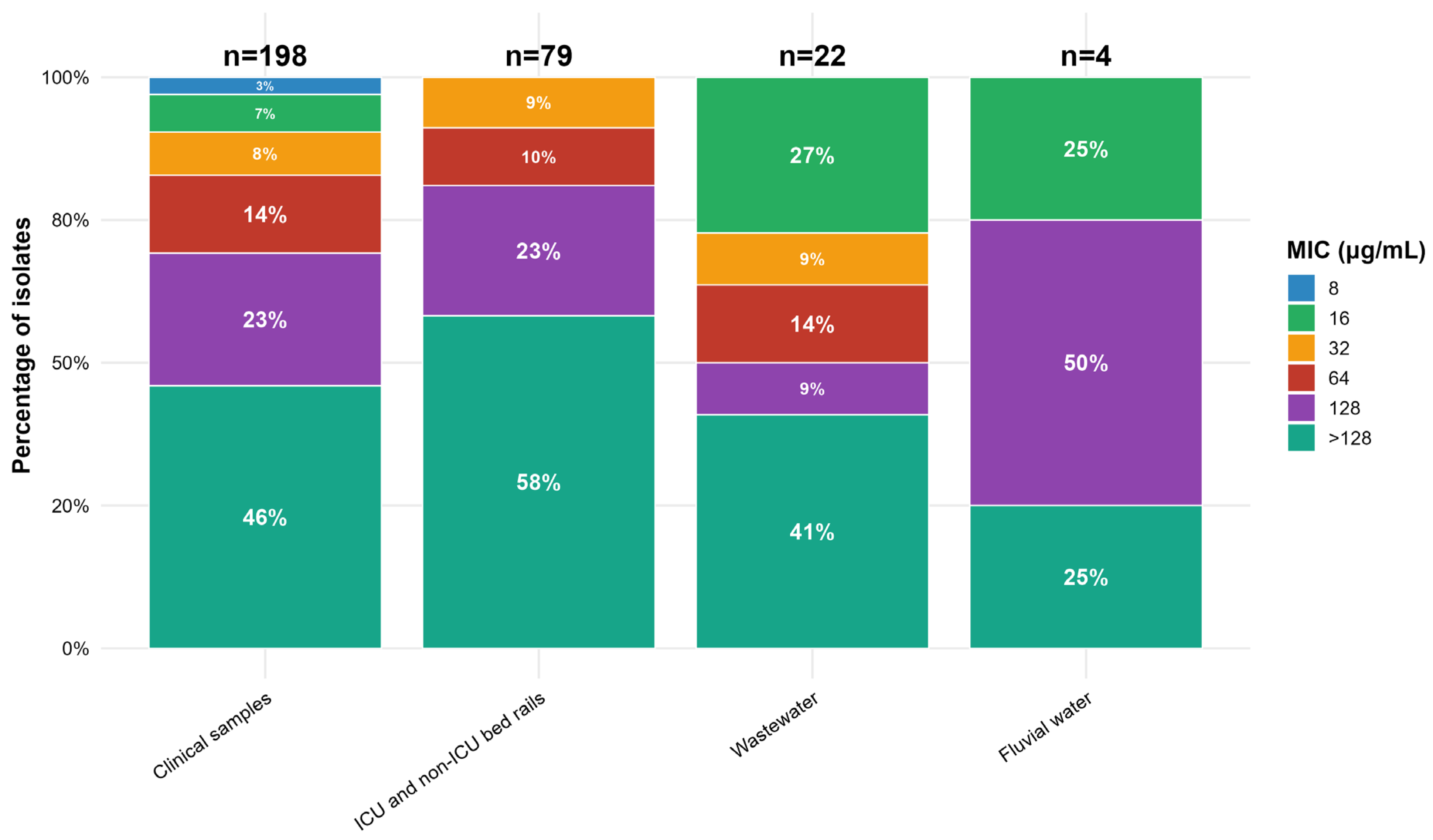
All presumptive Klebsiella colonies recovered on meropenem-supplemented MacConkey agar were confirmed as meropenem-resistant by MIC. Phenotypic and typing analyses were then performed exclusively on these isolates. Most isolates (70.95%) had MIC values for meropenem ≥ 128 μg/mL, and high meropenem MIC values were found regardless of origin (Figure 1). The MIC50 and MIC90 for meropenem in clinical sample isolates were 128 and > 128 μg/mL, respectively. In bed rail sample isolates, the MIC50 and MIC90 values were both > 128 μg/mL.
Figure 1. Stacked bar chart of meropenem minimum inhibitory concentration (MIC) categories (8, 16, 32, 64, 128, >128 µg/mL) across Klebsiella spp. isolated from clinical samples, ICU/non-ICU bed rails, wastewater, and fluvial water. Sample sizes (n) are shown above each bar, and segment labels indicate within-source percentages.
The majority of Klebsiella spp. isolates were susceptible to polymyxin B (n = 235, 77.6%). The polymyxin B resistance rates found in the different sources were 41.8% of bed rail isolates (n = 33), 25% of stream isolates (n = 1), 15.1% of clinical isolates (n = 30), and 18.2% of hospital wastewater isolates (n = 4) (Supplementary Table S1). A significantly higher proportion of polymyxin B-resistant Klebsiella spp. isolates was observed among samples from bed rails compared to other sources (p = 0.0007). Pairwise comparisons confirmed that the proportion of isolates resistant to polymyxin B was significantly higher in samples from bed rails compared to those from clinical samples (adjusted p = 0.027) and wastewater samples (adjusted p = 0.006). Among the clinical isolates, MIC50 and MIC90 values for polymyxin B were ≤1 and 8 μg/mL, respectively, whereas 2 and 16 μg/mL were found for the bed rails isolates, respectively.
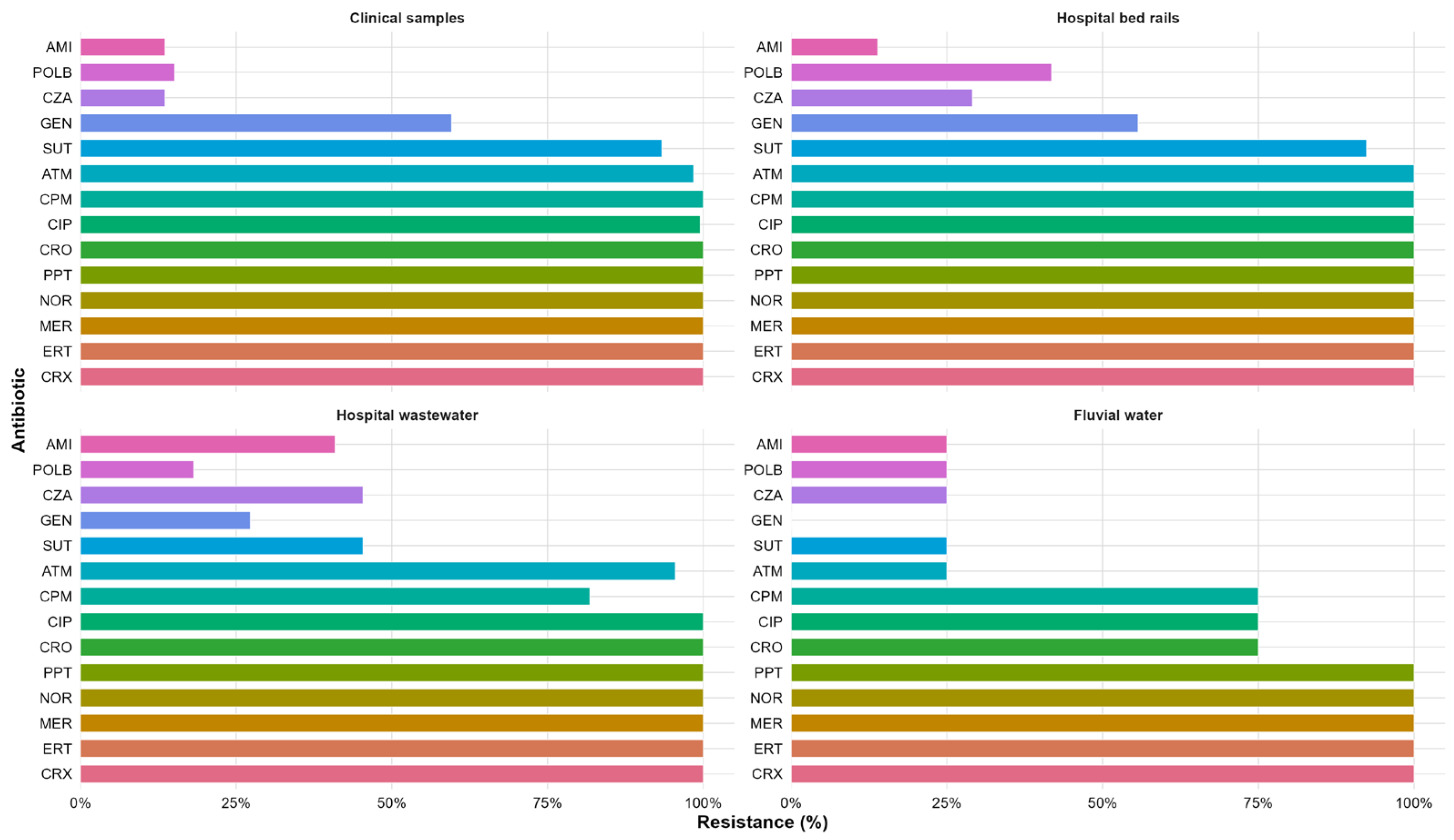
All carbapenem-resistant Klebsiella spp. isolates were also tested for susceptibility to seven other classes of antibiotics using the disk diffusion method. All clinical isolates were also resistant to cephalosporins, piperacillin-tazobactam, and norfloxacin. High resistance rates were found for ciprofloxacin (99.5%), aztreonam (98.5%), and sulfamethoxazole-trimethoprim (93.4%). Resistance rates were 59.6% for gentamicin, 25.8% for ceftazidime-avibactam, and 13.6% for amikacin (Figure 2). Resistance was significantly lower to amikacin, ceftazidime-avibactam, and gentamicin among the isolates from clinical simples (p < 0.0001).
Figure 2. Grouped bar chart showing the percentages of antimicrobial resistance among Klebsiella spp. isolates by antibiotic and source. The antibiotics tested were cefepime (CPM), cefuroxime (CRX), ceftriaxone (CRO), aztreonam (ATM), ertapenem (ERT), meropenem (MER), gentamicin (GEN), amikacin (AMI), ciprofloxacin (CIP), norfloxacin (NOR), sulfamethoxazole-trimethoprim (SUT), ceftazidime-avibactam (CZA), piperacillin-tazobactam (PPT), and polymyxin B (POLB).
The highest resistance rates were found among isolates from bed rails. All isolates were resistant to the tested β-lactams and fluoroquinolones, as well as piperacillin-tazobactam. The other resistance rates found were 92.4% for sulfamethoxazole-trimethoprim, 55.7% for gentamicin, 29.1% for ceftazidime-avibactam, and 13.9% for amikacin (Figure 2). Similar to the isolates from clinical samples, isolates from bed rails showed significantly lower resistance to gentamicin, amikacin, and ceftazidime-avibactam (p < 0.0001).
All the isolates from hospital wastewater were resistant to fluoroquinolones and piperacillin-tazobactam, and none were susceptible to β-lactams. The resistance rates to other antibiotics were as follows: 45.4% for sulfamethoxazole-trimethoprim and ceftazidime-avibactam, 40.9% for amikacin, and 27.3% for gentamicin (Figure 2). Resistance rates to gentamicin, amikacin, ceftazidime-avibactam and sulfamethoxazole-trimethoprim were significantly lower when compared to several other antibiotics in the isolates from wastewater (p < 0.05). Wastewater isolates showed significantly lower resistance rates to gentamicin and sulfamethoxazole-trimethoprim compared with clinical and bed rail isolates (p < 0.05). In contrast, they displayed significantly higher resistance rates to amikacin (p < 0.05).
Unlike the other sources, isolates from the stream water samples were susceptible to cephalosporins, and 50% were also susceptible to aztreonam. However, all isolates were resistant to carbapenems, norfloxacin, and piperacillin-tazobactam. Most isolates (75%) were resistant to ciprofloxacin, in contrast to 25% that were resistant to amikacin, sulfamethoxazole-trimethoprim, and ceftazidime-avibactam. None of the isolates were resistant to gentamicin (Figure 2).
All Klebsiella spp. isolates were classified considering their resistance profile according to the Magiorakos et al. []. Most isolates (95.4%) were classified as XDR (n = 289), 3.3% as MDR (n = 10), and 1.3% as PDR (n = 4). The PDR isolates were obtained from bed rails in the ICU (n = 3) and wastewater (n = 1). Detailed results of antimicrobial susceptibility testing are presented in Table S1 of the Supplementary Materials.

2.3. Biofilm Formation

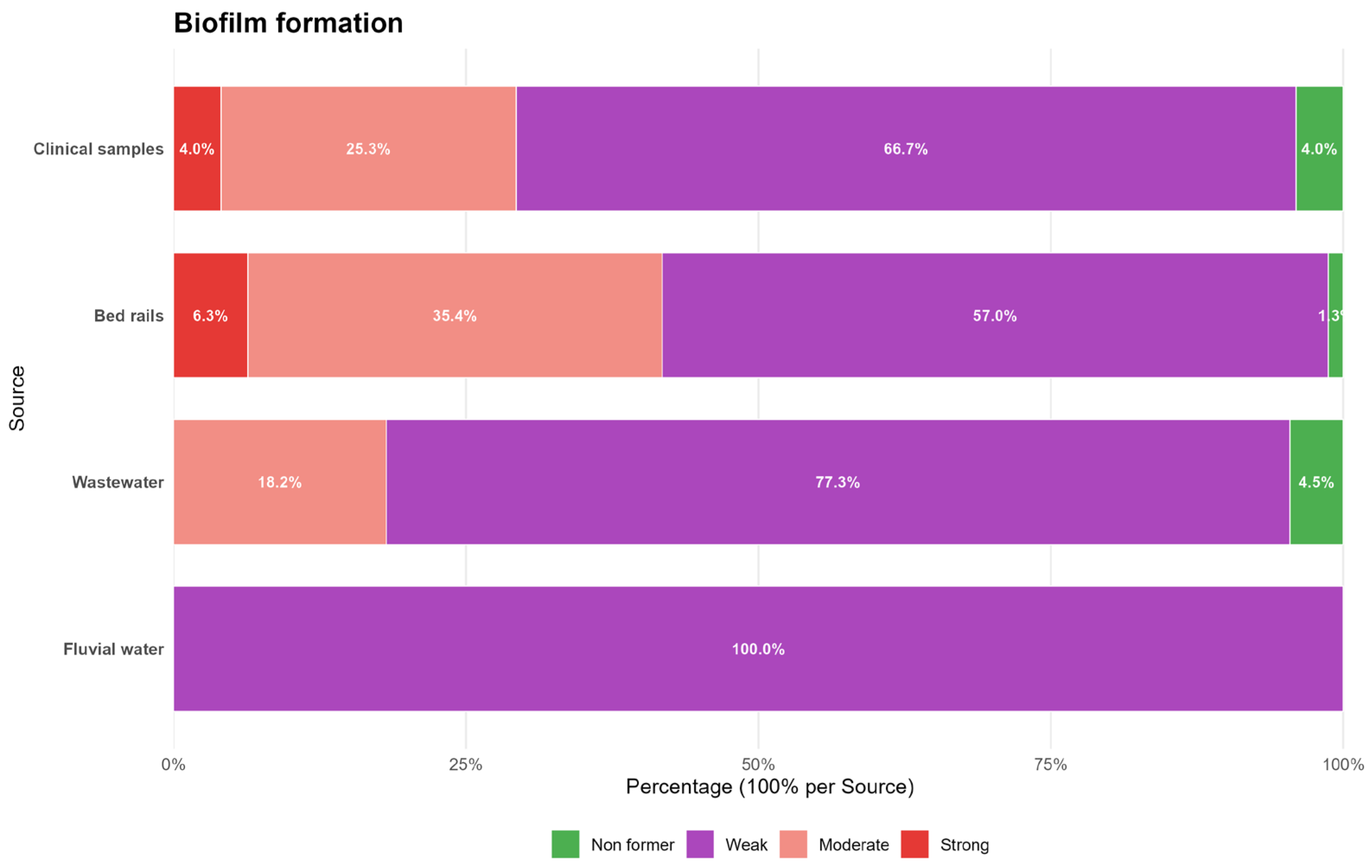
The bacterial isolates were analyzed for their ability to form biofilm. Among them, 3.3% (n = 10) were classified as non-biofilm formers, 65.3% (n = 198) as weak formers, 27.1% (n = 82) as moderate formers, and 4.3% (n = 13) as strong formers (Figure 3). No statistically significant association was observed between the biofilm formation classification and the source of Klebsiella spp. isolates (p = 0.092) or polymyxin B resistance among Klebsiella spp. isolates (p = 0.990).
Figure 3. Distribution of biofilm phenotypes (non-former, weak, moderate, strong) among Klebsiella spp. isolates from clinical samples, bed rails, hospital wastewater, and fluvial water, displayed as a 100% stacked bar chart normalized within each source.

2.4. K Typing

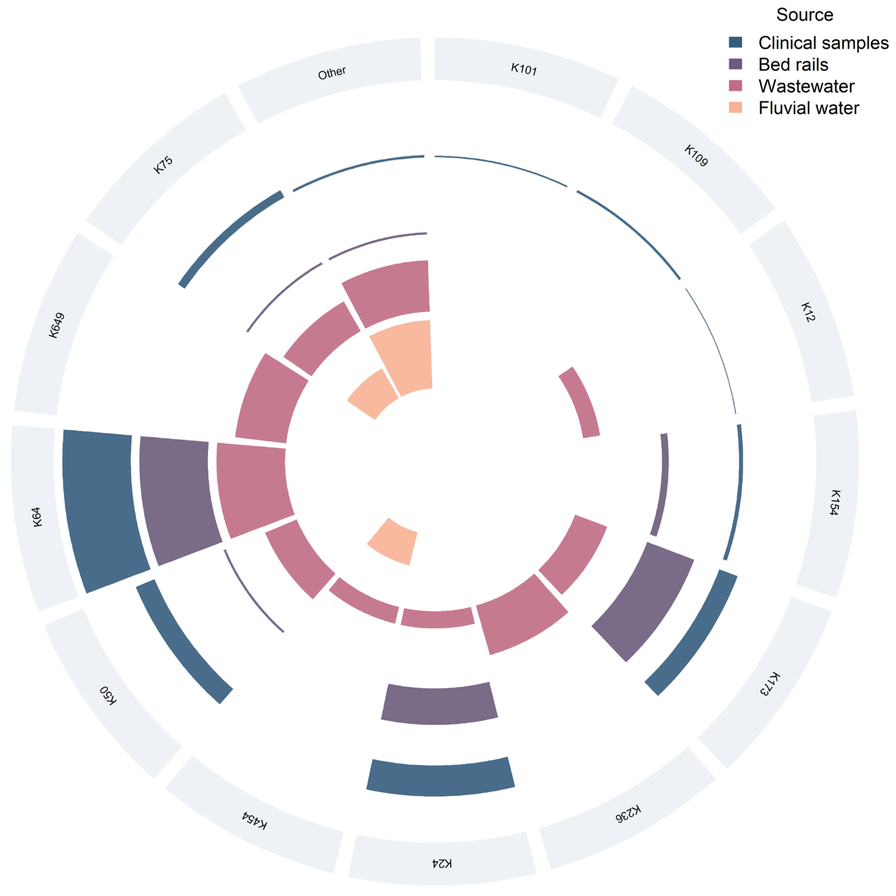
A total of 21 distinct K types was identified among the 295 Klebsiella (294 K. pneumoniae and 1 K. variicola) isolates analyzed (Figure 4). The overall distribution was highly uneven, with a marked predominance of K64 (40%), followed by K24 (18.6%), K173 (16.3%), and K50 (9.5%). Together, these four types accounted for approximately 84% of all isolates. Less common K types comprised K75 (4.75%) and K154 (2.7%); K109, K236, and K649 (1.0% each); K101, K12, K269, K3, and K167 (0.7% each); and K225, K454, K579, K66, K258, K359, and K925 (0.3% each).
Figure 4. Circular stacked chart showing capsular K-type distribution by source. Each labeled sector represents a K type; colored arcs indicate contributions from clinical samples, bed rails, hospital wastewater, and fluvial water. Arc length is proportional to the number of isolates per K type and source.
Among clinical isolates, 12 distinct K types were observed. K64 was most prevalent (n = 84; 42.9%), followed by K24 (n = 38; 19.4%), K50 (n = 25; 12.8%), K173 (n = 24; 12.2%), and K75 (n = 10; 5.1%). Less frequent types included K154 (n = 5; 2.55%), K109 (n = 3; 1.5%), and K101, K12, K27, K330, and K66 (≤1.0% each).
In bed-rail isolates (ICU and non-ICU combined), K64 was dominant (n = 30), followed by K173 (n = 22), K24 (n = 16), K154 (n = 3), K50, K75 and K 167 (n = 1 each). When analyzed separately, ICU surfaces (n = 59) revealed six distinct K types, with K64 and K173 being co-dominant (n = 21 each; 35.6%), followed by K24 (n = 14; 23.7%); K50, K154, and K167 occurred at lower frequencies (n = 1 each). In non-ICU areas (n = 15), K64 predominated (n = 9; 60%), whereas K24 and K154 (n = 2 each) and K75 and K173 (n = 1 each) were less frequent.
Among wastewater isolates, 12 K types were detected; K64, K236 and K649 were most frequent, whereas K173, K50, K75, K12, K24, K269, K301, K454 and K505 were less common. In fluvial water, four K types—K75, K225, K454, and K579—were identified.
Notably, K75 was the only K type found across all sources, including clinical isolates, bed-rail surfaces (non-ICU), wastewater, and fluvial water.
Polymyxin B resistance mirrored the overall K-type distribution—led by K64 (n = 27; 40.9%), followed by K24 (n = 16; 24.2%), K50 (n = 8; 12.1%), and K173 (n = 7; 10.6%)—with resistant phenotypes rare in other K types. No significant association between K type and polymyxin B resistance was detected (χ2(20) = 18.907, p = 0.528). All four PDR isolates belonged to different K types: two K24, one K50, and one K173.
Regarding biofilm formation, weak producers were most frequent, followed by moderate, strong, and non-formers. Within the weak biofilm group, K64 predominated (n = 74; 39.8%), followed by K24 (n = 37; 19.9%) and K173 (n = 29; 15.6%). Among moderate biofilm formers, the same K types were most represented—K64 (n = 39; 48.75%), K173 (n = 15; 18.75%), and K24 (n = 10; 12.5%). Strong producers were less frequent but showed a similar pattern (K64, K24, and K173, each 23.1%; n = 3), whereas non-formers were scarce, with K24 (n = 4; 50%) being the most common. A chi-square test of independence indicated a global association between K type and biofilm category (χ2(60) = 93.116, p = 0.003956); however, no pairwise differences remained significant after Bonferroni correction. Overall, again, the frequency pattern mirrored the overall K-type distribution in the collection, with no evidence of enrichment of any single K type in a specific biofilm category.

3. Discussion

All Klebsiella spp. isolates analyzed in this study were carbapenem-resistant and, in most cases, exhibited XDR phenotypes, with a few PDR isolates identified according to the criteria of Magiorakos et al. []. This resistance profile spanned multiple classes of both β-lactam and non-β-lactam agents, reflecting constrained therapeutic options and the potential for persistence and dissemination within healthcare environments. These findings align with worldwide surveillance data documenting the rising prevalence of carbapenem-resistant Klebsiella pneumoniae (CRKP) in both high- and middle-income countries [,,]. In Brazil, resistance trends have been equally concerning, with carbapenem resistance escalating from below 10% in 2011 to over 30% in 2015, alongside a rise in polymyxin B resistance from 0% to 27.1% [].
Phenotypically, we observed nearly universal resistance to β-lactams, including third- and fourth-generation cephalosporins and aztreonam, consistent with studies attributing such patterns to co-production of carbapenemases and ESBLs [,]. High fluoroquinolone resistance was also observed, in agreement with previous investigations that associate these phenotypes with mutations in gyrA and parC as well as plasmid-mediated qnr determinants [,]. Our study likewise showed almost complete resistance to trimethoprim–sulfamethoxazole (STX) and piperacillin–tazobactam (PPT), a finding comparable to global CRE reports. Residual susceptibility to amikacin, gentamicin, and ceftazidime–avibactam (CZA) was detected, corroborating recent evidence that these remain among the few active options against certain carbapenemase-producing strains [,,]. Polymyxin B also retained activity in a substantial proportion of isolates, supporting its continued role as last-resort agent, provided its use is judicious and closely monitored for toxicity []. Nevertheless, the high proportion of polymyxin B-resistant isolates recovered from hospital surfaces (41.8%), particularly from ICU beds, remains a matter of special concern. These findings are consistent with Brazilian and international evidence demonstrating persistence of resistant isolates on inanimate surfaces [,]. Environmental persistence is further supported by studies where disinfection reduced microbial load in only a minority of sites, emphasizing the resilience of hospital-associated Gram-negative bacteria []. Expanding on these observations, selective pressure from disinfectants such as chlorhexidine may select for decreased susceptibility and co-select plasmid-borne resistance determinants among Gram-negative bacteria [].
Collectively, our data demonstrate that environmental reservoirs of multidrug-resistant bacteria extend beyond patients and invasive devices, encompassing inanimate surfaces that silently sustain transmission. The effectiveness of disinfection depends on biocide choice, application method, and capacity to disrupt biofilms. Evidence indicates that conventional cleaning may be insufficient, highlighting the importance of integrating environmental surveillance, staff training, and targeted interventions into infection control programs []. In response to the detection of XDR isolates on bed rails, the hospital strengthened both terminal and routine cleaning protocols, upgraded disinfection barriers for high-touch surfaces, and expanded environmental microbiological surveillance for continuous monitoring. In parallel, contact precaution workflows, staff training, and antimicrobial management were revised—with an emphasis on stewardship—to reduce selective pressure and prevent the persistence of resistant clones in the care environment.
Our analysis also underscores the critical role of biofilm formation in bacterial persistence and resistance dissemination. Biofilms facilitate survival on inanimate surfaces despite cleaning and provide niches for horizontal gene transfer (HGT) [,]. Notably, K. pneumoniae biofilms can survive on dry surfaces for up to four weeks, further underscoring their role in persistence and transmission within healthcare environments []. In our series, 96.7% of carbapenem-resistant Klebsiella spp. isolates produced biofilms. Compared with previous reports, our isolates showed a higher proportion of weak producers, though even low-density biofilms can act as reservoirs for HGT [,]. Importantly, the biofilm matrix enhances proximity between cells and promotes plasmid transfer [,]. Within biofilm communities, additional mechanisms such as outer membrane vesicles and persister cells further enhance genetic exchange and long-term survival of K. pneumoniae under antimicrobial stress [,]. Mixed-species biofilms amplify these effects, enabling plasmid-mediated transfer of carbapenemase and colistin resistance genes across Enterobacterales []. Therefore, the high prevalence of biofilm producers in our study indicates a strong potential for persistence and genetic dissemination among multidrug-resistant Klebsiella spp., encompassing but not limited to carbapenem- and polymyxin-resistant strains. Additionally, other characteristics, such as capsular composition and production level, may interact synergistically to enhance environmental survival and spread [].
Capsular typing provided valuable insight into the distribution and characteristics of carbapenem-resistant K. pneumoniae isolates recovered from clinical and environmental sources. In our dataset, four capsular types (K64, K24, K173, and K50) together accounted for 84.4% of all isolates, being detected in clinical samples, bed rails, and wastewater. Notably, K75 was the only K type identified in all sources, although without apparent association with hypervirulence or epidemiological predominance. K64 was the most frequent capsular type, consistent with its recurrent identification in hospital-associated carbapenem-resistant lineages, often linked to ST11 [,]. K24 was also detected in both clinical and environmental isolates, supporting its persistence across different settings [,]. By contrast, K173 and K50 were found less frequently and have been only sparsely reported in surveillance studies, suggesting either limited dissemination or regional occurrence. The recurrence of K64 and K24 in hospital-related isolates, together with the detection of K75 across all sampled sources, indicates continuous interconnection between clinical and environmental reservoirs. Therefore, the data presented herein is consistent with the persistence of specific capsular lineages under variable selective pressures, highlighting the importance of antimicrobial resistance stewardship within a One Health framework.
Our findings also revealed marked differences between isolates from surfaces, patients, and hospital sewage. Bacteria from the intrahospital environment showed higher resistance to aminoglycosides and STX than those from sewage, reflecting the stronger selective pressure of clinical settings where broad-spectrum antimicrobials are heavily used [,]. Sewage-derived isolates displayed lower resistance, consistent with wastewater studies reporting low gentamicin resistance in E. coli [] and higher aminoglycoside susceptibility in Enterobacterales []. These observations are consistent with the influence of microbial community interactions and selective conditions shaping AMR persistence beyond antibiotic exposure alone []. Consequently, ARGs may accumulate and persist in sewage environments even when phenotypic resistance among isolated bacteria remains low. Nonetheless, selective forces in wastewater cannot be dismissed: antibiotic residues, heavy metals, and disinfectants shape microbial communities and may favor resistance maintenance, especially within biofilms []. Accordingly, hospital sewage consistently carries higher loads of resistant bacteria and ARGs than other sources [,].
Extending beyond hospitals, we also examined carbapenem-resistant Klebsiella spp. from stream waters, which comprised fewer isolates and exhibited a heterogeneous resistance profile: uniform carbapenem resistance, consistent gentamicin susceptibility, and variable cephalosporin resistance patterns. The stream evaluated, although not directly connected to hospital effluents, is impacted by urban discharges []. Fluvial environments thus act as dynamic reservoirs integrating resistomes from multiple anthropogenic sources [,]. Urban streams can amplify ARG dissemination into the broader environment and nearby populations [,]. The detection of carbapenem-resistant isolates with preserved polymyxin susceptibility is consistent with Brazilian environmental data where polymyxin resistance was rare []. Sites with higher organic contamination, including fecal-origin material, exhibited clinically relevant carbapenemases, suggesting that heterogeneous resistance profiles may derive partly from HGT involving environmental bacteria [].
Methodologically, the crystal violet assay measures total biomass of biofilms without assessing cell viability or architecture. Variations in washing, fixation, cut-offs, and microtiter conditions may alter classifications and explain inter-laboratory variability [,]. In addition, since the assay was performed on polystyrene microplates under standardized laboratory conditions, it reflects the potential for biofilm formation rather than surface-specific behavior that may occur on clinical materials such as stainless steel or medical-grade plastics. Even considering these potential constraints, biofilms nonetheless emerged as a key mechanism of carbapenem-resistant Klebsiella persistence and dissemination, with 96.7% of isolates identified as biofilm producers, predominantly weak to moderate. While Rahdar et al. reported an association between stronger biofilm formation and resistance [], our results align with Cusumano et al. [] and Zheng et al. [], showing that biofilm intensity does not universally predict resistance. Although no association was observed with polymyxin B, the high prevalence of ability to form biofilm among XDR strains suggests an important role in antimicrobial resistance persistence. This observation is consistent with reports emphasizing that biofilm–resistance relationships are antibiotic-specific and clonal-context dependent rather than universal traits [].

4. Materials and Methods

4.1. Clinical Setting

The study was conducted over six months in a 335-bed tertiary care university teaching hospital located in the southern Brazil, with a total area of approximately 61,000 m2 and a 59-bed ICUs. In front of the hospital, there is a stream with a length of 17,830 m (550 m from the source of the stream) [], and there are no drainage or pipeline connections linking the hospital to this stream.

4.2. Sampling in the Hospital Environment

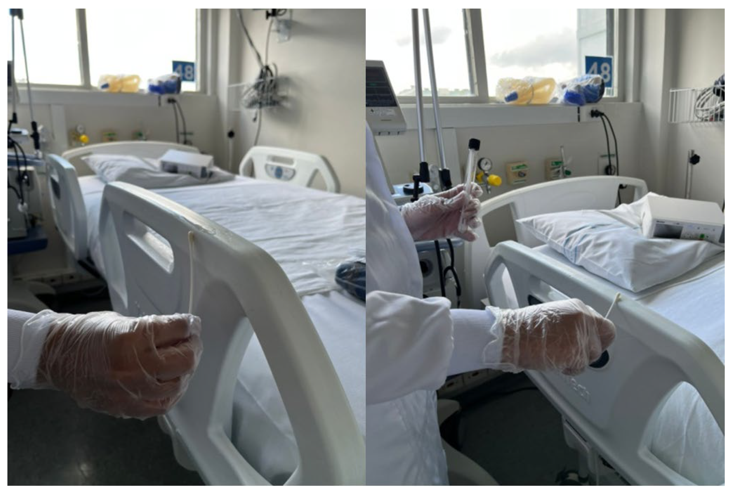
Samples were collected weekly from the bed rails of general adult ICU and non-ICU units. The entire lateral surface of the bed was sampled with a swab moistened with 0.85% saline (Labsynth, Diadema, Brazil) at least 3 h after the disinfection of the site (Figure 5). The swabs were placed in 0.85% saline and transported immediately to the laboratory. Following this, the swabs were inoculated in Brain Heart Infusion (BHI) broth (Kasvi, Madrid, Spain) with meropenem (1 μg/mL) (cat. Y0001252, European Directorate for the Quality of Medicines & Healthcare, Strasbourg, France) and subsequently incubated at 36 ± 1 °C for 12–18 h. BHI cultures were streaked on MacConkey agar (Kasvi, Madrid, Spain) with meropenem (1 μg/mL) and were incubated at 36 ± 1 °C for 18–24 h. Isolates showing Triple Sugar Iron (TSI) (Kasvi, Madrid, Spain) profiles compatible with Klebsiella spp. were stored at −80 °C in skim milk with 10% glycerol for further identification by matrix-assisted laser desorption/ionization time-of-flight (MALDI-TOF).
Figure 5. Representative images of the sample collection performed on the entire surface of the bed rails.

4.3. Water Sample Collections

During the same period, water samples were collected every two weeks, from the hospital domestic sewage (untreated influent) and from the stream located in front of the hospital. The hospital sewage samples were taken from a dedicated hospital sewer line manhole, prior to its junction with the municipal sewer. At each sampling point, a 50 mL water sample was collected aseptically into sterile glass vial. The collected samples were transported to the laboratory and then filtered using a vacuum pump with a 0.22 μm membrane (Sartorius, Göttingen, Germany). The membrane was placed in a sterile tube containing 10 mL of 0.85% saline for vortexing and release of microbial cells. Serial dilutions were made up to 10−3, considering the tube with the 0.22 μm membrane as the first dilution. Then, 100 μL of each dilution was spread on the surface of MacConkey agar with meropenem (1 μg/mL) using a Drigaslski loop. Colonies with a biochemical profile compatible with Klebsiella spp. were included in this study and stored at −80 °C in skim milk with 10% glycerol for subsequent identification by MALDI-TOF.

4.4. Clinical Isolates

The hospital laboratory provided the carbapenem-resistant Klebsiella spp. isolated from hospitalized patients and those from surveillance samples collected on patient admission during the same six months. Only one isolate per patient was included. The isolates were obtained from the following clinical specimens: urine (47%), tracheal aspirate (21.7%), blood culture (15.7%), abdominal fluid (3.5%), sputum (3.1%), drainage fluid (2.5%), surgical wound (2%), catheter (1%), and others (3.5%). The laboratory identified the isolates using the BD Phoenix M50 system (Becton Dickinson, Franklin Lakes, NJ, USA) and determined the antibiotic susceptibility by disk diffusion according to the guidelines of the Brazilian Committee on Antimicrobial Susceptibility (BrCAST) (https://brcast.org.br/wp-content/uploads/2022/09/Tabela-pontos-de-corte-clinico-BrCAST-01-02-2025.pdf, accessed on 1 February 2025) and the European Committee on Antimicrobial Susceptibility Testing (EUCAST) version 15.0 (valid from 1 January 2025). The isolates were stored at −80 °C in skim milk with 10% glycerol for further confirmation by MALDI-TOF.

4.5. Identification of Bacterial Isolates

Bacterial isolates that showed biochemical profiles compatible with Klebsiella spp. from all sampled sites were identified by MALDI-TOF mass spectrometry (Microflex LT, Bruker Daltonik®, Bremen, Germany). Bacterial isolates were cultured on BHI agar (Kasvi, Spain) and incubated at 37 °C for 18 to 24 h. Subsequently, 1 mL of the bacterial suspension was centrifuged at 5000 rpm for 5 min. The resulting pellet was treated with 70% ethanol and centrifuged again at 13,000 rpm for 2 min. The pellet was then transferred onto a stainless-steel MALDI target plate and overlaid with a matrix solution consisting of α-cyano-4-hydroxycinnamic acid (10 mg/mL in 50% acetonitrile and 2.5% trifluoroacetic acid). Each bacterial isolate was spotted in triplicate, and three independent readings were performed per sample. Protein spectra were acquired using FlexControl 3.3 software (Bruker Daltonik) with the MTB_autoX acquisition method, covering a mass range of 2 to 20 kDa. Spectral analysis was conducted using BioTyper 3.0 software (Bruker Daltonik®) by comparing the obtained profiles to those stored in the system’s reference database. Interpretation of identification scores followed the manufacturer’s criteria: scores ≥2.0 were considered reliable for species-level identification, while scores between 1.7 and <2.0 were accepted for genus-level assignments only. Each bacterial isolate was spotted in triplicate, and three independent readings were performed per sample.

4.6. Antimicrobial Susceptibility Testing

The minimum inhibitory concentration (MIC) of meropenem was determined by cation-adjusted broth microdilution according to the guidelines of the BrCAST and EUCAST, using a concentration range of 0.5 to 128 μg/mL. MIC assays were performed in triplicate for each Klebsiella spp. isolate. Escherichia coli ATCC 25922 was used as a quality control strain. In addition, a positive control with Mueller Hinton broth (Kasvi, Madrid, Spain) and 1 μL of bacterial dilution was made for each isolate without the addition of the antimicrobial. Also, a negative control was included for each plate using the Mueller Hinton broth culture medium only. The MIC50 and MIC90 values were calculated for the isolates from clinical samples and bed rails.
The isolates not susceptible to meropenem were characterized regarding susceptibility to other antibiotics. The MIC values for polymyxin B (cat. Y0000355, European Directorate for the Quality of Medicines & Healthcare, Strasbourg, France) were determined in the range of 0.5 to 32 μg/mL using the same conditions as above. The disk diffusion method according to EUCAST guidelines was used to evaluate the susceptibility to the following antibiotics: cefepime (CPM), cefuroxime (CRX), ceftriaxone (CRO), aztreonam (ATM), ertapenem (ERT), meropenem (MER), gentamicin (GEN), amikacin (AMI), ciprofloxacin (CIP), norfloxacin (NOR), sulfamethoxazole-trimethoprim (SUT), piperacillin-tazobactam (PPT), and ceftazidime-avibactam (CZA). Escherichia coli ATCC 25922 was used as a reference strain for quality control during the analyses, and the results were interpreted according to the criteria of the EUCAST guidelines. Isolates were classified as multidrug-resistant (MDR), extensively resistant (XDR) or pan-resistant (PDR) according to the classification described by Margiorakos et al. [].

4.7. Determination of the Ability to Form Biofilm

The ability to form biofilm in polystyrene microplates was determined by the violet crystal method []. Each Klebsiella spp. isolate was grown in Lysogeny Broth (LB) (Kasvi, Madrid, Spain) overnight and then diluted to approximately 106 colony forming units. Subsequently, 1 µL of this dilution was inoculated into each well containing 200 µL of LB and incubated at 36 ± 1 °C for 48 h. To eliminate planktonic cells, the wells were then washed twice with 200 μL of a phosphate-buffered saline (PBS) solution [8 g/L NaCl, 1.44 g/L Na2HPO4, 0.24 g/L KH2PO4 and 0.2 g/L KCl]. The microplates were dried at 60 °C for 15 min and then stained with 0.1% violet crystal for 5 min. After staining, the dye was removed, the wells were washed twice with 200 µL of PBS, and the microplate was incubated at 60 °C for 1 h. Subsequently, crystal violet-stained biofilms were solubilized with absolute ethanol for 15 min at room temperature, and the biomass was quantified at an optical density (OD) of 570 using a SpectraMax 190 microplate reader (Molecular Devices, San Jose, CA, USA) [].
Klebsiella spp. isolates were classified as non-biofilm formers (OD ≤ ODc), weak biofilm formers (ODc < OD ≤ 2ODc), moderate biofilm formers (2ODc < OD ≤ 4ODc), and strong biofilm formers (4ODc < OD) according to Stepanovic et al. []. The cut-off OD (ODc) is the mean OD plus three times the standard deviation of the negative control. For this procedure, the Salmonella Typhimurium ATCC 14028 strain, classified as a weak biofilm-forming strain, was used as the positive control and LB broth as the negative control.

4.8. Capsular Typing

Klebsiella pneumoniae and K. variicola isolates were subjected to capsular typing through wzi gene sequencing, following the protocol described by Brisse et al. (2013) []. The wzi gene encodes an outer membrane protein involved in capsule anchoring, whose sequence variability allows discrimination among different capsular types (K types).
Genomic DNA was purified using the PureLink Genomic DNA Mini Kit (Thermo Fisher Scientific, Waltham, MA, USA), according to the manufacturer’s instructions. DNA quality and concentration were assessed with a NanoDrop 2000C spectrophotometer (NanoDrop Technologies, Wilmington, DE, USA). All DNA samples were normalized to a final concentration of 10 ng/μL and stored at −20 °C until use. A fragment of approximately 580 bp of the wzi gene was amplified by PCR using the primers wzi_for2 (5′-GTG CCG CGA GCG CTT TCT ATC TTG GTA TTC C-3′) and wzi_rev (5′-GAG AGC CAC TGG TTC CAG AA[C/T] TT[C/G] ACC GC-3′). Each 25 μL reaction contained 0.2 μM of each primer, 1× GoTaq G2 Master Mix (Promega, Madison, WI, USA), 20 ng of template DNA, and nuclease-free water (Ambion, Austin, TX, USA) to complete the volume. PCR cycling conditions consisted of an initial denaturation at 94 °C for 2 min, followed by 30 cycles of 94 °C for 30 s, 55 °C for 40 s, and 72 °C for 30 s, with a final extension at 72 °C for 5 min.
PCR products were separated by electrophoresis in a 1.5% agarose gel for 1 h at 60 V in 1× Tris-acetate-EDTA (TAE) buffer and stained with GelRed (Biotium, Fremont, CA, USA). A 100 bp DNA ladder (Invitrogen, Carlsbad, CA, USA) was used as a molecular mass marker. DNA bands were visualized using an L-Pix Touch Transilluminator (Loccus Biotecnologia, Cotia, Brazil).
Amplicons were sequenced in both forward and reverse directions, in duplicate, using the BigDye Terminator kit and capillary electrophoresis on a SeqStudio sequencer (Thermo Fisher Scientific) at INCQS/FIOCRUZ (Rio de Janeiro, Brazil). Chromatogram quality was evaluated with SeqScape 4.0 software (Thermo Fisher Scientific). The resulting sequences were submitted to the Klebsiella Pasteur MLST database (http://bigsdb.pasteur.fr/klebsiella/, accessed on 10 October 2025) for allele assignment and K-type determination.

4.9. Statistical Analysis

To evaluate differences in polymyxin B resistance among isolates from different sources, a Chi-square test of independence was performed. Pairwise comparisons of resistance proportions were subsequently conducted using two-proportion Z-tests with Bonferroni correction for multiple testing. Pairwise comparisons of resistance rates among sources (clinical, bed rails and wastewater) were performed for each antibiotic using Fisher’s Exact Test, with Bonferroni correction applied for multiple testing. Fisher’s Exact Test was also used to perform pairwise comparisons of resistance rates among antibiotics and identify significant differences in antimicrobial susceptibility rates within each source. Isolates from the stream water source were excluded from the statistical analysis due to the low number of samples (n = 4), which could compromise the reliability of the comparisons. The associations between biofilm-forming categories and isolate sources and polymyxin B resistance were assessed using a Chi-square test of independence. All statistical analyses were conducted using Python (version 3.x), with the SciPy and statsmodels libraries, assisted by ChatGPT 4.0. A significance level of p < 0.05 was adopted for all tests. To visualize proportional distributions, stacked bar charts were generated using the ggplot2 package in R (v4.5.0). These charts displayed the distributions of resistance profiles, biofilm formation, and meropenem MIC categories, normalized to 100% for each source, with percentage values annotated. Furthermore, the relationship between K-type and source was illustrated using a Circos plot created with the circlize package in R (v4.5.0).

5. Conclusions

This study provides an integrated overview of carbapenem-resistant K. pneumoniae from clinical and hospital-associated sources, with the inclusion of a few environmental isolates under a One Health perspective. Most isolates exhibited extensively drug-resistant (XDR) profiles, with residual susceptibility restricted to a few therapeutic options such as amikacin, gentamicin, and ceftazidime–avibactam. The near-universal ability to form biofilms observed among isolates supports their persistence on inanimate surfaces and their potential to sustain horizontal gene transfer. Capsular typing revealed a predominance of specific K types—mainly K64, K24, K173, and K50—detected across different origins, suggesting the persistence of adaptable lineages under variable selective pressures. Together, these results provide complementary insights into the phenotypic and structural features associated with the persistence of carbapenem-resistant K. pneumoniae in healthcare-related environments, supporting continued monitoring of high-risk lineages and their resistance determinants.

Supplementary Materials

The following supporting information can be downloaded at https://www.mdpi.com/article/10.3390/antibiotics14111140/s1: Table S1: Complete dataset of Klebsiella spp. isolates, including identification, antimicrobial resistance profiles, biofilm classification, and capsular typing.

Author Contributions

Conceptualization: S.D.d.O. Methodology: K.V.M.M., J.M.P., T.N.d.C., G.L.B., K.N.d.A.P., A.B.d.S., G.C., Â.C.d.S., A.S.M. and C.F. Formal analysis: S.D.d.O. Investigation: K.V.M.M., A.B.d.S., D.R.F., A.B.M., K.B., M.M.C., C.A.S.F., R.M.-S. and S.D.d.O. Resources: G.C. Data curation: K.V.M.M., K.N.d.A.P. and A.B.d.S. Writing—original draft: K.V.M.M. Writing—review & editing: D.R.F., K.B., M.M.C., C.A.S.F., R.M.-S. and S.D.d.O. Supervision: S.D.d.O. Project administration: R.M.-S. and S.D.d.O. Funding acquisition: S.D.d.O. All authors have read and agreed to the published version of the manuscript.

Funding

This work was supported by the State of Rio Grande do Sul through Fundação de Amparo à Pesquisa do Rio Grande do Sul (FAPERGS) (grant number 23/2551-0000506-9) and by the Coordenação de Aperfeiçoamento de Pessoal de Nível Superior—Brasil (CAPES)—[Finance Code 001 in the form of a scholarship granted to T.N.d.C. and A.B.d.S.]. S.D.d.O. (grant number 311162/2023-1) and R.M.-S. (grant number 309387/2022-1) are Research Career Awarded of the Conselho Nacional de Desenvolvimento Científico e Tecnológico (CNPq).

Institutional Review Board Statement

The study was approved by the Research Ethics Committee of PUCRS (protocol CAAE 68800923.8.1001.5336). All methods were carried out in accordance with relevant institutional guidelines and regulations.

Data Availability Statement

All datasets are provided in the Supplementary Information.

Conflicts of Interest

The authors declare no competing interests.

Abbreviations

The following abbreviations are used in this manuscript:
AMRAntimicrobial Resistance
AMROAmericas Regional Office
ARGAntibiotic resistance gene
ATCCAmerican Type Culture Collection
BigDyeBigDye Terminator Sequencing Chemistry
CMY-2Cephamycinase type 2
CRECarbapenem-resistant enterobacteria
CRKPCarbapenem-resistant Klebsiella pneumoniae
CZACeftazidime–avibactam
DHA-1Dhahran Hospital AmpC type 1
ESBLExtended-spectrum β-lactamase
EUCASTEuropean Committee on Antimicrobial Susceptibility Testing
FiocruzFundação Oswaldo Cruz
gyrADNA gyrase subunit A gene
HAIHealthcare-Associated Infection
HGTHorizontal Gene Transfer
ICUIntensive Care Unit
INCQSInstituto Nacional de Controle de Qualidade em Saúde
KPCKlebsiella pneumoniae carbapenemase
K-typeCapsular type
L-Ara4N4-Amino-4-Deoxy-L-Arabinose
LPSLipopolysaccharide
MALDI-TOFMatrix-Assisted Laser Desorption/Ionization—Time of Flight
mcr-1Mobilized colistin resistance gene-1
MDRMulti-drug-resistant
MGEMobile Genetic Element
MICMinimum inhibitory concentration
MTB_autoXAn automated acquisition method used in MALDI-TOF for microbial identification, ensuring standardized spectral data collection
NDMNew Delhi metallo-β-lactamase
ODOptical Density
OXA-48Oxacillinase-48
PAHOPan American Health Organization
parCDNA topoisomerase IV subunit C gene
PBSPhosphate-Buffered Saline
PDRPan-drug-resistant
PhoPQTwo-Component Regulatory System PhoPQ
PmrABTwo-Component Regulatory System PmrAB
POLBPolymyxin B
PPTPiperacillin–tazobactam
qnrQuinolone resistance gene
ReLAVRALatin American Network for Antimicrobial Resistance Surveillance
SciPyScientific Python
STXSulfamethoxazole-trimethoprim
TAETris-Acetate-EDTA Buffer
TSITriple Sugar Iron
XDRExtensively drug-resistant

References

  1. Murray, C.J.L.; Ikuta, K.S.; Sharara, F.; Swetschinski, L.; Robles Aguilar, G.; Gray, A.; Han, C.; Bisignano, C.; Rao, P.; Wool, E.; et al. Global burden of bacterial antimicrobial resistance in 2019: A systematic analysis. Lancet 2022, 399, 629–655. [Google Scholar] [CrossRef] [PubMed]
  2. Robles Aguilar, G.; Swetschinski, L.R.; Weaver, N.D.; Ikuta, K.S.; Mestrovic, T.; Gray, A.P.; Chung, E.; Wool, E.E.; Han, C.; Hayoon, A.G.; et al. The burden of antimicrobial resistance in the Americas in 2019: A cross-country systematic analysis. Lancet Reg. Health Am. 2023, 25, 100561. [Google Scholar] [CrossRef]
  3. Pan American Health Organization. Magnitude and Trends of Antimicrobial Resistance in Latin America: ReLAVRA 2014–2016; PAHO: Washington, DC, USA, 2018; Available online: https://www.paho.org/en/documents/magnitude-and-trends-antimicrobial-resistance-latin-america-relavra-2014-2015-2016 (accessed on 7 October 2025).
  4. Fabre, V.; Cosgrove, S.E.; Secaira, C.; Tapia Torrez, J.C.; Lessa, F.C.; Patel, T.S.; Quiros, R.; Caceres, D.H.; Perz, J.F.; Cardo, D.; et al. Antimicrobial stewardship in Latin America: Past, present, and future. Antimicrob. Steward. Healthc. Epidemiol. 2022, 2, e68. [Google Scholar] [CrossRef]
  5. Pillonetto, M.; Wink, P.L.; Melano, R.G.; Jiménez-Pearson, M.A.; Melgarejo Touchet, N.L.; Saavedra Rojas, S.Y.; Kulek, D.N.O.; Abreu, A.L.; Peral, R.T.; Miorando, R.; et al. Carbapenemases producing gram-negative bacteria surveillance in Latin America and the Caribbean: A retrospective observational study from 2015 to 2020. Lancet Reg. Health Am. 2025, 49, 101185. [Google Scholar] [CrossRef]
  6. World Health Organization. WHO Publishes List of Bacteria for Which New Antibiotics are Urgently Needed. Available online: http://www.who.int/news-room/detail/27-02-2017-who-publishes-list-of-bacteria-for-which-new-antibiotics-are-urgently-needed (accessed on 1 March 2025).
  7. Assefa, M.; Amare, A. Biofilm-Associated Multi-Drug Resistance in Hospital-Acquired Infections. Infect. Drug Resist. 2022, 15, 5061–5068. [Google Scholar] [CrossRef]
  8. Maillard, J.Y.; Centeleghe, I. How biofilm changes our understanding of cleaning and disinfection. Antimicrob. Resist. Infect. Control 2023, 7, 12. [Google Scholar] [CrossRef]
  9. Vickery, K. Special Issue: Microbial Biofilms in Healthcare: Formation, prevention and treatment. Materials 2019, 12, 2001. [Google Scholar] [CrossRef]
  10. Nirwati, H.; Sinanjung, K.; Fahrunissa, F.; Wijaya, F.; Napitupulu, S.; Hati, V. Biofilm formation and antibiotic resistance of Klebsiella pneumoniae isolated from clinical samples in a tertiary care hospital, Klaten, Indonesia. BMC Proc. 2019, 13, 11–20. [Google Scholar] [CrossRef]
  11. Nkemngong, C.; Teska, P. Biofilms, mobile genetic elements and the persistence of pathogens on environmental surfaces in healthcare and food processing environments. Front. Microbiol. 2024, 15, 1405428. [Google Scholar] [CrossRef]
  12. Jamal, M.; Ahmad, W.; Andleeb, S.; Jalil, F.; Imran, M.; Nawaz, M.A.; Hussain, T.; Ali, M.; Rafiq, M.; Kamil, M.A.; et al. Bacterial biofilm and associated infections. J. Chin. Med. Assoc. 2018, 81, 7–11. [Google Scholar] [CrossRef]
  13. Guerra, M.E.S.; Destro, G.; Vieira, B.; Lima, A.S.; Ferraz, L.F.C.; Hakansson, A.P.; Darrieux, M.; Converso, T.R. Klebsiella pneumoniae biofilms and their role in disease pathogenesis. Front. Cell. Infect. Microbiol. 2022, 12, 877995. [Google Scholar] [CrossRef]
  14. Gashaw, M.; Gudina, E.K.; Tadesse, W.; Froeschl, G.; Ali, S.; Seeholzer, T.; Kroidl, A.; Wieser, A. Hospital wastes as potential sources for multidrug-resistant ESBL-producing bacteria at a tertiary hospital in Ethiopia. Antibiotics 2024, 13, 374. [Google Scholar] [CrossRef]
  15. Irfan, M.; Almotiri, A.; AlZeyadi, Z.A. Antimicrobial resistance and β-lactamase production in clinically significant Gram-negative bacteria isolated from hospital and municipal wastewater. Antibiotics 2023, 12, 653. [Google Scholar] [CrossRef] [PubMed]
  16. Nakamura-Silva, R.; Dias, L.L.; Sousa, R.C.; Fujimoto, R.Y.; Pitondo-Silva, A. Multidrug-resistant and potentially pathogenic Enterobacteriaceae found in a tertiary hospital sewage in southeastern Brazil. Environ. Monit. Assess. 2022, 194, 782. [Google Scholar] [CrossRef] [PubMed]
  17. Davidova-Gerzova, L.; Lausova, J.; Sukkar, I.; Nesporova, K.; Nechutna, L.; Vlkova, K.; Chudejova, K.; Krutova, M.; Palkovicova, J.; Kaspar, J.; et al. Hospital and community wastewater as a source of multidrug-resistant ESBL-producing Escherichia coli. Front. Cell. Infect. Microbiol. 2023, 13, 1184081. [Google Scholar] [CrossRef]
  18. Karkman, A.; Do, T.T.; Walsh, F.; Virta, M.P.J. Antibiotic-resistance genes in waste water. Trends Microbiol. 2018, 26, 220–228. [Google Scholar] [CrossRef]
  19. Kalaiselvi, K.; Mangayarkarasi, V.; Balakrishnan, D.; Chitraleka, V. Survival of antibacterial resistance microbes in hospital-generated recycled wastewater. J. Water Health 2016, 14, 942–949. [Google Scholar] [CrossRef]
  20. Ashurst, J.V.; Dawson, A. Klebsiella pneumoniae. In StatPearls; StatPearls Publishing: Treasure Island, FL, USA, 2023. Available online: http://www.ncbi.nlm.nih.gov/books/NBK519004/ (accessed on 17 June 2025).
  21. Bush, K. β-lactam antibiotics. In Antibiotic and Chemotherapy, 9th ed.; Finch, R.G., Greenwood, D., Norrby, S.R., Whitley, R.J., Eds.; Elsevier: London, UK, 2010; pp. 200–225. [Google Scholar] [CrossRef]
  22. Opal, S.M.; Pop-Vicas, A. Molecular mechanisms of antibiotic resistance in bacteria. In Mandell, Douglas, and Bennett’s Principles and Practice of Infectious Diseases, 8th ed.; Bennett, J.E., Dolin, R., Blaser, M.J., Eds.; Elsevier: Philadelphia, PA, USA, 2015; pp. 235–251.e3. Available online: https://linkinghub.elsevier.com/retrieve/pii/B9781455748013000187 (accessed on 23 July 2025).
  23. Rawat, D.; Nair, D. Extended-spectrum β-lactamases in gram negative bacteria. J. Glob. Infect. Dis. 2010, 2, 263–274. [Google Scholar] [CrossRef]
  24. Lavagnoli, L.S.; Bassetti, B.R.; Kaiser, T.D.L.; Kutz, K.M.; Cerutti Junior, C. Factors associated with acquisition of carbapenem-resistant Enterobacteriaceae. Rev. Lat. Am. Enferm. 2017, 25, e2935. [Google Scholar] [CrossRef]
  25. Nordmann, P.; Poirel, L. Epidemiology and diagnostics of carbapenem resistance in gram-negative bacteria. Clin. Infect. Dis. 2019, 69, S521–S528. [Google Scholar] [CrossRef]
  26. Wong, J.L.C.; Romano, M.; Kerry, L.E.; Kwong, H.-S.; Low, W.-W.; Brett, S.J.; Clements, A.; Beis, K.; Frankel, G. OmpK36-mediated carbapenem resistance attenuates ST258 Klebsiella pneumoniae in vivo. Nat. Commun. 2019, 10, 3957. [Google Scholar] [CrossRef]
  27. Jacoby, G.A. AmpC β-lactamases. Clin. Microbiol. Rev. 2009, 22, 161–182. [Google Scholar] [CrossRef] [PubMed]
  28. Teng, J.; Walters, L.; Leong, L.E.X.; Smith, K.; Amato, M.; Chen, X.; Turra, M.; Warner, M.S.; Papanicolas, L.E. Characterizing Escherichia coli carrying plasmid-mediated AmpC β-lactamases to optimize detection in a diagnostic laboratory setting. Microbiol. Spectr. 2024, 12, e0093324. [Google Scholar] [CrossRef] [PubMed]
  29. Trimble, M.J.; Mlynárčik, P.; Kolář, M.; Hancock, R.E.W. Polymyxin: Alternative mechanisms of action and resistance. Cold Spring Harb. Perspect. Med. 2016, 6, a025288. [Google Scholar] [CrossRef]
  30. Silva, K.E.D.; Rossato, L.; Leite, A.F.; Simionatto, S. Overview of polymyxin resistance in Enterobacteriaceae. Rev. Soc. Bras. Med. Trop. 2022, 55, e0349-2021. [Google Scholar] [CrossRef]
  31. Vaara, M. Polymyxins and their potential next generation as therapeutic antibiotics. Front. Microbiol. 2019, 10, 1689. [Google Scholar] [CrossRef]
  32. Moffatt, J.H.; Harper, M.; Boyce, J.D. Mechanisms of polymyxin resistance. In Polymyxin Antibiotics: From Laboratory Bench to Bedside; Li, J., Nation, R.L., Kaye, K.S., Eds.; Advances in Experimental Medicine and Biology; Springer: Cham, Switzerland, 2019; Volume 1145, pp. 55–71. Available online: http://link.springer.com/10.1007/978-3-030-16373-0_5 (accessed on 23 July 2025).
  33. Liu, Y.-Y.; Wang, Y.; Walsh, T.R.; Yi, L.-X.; Zhang, R.; Spencer, J.; Doi, Y.; Tian, G.; Dong, B.; Huang, X.; et al. Emergence of plasmid-mediated colistin resistance mechanism MCR-1 in animals and human beings in China: A microbiological and molecular biological study. Lancet Infect. Dis. 2016, 16, 161–168. [Google Scholar] [CrossRef]
  34. Gallo, S.W.; Figueiredo, T.P.; Bessa, M.C.; Pagnussatti, V.E.; Ferreira, C.A.; Oliveira, S.D. Isolation and characterization of Stenotrophomonas maltophilia isolates from a Brazilian hospital. Microb. Drug Resist. 2016, 22, 688–695. [Google Scholar] [CrossRef]
  35. Raro, O.H.F.; Gallo, S.W.; Ferreira, C.A.S.; Oliveira, S.D. Carbapenem-resistant Acinetobacter baumannii contamination in an intensive care unit. Rev. Soc. Bras. Med. Trop. 2017, 50, 167–172. [Google Scholar] [CrossRef]
  36. Magiorakos, A.P.; Srinivasan, A.; Carey, R.B.; Carmeli, Y.; Falagas, M.E.; Giske, C.G.; Harbarth, S.; Hindler, J.F.; Kahlmeter, G.; Olsson-Liljequist, B.; et al. Multidrug-resistant, extensively drug-resistant and pandrug-resistant bacteria: An international expert proposal for interim standard definitions for acquired resistance. Clin. Microbiol. Infect. 2012, 18, 268–281. [Google Scholar] [CrossRef]
  37. Karampatakis, T.; Tsergouli, K.; Behzadi, P. Carbapenem-resistant Klebsiella pneumoniae: Virulence factors, molecular epidemiology and latest updates in treatment options. Antibiotics 2023, 12, 234. [Google Scholar] [CrossRef] [PubMed]
  38. Galani, I.; Nafplioti, K.; Adamou, P.; Karaiskos, I.; Giamarellou, H.; Souli, M. Nationwide epidemiology of carbapenem-resistant Klebsiella pneumoniae isolates from Greek hospitals, with regards to plazomicin and aminoglycoside resistance. BMC Infect. Dis. 2019, 19, 167. [Google Scholar] [CrossRef]
  39. Bartolleti, F.; Seco, B.M.S.; Capuzzo dos Santos, C.; Felipe, C.B.; Lemo, M.E.B.; Alves, T.S.; Passadore, L.F.; Mimica, M.J.; Sampaio, S.C.F.; Zavascki, A.P.; et al. Polymyxin B resistance in carbapenem-resistant Klebsiella pneumoniae, São Paulo, Brazil. Emerg. Infect. Dis. 2016, 22, 1849–1851. [Google Scholar] [CrossRef]
  40. Aiesh, B.M.; Natsheh, M.; Amar, M.; AbuTaha, S.; Qadi, M.; AbuTaha, A.; Sabateen, A.; Zyoud, S.H. Epidemiology and clinical characteristics of patients with healthcare-acquired multidrug-resistant Gram-negative bacilli: A retrospective study from a tertiary care hospital. Sci. Rep. 2024, 14, 3022. [Google Scholar] [CrossRef]
  41. Ran, X.; Chen, X.; Wang, C.; Wang, H.; Xie, W.; Jing, C. Carbapenem-resistant Klebsiella pneumoniae infections in Chinese children: In vitro activities of ceftazidime–avibactam and aztreonam–avibactam against carbapenemase-producing strains in a two-center study. Front. Cell. Infect. Microbiol. 2025, 15, 1545999. [Google Scholar] [CrossRef]
  42. Liu, W.; Lin, D.; Wu, N. Analysis of the prevalence, drug resistance genes and evolution of carbapenem-resistant Klebsiella pneumoniae in Lishui, China from 2015 to 2024. Jundishapur J. Microbiol. 2025, 18, e157871. [Google Scholar] [CrossRef]
  43. Khosravi, A.D.; Meghdadi, H.; Saki, M.; Bakhtiyariniya, P.; Heidari, R.; Akrami, S.; Rahbar, M.; Soltani, F.; Shahi, F.; Moosavian, M.; et al. Molecular identification of plasmid-mediated quinolone resistance genes among Klebsiella pneumoniae clinical isolates from southwest Iran. BMC Infect. Dis. 2025, 25, 222. [Google Scholar] [CrossRef]
  44. Shahid, M.; Ahmad, N.; Saeed, N.K.; Shadab, M.; Joji, R.M.; Al-Mahmeed, A.; Bindayna, K.M.; Yusuf, M.A.; Qureshi, A.; Al Maslamani, M.; et al. Clinical carbapenem-resistant Klebsiella pneumoniae isolates simultaneously harboring blaNDM-1, blaOXA types and qnrS genes from the Kingdom of Bahrain: Resistance profile and genetic environment. Front. Cell. Infect. Microbiol. 2022, 12, 1033305. [Google Scholar] [CrossRef]
  45. Livermore, D.M.; Meunier, D.; Hopkins, K.L.; Doumith, M.; Hill, R.; Pike, R.; Woodford, N. Activity of ceftazidime/avibactam against problem Enterobacteriaceae and Pseudomonas aeruginosa in the UK, 2015–2016. J. Antimicrob. Chemother. 2018, 73, 648–657. [Google Scholar] [CrossRef]
  46. Ahmed, O.B.; Asghar, A.H.; Bamaga, M.; Bahwerth, F.S.; Ibrahim, M.E.; Alghamdi, M.S.; Alzahrani, A.S.; Al-Hakami, A.M.; Alhazmi, H.A.; Alshareef, W.A.; et al. Characterization of aminoglycoside resistance genes in multidrug-resistant Klebsiella pneumoniae collected from tertiary hospitals during the COVID-19 pandemic. PLoS ONE 2023, 18, e0289359. [Google Scholar] [CrossRef]
  47. Xie, M.; Zhang, Y.; Chen, K.; Dong, N.; Zhou, H.; Huang, Y.; Wu, J.; Li, R.; Xu, C.; Chen, Y.; et al. Increasing polymyxin resistance in clinical carbapenem-resistant Klebsiella pneumoniae strains in China between 2000 and 2023. Commun. Med. 2025, 5, 73. [Google Scholar] [CrossRef] [PubMed]
  48. Conceição-Neto, O.C.; da Costa, B.S.; Pontes, L.D.S.; Silveira, M.C.; Justo-da-Silva, L.H.; de Oliveira Santos, I.C.; Silva, R.F.; Reis, C.M.F.; Carvalho-Assef, A.P.D.; Asensi, M.D.; et al. Polymyxin resistance in clinical isolates of Klebsiella pneumoniae in Brazil: Update on molecular mechanisms, clonal dissemination and relationship with KPC-producing strains. Front. Cell. Infect. Microbiol. 2022, 12, 898125. [Google Scholar] [CrossRef]
  49. Nieto-Rosado, M.; Sands, K.; Portal, E.A.R.; Thomson, K.M.; Carvalho, M.J.; Mathias, J.; Phee, L.M.; Kinsman, T.; Smith, C.; Mathur, S.; et al. Colonisation of hospital surfaces from low- and middle-income countries by extended spectrum β-lactamase- and carbapenemase-producing bacteria. Nat. Commun. 2024, 15, 2758. [Google Scholar] [CrossRef]
  50. Santos-Junior, A.G.; Ferreira, A.M.; Frota, O.P.; Rigotti, M.A.; Barcelos, L.D.S.; Lopes de Sousa, A.F.; Oliveira, A.C. Effectiveness of surface cleaning and disinfection in a Brazilian healthcare facility. Open Nurs. J. 2018, 12, 36–44. [Google Scholar] [CrossRef]
  51. Kampf, G. Adaptive bacterial response to low level chlorhexidine exposure and its implications for hand hygiene. Microb. Cell 2019, 6, 307–320. [Google Scholar] [CrossRef]
  52. Element, S.J.; Moran, R.A.; Beattie, E.; Hall, R.J.; van Schaik, W.; Buckner, M.M.C. Growth in a biofilm promotes conjugation of a blaNDM-1-bearing plasmid between Klebsiella pneumoniae strains. mSphere 2023, 8, e00170-23. [Google Scholar] [CrossRef]
  53. Liu, H.Y.; Prentice, E.L.; Webber, M.A. Mechanisms of antimicrobial resistance in biofilms. NPJ Antimicrob. Resist. 2024, 2, 27. [Google Scholar] [CrossRef]
  54. Centeleghe, I.; Norville, P.; Hughes, L.; Maillard, J.Y. Klebsiella pneumoniae survives on surfaces as a dry biofilm. Am. J. Infect. Control 2023, 51, 1157–1162. [Google Scholar] [CrossRef]
  55. Ramos-Vivas, J.; Chapartegui-González, I.; Fernández-Martínez, M.; González-Rico, C.; Fortún, J.; Escudero, R.; Marco, F.; Linares, L.; Montejo, M.; Aranzamendi, M.; et al. Biofilm formation by multidrug resistant Enterobacteriaceae strains isolated from solid organ transplant recipients. Sci. Rep. 2019, 9, 8928. [Google Scholar] [CrossRef]
  56. Di Domenico, E.G.; Cavallo, I.; Sivori, F.; Marchesi, F.; Prignano, G.; Pimpinelli, F.; Sperduti, I.; Pelagalli, L.; Di Salvo, F.; Celesti, I.; et al. Biofilm production by carbapenem-resistant Klebsiella pneumoniae significantly increases the risk of death in oncological patients. Front. Cell. Infect. Microbiol. 2020, 10, 561741. [Google Scholar] [CrossRef]
  57. Michaelis, C.; Grohmann, E. Horizontal gene transfer of antibiotic resistance genes in biofilms. Antibiotics 2023, 12, 328. [Google Scholar] [CrossRef] [PubMed]
  58. Flemming, H.C.; Baveye, P.; Neu, T.R.; Stoodley, P.; Szewzyk, U.; Wingender, J. Who put the film in biofilm? The migration of a term from wastewater engineering to medicine and beyond. npj Biofilms Microbiomes 2021, 7, 10. [Google Scholar] [CrossRef] [PubMed]
  59. Ramakrishnan, R.; Nair, A.V.; Parmar, K.; Rajmani, R.S.; Chakravortty, D.; Das, D.; De, U.C.; Majumdar, S.; Pati, S.; Mukherjee, A.; et al. Combating biofilm-associated Klebsiella pneumoniae infections using a bovine microbial enzyme. npj Biofilms Microbiomes 2024, 10, 119. [Google Scholar] [CrossRef]
  60. Yao, Y.; Imirzalioglu, C.; Falgenhauer, L.; Falgenhauer, J.; Heinmüller, P.; Domann, E.; Chakraborty, T. Plasmid-mediated spread of carbapenem resistance in Enterobacterales: A three-year genome-based survey. Antibiotics 2024, 13, 682. [Google Scholar] [CrossRef]
  61. Zhang, Y.; Jin, L.; Ouyang, P.; Wang, Q.; Wang, R.; Wang, J.; Gao, H.; Wang, X.; Wang, H.; Kang, H.; et al. Evolution of hypervirulence in carbapenem-resistant Klebsiella pneumoniae in China: A multicentre, molecular epidemiological analysis. J. Antimicrob. Chemother. 2020, 75, 327–336. [Google Scholar] [CrossRef]
  62. Thorpe, H.A.; Booton, R.; Kallonen, T.; Gibbon, M.J.; Couto, N.; Passet, V.; López-Fernández, S.; Rodrigues, C.; Matthews, L.; Mitchell, S.; et al. A large-scale genomic snapshot of Klebsiella spp. isolates in Northern Italy reveals limited transmission between clinical and non-clinical settings. Nat. Microbiol. 2022, 7, 2054–2067. [Google Scholar] [CrossRef]
  63. Zhao, L.; Xia, X.; Yuan, T.; Zhu, J.; Shen, Z.; Li, M. Molecular epidemiology of antimicrobial resistance, virulence and capsular serotypes of carbapenemase-carrying Klebsiella pneumoniae in China. Antibiotics 2022, 11, 1100. [Google Scholar] [CrossRef]
  64. Lam, M.M.C.; Wick, R.R.; Watts, S.C.; Cerdeira, L.T.; Wyres, K.L.; Holt, K.E. A genomic surveillance framework and genotyping tool for Klebsiella pneumoniae and its related species complex. Nat. Commun. 2021, 12, 4188. [Google Scholar] [CrossRef]
  65. Jiménez Pearson, M.A.; Galas, M.; Corso, A.; Hormazábal, J.C.; Duarte Valderrama, C.; Salgado Marcano, N.; Castañeda, N.; Pasterán, F.; Petroni, A.; López, C.; et al. Consenso latinoamericano para definir, categorizar y notificar patógenos multirresistentes, con resistencia extendida o panresistentes. Rev. Panam. Salud Pública 2019, 43, 65. [Google Scholar] [CrossRef]
  66. Carlet, J.; Jarlier, V.; Harbarth, S.; Voss, A.; Goossens, H.; Pittet, D.; Pulcini, C.; Kluytmans, J.; Rice, L.B.; Struelens, M.J.; et al. Ready for a world without antibiotics? The pensières antibiotic resistance call to action. Antimicrob. Resist. Infect. Control 2012, 1, 11. [Google Scholar] [CrossRef]
  67. Abdelgalel, R.R.; Ibrahem, R.A.; Mohamed, D.S.; Ahmed, A.B.F. Multidrug-resistant Escherichia coli in wastewater sources: A comparative study and identification of resistance hotspots. BMC Microbiol. 2025, 25, 498. [Google Scholar] [CrossRef]
  68. Bartley, P.S.; Domitrovic, T.N.; Moretto, V.T.; Santos, C.S.; Ponce-Terashima, R.; Reis, M.G.; Riley, L.W. Antibiotic resistance in Enterobacteriaceae from surface waters in urban Brazil highlights the risks of poor sanitation. Am. J. Trop. Med. Hyg. 2019, 100, 1369–1377. [Google Scholar] [CrossRef]
  69. Fang, P.; Elena, A.X.; Kunath, M.A.; Berendonk, T.U.; Klümper, U. Reduced selection for antibiotic resistance in community context is maintained despite pressure by additional antibiotics. ISME Commun. 2023, 3, 52. [Google Scholar] [CrossRef] [PubMed]
  70. La Rosa, M.C.; Maugeri, A.; Favara, G.; La Mastra, C.; Magnano San Lio, R.; Barchitta, M.; Agodi, A. The impact of wastewater on antimicrobial resistance: A scoping review of transmission pathways and contributing factors. Antibiotics 2025, 14, 131. [Google Scholar] [CrossRef] [PubMed]
  71. Hassoun-Kheir, N.; Stabholz, Y.; Kreft, J.U.; De La Cruz, R.; Romalde, J.L.; Nesme, J.; Graham, D.W.; Paul, M.; Ploy, M.C.; Wuijts, S.; et al. Comparison of antibiotic-resistant bacteria and antibiotic resistance genes abundance in hospital and community wastewater: A systematic review. Sci. Total Environ. 2020, 743, 140804. [Google Scholar] [CrossRef]
  72. Zagui, G.S.; Tonani, K.A.A.; Fregonesi, B.M.; Machado, G.P.; Silva, T.V.; Andrade, L.N.; Darini, A.L.C.; Oliveira-Pinto, C.; Rocha, D.J.P.G.; Ribeiro, R.A.; et al. Tertiary hospital sewage as reservoir of bacteria expressing MDR phenotype in Brazil. Braz. J. Biol. 2022, 82, e234471. [Google Scholar] [CrossRef]
  73. Fontoura, N.; Brun, G.; Figueira, C.; Frankeberg, C.; dos Santos, F.; Campos, M.; Silva, R.; Souza, T.; Almeida, P.; Vieira, L.; et al. Diagnóstico e Monitoramento Ambiental do Arroio Dilúvio (eixo Ipiranga); Instituto do Meio Ambiente da PUCRS: Porto Alegre, Brazil; Available online: https://www.pucrs.br/ima/projetos/projetos-concluidos/diagnostico-e-monitoramento-ambiental-do-arroio-diluvio-eixo-ipiranga/ (accessed on 17 August 2025).
  74. Mills, M.; Wittum, T.; Lee, J. Dynamic microbiome and mobile resistome are revealed in river biofilms from a multi-use watershed through long-read sequencing. Front. Environ. Sci. 2024, 12, 1440635. [Google Scholar] [CrossRef]
  75. Cedeño-Muñoz, J.S.; Aransiola, S.A.; Reddy, K.V.; Ranjit, P.; Victor-Ekwebelem, M.O.; Oyedele, O.J.; Adeola, O.; Ajiboye, T.; Ibe, C.; Eze, C.; et al. Antibiotic resistant bacteria and antibiotic resistance genes as contaminants of emerging concern: Occurrences, impacts, mitigations and future guidelines. Sci. Total Environ. 2024, 952, 175906. [Google Scholar] [CrossRef]
  76. O’Malley, K.; McDonald, W.; McNamara, P. Antibiotic resistance in urban stormwater: A review of the dissemination of resistance elements, their impact, and management opportunities. Environ. Sci. Water Res. Technol. 2023, 9, 2188–2212. [Google Scholar] [CrossRef]
  77. Hamilton, K.A.; Garner, E.; Joshi, S.; Ahmed, W.; Ashbolt, N.; Medema, G.; Graham, D.W.; Rose, J.B.; Wuijts, S.; Manaia, C.M.; et al. Antimicrobial-resistant microorganisms and their genetic determinants in stormwater: A systematic review. Curr. Opin. Environ. Sci. Health 2020, 16, 101–112. [Google Scholar] [CrossRef]
  78. Poirel, L.; Héritier, C.; Nordmann, P. Chromosome-encoded Ambler class D β-lactamase of Shewanella oneidensis as a progenitor of carbapenem-hydrolyzing oxacillinase. Antimicrob. Agents Chemother. 2004, 48, 348–351. [Google Scholar] [CrossRef]
  79. Allkja, J.; Van Charante, F.; Aizawa, J.; Reigada, I.; Guarch-Pérez, C.; Vazquez-Rodriguez, J.A.; Shainheit, M.G.; Anwar, S.; Giske, C.G.; Martínez, J.L.; et al. Interlaboratory study for the evaluation of three microtiter plate-based biofilm quantification methods. Sci. Rep. 2021, 11, 13779. [Google Scholar] [CrossRef]
  80. Kragh, K.N.; Alhede, M.; Kvich, L.; Bjarnsholt, T. Into the well—A close look at the complex structures of a microtiter biofilm and the crystal violet assay. Biofilm 2019, 1, 100006. [Google Scholar] [CrossRef]
  81. Ali Rahdar, H.; Shiri Malekabad, E.; Dadashi, A.R.; Takei, E.; Keikha, M.; Kazemian, H.; Mohammadi, F.; Goudarzi, M.; Goudarzi, H.; Heidary, M.; et al. Correlation between biofilm formation and carbapenem resistance among clinical isolates of Klebsiella pneumoniae. Ethiop. J. Health Sci. 2019, 29, 761–768. [Google Scholar] [CrossRef]
  82. Cusumano, J.A.; Caffrey, A.R.; Daffinee, K.E.; Luther, M.K.; Lopes, V.; LaPlante, K.L. Weak biofilm formation among carbapenem-resistant Klebsiella pneumoniae. Diagn. Microbiol. Infect. Dis. 2019, 95, 114877. [Google Scholar] [CrossRef] [PubMed]
  83. Zheng, J.X.; Lin, Z.W.; Chen, C.; Chen, Z.; Lin, F.J.; Wu, Y.; Yang, X.; Jiang, Y.; Qu, T.; Wang, L.; et al. Biofilm formation in Klebsiella pneumoniae bacteremia strains was found to be associated with CC23 and the presence of wcaG. Front. Cell. Infect. Microbiol. 2018, 8, 21. [Google Scholar] [CrossRef] [PubMed]
  84. Li, L.; Gao, X.; Li, M.; Liu, Y.; Ma, J.; Wang, X.; Zhang, H.; Zhao, J.; Xu, Y.; Yu, J.; et al. Relationship between biofilm formation and antibiotic resistance of Klebsiella pneumoniae and updates on antibiofilm therapeutic strategies. Front. Cell. Infect. Microbiol. 2024, 14, 1324895. [Google Scholar] [CrossRef]
  85. Junqueira, I. Morfometria Comparativa de Três Arroios Afluentes ao Lago Guaíba no Município de Porto Alegre/RS/Brasil, com Uso da Tecnologia Sistema de Informações Geográficas (SIG); Instituto de Pesquisas Hidráulicas, Universidade Federal do Rio Grande do Sul: Porto Alegre, Brazil, 2019; Available online: https://professor.ufrgs.br/sites/default/files/collischonn/files/artigo_isabel.pdf (accessed on 17 August 2025).
  86. Drescher, S.P.M.; Gallo, S.W.; Ferreira, P.M.A.; Ferreira, C.A.S.; Oliveira, S.D.D. Salmonella enterica persister cells form unstable small colony variants after in vitro exposure to ciprofloxacin. Sci. Rep. 2019, 9, 7232. [Google Scholar] [CrossRef]
  87. Stepanović, S.; Vuković, D.; Dakić, I.; Savić, B.; Švabić-Vlahović, M. A modified microtiter-plate test for quantification of staphylococcal biofilm formation. J. Microbiol. Methods 2000, 40, 175–179. [Google Scholar] [CrossRef]
  88. Brisse, S.; Passet, V.; Haugaard, A.B.; Babosan, A.; Kassis-Chikhani, N.; Struve, C.; Decré, D. wzi gene sequencing, a rapid method for determination of capsular type for Klebsiella strains. J. Clin. Microbiol. 2013, 51, 4073–4078. [Google Scholar] [CrossRef]
Disclaimer/Publisher’s Note: The statements, opinions and data contained in all publications are solely those of the individual author(s) and contributor(s) and not of MDPI and/or the editor(s). MDPI and/or the editor(s) disclaim responsibility for any injury to people or property resulting from any ideas, methods, instructions or products referred to in the content.

Article Metrics

Citations

Article Access Statistics

Multiple requests from the same IP address are counted as one view.