Article

A Predicted Helix-Turn-Helix Core Is Critical for Bacteriophage Kil Peptide to Disrupt Escherichia coli Cell Division

by
Arindam Naha
,
Todd A. Cameron
and
William Margolin
*
Department of Microbiology and Molecular Genetics, UTHealth-Houston, Houston, TX 77030, USA
*
Author to whom correspondence should be addressed.
Current address: Department of Molecular Physiology and Biological Physics, University of Virginia School of Medicine, Sheridan G. Snyder Translational Research Building, 480 Ray C. Hunt Drive, Charlottesville, VA 22903, USA.
Antibiotics 2025, 14(1), 52; https://doi.org/10.3390/antibiotics14010052
Submission received: 12 December 2024 / Revised: 3 January 2025 / Accepted: 5 January 2025 / Published: 8 January 2025
(This article belongs to the Section Antimicrobial Peptides)

Abstract

:
Background/objectives: FtsZ, a eukaryotic tubulin homolog and an essential component of the bacterial divisome, is the target of numerous antimicrobial compounds as well as proteins and peptides, most of which inhibit FtsZ polymerization dynamics. We previously showed that the Kil peptide from bacteriophage λ inhibits Escherichia coli cell division by disrupting FtsZ ring assembly, and this inhibition requires the presence of the essential FtsZ membrane anchor protein ZipA. Methods: To investigate Kil’s molecular mechanism further, we employed deletions, truncations, and molecular modeling to identify the minimal residues necessary for its activity. Results: Modeling suggested that Kil’s core segment folds into a helix-turn-helix (HTH) structure. Deleting either the C-terminal 11 residues or the N-terminal 5 residues of Kil still allowed the inhibition of E. coli cell division, but removing both termini nearly abolished this activity, indicating that a minimal region within the Kil HTH core is essential for its structure and function. Another Kil-like peptide from a closely related enterobacterial phage also disrupted FtsZ ring assembly and required ZipA for this activity. Consistent with its broader activity against FtsZ, λ Kil was able to efficiently inhibit cell division of a uropathogenic E. coli (UPEC) strain. Conclusions: Understanding the structure and function of Kil and similar peptides can potentially reveal additional ways to target FtsZ for antimicrobial therapies and elucidate how FtsZ functions in bacterial cell division.

1. Introduction

One of the most formidable challenges a bacterial cell faces during survival and proliferation is to faithfully duplicate and partition its genome and then divide into two identical daughter cells, a process known as cytokinesis [1,2]. Binary fission, the primary method of bacterial cell division, is driven by a multiprotein complex called the divisome. The formation of the divisome begins with the polymerization of FtsZ monomers into protofilaments, resulting in the formation of a ring-like structure, known as the Z-ring, at the cell’s midpoint [3]. This Z-ring forms at the site of future division, marking the first crucial step in bacterial cytokinesis. Following Z-ring assembly, the remaining divisome proteins are recruited through multiple interactions [4,5].
Proper localization and recruitment of the Z-ring are critical and intricately regulated. The assembly and function of the Z-ring during cell division relies on the interaction of FtsZ polymers with the cytoplasmic membrane (CM). FtsZ lacks an intrinsic membrane-binding ability, requiring the involvement of adaptor proteins that bridge FtsZ and the membrane [6]. These anchors attach FtsZ to the membrane and organize it into higher-order structures that enhance cell division. Key membrane tethers, such as FtsA and SepF, are conserved across diverse bacterial species [6]. They attach to the membrane using an amphipathic helix, forming various oligomeric structures [7,8,9,10]. In contrast, less-conserved proteins like EzrA and ZipA possess transmembrane domains and interact with FtsZ through multiple binding sites, forming extended structures [11,12,13,14].
In γ-proteobacteria like Escherichia coli, both FtsA and ZipA are essential for cell division. These proteins independently tether FtsZ polymers to the CM. While either FtsA or ZipA can localize Z rings to the midcell, both are required for functional divisome assembly and septum formation [15]. The absence of both proteins prevents Z ring formation entirely, underscoring the necessity of at least one tether for FtsZ’s membrane attachment and Z ring structure.
Bacterial antibiotic resistance is a global threat to human health [16]. As bacteria quickly develop resistance, our arsenal of effective drugs is depleted, necessitating the discovery of new antibiotic targets. One very promising target is the bacterial divisome [17]. Due to its pivotal role in bacterial cell division and highly conserved sequence homology across prokaryotes [18], FtsZ, a bacterial homolog of tubulin and the divisome’s keystone has emerged as an attractive target for antibiotic development [19]. Consistent with its key role, many naturally occurring small molecules and proteins are known to bind to FtsZ and perturb its essential function [20]. Inhibiting FtsZ activity results in the disruption of bacterial cytokinesis: rod-shaped cells such as Escherichia coli elongate into filaments and eventually lyse, resulting in total loss of viability [21].
For billions of years, bacteria and their phages (bacteriophages) have been engaged in an evolutionary arms race [22]. After several decades of study, we now know that some bacteriophage-encoded peptides interfere with host cell morphology or cytokinesis by targeting FtsZ during viral infection, leading to cell filamentation and death [23,24,25,26]. One of the best studied is Kil, a 47 amino acid peptide produced by bacteriophage lambda (λ) during lytic growth. It was previously shown that Kil inhibits E. coli cell division by perturbing Z-ring assembly, and this inhibition requires the essential FtsZ membrane tether ZipA [25,27]. In this study, as a first step to define Kil’s molecular mechanism of action, we have used molecular modeling and mutagenesis to define the minimal residues required for Kil’s cell division inhibition activity. We also tested the ability of a related Kil-like phage-encoded peptide to inhibit E. coli cell division, as well as the ability of λ Kil to inhibit cell division of uropathogenic E. coli (UPEC).

2. Results

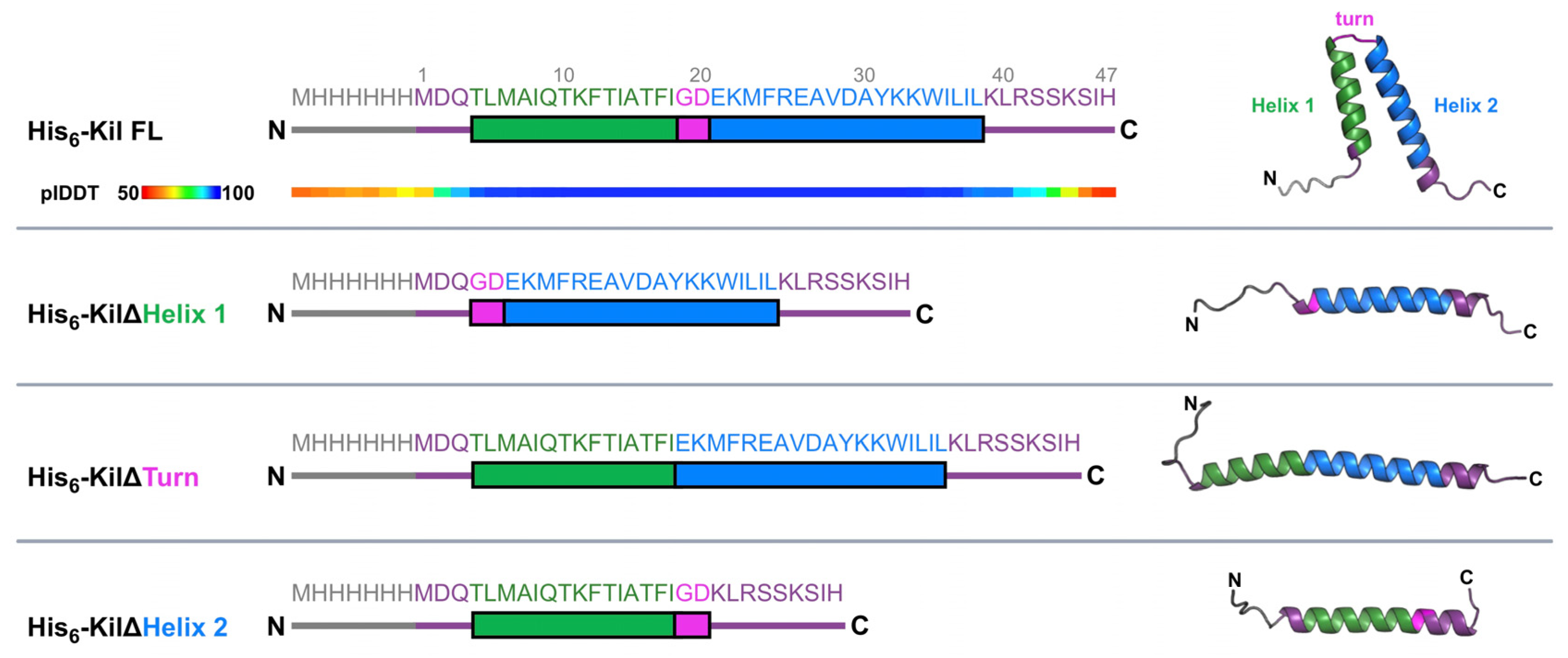
2.1. Kil Requires a Predicted Helix-Turn-Helix Structural Element for Its Anti-FtsZ Activity

Modeling by AlphaFold 3 [28] indicated that the His6-tagged λ Kil peptide is predicted to form a helix-turn-helix (HTH) structure (Figure 1, top). Residues 4–38 were predicted with very high confidence by AlphaFold 3 (predicted local distance difference test, plDDT, value > 90), including the N-terminal (aa 4–18) and C-terminal (aa 21–38) domains forming the two arms of the HTH structure, helix 1 and 2, respectively. Both helices are interlinked by a turn comprising glycine and aspartic acid (G19-D20).
To acquire more details about the functional importance of this HTH structure in retaining Kil’s anti-FtsZ activity, we generated N-terminally His6-tagged Kil variants in which the residues comprising helix 1, helix 2, or the inter-helix turn were deleted. As expected, when these HTH deletion variants were modeled using AlphaFold 3 (Figure 1), the resulting structures lacked Kil’s signature HTH core. We then investigated the ability of these His-tagged Kil derivatives to inhibit cytokinesis in the E. coli K-12 strain WM1074 when expressed from the arabinose-inducible araBAD promoter on the multi-copy plasmid pBAD24.
Microscopic examination of WM1074 cells expressing full-length (FL) Kil or the deletion variants demonstrated that an intact HTH core is indispensable for Kil’s activity (Figure 2A). Under the repressing condition, where the pBAD promoter is shut off in the presence of glucose, the bacterial cells divided normally. Following arabinose induction, His-tagged FL-Kil produced from pBAD24 inhibited cytokinesis and formed filamentous cells as previously described [25]. In contrast, we observed a complete suppression of cell division inhibition in E. coli cells expressing either of the His-tagged Kil HTH deletion variants (Figure 2A, ΔHelix1, ΔHelix2, or ΔTurn).
Figure 2. The ability of the λ Kil peptide to inhibit cell division requires an intact HTH core. (A) DIC (differential interference contrast) microscopic images of WT E. coli strain WM1074 expressing His-tagged FL Kil or its HTH deletion variants from pBAD24 under repressing (0.2% glucose, Glu) or inducing (0.2% arabinose, Ara) conditions. All derivatives are fused to an N-terminal His epitope. (A,B) Linear schematics and AlphaFold 3-modeled structures of the various derivatives are presented below and above each column of corresponding micrographs. Scale bars, 5 µm. (B) Fluorescence microscopic images of strain WM4952 expressing zapA-GFP plus either FL Kil or HTH deletion derivatives of Kil as described in part A. Arrowheads highlight intact Z-rings. (C) Immunoblot of His-FLAG-FL Kil (lanes 1 and 4) and His-FLAG-Kil variants with either helix 1 (lanes 2 and 5) or helix 2 (lanes 3 and 6) deleted, expressed from a phage T7 promoter in pET15b ± IPTG induction in strain WM5017 (ΔzipA ftsA*) to suppress Kil toxicity and probed with α-FLAG to detect expression of the tagged Kil derivatives. The predicted sizes of the His-FLAG-tagged FL, Δhelix 1, or Δhelix 2 Kil derivatives are 10.5, 8.8, and 8.2 kDa, respectively. A portion of the Ponceau S-stained blot is shown below the immunoblot as a loading control.
Antibiotics 14 00052 g002
We further investigated whether the resistance of E. coli cells to Kil HTH deletion variants was due to unperturbed Z-ring assembly, hypothesizing that HTH variants might lose their ability to bind and/or inhibit FtsZ polymerization. To evaluate Z-ring localization, we used E. coli cells expressing ZapA-GFP at the native locus (WM4952) as a proxy, as ZapA strongly localizes to the divisome through direct interactions with FtsZ [29,30]. As expected, WM4952 cells expressing His-tagged full-length (FL) Kil exhibited ZapA-GFP dispersed throughout the filamentous cells, indicating Z-ring disintegration (Figure 2B). In stark contrast, E. coli cells producing HTH variants exhibited intact Z rings one hour after arabinose induction.
Despite repeated attempts, we were unable to detect His-tagged Kil derivatives in either E. coli WM1074 or WM4952 by immunoblotting. Therefore, to quantitatively measure intracellular accumulation levels of the Kil derivatives, we expressed His-FLAG-tagged FL Kil and two of the HTH variants, ΔHelix1 and ΔHelix2, from the high-expression vector pET15b. We used a ΔzipA strain carrying the ftsA* allele previously used for Kil purification [25,27] because Kil activity is ZipA-dependent and thus is less toxic to cells lacking ZipA. However, as ZipA is essential for E. coli cell division, to permit viability we combined ΔzipA with a gain-of-function allele in another FtsZ anchor protein, FtsAR286W (ftsA*) that bypasses the need for several normally essential cell division proteins, including ZipA and FtsK [31]. Notably, we found that both Kil variants, particularly Kil ΔHelix2, were unstable in the cell lysates extracted from BL21 ΔzipA ftsA* recombinant strains (Figure 2C). This reduction in stability may also contribute to the loss of protein activity. Unfortunately, we could not generate a pET15b His-FLAG-KilΔTurn despite repeated efforts. Together, this result supports the idea that an unperturbed HTH core is necessary for Kil’s stability and anti-FtsZ activity in E. coli cells.

2.2. Identifying the Minimal Region of Kil That Inhibits Cell Division

Given that residues 4–38 of Kil are predicted to form the HTH structure (Figure 1), we hypothesized that truncations at the N-terminal and C-terminal ends of the 47-residue Kil sequence might retain functional activity. To test this hypothesis, we generated several truncations of the N-terminus and C-terminus of the His-tagged full-length (FL) Kil peptide that retained all but one or two residues of the HTH structure, modeled their structures with AlphaFold 3 (Figure 3A), and functionally expressed these variants. We then compared the ability of these truncated versions to induce filamentous cell formation, indicative of cell division inhibition, with that of the His-Kil FL peptide.
Figure 3. Shorter λ Kil truncations differentially inhibit E. coli cytokinesis. (A) Additional truncation derivatives of His-tagged λ Kil are diagrammed, using the same color schemes as in Figure 1. (B) DIC microscopic images of WM1074 expressing His-tagged Kil truncations from pBAD24 under repressing (0.2% glucose, Glu) or inducing (0.2% arabinose, Ara) conditions. Scale bar, 5 µm. AlphaFold 3-modeled structures of the Kil derivatives are included. (C) Fluorescence microscopic images of strain XTL937 expressing ZipA-GFP at its native chromosomal locus along with the indicated His-tagged Kil derivatives in pBAD24 are shown. Arrowheads indicate intact Z-rings. Schematic diagrams of the Kil derivatives are presented at the bottom of each corresponding micrograph, along with AlphaFold 3-modeled structures. Scale bar, 5 µm. (D) Quantitation of ZipA-GFP rings from the strains shown in panel C induced with arabinose.
Antibiotics 14 00052 g003
Our experiments revealed that deletions of up to 11 amino acids from the C-terminus (KilΔC11, corresponding to residues 1–36) or the first 5 amino acids from the N-terminus (KilΔN5, corresponding to residues 6–47) did not impair Kil’s ability to inhibit E. coli cell division (Figure 3B). However, simultaneous deletion of the N-terminal 5 amino acids and the 11 C-terminal amino acids (KilΔN5ΔC11, corresponding to residues 6–36) completely abolished Kil activity, resulting in short, dividing cells (Figure 3B). Using ZipA-GFP as a proxy for Z rings, we found that induction of KilΔC11 or Kil∆N5 from pBAD24 disassembled most Z rings, although ~10-fold less efficiently compared with His-FL Kil, whereas induction of KilΔN5ΔC11 left Z rings intact in all cells, as expected (Figure 3C,D). Induction of KilΔN5 (1 intact Z ring per ~20 µm of cell length) was slightly less effective at disassembling Z rings compared with Kil∆C11 (1 intact Z ring per ~40 µm of cell length) (Figure 3C,D), but both were potent at inhibiting Z ring formation. Similar patterns of disruption were observed after the induction of the Kil derivatives in the WM4952 strain expressing ZapA-GFP (Figure S1).

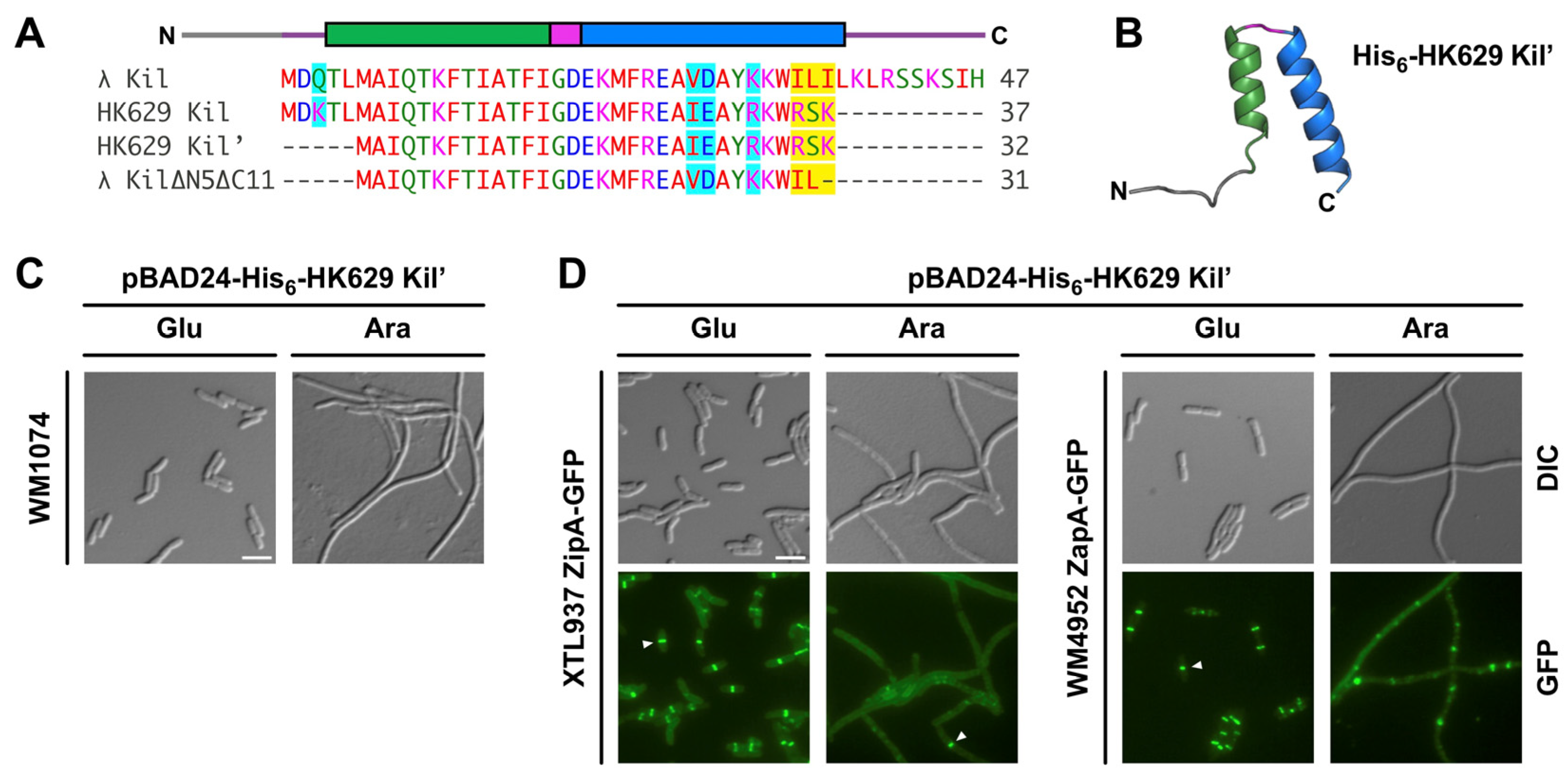
2.3. Kil Peptide from Enterobacteria Phage HK629 Shares a Common Mechanism to Inhibit Cytokinesis in E. coli

Kil peptides are diverse and widely distributed among enterobacterial λ-like phages. Bacteriophage-encoded Kil family peptides likely evolved as potent inhibitors of the E. coli divisome during phage infection, making them attractive models for how FtsZ can be efficiently inhibited. This family consists of several bacteriophage λ Kil-like sequences from both phages and bacteria (Figure S2). For example, a 37 amino acid peptide encoded by enterobacterial lambdoid phage HK629 shares significant sequence similarity with λ Kil, including most of the λ Kil HTH core (Figure 4A). As a result, HK629 Kil closely resembles λ KilΔC11 except that HK629 Kil’s C terminus consists of the highly polar triplet RSK instead of the aliphatic residues that terminate λ KilΔC11. Curiously, a similar RSSK motif exists in the C-terminus of full length λ Kil, so we hypothesized that RSK in HK629 Kil may be of functional importance. Notably, a five amino acid N-terminal truncation of HK629 Kil, which we term HK629 Kil’, is nearly identical to λ KilΔN5ΔC11 aside from the presence of the RSK triplet sequence. Therefore, we set out to determine whether HK629 Kil’ would successfully inhibit bacterial cell division like λ Kil.
Figure 4. Comparing cell division inhibition activities of Kil from phage HK629 and λ. (A) Sequence alignment of Kil peptides produced by bacteriophages λ and HK629 using the Clustal Omega multiple sequence alignment tool. HK629 Kil has several potential initiator methionines; longer (Kil) and shorter (Kil’) versions are depicted. Similar amino acids are highlighted in cyan and non-conservative amino acid positions are highlighted in yellow. λ Kil HTH domains are depicted above the alignment. (B) Predicted structure of His-tagged HK629 Kil’ from AlphaFold 3, showing a HTH fold similar to λ Kil. (C) DIC microscopy of pBAD24-His-HK629-Kil’ expressed in WT strain WM1074. Scale bar, 5 µm. (D) DIC (top) or fluorescence (bottom) microscopy of pBAD24-His-HK629-Kil’ expressed in strain XTL937 with ZipA-GFP co-expressed from the native locus (left panels) or in strain WM4952 expressing chromosomal ZapA-GFP (right panels). Expression of His-HK629 Kil’ was either repressed (Glu) or induced (Ara), showing that HK629 Kil’ inhibits cell division and disrupts Z rings. Arrowheads highlight intact Z-rings. Scale bar, 5 µm.
Antibiotics 14 00052 g004
As expected, AlphaFold 3 predicted that a His-tagged 39-residue version of HK629 Kil’ can form an HTH structure similar to that of λ Kil (Figure 4B). Next, we expressed the His-HK629 Kil’ (analogous to His-λ KilΔN5ΔC11) from pBAD24, with the hypothesis that this would act to inhibit E. coli cell division. Indeed, the induction of His-HK629 Kil’ in E. coli WM1074 resulted in efficient inhibition of cytokinesis (Figure 4C). As with λ Kil, HK629 Kil’ disrupted most Z rings, as measured by fluorescence microscopy of cells expressing HK629 Kil’ along with either ZipA-GFP or ZapA-GFP expressed from their native chromosomal loci as proxies for Z rings (Figure 4D).
To investigate whether HK629 Kil’ requires ZipA to inhibit E. coli cell division like λ Kil, we expressed His-HK629 Kil’ in wild-type (WT) zipA ftsA* or ΔzipA ftsA* cells. Indeed, HK629 Kil’ cell division inhibition activity was suppressed in ΔzipA ftsA* cells (Figure 5). This strongly suggests that ZipA is also required for the effect of HK629 Kil’ on FtsZ assembly and subsequent bacterial cytokinesis.
Figure 5. HK629 Kil’ requires ZipA for its cell division inhibition activity. DIC microscopic images of E. coli WM1657 (ΔzipA ftsA*) or WM1659 (zipA+ ftsA*) expressing His-tagged FL λ Kil or HK629 Kil’ from pBAD24 under repressing (0.2% glucose, Glu) or inducing (0.2% arabinose, Ara) conditions. Cells harboring the FtsAR286W variant (FtsA*) that allows bypass of zipA (ΔzipA::aph) are resistant to the inhibitory effects of λ Kil or HK629 Kil’, but are not resistant when containing FtsAR286W in a zipA+ background. Ribbon diagrams of both Kil peptides are shown as modeled by AlphaFold 3. Scale bar, 5 µm.
Antibiotics 14 00052 g005

2.4. Kil Inhibits Cytokinesis of a Pathogenic E. coli Strain

Uropathogenic E. coli (UPEC) strains are the primary causative agents of urinary tract infections (UTIs), posing a significant health concern in the United States [32]. The UTI89 strain is widely used to study UPEC physiology and virulence during UTI pathogenesis. To investigate the potential of Kil peptides as an alternative to traditional antibiotics, we introduced pBAD24-His-Kil or -His-HK629 Kil’ into UTI89 and induced the expression of the kil genes with arabinose. This resulted in the inhibition of cell division and filamentation in bacterial cells expressing His-tagged full-length (FL) Kil or HK629 Kil’ peptides (Figure 6). In contrast, E. coli cells carrying an empty plasmid showed no change in cell length. These findings suggest that Kil peptides can be potent cell division disruptors in pathogenic strains of E. coli.
Figure 6. Kil peptides produced from phages HK629 or λ inhibit cell division of Uropathogenic E. coli (UPEC) strain UTI89. DIC microscopic images of E. coli cells expressing His-tagged FL λ Kil or HK629 Kil’ from pBAD24 under repressing (0.2% glucose, Glu) or inducing (0.2% arabinose, Ara) conditions. Ribbon diagrams of both Kil peptides are shown as modeled by AlphaFold 3. Scale bar, 5 µm.
Antibiotics 14 00052 g006

3. Discussion

In this study, we explored the structural and functional features of bacteriophage-derived Kil peptides and assessed their inhibitory effects on cell division of both E. coli K-12 and a pathogenic UPEC strain. Structural analysis of the λ Kil peptide revealed a conserved HTH motif essential for its function. Deletion of any single component of the HTH structure, including the two residues forming the turn, resulted in a complete loss of Kil activity. In contrast, deletion of the N-terminal five residues or the C-terminal 11 residues at the ends of the two helices reduced, but did not eliminate, Kil’s inhibitory function (Figure 3D). However, λ Kil minimally requires an intact N- or C-terminus, as truncating both simultaneously eliminated Kil’s activity. Intriguingly, these N- and C-terminal truncations did not affect the localization of ZipA-GFP equally; the ΔC11 truncation resulted in completely dispersed ZipA-GFP localization, whereas the ΔN5 truncation permitted localized accumulations of ZipA-GFP (Figure 3C), suggesting reduced ZipA disruption by this truncation. Based on the requirement of ZipA for Kil’s inhibition of FtsZ, we speculate that the two helices in Kil’s HTH may each target ZipA and FtsZ independently. Such a mechanism would result in a two-pronged inhibition of cell division by disrupting both FtsZ polymer dynamics and ZipA’s functions in FtsZ membrane tethering and divisome assembly. An analogous two-pronged inhibitory mechanism involves disruption of both FtsZ and MreB function by the E. coli CbtA toxin [33]. Understanding how Kil acts on FtsZ and ZipA may shed further light on how FtsZ normally interacts with ZipA and other proteins during bacterial cell division.
The structural and functional conservation of Kil peptides across different bacteriophages further suggests a common mechanism of action. In support of this, the Kil peptide from enterobacteria phage HK629 exhibited similar cell division inhibition as λ Kil, consistent with its similar HTH fold. HK629 Kil’ is the smallest Kil that retains function in our experiments. The key difference between HK629 Kil’ and the inactive Kil ΔN5ΔC11 truncation is the charges on the C-terminus of the peptides. Despite its significantly shorter length, HK629 Kil’ retains a C-terminal RSK motif resembling a similar RSSK motif found in the C-terminus of full-length λ Kil. An RSK or RSSK motif in this position is found in nearly all λ Kil homologs (Figure S2), suggesting it serves an important role in protein function. Indeed, the λ KilΔC11 truncation allowed nearly 10 times as many intact Z-rings compared to the FL Kil protein (Figure 3D). Removal of this motif from λ Kil in combination with the N-terminal truncation in λ Kil ΔN5ΔC11 may be responsible for the full inactivation of λ Kil, perhaps due to simultaneous disruption of both parts of the two-pronged FtsZ and ZipA binding mechanism theorized above. However, this hypothesis will need to be validated in the future, and further experiments will be required to unveil the mechanistic details of the E. coli cell division inhibition by HK629 Kil’.
The cross-species activity of Kil highlights the potential of Kil peptides as antibacterial agents. In the context of UPEC, a major causative agent of UTIs, induction of filamentation and inhibition of cell division in UTI89 cells expressing His-tagged FL Kil or HK629 Kil’ peptides confirmed their efficacy in targeting and disrupting the bacterial division machinery. This finding is particularly pertinent given the rising concern of antimicrobial resistance (AMR) among UPEC strains. The effect of bacteriophage-encoded peptides on FtsZ localization during infection by pathogenic E. coli was also recently investigated in a neonatal meningitis model system [34].
In summary, our study provides initial evidence for the efficacy of phage-derived Kil peptides as alternative antimicrobial agents. Their structural conservation and ability to inhibit cell division in pathogenic bacteria underscore their therapeutic potential. It would be interesting to see if the Kil peptides are active against other bacterial pathogens including Mycobacterium tuberculosis, where both FtsA and ZipA are absent and SepF is the essential membrane tether for FtsZ [6]. Future research should focus on elucidating the precise molecular mechanisms of Kil peptide action and optimizing their stability and delivery.

4. Materials and Methods

4.1. Bacterial Strains and Growth Conditions

All bacterial strains used in this study are listed in Table 1. Competent cells of XL1-Blue, UTI89, WM4952, XTL937, WM1657, and WM1659 strains were prepared from Luria-Bertani (LB) broths supplemented with 25% (v/v) glycerol stored at −80 °C. Recombinant E. coli strains ANWM1-ANWM34 were prepared by transforming WM1074, UTI89, and the genetically engineered strains WM4952, XTL937, WM1657, and WM1659 with pBAD24/pET15b-derived plasmids producing the full length (FL) Kil peptide or the deleted variants. Microscopic experiments were performed in triplicate using individual colonies isolated from fresh transformation plates. For cultivation, strains were grown in LB medium with vigorous aeration (200 rpm) at either 30 °C or 37 °C, as specified. Appropriate antibiotics were added as follows: carbenicillin (100 µg/mL), kanamycin (50 µg/mL), or chloramphenicol (15 µg/mL). Gene expression was regulated using IPTG (isopropyl-β-D-1-thiogalactopyranoside), 0.2% D-(+)-Glucose, and 0.2% L-(+)-arabinose (MilliporeSigma, Burlington, MA, USA).
For IPTG inductions of pET15b vector-expressed Kil derivatives, overnight bacterial cultures were diluted 1:100 in fresh LB medium and grown to an optical density at 600 nm (OD600) of 0.7. Protein expression was induced by adding 1 mM IPTG, and the cultures were incubated for four hours at 30 °C. Bacterial cells were harvested by spinning at 7000 rpm. Supernatants were carefully removed, and cell pellets were washed in buffer (50 mM sodium phosphate pH 8.0, 300 mM NaCl), re-centrifuged, and stored at −80 °C until used for follow-up experiments. For arabinose-inducible protein expression, bacteria were initially grown overnight in an LB medium containing 0.2% glucose (Glu, repressing condition). The overnight cultures were diluted 100 times in 1 mL of pre-warmed LB + Glu. Once the OD600 value reached ~0.35, the cultures were split into two 500 µL aliquots, pelleted, washed with prewarmed LB, and resuspended in LB with 0.2% arabinose (Ara, inducing condition) and Glu. Following the addition of inducers, cultures were further grown for 40 min and then observed under light and fluorescence microscopy.

4.2. Plasmid Construction

All plasmids and primers used in this study are described in Table 1 and Table 2, respectively. Recombinant plasmids were constructed using standard molecular cloning techniques. Site-directed mutagenesis (SDM) was employed to generate Kil HTH deletion variants and shorter truncations on pBAD24 using the templates and primers specified in Table 2. All constructs were verified by DNA sequencing using plasmid-specific primers.

4.3. Microscopy

Cells were mounted on agarose pads and imaged using an Olympus BX63 microscope (Tokyo, Japan) equipped with a Hamamatsu C11440 ORCA-Spark digital complementary metal oxide semiconductor (CMOS) camera (Hamamatsu, Japan) and cellSens Dimensions 4.2 software (Olympus). Image analysis was performed using ImageJ 2.14.0/1.54f software.

4.4. Immunoblot Analysis

To prepare whole-cell lysates, bacterial cell pellets were suspended in 1X SDS-PAGE sample buffer. Samples were boiled for 10 min prior to separation by SDS-polyacrylamide gel electrophoresis (PAGE) using 4–20% precast polyacrylamide gels (Bio-Rad, Hercules, CA, USA, #4561094) with Laemmli running buffer.
Spectra Multicolor Low Range Protein Ladder (Thermo Scientific 26628, Waltham, MA, USA) was used as the molecular weight standard. Samples were transferred to nitrocellulose membranes (Bio-Rad 1620112) using a Trans-Blot SD semi-dry transfer cell (Bio-Rad) at 15 V for 25 min in 1X Tris-Glycine transfer buffer (25 mM Tris, 192 mM glycine, pH 8.3, 20% methanol). Blots were stained with Ponceau stain for assessment of total protein levels, then blocked using 4% bovine serum albumin (BSA) in Tris-buffered saline with Tween 20 (TBST) and incubated with mouse anti-FLAG antibodies (1:5000, MilliporeSigma F1804). After washes, blots were incubated with goat anti-mouse antibody labeled with horseradish peroxidase (1:5000, Bio-Rad 1706516), developed with Clarity ECL Western Blotting Substrates (Bio-Rad 1705061), and imaged using a ChemiDoc MP imaging system (Bio-Rad).

Supplementary Materials

The following supporting information can be downloaded at: https://www.mdpi.com/article/10.3390/antibiotics14010052/s1, Figure S1. Shorter λ Kil truncations also inhibit E. coli cytokinesis.; Figure S2. Multiple sequence alignment of λ Kil-like protein sequences.

Author Contributions

A.N., conceptualization, formal analysis, investigation, methodology, validation, visualization, writing—original draft, writing—review and editing; T.A.C., formal analysis, methodology, validation, writing—review and editing; W.M., conceptualization, data curation, Formal analysis, funding acquisition, investigation, project administration, resources, supervision, validation, writing—original draft, writing—review and editing. All authors have read and agreed to the published version of the manuscript.

Funding

This work was supported by the National Institutes of Health (NIH) Grant AI171856. Part of this work was presented at the annual meeting of the American Society for Microbiology (ASM Microbe 2023), and at the Gulf Coast Consortia AMR conference (GCC-AMR 2024).

Institutional Review Board Statement

Not applicable.

Informed Consent Statement

Not applicable.

Data Availability Statement

The data presented in this study are openly available in FigShare at doi:10.6084/m9.figshare.28012280.

Acknowledgments

The authors thank Jennifer N. Walker for providing the UTI89 strain, Xintian Li for providing the XTL937 strain, and Xu Wang and Maria A. Schumacher for useful insights into Kil structures.

Conflicts of Interest

The authors declare no conflicts of interest.

References

  1. Errington, J.; Daniel, R.A.; Scheffers, D.J. Cytokinesis in bacteria. Microbiol. Mol. Biol. Rev. 2003, 67, 52–65. [Google Scholar] [CrossRef]
  2. Du, S.; Lutkenhaus, J. At the heart of bacterial cytokinesis: The Z ring. Trends Microbiol. 2019, 27, 781–791. [Google Scholar] [CrossRef]
  3. Bi, E.; Lutkenhaus, J. FtsZ ring structure associated with division in Escherichia coli. Nature 1991, 354, 161–164. [Google Scholar] [CrossRef] [PubMed]
  4. Goehring, N.W.; Beckwith, J. Diverse paths to midcell: Assembly of the bacterial cell division machinery. Curr. Biol. 2005, 15, 514–526. [Google Scholar] [CrossRef]
  5. Cameron, T.A.; Margolin, W. Insights into the assembly and regulation of the bacterial divisome. Nat. Rev. Microbiol. 2024, 22, 33–45. [Google Scholar] [CrossRef] [PubMed]
  6. Naha, A.; Haeusser, D.P.; Margolin, W. Anchors: A way for FtsZ filaments to stay membrane bound. Mol. Microbiol. 2023, 120, 525–538. [Google Scholar] [CrossRef] [PubMed]
  7. Duman, R.; Ishikawa, S.; Celik, I.; Strahl, H.; Ogasawara, N.; Troc, P.; Lowe, J.; Hamoen, L.W. Structural and genetic analyses reveal the protein SepF as a new membrane anchor for the Z ring. Proc. Natl. Acad. Sci. USA 2013, 110, E4601–E4610. [Google Scholar] [CrossRef] [PubMed]
  8. Wenzel, M.; Celik Gulsoy, I.N.; Gao, Y.; Teng, Z.; Willemse, J.; Middelkamp, M.; van Rosmalen, M.G.M.; Larsen, P.W.B.; van der Wel, N.N.; Wuite, G.J.L.; et al. Control of septum thickness by the curvature of SepF polymers. Proc. Natl. Acad. Sci. USA 2021, 118, e2002635118. [Google Scholar] [CrossRef] [PubMed]
  9. Schoenemann, K.M.; Krupka, M.; Rowlett, V.W.; Distelhorst, S.L.; Hu, B.; Margolin, W. Gain-of-function variants of FtsA form diverse oligomeric structures on lipids and enhance FtsZ protofilament bundling. Mol. Microbiol. 2018, 109, 676–693. [Google Scholar] [CrossRef]
  10. Nierhaus, T.; McLaughlin, S.H.; Bürmann, F.; Kureisaite-Ciziene, D.; Maslen, S.L.; Skehel, J.M.; Yu, C.W.H.; Freund, S.M.V.; Funke, L.F.H.; Chin, J.W.; et al. Bacterial divisome protein FtsA forms curved antiparallel double filaments when binding to FtsN. Nat. Microbiol. 2022, 7, 1686–1701. [Google Scholar] [CrossRef] [PubMed]
  11. Du, S.; Park, K.-T.; Lutkenhaus, J. Oligomerization of FtsZ converts the FtsZ Tail Motif (CCTP) into a multivalent ligand with high avidity for partners ZipA and SlmA. Mol. Microbiol. 2014, 95, 173–188. [Google Scholar] [CrossRef] [PubMed]
  12. Hernandez-Rocamora, V.M.; Garcia-Montanes, C.; Rivas, G.; Llorca, O. Reconstitution of the Escherichia coli cell division ZipA-FtsZ complexes in nanodiscs as revealed by electron microscopy. J. Struct. Biol. 2012, 180, 531–538. [Google Scholar] [CrossRef] [PubMed]
  13. Hale, C.A.; Rhee, A.C.; de Boer, P.A. ZipA-induced bundling of FtsZ polymers mediated by an interaction between C-terminal domains. J. Bacteriol. 2000, 182, 5153–5166. [Google Scholar] [CrossRef] [PubMed]
  14. Haeusser, D.P.; Schwartz, R.L.; Smith, A.M.; Oates, M.E.; Levin, P.A. EzrA prevents aberrant cell division by modulating assembly of the cytoskeletal protein FtsZ. Mol. Microbiol. 2004, 52, 801–814. [Google Scholar] [CrossRef] [PubMed]
  15. Pichoff, S.; Lutkenhaus, J. Unique and overlapping roles for ZipA and FtsA in septal ring assembly in Escherichia coli. EMBO J. 2002, 21, 685–693. [Google Scholar] [CrossRef] [PubMed]
  16. Global burden of bacterial antimicrobial resistance in 2019: A systematic analysis. Lancet 2022, 399, 629–655. [CrossRef]
  17. Sass, P.; Brotz-Oesterhelt, H. Bacterial cell division as a target for new antibiotics. Curr. Opin. Microbiol. 2013, 16, 522–530. [Google Scholar] [CrossRef] [PubMed]
  18. Battaje, R.R.; Piyush, R.; Pratap, V.; Panda, D. Models versus pathogens: How conserved is the FtsZ in bacteria? Biosci. Rep. 2023, 43, BSR20221664. [Google Scholar] [CrossRef]
  19. Wang, Y.-T.; Liu, L.-T.; Hou, B.; Yao, C.-M.; Wang, X.-F.; Lu, B. Recent advances in studies on FtsZ inhibitors. Biochem. Pharmacol. 2024, 230, 116551. [Google Scholar] [CrossRef] [PubMed]
  20. Han, H.; Wang, Z.; Li, T.; Teng, D.; Mao, R.; Hao, Y.; Yang, N.; Wang, X.; Wang, J. Recent progress of bacterial FtsZ inhibitors with a focus on peptides. FEBS J. 2021, 288, 1091–1106. [Google Scholar] [CrossRef] [PubMed]
  21. Hirota, Y.; Ryter, A.; Jacob, F. Thermosensitive mutants of Escherichia coli affected in the process of dna synthesis and cellular division. Cold Spring Harb. Symp. Quant. Biol. 1968, 33, 677–694. [Google Scholar] [CrossRef] [PubMed]
  22. Georjon, H.; Bernheim, A. The highly diverse antiphage defence systems of bacteria. Nat. Rev. Microbiol. 2023, 21, 686–700. [Google Scholar] [CrossRef]
  23. Conter, A.; Bouché, J.P.; Dassain, M. Identification of a new inhibitor of essential division gene ftsZ as the kil gene of defective prophage Rac. J. Bacteriol. 1996, 178, 5100–5104. [Google Scholar] [CrossRef] [PubMed]
  24. Kiro, R.; Molshanski-Mor, S.; Yosef, I.; Milam, S.L.; Erickson, H.P.; Qimron, U. Gene product 0.4 increases bacteriophage T7 competitiveness by inhibiting host cell division. Proc. Natl. Acad. Sci. USA 2013, 110, 19549–19554. [Google Scholar] [CrossRef]
  25. Haeusser, D.P.; Hoashi, M.; Weaver, A.; Brown, N.; Pan, J.; Sawitzke, J.A.; Thomason, L.C.; Court, D.L.; Margolin, W. The Kil peptide of bacteriophage lambda blocks Escherichia coli cytokinesis via ZipA-dependent inhibition of FtsZ assembly. PLoS Genet. 2014, 10, e1004217. [Google Scholar] [CrossRef]
  26. Ragunathan, P.T.; Vanderpool, C.K. Cryptic-prophage-encoded small protein DicB protects Escherichia coli from phage infection by inhibiting inner membrane receptor proteins. J. Bacteriol. 2019, 201, 1–16. [Google Scholar] [CrossRef] [PubMed]
  27. Hernandez-Rocamora, V.M.; Alfonso, C.; Margolin, W.; Zorrilla, S.; Rivas, G. Evidence that bacteriophage lambda Kil peptide inhibits bacterial cell division by disrupting FtsZ protofilaments and sequestering protein subunits. J. Biol. Chem. 2015, 290, 20325–20335. [Google Scholar] [CrossRef]
  28. Abramson, J.; Adler, J.; Dunger, J.; Evans, R.; Green, T.; Pritzel, A.; Ronneberger, O.; Willmore, L.; Ballard, A.J.; Bambrick, J.; et al. Accurate structure prediction of biomolecular interactions with AlphaFold 3. Nature 2024, 630, 493–500. [Google Scholar] [CrossRef]
  29. Low, H.H.; Moncrieffe, M.C.; Löwe, J. The crystal structure of ZapA and its modulation of FtsZ polymerisation. J. Mol. Biol. 2004, 341, 839–852. [Google Scholar] [CrossRef] [PubMed]
  30. Galli, E.; Gerdes, K. Spatial resolution of two bacterial cell division proteins: ZapA recruits ZapB to the inner face of the Z-ring. Mol. Microbiol. 2010, 76, 1514–1526. [Google Scholar] [CrossRef] [PubMed]
  31. Geissler, B.; Margolin, W. Evidence for functional overlap among multiple bacterial cell division proteins: Compensating for the loss of FtsK. Mol. Microbiol. 2005, 58, 596–612. [Google Scholar] [CrossRef] [PubMed]
  32. Flores-Mireles, A.L.; Walker, J.N.; Caparon, M.; Hultgren, S.J. Urinary tract infections: Epidemiology, mechanisms of infection and treatment options. Nat. Rev. Microbiol. 2015, 13, 269–284. [Google Scholar] [CrossRef]
  33. Heller, D.M.; Tavag, M.; Hochschild, A. CbtA toxin of Escherichia coli inhibits cell division and cell elongation via direct and independent interactions with FtsZ and MreB. PLoS Genet. 2017, 13, e1007007. [Google Scholar] [CrossRef]
  34. Dhanoa, G.; Kushnir, I.; Qimron, U.; Roper, D.I.; Sagona, A.P. Investigating the effect of bacteriophages on bacterial FtsZ localisation. Front. Cell. Infect. Microbiol. 2022, 12, 863712. [Google Scholar] [CrossRef] [PubMed]
  35. Geissler, B.; Elraheb, D.; Margolin, W. A gain of function mutation in ftsA bypasses the requirement for the essential cell division gene zipA in Escherichia coli. Proc. Natl. Acad. Sci. USA 2003, 100, 4197–4202. [Google Scholar] [CrossRef] [PubMed]
  36. Tsang, M.J.; Bernhardt, T.G. A role for the FtsQLB complex in cytokinetic ring activation revealed by an ftsL allele that accelerates division. Mol. Microbiol. 2015, 95, 925–944. [Google Scholar] [CrossRef] [PubMed]
  37. Guzman, L.M.; Belin, D.; Carson, M.J.; Beckwith, J. Tight regulation, modulation, and high-level expression by vectors containing the arabinose pBAD promoter. J. Bacteriol. 1995, 177, 4121–4130. [Google Scholar] [CrossRef] [PubMed]
Figure 1. Schematic representation of the λ Kil peptide and deletion derivatives. Green and blue bars indicate helix 1 and 2, respectively, with the turn highlighted in magenta and the 6xHis tag highlighted in grey. This color scheme is the same for Figure 2, Figure 3, Figure 4, Figure 5 and Figure 6. The full-length (FL) Kil peptide with its intact HTH domains is depicted in the top row, followed by derivatives deleted for helix 1, the turn, or helix 2, shown below. Amino acid numbering of the native Kil peptide is shown above the FL sequence. Ribbon diagrams of each His-tagged Kil derivative are shown as high confidence predictions from AlphaFold 3 with corresponding colors representing the different domains. The predicted local difference test (plDDT) confidence values for each residue of the Kil peptide are shown below the FL Kil peptide schematic.
Antibiotics 14 00052 g001
Table 1. Bacterial strains and plasmids used in this study.
Strain/PlasmidGenotype and/or PhenotypeReference/Source
UTI89Uropathogenic E. coli (UPEC)[32]
XL1-BluerecA1 endA1 gyrA96 thi-1 hsdR17 supE44 relA1 lac [F’ proAB lacIqZ∆M15 Tn10 (Tetr)]Cloning host, lab collection
XTL937zipA-gfp at the native locusXintian Li
WM1074MG1655: ilvG rpb-50 rph-1 ΔlacU169Lab collection
WM1657 WM1074 ftsAR286W ΔzipA::aph (ΔzipA::kan)[35]
WM1659WM1074 ftsAR286W (ftsA*)[35]
WM4952MT78 (leuA::Tn10 ftsA°, zapA-gfp, pSC101(ts)-PR-ftsA[36]
WM5017BL21(DE3) ftsAR286W (ftsA*) sac284-Tn10 [pBS58] ΔzipA::aph[25]
ANWM1WM1074 pANWM1This study
ANWM2WM1074 pANWM2This study
ANWM3WM1074 pANWM3This study
ANWM4WM1074 pANWM4This study
ANWM5WM1074 pANWM5This study
ANWM6WM1074 pANWM6This study
ANWM7WM1074 pANWM7This study
ANWM8WM1074 pANWM8This study
ANWM9WM1074 pANWM9This study
ANWM10WM4952 pANWM1This study
ANWM11WM4952 pANWM2This study
ANWM12WM4952 pANWM3This study
ANWM15WM4952 pANWM4This study
ANWM16WM4952 pANWM5This study
ANWM17WM4952 pANWM7This study
ANWM18WM4952 pANWM8This study
ANWM19XTL937 pANWM1This study
ANWM20XTL937 pANWM5This study
ANWM21XTL937 pANWM7This study
ANWM22XTL937 pANWM8This study
ANWM23WM1074 pANWM9This study
ANWM24XTL937 pANWM9This study
ANWM25WM4952 pANWM9This study
ANWM26WM1657 pANWM9This study
ANWM27WM1659 pANWM9This study
ANWM28WM1657 pANWM1This study
ANWM29WM1659 pANWM1This study
ANWM30UTI89 pBAD24This study
ANWM31UTI89 pANWM1This study
ANWM32UTI89 pANWM9This study
WM5018WM5017 pDH149[25]
ANWM33WM5017 pANWM10This study
ANWM34WM5017 pANWM11This study
pBAD24Arabinose inducible expression vector[37]
pANWM1pBAD24-His6-Kil (Full length Kil; 47 aa)This study
pANWM2pBAD24-His6-KilΔHelix1 (aa 4–18 deleted)This study
pANWM3pBAD24-His6-KilΔTurn (aa G19D20 deleted)This study
pANWM4pBAD24-His6-KilΔHelix2 (aa 21–38 deleted)This study
pANWM5pBAD24-His6-KilΔN5 (Kil6–47, aa 1–5 deleted)This study
pANWM6pBAD24-His6-KilΔC5 (Kil1–42, aa 43–47 deleted)This study
pANWM7pBAD24-His6-KilΔC11 (Kil1–36, aa 37–47 deleted)This study
pANWM8pBAD24-His6-Kil ΔN5ΔC11 (Kil6–36, aa 1–5, 37–47 deleted)This study
pANWM9pBAD24-His6-HK629 Kil’ (aa 6–32)This study
pDH149 pET15b- His6-FLAG-Kil[25]
pANWM10pET15b- His6-FLAG-Kil ΔHelix1This study
pANWM11pET15b- His6-FLAG-Kil ΔHelix2 This study
Table 2. Oligonucleotides used in this study.
OligonucleotideSequence 5′-3′Template Notes
WM2688AGCCGGAATTCCGGATGCATCATCATCATCATCACpRR48-Kil, HK629Kil’
oligo
5′ EcoRI site, Used as forward primer (Fp) for pBAD24-his6-kil and pBAD24-his6-hk629kil’ cloning
WM2689GGAACAAGCTTTTAGTGAATGCTTTTGCTTGATCpRR48-Kil3′ HindIII site, used as a reverse primer (Rp) for pBAD24-his6-kil cloning
WM2759GGCGATGAAAAGATGTTTCpANWM1, pDH149KilΔHelix1 SDM Fp
WM2760TTGATCCATGTGATGATGpANWM1His6-KilΔHelix1 SDM Rp
WM2778TTGATCCATACGCTGGATTTTGpDH149His6 -FLAG-KilΔHelix1 SDM Rp
WM2757GAAAAGATGTTTCGTGAAGpANWM1KilΔTurn SDM Fp
WM2758AATAAAAGTGGCGATAGTGpANWM1KilΔTurn SDM Rp
WM2761AAACTGAGATCAAGCAAAAGpANWM1, pDH149KilΔHelix2 SDM Fp
WM2762 ATCGCCAATAAAAGTGGCpANWM1, pDH149KilΔHelix2 SDM Rp
WM2753TAAAAGCTTGGCTGTTTTGpANWM1KilΔC SDM Fp
WM2709TGATCTCAGTTTCAGTATTAATATCpANWM1KilΔC5 SDM Rp
WM2655TAATATCCATTTTTTATAAGCGTCpANWM1KilΔC11 SDM Rp
WM2748ATGGCTATCCAGACTAAATTCpANWM1His6-KilΔN5 SDM Fp
WM2749GTGATGATGATGATGATGCpANWM1His6-KilΔN5 SDM Rp
WM2813GGAACAAGCTTTCATTTTGACCTCCA HK629Kil’
oligo
3′ HindIII site, Used along with WM2688 for pBAD24-his6-HK629kil’ cloning as Rp
HK629Kil’ OligoggGAATTCcggATGCATCATCATCATCATCACATGGC
TATCCAGACTAAATTCACTATCGCCACTTTTATTGG
CGATGAAAAGATGTTTCGTGAGGCCATCGAAGCCT
ACAGAAAATGGAGGTCAAAATGAaAGCTTgg
His-tagged hk629kil’ oligo synthesized, Azenta Life Sciences
pBAD ForwardATGCCATAGCATTTTTATCCpBAD24 derived clonesSequencing primers from Azenta Life Sciences
pBAD ReverseGATTTAATCTGTATCAGG
T7 TermGCTAGTTATTGCTCAGCGGpET15b derived clonesSequencing primers from Azenta Life Sciences
T7TAATACGACTCACTATAGGG
Disclaimer/Publisher’s Note: The statements, opinions and data contained in all publications are solely those of the individual author(s) and contributor(s) and not of MDPI and/or the editor(s). MDPI and/or the editor(s) disclaim responsibility for any injury to people or property resulting from any ideas, methods, instructions or products referred to in the content.

