Article

Phylotypic Profiling, Distribution of Pathogenicity Island Markers, and Antimicrobial Susceptibility of Escherichia coli Isolated from Retail Chicken Meat and Humans

1
Department of Bacteriology, Mycology, and Immunology, Faculty of Veterinary Medicine, Mansoura University, Mansoura 35516, Egypt
2
Hygiene and Zoonoses Department, Faculty of Veterinary Medicine, Mansoura University, Mansoura 35516, Egypt
3
Department of Bacteriology, Animal Health Research Institute (AHRI), Mansoura Branch, Agriculture Research Center (ARC), Mansoura 35516, Egypt
4
Biochemistry Department, Faculty of Medicine Damietta, Al-Azhar University, Cairo 11651, Egypt
5
Clinical Laboratory Sciences Department, Turabah University College, Taif University, Taif 21995, Saudi Arabia
6
Biology Department, Turabah University College, Taif University, Al Hawiyah 21995, Saudi Arabia
7
Faculty of Veterinary Medicine, Mansoura University, Mansoura 35516, Egypt
8
Department of Pharmacology, Faculty of Veterinary Medicine, Benha University, Moshtohor, Toukh 13736, Egypt
*
Author to whom correspondence should be addressed.
Antibiotics 2022, 11(9), 1197; https://doi.org/10.3390/antibiotics11091197
Submission received: 19 July 2022 / Revised: 20 August 2022 / Accepted: 30 August 2022 / Published: 4 September 2022

Abstract

:
Escherichia coli (E.coli) found in retail chicken meat could be causing a wide range of infections in humans and constitute a potential risk. This study aimed to evaluate 60 E. coli isolates from retail chicken meat (n = 34) and human urinary tract infections (UTIs, n = 26) for phylogenetic diversity, presence of pathogenicity island (PAI) markers, antimicrobial susceptibility phenotypes, and antimicrobial resistance genes, and to evaluate their biofilm formation capacity. In that context, confirmed E.coli isolates were subjected to phylogrouping analysis using triplex PCR, antimicrobial susceptibility testing using the Kirby–Bauer disc diffusion method; PAI distribution was investigated by using two multiplex PCRs. Most of the chicken isolates (22/34, 64.7%) were identified as commensal E. coli (A and B1), while 12 isolates (35.3%) were classified as pathogenic virulent E. coli (B2 and D). Similarly, the commensal group dominated in human isolates. Overall, 23 PAIs were detected in the chicken isolates; among them, 39.1% (9/23) were assigned to group B1, 34.8% (8/23) to group A, 4.34% (1/23) to group B2, and 21.7% (5/23) to group D. However, 25 PAIs were identified from the human isolates. PAI IV536 was the most prevalent (55.9%, 69.2%) PAI detected in both sources. In total, 37 (61.7%) isolates of the chicken and human isolates were biofilm producers. Noticeably, 100% of E. coli isolates were resistant to penicillin and rifamycin. Markedly, all E. coli isolates displayed multiple antibiotic resistance (MAR) phenotypes, and the multiple antibiotic resistance index (MARI) among E. coli isolates ranged between 0.5 and 1. Several antibiotic resistance genes (ARGs) were identified by a PCR assay; the sul2 gene was the most prevalent (38/60, 63.3%) from both sources. Interestingly, a significant positive association (r = 0.31) between biofilm production and resistance to quinolones by the qnr gene was found by the correlation analysis. These findings were suggestive of the transmission of PAI markers and antibiotic resistance genes from poultry to humans or humans to humans through the food chain. To avoid the spread of virulent and multidrug-resistant E. coli, intensive surveillance of retail chicken meat markets is required.

1. Introduction

The majority of Escherichia coli (E. coli) is found in the gastrointestinal tracts of both animals and humans as a commensal microbiota [1,2]. While certain E. coli strains can cause a wide range of diseases in humans and animals. [1,3], due to the increase in community-acquired human urinary tract infections (UTIs) caused by multiple antibiotic resistance (MAR) E. coli, it is very important to recognize the most probable sources of urovirulent E. coli involved in such infections. Intestinal carriage of pathogenic and multidrug-resistant (MDR) Escherichia coli (E. coli) strains in food-producing animals and contamination of retail meat may contribute to increased incidences of infections by these strains in humans. Many previous studies have reported a close genetic relatedness between E. coli isolates from poultry and humans depending on phylogenetic groups, serogroups, and virulence gene profiles [4,5,6,7,8].
Poultry meat is a potential source of E. coli infection in humans that results in foodborne illnesses. E. coli is thought to enter a human host through the consumption of contaminated poultry meat. Pathogenic E. coli is found naturally in the guts of birds and is transferred through intestinal content, contaminating chicken carcasses during unsanitary slaughtering and processing activities, which poses a zoonotic and health hazard to people [9]. Human-to-human transmission through food is another way for poultry meat to become contaminated. Handlers transmit E. coli onto the meat during the preparation process, resulting in meat contamination.
Generally, E. coli strains are classified into four phylogenetic groups: A, B1, B2, and D [10]. According to ECOR phylogenetic grouping and virulence characteristics, both commensal and extraintestinal pathogenic E. coli typically differ [11]. Phylogenetic group B2 and group D are related to pathogenic extraintestinal E. coli isolates; however, groups A and B1 are classified as commensal E. coli isolates.
Pathogenic E. coli strains have virulence factors [12] clustered on pathogenicity islands (PAIs) that allow them to cause infection [13,14,15]. These PAIs are a sequence of DNA <10 kb in size [16,17]. They were first identified in UPEC strain 536 [16]. PAIs and their virulence genes are disseminated between bacterial populations through horizontal transfer [15]. E. coli strains (E. coli 536, E. coli J96, E. coli CFT073) encode many PAIs; for example, PAIs I to IV from strain 536 encode fimbriae (P fimbriae, P-related fimbriae, S fimbriae, α-hemolysin, iron siderophore system, yersiniabactin siderophore system, CS12 fimbriae, and F17-like fimbrial adhesion). Furthermore, PAIs IJ96 and IIJ96 include Prs-fimbriae, cytotoxic necrotizing factor 1, and α-hemolysin. PAIs ICFT073 and IICFT073 include aerobactin, P fimbriae, iron-regulated genes, and α-hemolysin [18]. Pathogenic ExPEC strains usually possess PAIs and belong to phylogenetic groups B2 and D [19].
During the past two decades, the development of antimicrobial resistance in Enterobacteriaceae has increased worldwide. Since E. coli is present as a gut commensal in humans and animals, it has become one of the microorganisms that are commonly resistant to antimicrobials. Antibiotic resistance is a rapidly growing problem due to its ability to mutate, acquire, and transmit plasmids and other mobile genetic elements that encode resistance genes [20]. In humans, ExPEC are responsible for millions of episodes of urinary tract infections (UTIs) in the United States, about 36,000 deaths from sepsis, and billions of dollars in increased healthcare costs annually [21]. Emerging antimicrobial resistance results in complications of the infections and treatment. In the poultry sector, the indiscriminate use of antimicrobials given to animals in food at subtherapeutic doses for growth promotion and prophylaxis imposes selective pressure on the organism that leads to the development of antimicrobial resistance. The emergence of antimicrobial resistance among E. coli of animal origin constitutes serious public health implications. Various studies reported that animals were the source of antimicrobial-resistant E. coli in humans [22,23,24]. Additionally, the ability of E. coli to produce biofilms is another source of antimicrobial resistance [25]. Biofilms can stick to the surface of numerous objects for long periods, allowing resistance genes to be transferred, which is facilitated by a protective matrix that potentiates this effect [26].
In Egypt, there is a scarcity of information on the phylogenetic grouping, virulence, resistant phenotypes, and distribution of pathogenicity island markers shared between chickens and humans. Even though PAI distributions have been investigated extensively in E.coli isolated from human UTI infections, little information is available regarding their characterization from poultry meat. Thus, the current study aimed to evaluate E. coli strains for phylogenetic profiles, antimicrobial resistance, presence of PAI markers, and biofilm formation from chicken and humans, as well as to analyze the link among the existence of the PAIs and the phylogenetic groups from both sources.

2. Results

2.1. Prevalence of Phylogenetic Groups and PAIs among the Examined E. coli Isolates

Out of the total examined human and retail chicken meat samples (n = 172), 60 E. coli isolates were recovered with an overall prevalence of 34.9% (60/172). The recovery rates of E. coli from the human and chicken samples were 38.8% (26/67) and 32.4% (34/105), respectively. A PCR technique targeting a species-specific primer was used to confirm all E. coli isolates at the species level (Figure S1). The distribution of the four phylogroups among the chicken and human isolates was as follows: B1 (13 (38.3%) and 10 (38.5%)), A (9 (26.5%) and 4 (15.4%)), B2 (7 (20.6%) and 7 (26.9%)), and D (5 (14.7%) and 5 (19.2%)). The findings also revealed that commensal phylogroups (A and B1) were found in higher frequencies in both the chicken (22/34, 64.7%) and human (14/26, 53.8%) isolates compared to the pathogenic phylogroups (B2 and D; Table 1, Figure S2). PCR screening of pathogenic island genetic markers displayed the existence of 23 and 25 PAIs from the chicken and human isolates, respectively (Table 2). Of the 23 PAIs identified in the chicken isolates, PAI IV536 was the most prevalent PAI (19, 55.9%) followed by PAI IICFT073 (4, 11.8%), while PAI III536, PAI J196, PAI ICFT073, PAI II536, PAI I536, and PAI 11J196 were not identified. Among the human isolates, PAI IV536, PAI IICFT073, and PAI J196 were detected at a prevalence of 69.2% (18/26), 19.2% (5/26), and 7.7% (2/26), respectively. PAI III536, PAI II536, PAI ICFT073, PAI I536, and PAI IIJ196 were not identified (Figure 1 and Figure 2).

2.2. Distribution of PAIs among the Examined E. coli Isolates in Relation to Phylogenetic Group

The distribution of PAIs in association with E. coli phylogroups was determined (Table 3). Higher frequencies of PAIs in the chicken isolates were detected in phylogroup B1 (9/23; 39.1%) followed by A (8/23, 34.8%), D (5/23, 21.7%), and B2 (1/23, 4.34%). In the human isolates, the distribution of PAIs in phylogenetic groups was as follows: 40% (10/25) of PAIs in phylogroup B1, 28% (7/25) in B2, 20% (5/25) in A, and 12% (3/25) in D. More than half of the identified PAIs from chicken (17/23, 73.9%) and human (15/25, 60%) isolates were assigned to the commensal phylogroups A and B1. Of the eight PAIs screened in this study, PAI IV536 was identified in all phylogenetic groups in isolates from both sources, PAI IICFT073 was characterized in three phylogenetic groups from both sources isolates, and PAI J196 was assigned to two phylogenetic groups among the human isolates and was not identified in the chicken isolates (Table 3).

2.3. Antimicrobial Resistance Phenotypes, Genotypes, and in Vitro Biofilm Production of the Examined E. coli Isolates

The antimicrobial resistance phenotypes for the chicken and human E. coli isolates were determined. In both sources, 100% of E. coli isolates were resistant to penicillin and higher frequencies of antimicrobial resistance also were found to rifamycin (97.1% and 100%), cefuroxime (88.2% and 96.2%), cefoperazone (85.3% and 80.8%), ciprofloxacin (85.3% and 84.6%), nalidixic acid (88.2% and 92.3%), neomycin (82.4% and 92.3%), trimethoprim–sulfamethoxazole (88.2% and 76.9%), and streptomycin (73.5% and 76.9%) in the chicken and human isolates, respectively. Antimicrobial resistance to certain antimicrobials varied according to the source of the isolates. For instance, amikacin resistance was observed in 23.5% and 100% of the chicken and human isolates, respectively (Table 4). Significantly, all E. coli isolates from the human and chicken sources exhibited multiple antibiotic resistance. Upon calculation of MAR index values, which range from 0 to 1, all antibiotypes identified in this study had a MAR value >0.2 (Table 5). Interestingly, four isolates from chicken (n = 1) and human (n = 3) were pan-resistant to all antimicrobials used in this study. In the human and chicken isolates, 17 and 23 antibiotypes were identified, respectively. The most prevalent antibiotypes in the chicken isolates were (C, P, S, DA, STX, RA, N, NA, CXM, CEP, CIP) and (C, P, S, DA, STX, RA, N, NA, CXM, CEP, AZM, CIP), which represented approximately 30% of isolates. In human isolates, the (P, S, DA, STX, RA, N, AK, NA, CXM, CEP, AZM, CIP) antibiotype was the predominant one (5/26, 19.2%) (Table 5).
When screening E. coli isolates for the presence of five antibiotic resistance genes, sul2, blaTEM, aphA1, dfrA1, and qnrA were detected in 63.3%, 36.7%, 35%, 18.3%, and 20% respectively. Noticeably, the sul2 gene was found to be the most frequent (38/60, 63.3%) among E.coli isolates, with 85.3% (29/34) and 34.6% (9/26) from the chicken and human isolates, respectively. When compared to the human E. coli isolates, the presence of most of resistance genes was higher in the chicken isolates (Table 6, Figure S3).
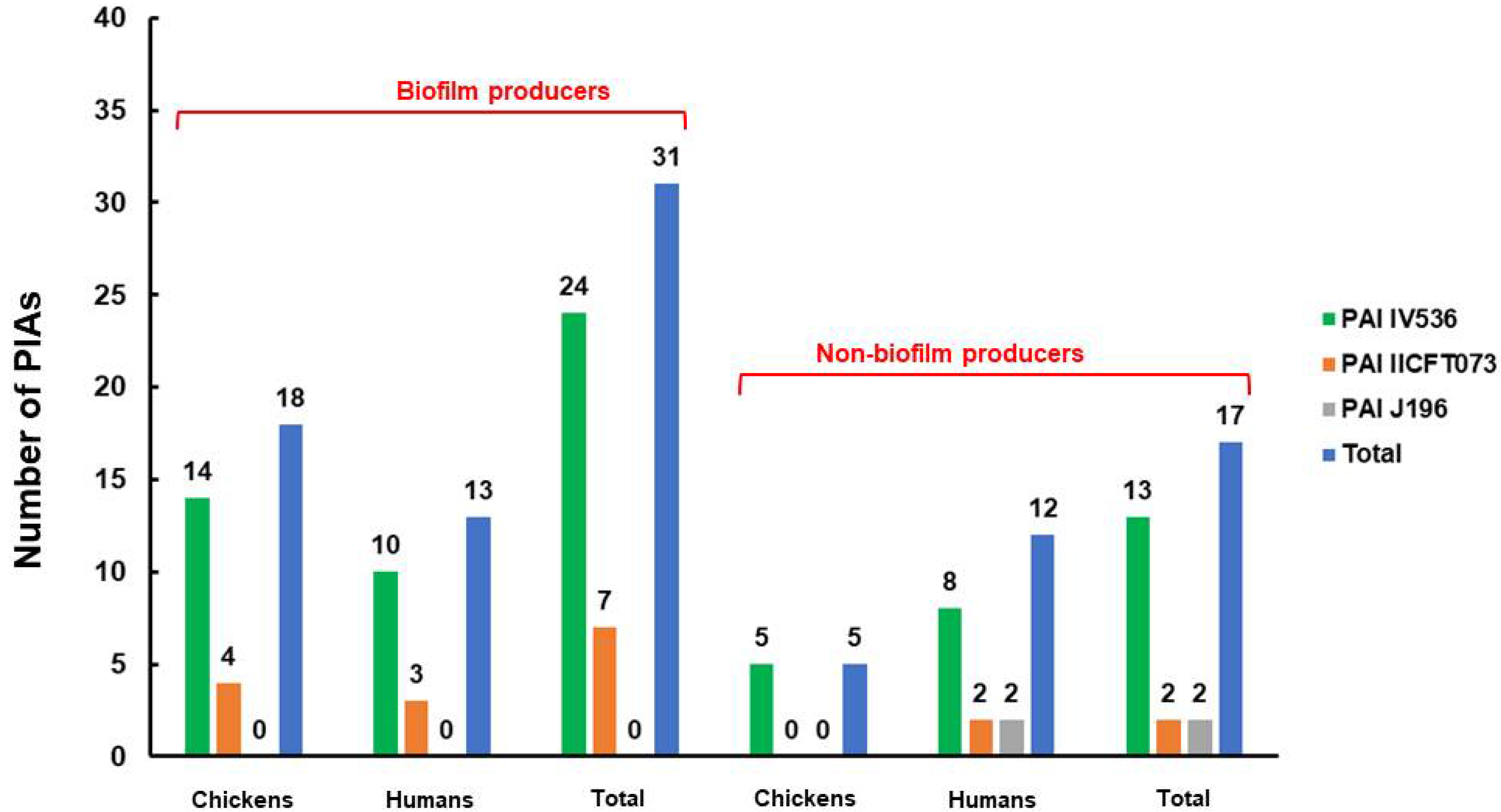
For both the human and chicken meat isolates, the dynamic mechanism of biofilm development was examined. In total, 37 (61.7%) isolates were found to be positive, including 23 chicken isolates and 14 human isolates. Among them, 7 (11.7%) were weak biofilm producers, 15 (25%) were moderate biofilm producers, and 15 (25%) were strong biofilm producers (Table 6). Interestingly, the biofilm-producer isolates carried most of the PAI markers compared to the non-biofilm-producer isolates. Out of 48 PAI markers identified in this study from both chicken (23) and human (25), 31 PAI markers were carried by the biofilm-producer isolates (Figure 3). Moreover, in the chicken isolates, most biofilm-producer isolates (16/23) were assigned to nonpathogenic groups A and B1, while in the human isolates, most biofilm-producer isolates were assigned to groups B1 and B2 (13/14). Noticeably, in the human isolates, all isolates of group D were non-biofilm producers (Figure 4). Concerning the association of antimicrobial resistance with biofilm production, the correlation analysis findings only revealed the existence of a significant positive association (r = 0.31) between biofilm production and resistance to quinolones by the qnr gene (Figure S4).
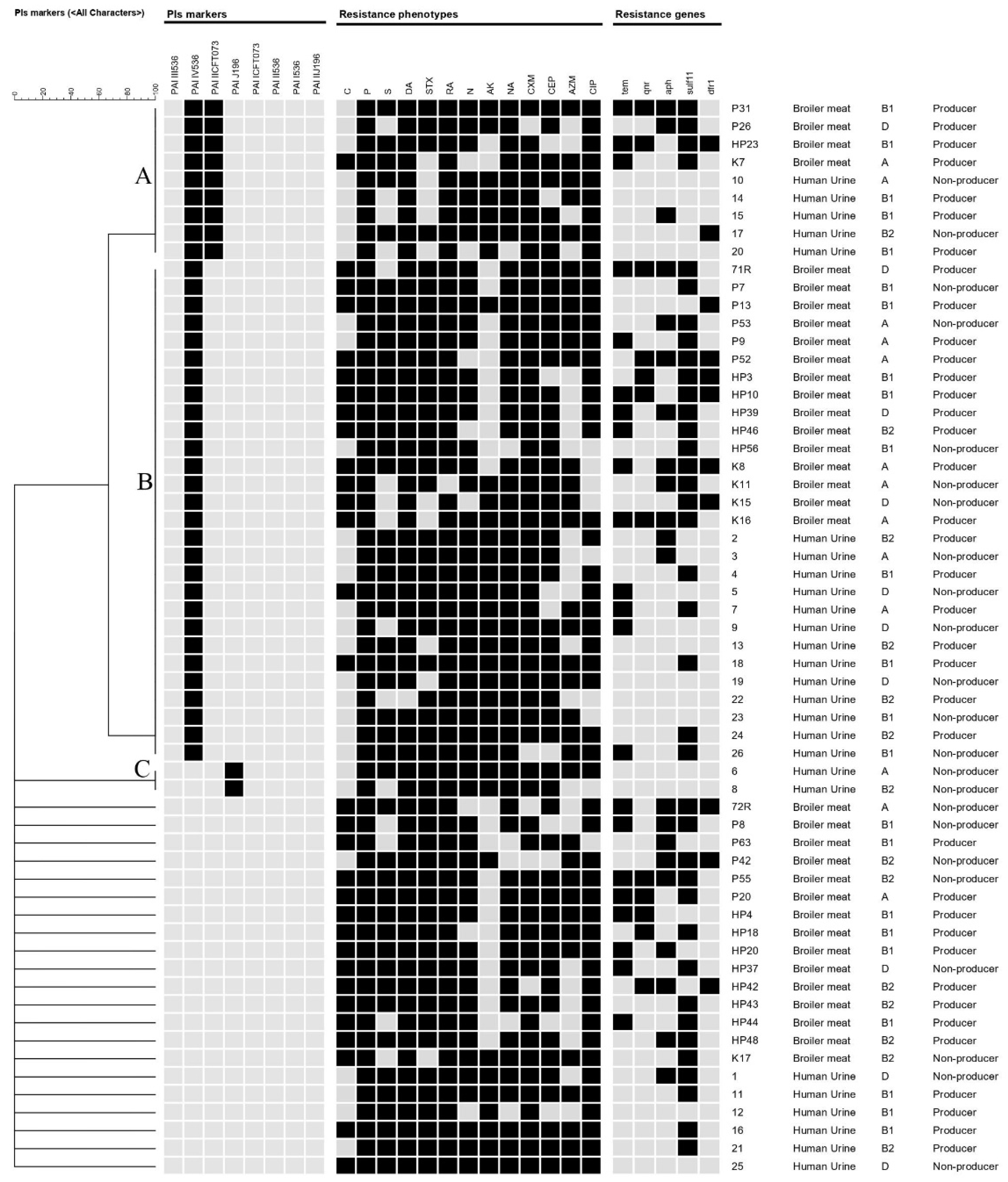
Hierarchal clustering of the chicken and human isolates was performed based on the presence/absence of PAIs among isolates. Chicken and human E. coli were grouped into three major clusters (A, B, and C); isolates in each cluster had identical profiles of PAIs (Figure 5). Both clusters A and B were composed of isolates from both sources, while cluster C contained two isolates sourced from human urine. Within each cluster, the distribution of antimicrobial resistances, phylogenetic groups, and biofilm production were not confined to a certain isolate source.

3. Discussion

Retail meats and poultry serve as an important vehicle for transmitting antimicrobial-resistant and/or pathogenic bacteria to consumers. Pathogenic bacteria with virulence and antimicrobial-resistance characteristics can contaminate poultry meat in large quantities during slaughtering procedures [27]. Food handlers may also transmit pathogenic E. coli from one person to another through food in processing plants. It was assumed that E. coli isolated from poultry and human were completely different; however, recent investigations of both intestinal and extraintestinal pathogenic E. coli isolated from both sources revealed common genetic features [4,5,6,7,8,28,29]. Serogrouping, phylogenetic grouping, virulence genotyping, antimicrobial sensitivity testing, and pathogenicity testing were used frequently to investigate this connection [28,30,31,32,33,34]. In this study, we collected meat and urine samples from chickens and humans, respectively, to study the phylogrouping, distribution of pathogenicity island markers, antimicrobial-resistant genotypes, and phenotypes shared between chickens and humans. Out of 105 fresh chicken meat and 67 urine samples from human urinary tract infections, 34 (32.38%) and 26 (38.8%) E.coli isolates were recovered, respectively. All the recovered E. coli isolates were subjected to PCR-based phylotyping that provided information about virulence based on their grouping into pathogenic and commensal groups. The E. coli isolates were grouped into four primary phylogenetic groups; namely, A, B1, B2, and D. The majority of pathogenic virulent E. coli isolates belonged to phylogenetic groups B2 and D [10]; the virulence genes were mostly associated with phylogroups B2 and D [35,36]. In this study, the presence of phylogroups B1 and A in approximately 38.3% and 26.5%, respectively, of chicken isolates, which was predominantly associated with commensal E. coli, could be attributed to the origin of these strains as commensals that acquired virulence-related genes. Similarly, commensal group A dominated among avian pathogenic E. coli strains in previous studies conducted in the USA, Germany, and Brazil [5,37,38]. On the other hand, pathogenic groups B2 and D were detected in 20.6%% and 14.7%, respectively. Both groups B2 and D are of public health concern because they are thought to be more pathogenic and virulent. As a result, pathogenic phylogenetic groups of chicken isolates could constitute a public health risk through direct contact or contaminated chicken products [38,39].
Regarding the human isolates, the phylotype distribution was as follows: B1 (10/26, 38.5%), B2 (7/26, 26.9%), D (5/26, 19.2%) and A (4/26, 15.4%), with predominance of phylogenetic group B1. This finding agreed with previously reported studies that showed higher frequencies of phylogroup B1 amongst E. coli isolates [40,41] and the infrequency of group A [42,43]. However, the phylogenetic group B2 was reported as the most prevalent group in another study of UTIs [44,45,46]. Many factors influence E. coli’s phylogenetic distribution, including geographical dispersion, host genetic factors, dietary factors, and medicine used [40,44,47].
The presence of different virulence genes on plasmids or chromosomal areas, known as pathogenicity islands (PAIs), is usually connected with E. coli’s potential to induce infection [48]. Many studies have focused on the characterization of PAIs from UPEC, but there is still a gap in the knowledge concerning their characterization in non-UPEC isolates. [49,50]. Therefore, in this study, we studied the distribution of PAI markers among both chicken and human isolates. Overall, 23 PAI markers were identified from chicken isolates, and PAI IV536 was the most prevalent PAI identified. The high frequency of PAI IV536 contributed to its stability because it was the first to be acquired and fixed on the chromosome, while the other PAIs were unstable and easy to lose [51].
Most of the human isolates (n = 20) harbored PAIs markers; likewise, a lower prevalence was reported in [14] and [52], while a higher occurrence of PAIs from E. coli isolated from UTIs was reported [12,53,54]. In this study, PAI IV536 (69.2%), PAI IICFT073 (19.2%), and PAI J196 (7.7%) were identified in human isolates. In detail, PAI IV536 was detected in 13 isolates, PAI J196 was detected in 2 isolates, and both PAI IV536 and PAI IICFT073 were identified in 5 isolates. In total, 20 isolates out of 26 human isolates (76.9%) harbored PAIs. The most frequent combination of PAI markers among chicken and human isolates was PAI IV536+PAI IICFT073. These findings were in agreement with previous reports on humans in Iran [53], China [55], Sweden [36], and Spain [56]. A similar distribution of PAIs with a predominance of PAI IV536 was reported in many studies [12,55,56,57,58,59].
The identification of PAI markers on a regular basis is thought to be a beneficial tool for detecting pathogenic E. coli pathotypes and, as a result, introducing new therapeutic targets [16]. The number of PAIs found in this investigation varied among phylogroups but was considerably high in groups A (34.8%) and B1 (39.1%) in the chicken and human isolates than in the other groups. However, prior research on E. coli isolates from UTIs indicated that the group B1, A, and D UPEC isolates had fewer PAI markers than group B2. [12,56].
PAIs are genomic parts that contain virulence genes responsible for the pathogenicity of bacteria. Although the majority of PAIs are not mobile, additional mobile genetic elements such as conjugative plasmids, bacteriophages, or integrative and conjugative elements (ICEs) can help them spread among bacterial species [18,60,61]. Based on the presence/absence of PAIs, we discovered that E. coli isolates from humans and chickens were clustered together. Most importantly, the existence of isolates that exhibited identical PAI patterns and that belonged to the commensal phylogroup B1 could have been the reason for the possible acquisition of PAIs by commensal E. coli [55].
Multiple antibiotic resistance in E. coli has emerged as a serious problem in both human and veterinary medicine. Most E. coli isolates were found to have a high frequency of resistance in this study. They were extremely resistant to β-lactams, sulfonamides, aminoglycosides, quinolones, lincosamide, fluoroquinolones, and rifampin regardless of the source of E. coli isolates, while slightly lower frequencies of resistance were detected against macrolide (azithromycin), amikacin, and chloramphenicol. These marked antibiotic resistances may contribute to the dissemination of resistance plasmids, which are involved in the spreading of resistance genes between veterinary and human healthcare [62]; specifically, those coding for extended-spectrum lactamases, 16S rRNA methylases, and plasmid-mediated quinolone resistance (PMQR), which confer resistance to broad-spectrum cephalosporins, aminoglycosides, and fluoroquinolones, respectively. Noticeably, MAR was found in all chicken and human isolates against three or more antimicrobial drugs. Furthermore, the MAR of retail chicken meat was greater than 0.6, indicating a high risk of human infection. Long-term exposure and/or indiscriminate use of various antimicrobials in poultry farms for preventive and therapeutic purposes, as well as in human urinary tract infection, may be responsible for these high levels of antimicrobial resistance, which pose major health risks to both animals and humans. Other theories suggest that the transfer of resistance genes via plasmids and other mobile genetic elements is a key process in the development of multiple antibiotic resistance in E. coli. Increased frequencies of sul2, blaTEM, and apha1 genes were found in the investigated isolates, which corresponded to higher resistance phenotypes to the respective antimicrobials (sulfamethoxazole trimethoprim, penicillin, and streptomycin). The pan-resistant strains were mostly assigned to commensal group B1 and were biofilm producers, while one human isolate was assigned to the pathogenic E.coli (group D). Regarding the distribution of PAIs, two strains harbored PAI IV536, while the others tested negative for PAIs. This finding highlighted the emergence of acquiring AMR between E. coli strains regardless of the origin or the phylogeny.
The main complication of E. coli infection is the formation of a biofilm, which is a significant virulence factor. In slaughterhouses and food-processing plants, biofilm growth can result in financial losses due to equipment and facility failures, as well as food spoilage. [63]. Furthermore, biofilm-forming bacteria pose a public health danger because they can adhere to a variety of surfaces and biofilm structures can be broken, resulting in the release of hazardous microbes and product contamination [64]. In humans, biofilms provide bacteria with a survival strategy by allowing them to make the best use of available nutrients while preventing access to antimicrobial drugs, antibodies, and white blood cells [65]. They have also been found to contain a high number of antibiotic-inactivating enzymes such as beta-lactamases, resulting in an antimicrobial resistance island [66]. Controlling infections induced by E. coli biofilms is difficult due to the extracellular matrix and the observed increased resistance to common antibiotics. In this study, 61.7% (37/60) of E.coli isolates were biofilm-formers, including 23 (67.4%) isolates from chickens and 14 (58.8%) isolates from humans. These findings were similar to those reported in strains from birds [67] and humans [68]. This highlighted the importance of investigating alternative therapeutics such as antiadhesion agents, phytochemicals, and nanomaterials for effective drug delivery in humans to limit biofilm formation, as well as the need to improve cleaning and disinfection measures at slaughterhouses.

4. Materials and Methods

4.1. Sample Collection and Conventional Isolation of E. coli

In this study, a total of 172 samples from humans (n = 67) and retail chicken meat (n = 105) were collected between January and July 2019 and examined for the presence of E. coli. An informed consent for the current study was obtained from patients before urine sampling. Midstream urine samples were taken from patients who were clinically diagnosed with urinary tract infections (UTIs) and admitted to the Specialized Medical Hospital in Mansoura, Egypt. The authors were not involved in the sampling process for urine from the patients. Retail chicken meats were randomly collected weekly from retail chicken shops in Mansoura, Egypt. Samples were transported under complete aseptic conditions in an icebox to the laboratory of the Department of Bacteriology, Mycology, and Immunology, Faculty of Veterinary Medicine, Mansoura University, Mansoura, and subjected to conventional bacteriological analysis.
First, 1 mL of urine sample was enriched in 9 mL of tryptic soy broth (TSB; Oxoid, UK) and incubated at 37 °C for 18 h. Retail chicken meat (25 g) was homogenized in TSB (225 mL) and ground in a stomacher, and the homogenate was incubated overnight at 37 °C. A loopful of the enriched broth from the retail chicken meat and human urine samples was plated onto the surface of MacConkey agar plates (HiMedia, Mumbai, India) and incubated at 37 °C for 18–24 h. Lactose fermenter colonies (pink colonies) were picked up from MacConkey agar and subcultured on Eosin Methylene Blue (EMB) agar (HiMedia, Mumbai, India). Presumptive E. coli colonies with the characteristic green color and metallic sheen were subjected to the standard morphological and biochemical tests [69].

4.2. Molecular Characterization of E. coli

In 200 μL of deionized water, three to five bacterial colonies were picked up, heated for 15 min, and then centrifuged for three min at 10,000 rpm. After that, the supernatants were transferred to sterile, clean Eppendorf tubes and kept until used as a DNA template [70]. To confirm the genus identity of presumptive E. coli isolates, a uniplex PCR assay was conducted using a genus-specific primer (Table 7) that targeted variable regions within the 16S small subunit rRNA gene [71].

4.3. PCR-based Phylotyping

Escherichia coli isolates were subjected to a phylogrouping analysis using triplex PCR as proposed by Clermont et al. [10] with three primer pairs: chuA.1–chuA.2, yjaA.1–yjaA.2, and TspE4C2.1–TspE4C2.2 (Table 7). Strains were assigned to four main phylogenetic groups (A, B1, B2, or D) according to the amplification of the three primer pairs [30]. A reaction mixture of 23 μL containing 3 μL of DNA template, 10 mL of 2X PCR master mix (Thermo scientific, Waltham, MA, USA), 4 μL of deionized free water, and 1 μL of every one of the six primers. PCR reactions were applied using 96-well plates and an Applied Biosystem 2720 thermal cycler; the cyclic PCR conditions were as follows: initial denaturation at 94°C for 5 min; 30 cycles of 94 °C for 30 sec, 55 °C for 30 sec, and 72 °C for 30 sec; and a final extension at 72 °C for 7 min. Amplicons (5 μL) were separated in 1% agarose gel stained with ethidium bromide and visualized under a UV transilluminator and a Gel Documentation System (cleaver scientific ltd UV gel documentation system, Rugby, Warwickshire, UK). The results were interrupted according to Bonacorsi et al. [77].

4.4. Detection of Pathogenicity Islands Markers

Eight PAIs in E. coli isolates were investigated in this study using two multiplex PCRs (1 and 2) according to Sabaté et al [56]. Three PAI markers were targeted (PAI III536, PAI IV536, and PAI IICFT073) in the first reaction, yielding 200, 300, and 400 bp, respectively; the reaction mixture measured 20 μL and contained 3 μL of DNA template, 10 μL of 2X PCR master mix (Thermo scientific, Waltham, MA, USA), 6 μL of deionized water, and 1 μL of each of the six primers. The second multiplex PCR targeted five PAI markers (PAI IIJ96, PAI I536, PAI II536, PAI ICFT073, and PAI IJ96), yielding 2300, 1800, 1000, 930, and 400 bp fragments, respectively. The PCR reaction was performed in a total volume of 20 μL containing 3 μL of DNA template, 10 μL of 2X PCR master mix (Thermo scientific, Waltham, MA, USA), 2 μL of deionized water, and 1 μL of each primer. The primer sequences and expected sizes of amplicons for each PCR assay are given in Table 7. Both multiplex PCRs were performed under the following conditions: 94 °C for 5 min; 30 cycles of 94 °C for 1 min, 55 °C for 1 min, and 72 °C for 1 min; and a final extension at 72 °C for 10 min.

4.5. Antimicrobial Susceptibility Testing

The Kirby–Bauer disc diffusion method was used to test the susceptibility of the E. coli isolates to antimicrobial drugs [78]. A total of 13 antimicrobial discs (Oxoid, Basingstoke, UK) were selected, including penicillin (P; 10 μg), cefuroxime (CXM; 30 μg), cefoperazone (CEP; 30 μg), amikacin (AK; 30 μg), streptomycin (S; 15 μg), neomycin (N; 5 μg), azithromycin (AZM; 15 μg), nalidixic acid (NAL; 30 μg), trimethoprim–sulfamethoxazole (SXT; 25 μg), clindamycin (DA; 2 μg), ciprofloxacin (CIP; 5 μg), chloramphenicol (C; 30 μg), and rifampin (RA; 5 μg). These antibiotics are widely used in clinical and veterinary medicine. The percentages, frequencies, and profile of antibiotic-resistant E. coli were obtained and phenotyped for their multiple antibiotic resistance using E. coli ATCC 25922 as a quality control. The isolates that exhibited resistance ≥3 of the antimicrobial classes were recorded. The MAR index was derived by dividing the total number of antimicrobial resistances for each isolate by the total number of tested antimicrobials [79]; this is a crucial indicator in determining the risk source of contamination that is a possible hazard to humans [80]. The results were interpreted according to the interpretative chart in [78].

4.6. Molecular Characterization of Antimicrobial Resistance Genes

E.coli isolates were screened using PCR for characterization of the antimicrobial resistance genes that encoded resistance to β-lactamase (blaTEM), quinolone (qnrA), trimethoprim (dfrA1), aminoglycoside (aphA1), and sulfonamide (sul1). For blaTEM, qnrA, and dfrA1, these were examined as previously described in [73,75,76], respectively, while aphA1 and sul2 were investigated according to the procedure by Maynard et al. [74]. A reaction mixture of 25 μL containing 5 μL of DNA template, 12.5 μL of 2X PCR master mix (Thermo scientific, United States), 5.5 μL of deionized free water, and one μL of each primer was performed.

4.7. Biofilm-Formation Assay

The capacity of the E. coli isolates to produce biofilms was assessed according to [81]. Briefly, E. coli isolates were grown individually in glass tubes containing TSB for 10 h at 28 °C. The inoculated broth was discarded hygienically and the tubes were dyed with a 1% crystal violet solution [82]. After 15 min, the stained tubes were washed with sterile distilled water. All isolates, including the uninoculated TSB used as a negative control, were examined in triplicate. The stained tubes were examined for a visible film lining their walls and bottoms.
The results for PAI marker screening, resistance phenotypes and genotypes, phylogenetic groups, and biofilm production were imported into the BioNumerics software (Applied Maths, Austin, TX, USA, version 7.6) to generate a dendrogram for clustering the examined isolates from humans and chicken meat. The dendrogram was generated based on the PAI results using the band-based Dice coefficient and the unweighted pair group method with arithmetic averages (UPGMA). A correlation analysis to determine the association of antimicrobial resistance phenotypes, resistance genotypes, and biofilm production among the examined isolates was performed as previously described [83,84] (add these references please).

5. Conclusions

The findings of this study were suggestive of the transmission of PAI markers and antibiotic resistance genes from poultry to humans or humans to humans through the food chain, which could be a potential source of human disease. Thus, to avoid the spread of virulent and multidrug-resistant E. coli, intensive surveillance of retail chicken meat markets is required.

Supplementary Materials

The following supporting information can be downloaded at: https://www.mdpi.com/2079-6382/11/9/1197/s1, Figure S1. Agarose gel electrophoresis showing amplification of 16S small subunit rRNA gene of E. coli at 200 bp. Figure S2. Agarose gel electrophoresis showing ECOR phylogenetic grouping of representative E. coli isolates according to PCR: lane M: 100 bp DNA ladder; lanes 2, 9, 10, and 13: phylogroup A; lanes 3–7: phylogroup B1; lanes 8 and 11: phylogroup B2; lane 1: phylogroup D. Figure S3. Agarose gel electrophoresis showing amplification of E.coli resistant genes: lane M: 100 bp DNA ladder; lane 1: blaTEM at 1150 bp; lane 2: sul2 at 722 bp; lane 3: aphA1 at 600 bp; lane 4: qnrA at 514 bp; lane 5: dfrA1 at 367 bp. Figure S4. Association of antimicrobial resistance phenotypes, resistance genotypes, and biofilm production among the examined E. coli isolates. Blue and red boxes denote positive and negative correlations, respectively. Significant associations were calculated at p < 0.05; blank boxes denote non-significant correlations.

Author Contributions

Conceptualization, A.S. and A.A. (Amal Awad); Data curation, A.S. and A.A. (Amal Awad); Formal analysis, A.S., H.R., M.M.S., S.M.A., A.A. (Amira Abugomaa), M.E. and A.A. (Amal Awad); Funding acquisition, M.M.S., S.M.A. and H.F.A.-H.; Investigation, A.S., H.R. and A.A. (Amal Awad); Methodology, A.S., M.A.E., A.M.H., M.M.S., H.F.A.-H., A.A. (Amira Abugomaa), M.E. and A.A. (Amal Awad); Project administration, A.S., H.R. and A.A. (Amal Awad); Resources, A.S., H.R. and A.A. (Amal Awad); Software, H.R., A.A. (Amira Abugomaa) and M.E.; Supervision, A.A. (Amal Awad); Validation, A.A. (Amal Awad); Visualization, A.A. (Amal Awad); Writing—original draft, A.S., H.R. and A.A.; Writing—review and editing, A.S., H.R., M.A.E., A.M.H., M.M.S., S.M.A., H.F.A.-H., A.A. (Amira Abugomaa), M.E. and A.A. (Amal Awad). All authors have read and agreed to the published version of the manuscript.

Funding

This study was supported by the Taif University Researchers Supporting Project (TURSP-2020/105), Taif University, Taif, Saudi Arabia.

Institutional Review Board Statement

The study was conducted according to the guidelines of the Declaration of Helsinki and approved by the Committee on the Ethics of Animal Experiments of the Faculty of Veterinary Medicine, Mansoura University (Permit numbers R/60 on 27 February 2021).

Informed Consent Statement

Not applicable.

Data Availability Statement

The datasets generated during and/or analyzed during the current study are available from the corresponding author on reasonable request.

Acknowledgments

We appreciate and thank Taif University for the financial support from the Taif University Researchers Supporting Project (TURSP-2020/105), Taif University, Taif, Saudi Arabia.

Conflicts of Interest

The authors declare that they have no competing interests.

Correction Statement

This article has been republished with a minor correction to the existing affiliation information. This change does not affect the scientific content of the article.

References

  1. Leimbach, A.; Hacker, J.; Dobrindt, U. E. coli as an All-Rounder: The thin line between commensalism and pathogenicity. In Between Pathogenicity and Commensalism; Dobrindt, U., Hacker, J.H., Svanborg, C., Eds.; Springer: Berlin/Heidelberg, Germany, 2013; pp. 3–32. [Google Scholar]
  2. Croxen, M.A.; Finlay, B.B. Molecular mechanisms of Escherichia coli pathogenicity. Nat. Rev. Microbiol. 2010, 8, 26–38. [Google Scholar] [CrossRef] [PubMed]
  3. Köhler, C.D.; Dobrindt, U. What defines extraintestinal pathogenic Escherichia coli? Int. J. Med. Microbiol. 2011, 301, 642–647. [Google Scholar] [CrossRef] [PubMed]
  4. Ewers, C.; Li, G.; Wilking, H.; Kieβling, S.; Alt, K.; Antáo, E.-M.; Laturnus, C.; Diehl, I.; Glodde, S.; Homeier, T.; et al. Avian pathogenic, uropathogenic, and newborn meningitis-causing Escherichia coli: How closely related are they? Int. J. Med. Microbiol. 2007, 297, 163–176. [Google Scholar] [CrossRef] [PubMed]
  5. Rodriguez-Siek, K.E.; Giddings, C.W.; Doetkott, C.; Johnson, T.J.; Fakhr, M.K.; Nolan, L.K. Comparison of Escherichia coli isolates implicated in human urinary tract infection and avian colibacillosis. Microbiology 2005, 151, 2097–2110. [Google Scholar] [CrossRef] [PubMed]
  6. Zhao, L.; Gao, S.; Huan, H.; Xu, X.; Zhu, X.; Yang, W.; Gao, Q.; Liu, X. Comparison of virulence factors and expression of specific genes between uropathogenic Escherichia coli and avian pathogenic E. coli in a murine urinary tract infection model and a chicken challenge model. Microbiol. Read. 2009, 155, 1634–1644. [Google Scholar] [CrossRef]
  7. Maluta, R.P.; Logue, C.M.; Casas, M.R.T.; Meng, T.; Guastalli, E.A.L.; Rojas, T.C.G.; Montelli, A.C.; Sadatsune, T.; de Carvalho, R.M.; Nolan, L.K.; et al. Overlapped sequence types (STs) and serogroups of avian pathogenic (APEC) and human extra-intestinal pathogenic (ExPEC) Escherichia coli isolated in Brazil. PLoS ONE 2014, 9, e105016. [Google Scholar] [CrossRef]
  8. Nandanwar, N.; Janssen, T.; Kühl, M.; Ahmed, N.; Ewers, C.; Wieler, L.H. Extraintestinal pathogenic Escherichia coli (ExPEC) of human and avian origin belonging to sequence type complex 95 (STC95) portray indistinguishable virulence features. Int. J. Med. Microbiol. 2014, 304, 835–842. [Google Scholar] [CrossRef] [PubMed]
  9. Bélanger, L.; Garenaux, A.; Harel, J.; Boulianne, M.; Nadeau, E.; Dozois, C.M. Escherichia coli from animal reservoirs as a potential source of human extraintestinal pathogenic E. Coli. FEMS Immunol. Med. Microbiol. 2011, 62, 1–10. [Google Scholar] [CrossRef]
  10. Clermont, O.; Bonacorsi, S.; Bingen, E. Rapid and simple determination of the Escherichia coli phylogenetic group. Appl. Environ. Microbiol. 2000, 66, 4555–4558. [Google Scholar] [CrossRef]
  11. Johnson, J.R.; Delavari, P.; Kuskowski, M.; Stell, A.L. Phylogenetic distribution of extraintestinal virulence-associated traits in Escherichia coli. J. Infect. Dis. 2001, 183, 78–88. [Google Scholar] [CrossRef]
  12. Navidinia, M.; Najareerayeh, S.; Fallah, F.; Bakhshi, B.; Adabian, S.; Alimehr, S.; Gholinejad, Z. Distribution of the Pathogenicity Islands Markers (PAIs) in uropathogenic E. coli isolated from children in Mofid children hospital. Arch. Pediatr. Infect. Dis. 2013, 1, 75–79. [Google Scholar] [CrossRef]
  13. Navidinia, M.; Peerayeh, S.N.; Fallah, F.; Bakhshi, B.; Sajadinia, R.S. Phylogenetic grouping and pathotypic comparison of urine and fecal Escherichia coli isolates from children with urinary tract infection. Braz. J. Microbiol. 2014, 45, 509–514. [Google Scholar] [CrossRef] [PubMed]
  14. Neamati, F.; Firoozeh, F.; Saffari, M.; Zibaei, M. Virulence genes and antimicrobial resistance pattern in uropathogenic Escherichia coli isolated from hospitalized patients in Kashan, Iran. Jundishapur. J. Microbiol. 2015, 8, e17514. [Google Scholar] [CrossRef]
  15. Hacker, J.; Blum-Oehler, G.; Mühldorfer, I.; Tschäpe, H. Pathogenicity islands of virulent bacteria: Structure, function and impact on microbial evolution. Mol. Microbiol. 1997, 23, 1089–1097. [Google Scholar] [CrossRef]
  16. Oelschlaeger, T.A.; Dobrindt, U.; Hacker, J. Pathogenicity islands of uropathogenic E. coli and the evolution of virulence. Int. J. Antimicrob. Agents 2002, 19, 517–521. [Google Scholar] [CrossRef] [PubMed]
  17. Tangi, S.C.; Tajbakhsh, E.; Soleimani, N.A.; Shahraki, M.M. Prevalence of pathogenicity island markers genes in uropathogenic Escherichia coli isolated from patients with urinary tract infectious. Asian Pac. J. Trop. Dis. 2015, 5, 662–666. [Google Scholar] [CrossRef]
  18. Schmidt, H.; Hensel, M. Pathogenicity islands in bacterial pathogenesis. Clin. Microbiol. Rev. 2004, 17, 14–56. [Google Scholar] [CrossRef]
  19. Bingen-Bidois, M.; Clermont, O.; Bonacorsi, S.; Terki, M.; Brahimi, N.; Loukil, C.; Barraud, D.; Bingen, E. Phylogenetic analysis and prevalence of urosepsis strains of escherichia coli bearing pathogenicity island-like domains. Infect. Immun. 2002, 70, 3216–3226. [Google Scholar] [CrossRef]
  20. Chroma, M.; Kolar, M. Genetic methods for detection of antibiotic resistance: Focus on extended-spectrum β-lactamases. Biomed. Pap. 2010, 154, 289–296. [Google Scholar] [CrossRef]
  21. Russo, T.A.; Johnson, J.R. Medical and economic impact of extraintestinal infections due to Escherichia coli: Focus on an increasingly important endemic problem. Microbes. Infect. 2003, 5, 449–456. [Google Scholar] [CrossRef]
  22. Johnson, T.J.; Nolan, L.K. Pathogenomics of the virulence plasmids of Escherichia coli. Microbiol. Mol. Biol. Rev. 2009, 73, 750–754. [Google Scholar] [CrossRef] [PubMed]
  23. Hammerum, A.M.; Heuer, O.E. Human Health Hazards from Antimicrobial-Resistant Escherichia coli of Animal Origin. Clin. Infect. Dis. 2009, 48, 916–921. [Google Scholar] [CrossRef] [PubMed]
  24. Overdevest, I.; Willemsen, I.; Rijnsburger, M.; Eustace, A.; Xu, L.; Hawkey, P.; Heck, M.; Savelkoul, P.; Vandenbroucke-Grauls, C.; van der Zwaluw, K.; et al. Extended-spectrum β-lactamase genes of Escherichia coli in chicken meat and humans, The Netherlands. Emerg. Infect. Dis. J. 2011, 17, 1216–1222. [Google Scholar] [CrossRef] [PubMed]
  25. Dewanti, R.; Wong, A.C.L. Influence of culture conditions on biofilm formation by Escherichia coli O157:H7. Int. J. Food Microbiol. 1995, 26, 147–164. [Google Scholar] [CrossRef]
  26. Berlanga, M.; Guerrero, R. Living together in biofilms: The microbial cell factory and its biotechnological implications. Microb. Cell Factories 2016, 15, 165. [Google Scholar] [CrossRef] [PubMed]
  27. Johnson, J.R.; Kuskowski, M.A.; Smith, K.; O’Bryan, T.T.; Tatini, S. Antimicrobial-resistant and extraintestinal pathogenic Escherichia coli in retail foods. J. Infect. Dis. 2005, 191, 1040–1049. [Google Scholar] [CrossRef] [PubMed]
  28. Mellata, M.; Johnson, J.R.; Curtiss, R., III. Escherichia coli isolates from commercial chicken meat and eggs cause sepsis, meningitis and urinary tract infection in rodent models of human infections. Zoonoses Public Health 2018, 65, 103–113. [Google Scholar] [CrossRef]
  29. Johnson, J.R.; Porter, S.B.; Johnston, B.; Thuras, P.; Clock, S.; Crupain, M.; Rangan, U. Extraintestinal pathogenic and antimicrobial-resistant Escherichia coli, including sequence type 131 (st131), from retail chicken breasts in the United States in 2013. Appl. Environ. Microbiol. 2017, 83, e02956-16. [Google Scholar] [CrossRef]
  30. Lima-Filho, J.V.; Martins, L.V.; Nascimento, D.C.d.O.; Ventura, R.F.; Batista, J.E.C.; Silva, A.F.B.; Ralph, M.T.; Vaz, R.V.; Rabello, C.B.-V.; Silva, I.d.M.M.d.; et al. Zoonotic potential of multidrug-resistant extraintestinal pathogenic Escherichia coli obtained from healthy poultry carcasses in Salvador, Brazil. Braz. J. Infect. Dis. 2013, 17, 54–61. [Google Scholar] [CrossRef]
  31. Giufrè, M.; Graziani, C.; Accogli, M.; Luzzi, I.; Busani, L.; Cerquetti, M.; Escherichia coli Study Group. Escherichia coli of human and avian origin: Detection of clonal groups associated with fluoroquinolone and multidrug resistance in Italy. J. Antimicrob. Chemother. 2012, 67, 860–867. [Google Scholar] [CrossRef]
  32. Kluytmans, J.A.J.W.; Overdevest, I.T.M.A.; Willemsen, I.; Kluytmans-van den Bergh, M.F.Q.; van der Zwaluw, K.; Heck, M.; Rijnsburger, M.; Vandenbroucke-Grauls, C.M.J.E.; Savelkoul, P.H.M.; Johnston, B.D.; et al. Extended-spectrum β-Lactamase–producing Escherichia coli from retail chicken meat and humans: Comparison of strains, plasmids, resistance genes, and virulence factors. Clin. Infect. Dis. 2012, 56, 478–487. [Google Scholar] [CrossRef] [PubMed]
  33. Tóth, I.; Dobrindt, U.; Koscsó, B.; Kósa, A.; Herpay, M.; Nagy, B. Genetic and phylogenetic analysis of avian extraintestinal and intestinal Escherichia coli. Acta Microbiol. Immunol. Hung. 2012, 59, 393–409. [Google Scholar] [CrossRef]
  34. Literak, I.; Reitschmied, T.; Bujnakova, D.; Dolejska, M.; Cizek, A.; Bardon, J.; Pokludova, L.; Alexa, P.; Halova, D.; Jamborova, I. Broilers as a source of quinolone-resistant and extraintestinal pathogenic Escherichia coli in the Czech Republic. Microb. Drug Resist. 2012, 19, 57–63. [Google Scholar] [CrossRef] [PubMed]
  35. Parham, N.J.; Pollard, S.J.; Chaudhuri, R.R.; Beatson, S.A.; Desvaux, M.; Russell, M.A.; Ruiz, J.; Fivian, A.; Vila, J.; Henderson, I.R. Prevalence of pathogenicity island IICFT073 genes among extraintestinal clinical isolates of Escherichia coli. J. Clin. Microbiol. 2005, 43, 2425–2434. [Google Scholar] [CrossRef]
  36. Ostblom, A.; Adlerberth, I.; Wold, A.E.; Nowrouzian, F.L. Pathogenicity island markers, virulence determinants malX and usp, and the capacity of Escherichia coli to persist in infants’ commensal microbiotas. Appl. Env. Microbiol. 2011, 77, 2303–2308. [Google Scholar] [CrossRef] [PubMed]
  37. Barbieri, N.L.; Oliveira, A.L.D.; Tejkowski, T.M.; Pavanelo, D.B.; Matter, L.B.; Pinheiro, S.R.S.; Vaz, T.M.I.; Nolan, L.K.; Logue, C.M.; Brito, B.G.D.; et al. Molecular Characterization and Clonal Relationships Among Escherichia coli Strains Isolated from Broiler Chickens with Colisepticemia. Foodborne Pathog. Dis. 2014, 12, 74–83. [Google Scholar] [CrossRef]
  38. Ewers, C.; Antão, E.-M.; Diehl, I.; Philipp, H.-C.; Wieler, L.H. Intestine and environment of the chicken as reservoirs for extraintestinal pathogenic Escherichia coli strains with zoonotic potential. Appl. Environ. Microbiol. 2009, 75, 184–192. [Google Scholar] [CrossRef]
  39. Cortés, P.; Blanc, V.; Mora, A.; Dahbi, G.; Blanco, J.E.; Blanco, M.; López, C.; Andreu, A.; Navarro, F.; Alonso, M.P.; et al. Isolation and characterization of potentially pathogenic antimicrobial-resistant Escherichia coli strains from chicken and pig farms in Spain. Appl. Environ. Microbiol. 2010, 76, 2799–2805. [Google Scholar] [CrossRef]
  40. Duriez, P.; Clermont, O.; Bonacorsi, S.; Bingen, E.; Chaventré, A.; Elion, J.; Picard, B.; Denamur, E. Commensal Escherichia coli isolates are phylogenetically distributed among geographically distinct human populations. Microbiology 2001, 147, 1671–1676. [Google Scholar] [CrossRef]
  41. Derakhshandeh, A.; Firouzi, R.; Moatamedifar, M.; Motamedi, A.; Bahadori, M.; Naziri, Z. Phylogenetic analysis of Escherichia coli strains isolated from human samples. Mol. Biol. Res. Commun. 2013, 2, 143–149. [Google Scholar]
  42. Bingen, E.; Picard, B.; Brahimi, N.; Mathy, S.; Desjardins, P.; Elion, J.; Denamur, E. Phylogenetic analysis of Escherichia coli strains causing neonatal meningitis suggests horizontal gene transfer from a predominant pool of highly virulent b2 group strains. J. Infect. Dis. 1998, 177, 642–650. [Google Scholar] [CrossRef] [PubMed]
  43. Johnson, J.R.; Manges, A.R.; O’Bryan, T.T.; Riley, L.W. A disseminated multidrug-resistant clonal group of uropathogenic Escherichia coli in pyelonephritis. Lancet 2002, 359, 2249–2251. [Google Scholar] [CrossRef]
  44. Sannes, M.R.; Kuskowski, M.A.; Owens, K.; Gajewski, A.; Johnson, J.R. Virulence factor profiles and phylogenetic background of Escherichia coli isolates from veterans with bacteremia and uninfected control subjects. J. Infect. Dis. 2004, 190, 2121–2128. [Google Scholar] [CrossRef]
  45. Zhang, L.; Foxman, B.; Marrs, C. Both urinary and rectal Escherichia coli isolates are dominated by strains of phylogenetic group B2. J. Clin. Microbiol. 2002, 40, 3951–3955. [Google Scholar] [CrossRef]
  46. Kuznetsova, M.V.; Maslennikova, I.L.; Pospelova, J.S.; Bertok D, Ž.; Erjavec, M.S. Differences in recipient ability of uropathogenic Escherichia coli strains in relation with their pathogenic potential. Infect. Genet. Evol. 2022, 97, 105160. [Google Scholar]
  47. Lee, J.C.; Lee, N.Y.; Lee, H.C.; Huang, W.H.; Tsui, K.C.; Chang, C.M.; Lee, C.C.; Chen, P.L.; Wu, C.J.; Hsueh, P.R.; et al. Clinical characteristics of urosepsis caused by extended-spectrum beta-lactamase-producing Escherichia coli or Klebsiella pneumonia and their emergence in the community. J. Microbiol. Immunol. Infect. 2012, 45, 127–133. [Google Scholar] [CrossRef] [PubMed]
  48. Ahmed, A.M.; Shimamoto, T.; Shimamoto, T. Molecular characterization of multidrug-resistant avian pathogenic Escherichia coli isolated from septicemic broilers. Int. J. Med. Microbiol. 2013, 303, 475–483. [Google Scholar] [CrossRef] [PubMed]
  49. Che, D.; Hasan, M.S.; Chen, B. Identifying pathogenicity islands in bacterial pathogenomics using computational approaches. Pathogens 2014, 3, 36–56. [Google Scholar] [CrossRef]
  50. Naderi, G.; Haghi, F.; Zeighami, H.; Hemati, F.; Masoumian, N. Distribution of pathogenicity island (PAI) markers and phylogenetic groups in diarrheagenic and commensal Escherichia coli from young children. Gastroenterol. Hepatol. Bed Bench 2016, 9, 316–324. [Google Scholar]
  51. Middendorf, B.; Hochhut, B.; Leipold, K.; Dobrindt, U.; Blum-Oehler, G.; Hacker, J. Instability of pathogenicity islands in uropathogenic Escherichia coli 536. J. Bacteriol. 2004, 186, 3086–3096. [Google Scholar] [CrossRef]
  52. Johnson, J.R.; Stell, A.L. Extended virulence genotypes of Escherichia coli strains from patients with urosepsis in relation to phylogeny and host compromise. J. Infect. Dis. 2000, 181, 261–272. [Google Scholar] [CrossRef]
  53. Samei, A.; Haghi, F.; Zeighami, H. Distribution of pathogenicity island markers in commensal and uropathogenic Escherichia coli isolates. Folia Microbiol. 2016, 61, 261–268. [Google Scholar] [CrossRef] [PubMed]
  54. Firoozeh, F.; Saffari, M.; Neamati, F.; Zibaei, M. Detection of virulence genes in Escherichia coli isolated from patients with cystitis and pyelonephritis. Int. J. Infect. Dis. 2014, 29, 219–222. [Google Scholar] [CrossRef] [PubMed]
  55. Li, B.; Sun, J.-Y.; Han, L.-Z.; Huang, X.-H.; Fu, Q.; Ni, Y.-X. Phylogenetic groups and pathogenicity island markers in fecal Escherichia coli isolates from asymptomatic humans in China. Appl. Environ. Microbiol. 2010, 76, 6698–6700. [Google Scholar] [CrossRef]
  56. Sabaté, M.; Moreno, E.; Pérez, T.; Andreu, A.; Prats, G. Pathogenicity island markers in commensal and uropathogenic Escherichia coli isolates. Clin. Microbiol. Infect. 2006, 12(9), 880–886. [Google Scholar] [CrossRef]
  57. Gultekin, E.O.; Gultekin, E.O.; Ulger, S.T.; Delialioğlu, N. Distribution of Pathogenicity Island Markers and Virulence Factors Genes of Extraintestinal Pathogenic Escherichia coli Isolates. Jundishapur J. Microbiol. 2022, 15, e121044. [Google Scholar]
  58. El-Shaer, S.; Abdel-Rhman, S.H.; Barwa, R.; Hassan, R. Genetic characterization of extended-spectrum β-Lactamase-and carbapenemase-producing Escherichia coli isolated from Egyptian hospitals and environments. PloS One 2021, 16, e0255219. [Google Scholar] [CrossRef] [PubMed]
  59. El-Mahdy, R.; Mahmoud, R.; Shrief, R. Characterization of E. coli phylogroups causing catheter-associated urinary tract infection. Infect. Drug Resist. 2021, 14, 3183. [Google Scholar] [CrossRef] [PubMed]
  60. Gal-Mor, O.; Finlay, B.B. Pathogenicity islands: A molecular toolbox for bacterial virulence. Cell Microbiol. 2006, 8, 1707–1719. [Google Scholar] [CrossRef]
  61. Desvaux, M.; Dalmasso, G.; Beyrouthy, R.; Barnich, N.; Delmas, J.; Bonnet, R. Pathogenicity factors of genomic islands in intestinal and extraintestinal Escherichia coli. Front. Microbiol. 2020, 11, 2065. [Google Scholar] [CrossRef]
  62. Carattoli, A. Plasmids and the spread of resistance. Int. J. Med. Microbiol. 2013, 303, 298–304. [Google Scholar] [CrossRef]
  63. Hung, C.S.; Henderson, J.P. Emerging concepts of biofilms in infectious diseases. Mo. Med. 2009, 106, 292–296. [Google Scholar]
  64. Costerton, J.W.; Lewandowski, Z.; Caldwell, D.E.; Korber, D.R.; Lappin-Scott, H.M. Microbial biofilms. Annu. Rev. Microbiol. 1995, 49, 711–745. [Google Scholar] [CrossRef] [PubMed]
  65. Nandakumar, V.; Chittaranjan, S.; Kurian, V.M.; Doble, M. Characteristics of bacterial biofilm associated with implant material in clinical practice. Polym. J. 2013, 45, 137–152. [Google Scholar] [CrossRef]
  66. Davies, J.; Davies, D. Origins and evolution of antibiotic resistance. Microbiol. Mol. Biol. Rev. MMBR 2010, 74, 417–433. [Google Scholar] [CrossRef] [PubMed]
  67. Skyberg, J.A.; Siek, K.E.; Doetkott, C.; Nolan, L.K. Biofilm formation by avian Escherichia coli in relation to media, source and phylogeny. J. Appl. Microbiol. 2007, 102, 548–554. [Google Scholar] [CrossRef]
  68. Katongole, P.; Nalubega, F.; Florence, N.C.; Asiimwe, B.; Andia, I. Biofilm formation, antimicrobial susceptibility and virulence genes of Uropathogenic Escherichia coli isolated from clinical isolates in Uganda. BMC Infect. Dis. 2020, 20, 453. [Google Scholar] [CrossRef]
  69. Quinn, P.; Markey, B.; Carter, M.; Donnelly, W.; Leonard, F. Veterinary Microbiology and Microbial Disease; Blackwell Science Company: Hoboken, NJ, USA; State University Press: Detroit, MI, USA, 2002. [Google Scholar]
  70. Alexopoulou, K.; Foka, A.; Petinaki, E.; Jelastopulu, E.; Dimitracopoulos, G.; Spiliopoulou, I. Comparison of two commercial methods with PCR restriction fragment length polymorphism of the tuf gene in the identification of coagulase-negative staphylococci. Lett. Appl. Microbiol. 2006, 43, 450–454. [Google Scholar] [CrossRef]
  71. Teichmann, A.; Agra, H.N.; de Souza Nunes, L.; da Rocha, M.P.; Renner, J.D.; Possuelo, L.G.; Carneiro, M.; Rieger, A.; Benitez, L.B.; Valim, A.R. Antibiotic resistance and detection of the sul2 gene in urinary isolates of Escherichia coli in patients from Brazil. J. Infect. Dev. Ctries. 2014, 8, 39–43. [Google Scholar] [CrossRef]
  72. Higgins, J.; Hohn, C.; Hornor, S.; Frana, M.; Denver, M.; Joerger, R. Genotyping of Escherichia coli from environmental and animal samples. J. Microbiol. Methods 2007, 70, 227–235. [Google Scholar] [CrossRef]
  73. Belaaouaj, A.; Lapoumeroulie, C.; Caniça, M.M.; Vedel, G.; Névot, P.; Krishnamoorthy, R.; Paul, G. Nucleotide sequences of the genes coding for the TEM-like β-lactamases IRT-1 and IRT-2 (formerly called TRI-1 and TRI-2). FEMS Microbiol. Lett. 1994, 120, 75–80. [Google Scholar] [CrossRef] [PubMed]
  74. Maynard, C.; Fairbrother, J.M.; Bekal, S.; Sanschagrin, F.; Levesque, R.C.; Brousseau, R.; Masson, L.; Larivière, S.; Harel, J. Antimicrobial Resistance Genes in Enterotoxigenic Escherichia coli O149:K91 Isolates Obtained over a 23-Year Period from Pigs. Antimicrob. Agents Chemother. 2003, 47, 3214–3221. [Google Scholar] [CrossRef] [PubMed]
  75. Toro, C.S.; FarfÁN, M.; Contreras, I.; Flores, O.; Navarro, N.; Mora, G.C.; Prado, V. Genetic analysis of antibiotic-resistance determinants in multidrug-resistant Shigella strains isolated from Chilean children. Epidemiol. Infect. 2005, 133, 81–86. [Google Scholar] [CrossRef] [PubMed]
  76. Robicsek, A.; Strahilevitz, J.; Sahm, D.F.; Jacoby, G.A.; Hooper, D.C. qnr prevalence in ceftazidime-resistant Enterobacteriaceae isolates from the United States. Antimicrob. Agents Chemother. 2006, 50, 2872–2874. [Google Scholar] [CrossRef] [PubMed]
  77. Bonacorsi, S.P.P.; Clermont, O.; Tinsley, C.; Le Gall, I.; Beaudoin, J.-C.; Elion, J.; Nassif, X.; Bingen, E. Identification of regions of the Escherichia coli chromosome specific for neonatal meningitis-associated strains. Infect. Immun. 2000, 68, 2096–2101. [Google Scholar] [CrossRef] [PubMed]
  78. CLSI. Performance Standards for Antimicrobial Susceptibility Testing: Twenty-Seventh Informational Supplement M100-S27; CLSI: Wayne, PA, USA, 2017. [Google Scholar]
  79. Titilawo, Y.; Sibanda, T.; Obi, L.; Okoh, A. Multiple antibiotic resistance indexing of Escherichia coli to identify high-risk sources of faecal contamination of water. Environ. Sci. Pollut. Res. 2015, 22, 10969–10980. [Google Scholar] [CrossRef]
  80. Krumperman, P.H. Multiple antibiotic resistance indexing of Escherichia coli to identify high-risk sources of fecal contamination of foods. Appl. Environ. Microbiol. 1983, 46, 165–170. [Google Scholar] [CrossRef]
  81. Kadam, S.R.; den Besten, H.M.W.; van der Veen, S.; Zwietering, M.H.; Moezelaar, R.; Abee, T. Diversity assessment of Listeria monocytogenes biofilm formation: Impact of growth condition, serotype and strain origin. Int. J. Food Microbiol. 2013, 165, 259–264. [Google Scholar] [CrossRef]
  82. Wolfe, A.J.; Millikan, D.S.; Campbell, J.M.; Visick, K.L. Vibrio fischeri sigma54 controls motility, biofilm formation, luminescence, and colonization. Appl. Env. Microbiol. 2004, 70, 2520–2524. [Google Scholar] [CrossRef]
  83. Sadat, A.; El-Sherbiny, H.; Zakaria, A.; Ramadan, H.; Awad, A. Prevalence, antibiogram and virulence characterization of Vibrio isolates from fish and shellfish in Egypt: A possible zoonotic hazard to humans. J. Appl. Microbiol. 2020, 131, 485–498. [Google Scholar]
  84. Sadat, A.; Shata, R.R.; Farag, A.M.M.; Ramadan, H.; Alkhedaide, A.; Soliman, M.M.; Elbadawy, M.; Abugomaa, A.; Awad, A. Prevalence and Characterization of PVL-Positive Staphylococcus aureus Isolated from Raw Cow’s Milk. Toxins 2022, 14, 97. [Google Scholar] [CrossRef]
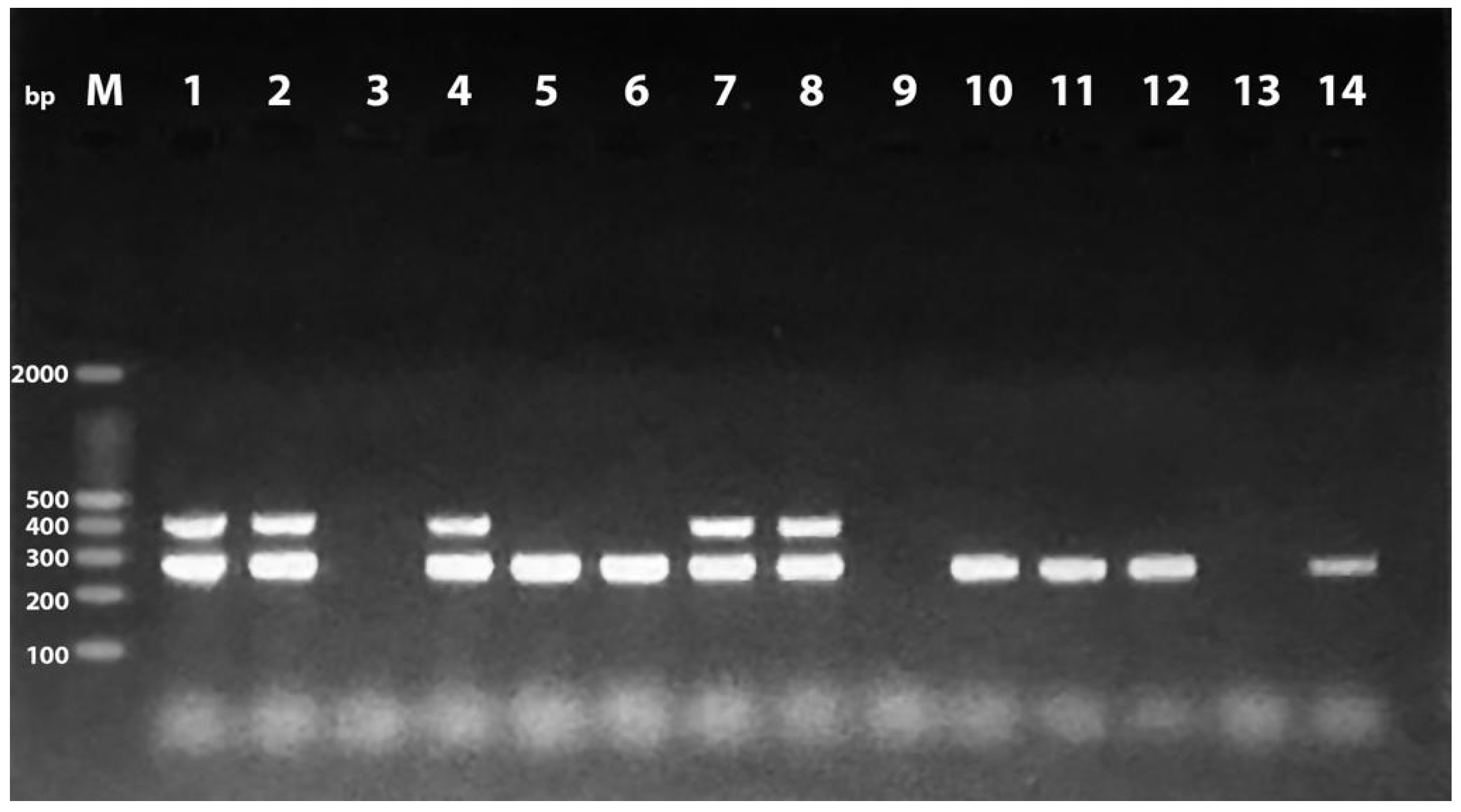
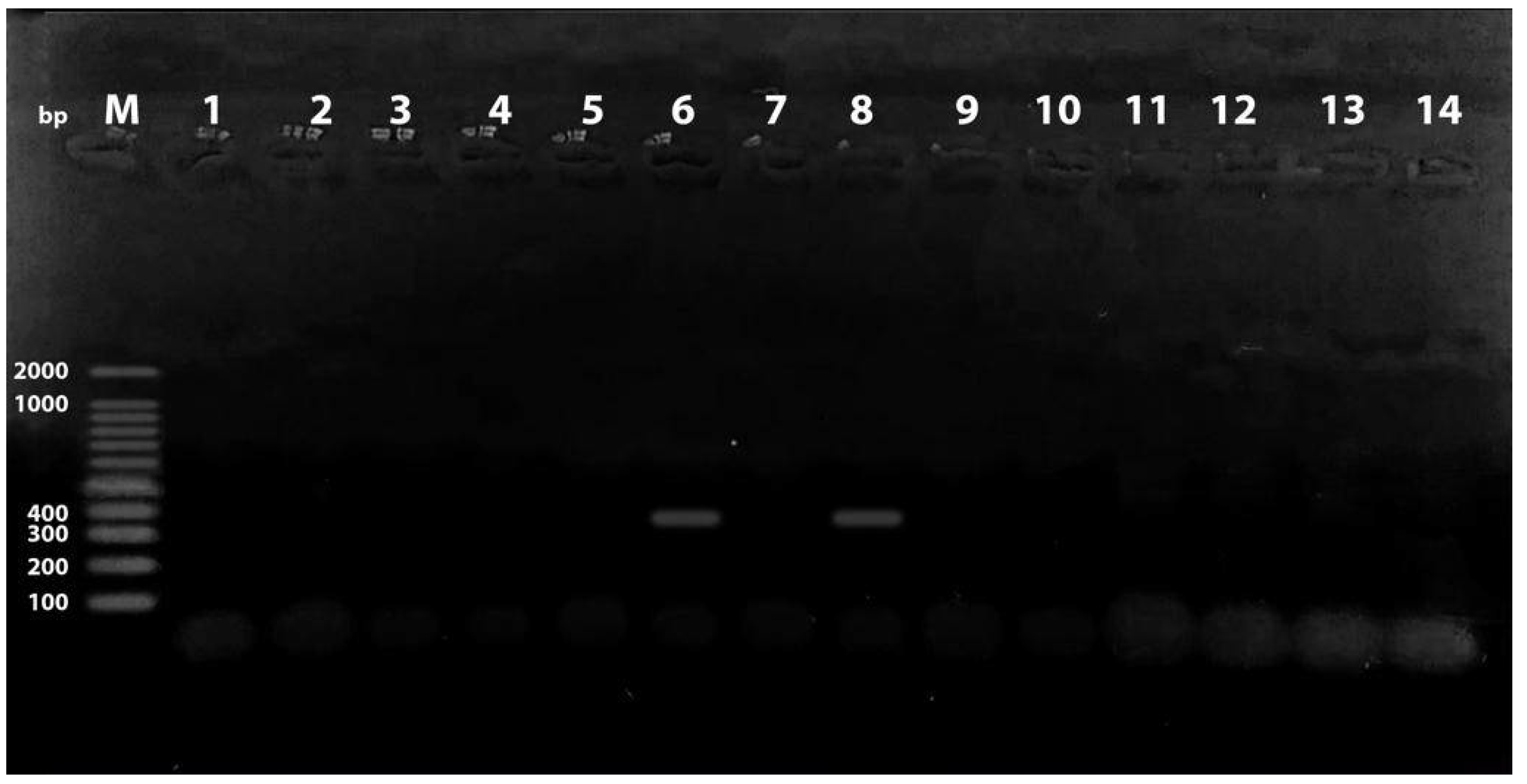
Figure 1. Multiplex PCR for identification of PAI IV536 and PAI IICFT073 at 300 and 400 bp. Lane M: 100 bp DNA ladder; lanes 1, 2, 4, 7, and 8: positive for both PAI IV536 and PAI IICFT073; lanes 5, 6, 10, 11, 12, and 14: positive for PAI IICFT073; lanes 3, 9, and 13: negative samples.
Antibiotics 11 01197 g001
Figure 2. Multiplex PCR showing amplification of PAI IJ96 at 400 bp. Lane M: 100 bp DNA ladder; lanes 6 and 8: positive samples; lanes 1–5, 7, and 9–14: negative samples.
Antibiotics 11 01197 g002
Figure 3. Distribution of phylogenetic groups among E. coli isolates in relation to biofilm production.
Antibiotics 11 01197 g003
Figure 4. Distribution of pathogenicity islands (PAIs) among E. coli isolates in relation to biofilm production.
Antibiotics 11 01197 g004
Figure 5. Hierarchal clustering of chicken and human isolates based on the existence of pathogenic islands (PAIs) using Dice coefficient and the unweighted pair group method (UPGMA) in BioNumerics software (Applied Maths, Austin, TX, USA, version 7.6). Black and gray squares represent positive and negative results, respectively, for PAIs, resistant phenotypes, and resistant genotypes. Phylogroups and biofilm production results were also determined.
Antibiotics 11 01197 g005
Table 1. Frequency distribution of phylogenetic groups among the examined E. coli isolates from chicken and human.
Source of Isolates Phylogenetic Group
Pathogenic PhylogroupsCommensal Phylogroups
Group B2Group DTotalGroup AGroup B1Total
Chicken (n = 34)7 (20.6%)5 (14.7%)12 (35.3%)9 (26.5%)13 (38.3%)22 (64.7%)
Human (n = 26)7 (26.9%)5 (19.2%)12 (46.2)4 (15.4%)10 (38.5%)14 (53.8%)
Table 2. Pathogenicity island (PAI) markers identified in E. coli isolates from chicken and human.
Source of IsolatesPAI III536PAI IV536PAI IICFT073PAI
J196
PAI
ICFT073
PAI II536PAI I536PAI IIJ196
Chicken-19 (55.9%)4 (11.8%)-----
Human-18 (69.2%)5 (19.2%)2(7.7%)----
Table 3. Distribution of pathogenicity islands (PAIs) among E. coli isolates in relation to phylogenetic group.
Isolates
PAIs/Phylogenetic GroupPAI III536PAI IV536PAI IICFT073PAI J196PAI ICFT073PAI II536PAI I536PAI IIJ196Total
Chicken (n = 34)B2 010000001 (4.34%)
D 041000005 (21.7%)
A 071000008 (34.8%)
B1 072000009 (39.1%)
Total01940000023 (100%)
Human (n = 26)B2051100007 (28%)
D030000003 (12%)
A031100005 (20%)
B10730000010 (40%)
Total01852000025 (100%)
Table 4. Results of antimicrobial susceptibility testing for the examined E. coli isolates.
Antibiotics FamilyDisc CodeCPDChicken (34)Human (26)
ResistantSensitiveResistantSensitive
Penicillin β-lactamP1034 (100%)0 (0.00%)26 (100%)0 (0.00%)
CefuroximeCephalosporinCXM3030 (88.2%)4 (11.8%)25 (96.2%)1 (3.9%)
CefoperazoneCEP3029 (85.3%)5 (14.7%)21 (80.8%)5 (19.2%)
AmikacinAminoglycosideAK308 (23.5%)26 (76.5%)26 (100%)0 (0.00%)
StreptomycinS1525 (73.5%)9 (26.5%)20 (76.9%)6 (23.1%)
NeomycinN528 (82.4%)6 (17.6%)24 ( 92.3%)2 ((7.7%)
AzithromycinMacrolideAZM1520 (58.8%)14 (41.2%)15 (57.7%)11 (42.3%)
Nalidixic acid QuinoloneNAL3030 (88.2%)4 (11.8%)24 (92.3%)2 (7.7%)
Trimethoprim–sulfamethoxazoleSulfonamideSXT2530 (88.2%)4 (11.8%)20 (76.9%)6 (23.1%)
ClindamycinLincosamideDA234 (100%)0 (0.00%)25 (96.2%)1 (3.8%)
Ciprofloxacin FluoroquinoloneCIP529 (85.3%)5 (14.7%)22 (84.6%)4 (15.4%)
Chloramphenicol PhenicolsC3027 (79.4%)7 (20.6%)4 (15.4%)22 (84.6%)
Rifampin RifamycinRifampin533 (97.1%)1 (2.9%)26 (100%)0 (0.00%)
Table 5. Different patterns of antimicrobial susceptibility detected for the E. coli isolates.
AntibiotypesResistance PatternIsolates No. (%)MARMAR Index
Chicken IC, P, DA, STX, RA, N, CXM, CIP1 (2.9%)80.62
IIP, S, DA, STX, RA, N, CXM, CEP1 (2.9%)60.62
IIIC, P, S, DA, SXT, RA, NA, CEP, CIP1 (2.9%)90.7
IVC, P, DA, STX, RA, N, NA, CXM, CIP1 (2.9%)90.7
VC, P, DA, STX, RA, N, CXM, CEP, AZM1 (2.9%)80.7
VIP, S, DA, STX, RA, N, AK, AZM, CIP1 (2.9%)80.7
VIIP, DA, STX, RA, N, AK, NA, CEP, CIP1 (2.9%)80.7
VIIIP, S, DA, STX, RA, N, NA, CXM, CIP1 (2.9%)80.7
IXC, P, DA, RA, AK, NA, CXM, CEP, AZM1 (2.9%)80.7
XC, P, S, DA, STX, RA, N, NA, CXM, CIP1 (2.9%)90.77
XIC, P, S, DA, STX, RA, N, NA, CEP, CIP1 (2.9%)90.77
XIIC, P, S, DA, STX, RA, NA, CXM, CEP, CIP1 (2.9%)90.77
XIIIC, P, S, DA, RA, NA, CXM, CEP, AZM, CIP1 (2.9%)90.77
XIVC, P, DA, STX, N, AK, NA, CXM, CEP, AZM 1 (2.9%)80.77
XVC, P, DA, STX, RA, N, NA, CXM, CEP, AZM, CIP 1 (2.9%)100.84
XVIP, S, DA, STX, RA, N, NA, CXM, CEP, AZM, CIP2 (5.8%)90.84
XVIIC, P, S, DA, STX, RA, NA, CXM, CEP, AZM. CIP2 (5.8%)100.84
XVIIIC, P, S, DA, STX, RA, N, NA, CXM, CEP, CIP5 (14.7%)90.84
XIXC, P, S, DA, STX, RA, N, NA, CXM, CEP, AZM1 (2.9%)90.84
XXC, P, DA, RA, N, AK, NA, CXM, CEP, AZM, CIP2 (5.8%)90.84
XXIC, P, S, DA, STX, RA, N, NA, CXM, CEP, AZM, CIP5 (17.7%)100.9
XXIIP, S, DA, STX, RA, N, AK, NA, CXM, CEP, AZM, CIP1 (2.9%)90.9
XXIIIC, P, S, DA, STX, RA, N, AK, NA, CXM, CEP, AZM, CIP1(2.9%)101
Human I P, DA, RA, AK, CXM, CEP, CIP1 (3.8%)60.54
IIP, S, DA, STX, RA, AK, CXM, CIP1 (3.8%)70.62
IIIP, STX, RA, N, AK, NA, CXM, CEP1 (3.8%)70.62
IVP, DA, SXT, RA, N, AK, NA, CXM, CEP1 (3.8%)80.7
VP, DA, RA, N, AK, NA, CXM, AZM, CIP1 (3.8%)80.7
VIP, DA, RA, N, AK, NA, CXM, CEP, CIP1 (3.8%)70.7
VIIP, S, DA, STX, RA, N, AK, NA, CXM, CEP1 (3.8%)80.77
VIIIP, S, DA, RA, N, AK, NA, CXM, CEP, CIP1 (3.8%)70.77
IXP, S, DA, STX, RA, N, AK, NA, AZM, CIP1 (3.8%)80.77
XP, S, DA, SXT, RA, N, AK, NA, CXM, CEP, CIP3 (11.5%) 80.85
XIC, P, S, DA, SXT, RA, N, AK, NA, CXM, CIP1 (3.8%)90.85
XIIP, S, DA, STX, RA, N, AK, NA, CXM, AZM, CIP1 (3.8%)100.85
XIIIP, DA, STX, RA, N, AK, NA, CXM, CEP, AZM, CIP1 (3.8%)90.85
XIVP, S, DA, RA, N, AK, NA, CXM, CEP, AZM, CIP2 (7.7%)80.85
XVP, S, DA, STX, RA, N, AK, NA, CXM, CEP, AZM1 (3.8%)80.85
XVIP, S, DA, STX, RA, N, AK, NA, CXM, CEP, AZM, CIP5 (19.2%)90.92
XVIIC, P, S, DA, STX, RA, N, AK, NA, CXM, CEP, AZM, CIP3 (11.5%)101
Table 6. Prevalence of antimicrobial resistance genes and biofilm production detected in E. coli isolates.
Point of DifferencesItems TestedChicken Isolates (34)Human Isolates (26)Total(60)
Resistant genesblaTEM18 (52.9%)4 (15.4%)22 (36.7%)
qnrA12 (35.3%)0 (0.00%)12 (20%)
aphA117 (50%)4 (15.4%)21 (35%)
sul229 (85.3%)9 (34.6%)38 (63.3%)
dfrA110 (29.4%)1 (3.8%)11 (18.3%)
Biofilm productionNegative11 (32.4%)12 (46.2%)23 (38.3%)
Weak positive4 (11.8%)3 (11.5%)7 (11.7%)
Moderate11 (32.4%)4 (15.4%)15 (25%)
Strong positive8 (23.5%)7 (26.9%)15 (25%)
Overall positive23 (67.6%)14 (53.8%)37 (61.7%)
Table 7. List of oligonucleotide primers used in the present study.
GenePrimer NamePrimer Sequences (5’ to 3’)(bp)Reference
16S rRNA16S-FF-GCGGACGGGTGAGTAATGT200[71]
16S-RR-TCATCCTCTCAGACCAGCTA
Pathogenicity Island (PAI) Genes
PAI I536I.9F-TAATGCCGGAGATTCATTGTC1800[56]
1.10R-AGGATTTGTCTCAGGGCTTT
PAI II536orf1 upF-CCATGTCCAAAGCTCGAGC1000
orf1 downR-CTACGTCAGGCTGGCTTTG
PAI III536sfaAI.1F-CGGGCATGCATCAATTATCTTTG200[3]
sfaAI.2R-TGTGTAGATGCAGTCACTCCG
PAI IV536IRP2 FPF-AAGGATTCGCTGTTACCGGAC300[3]
IRP2 RPR-TCGTCGGGCAGCGTTTCTTCT
PAI IICFT073cft073.2Ent1F-ATGGATGTTGTATCGCGCP400[56]
cft073.2Ent2R-ACGAGCATGTGGATCTGC
PAI J196papGIfF-TCGTGCTCAGGTCCGGAATTT400[13]
papGIRR-TGGCATCCCACATTATCG
PAI IIJ196HlydF-GGATCCATGAAAACATGGTTAATGGG2300
CnfR-GATATTTTTGTTGCCATTGGTTACC
PAI ICFT073RPAiF-GGACATCCTGTTACAGCACGCA930
RPAfR-TCGCCACCAATCACAGCCGAAC
Phylogenetic Island Genes
ChuAChuA-FF-GACGAACCAACGGTCAGGAT279[72]
ChuA-RR-TGCCGCCAGTACCAAAGACA
YjaAYjaA-FF-TGAAGTGTCAGGAGACGCTG211
YjaA-RR-ATGGAGAATGCGTTCCTCAAC
TspE4C2TspE4C2-FF-GAGTAATGTCGGGGCATTCA152
TspE4C2-RR-CGCGCCAACAAAGTATTACG
Antimicrobial Resistance Genes
blaTEMTem-FATTCTTGAAGACGAAAGG1150[73]
Tem-RACGCTCAGTGGAACGAAAAC
sul2Sul2-FCGGCATCGTCAACATAACC722[74]
Sul2-RGTGTGCGGATGAAGTCAG
dfrA1dfrA1-FGGAGTGCCAAAGGTGAACAGC367[75]
dfrA1-RGAGGCGAAGTCTTGGGTAAAAAC
qnrAQnrA-FATTTCTCACGCCAGGATTTG514[76]
QnrA-RGATCGGCAAAGGTTAGGTCA
aphA1aphA1-FATGGGCTCGCGATAATGTC600[74]
Publisher’s Note: MDPI stays neutral with regard to jurisdictional claims in published maps and institutional affiliations.