Article

Antibiotic Susceptibility and Minimum Inhibitory Concentration for Stenotrophomonas maltophilia Ocular Infections

1
Department of Ophthalmology, Chang Gung Memorial Hospital, Taoyuan 333, Taiwan
2
College of Medicine, Chang Gung University, Taoyuan 333, Taiwan
3
Department of Ophthalmology, Chang Gung Memorial Hospital, Keelung 204, Taiwan
*
Authors to whom correspondence should be addressed.
Antibiotics 2022, 11(11), 1457; https://doi.org/10.3390/antibiotics11111457
Submission received: 1 October 2022 / Revised: 19 October 2022 / Accepted: 21 October 2022 / Published: 22 October 2022

Abstract

:
Stenotrophomonas maltophilia (S. maltophilia) is a Gram-negative, opportunistic pathogen that can lead to ocular infections, such as keratitis and endophthalmitis. The purpose of this study was to determine the antibiotic susceptibility and minimum inhibitory concentrations (MICs) of S. maltophilia isolates from ocular infections and to evaluate the differences in antibiotic MICs between keratitis and endophthalmitis isolates. The disc diffusion method revealed that S. maltophilia isolates exhibited 91% susceptibility to levofloxacin and moxifloxacin and 61% susceptibility to trimethoprim–sulfamethoxazole (TMP–SMX). The E-test indicated that S. maltophilia isolates exhibited 40%, 100%, 72%, 91%, 91%, and 93% susceptibility to ceftazidime, tigecycline, TMP–SMX, levofloxacin, gatifloxacin, and moxifloxacin, respectively. The MIC90 values of amikacin, ceftazidime, cefuroxime, tigecycline, TMP–SMX, levofloxacin, gatifloxacin, and moxifloxacin were >256, >256, >256, 3, >32, 1, 2, and 0.75 µg/mL, respectively. The geometric mean MICs of ceftazidime, TMP–SMX, levofloxacin, gatifloxacin, and moxifloxacin were significantly lower for the keratitis isolates than for the endophthalmitis isolates (p = 0.0047, 0.003, 0.0029, 0.0003, and 0.0004, respectively). Fluoroquinolones showed higher susceptibility and lower MICs for the S. maltophilia isolates when compared with other antibiotics. Fluoroquinolones can be recommended for treating S. maltophilia ocular infections. Tigecycline and TMP–SMX could be alternative antibiotics for S. maltophilia ocular infections.

1. Introduction

Stenotrophomonas maltophilia is a Gram-negative, opportunistic, nosocomial pathogen that can lead to severe systemic diseases, such as bacteremia, pneumonia, soft-tissue infections, and endocarditis, especially among immunocompromised patients [1,2,3,4]. In ocular infections, S. maltophilia could cause keratitis, conjunctivitis, dacryocystitis, scleritis, and endophthalmitis, which may lead to ocular discomfort and visual impairment [5,6,7]. In Taiwan, the most commonly used antibiotics for vision-threatening ocular infections are topical 0.5% levofloxacin for bacterial keratitis and intravitreal ceftazidime (2.25 mg/0.1 mL) and vancomycin (1 mg/0.1 mL) for bacterial endophthalmitis. The treatment of ocular S. maltophilia infection has been challenging for ophthalmologists because the S. maltophilia strain is resistant to multiple antibiotics, including penicillins, third-generation cephalosporins, aminoglycosides, and imipenem [6,8,9,10] and has variable susceptibility to fluoroquinolones, tigecycline, and trimethoprim–sulfamethoxazole (TMP–SMX) [11,12,13]. One of the most widely discussed antimicrobial resistance mechanisms (ARMs) of S. maltophilia was the multidrug-resistance (MDR) efflux pumps [14]. The ATP-binding cassette efflux pump (SmrA), one of the MDR efflux pumps, may contribute to the resistance of fluoroquinolones and tetracycline. Other efflux pumps, such as SmeDEF, one of the most well-known efflux pumps in the resistance nodulation division (RND) family, may also affect the susceptibility to antimicrobials, such as fluoroquinolones, tetracycline, and TMP-SMX. Other ARMs of S. maltophilia include β-Lactamases, aminoglycoside-inactivating enzymes, RNase G inactivation, the class I integrons, and the biocide resistance [15,16].
Minimum inhibitory concentrations (MICs) are defined as the lowest concentrations of antimicrobials that will inhibit the visible growth of a microorganism after overnight incubation [17]. It was used to determine the susceptibility of antimicrobials and found the potential treatment for microorganisms. As far as we were aware, reports on the antibiotic MICs for S. maltophilia isolates collected from other ocular infections are rare, and whether the MICs for such isolates are similar to those for endophthalmitis isolates is unclear [18,19]. Accordingly, the aim of the present study was to investigate the antibiotic susceptibility—analyzed using MICs—of S. maltophilia isolates collected from patients with ocular infections over a 10-year period.

2. Materials and Methods

The study design conformed to the principles of the Declaration of Helsinki and was approved by the Institutional Review Board of Chang Gung Memorial Hospital (IRB number: 202000181B0, 2020/02/06v1), Taoyuan, Taiwan. S. maltophilia isolates were retrospectively collected from patients with ocular infections from 1 January 2010 to 30 April 2019. For keratitis, the isolates were collected through corneal scraping; for endophthalmitis, the isolates were collected from anterior chamber or vitreous samples obtained through tapping or vitrectomy.

2.1. Antibiotic Susceptibility Testing

All microbiology investigations were performed at the microbiology department of Chang Gung Memorial Hospital, Taoyuan, Taiwan. Different methods were used to identify bacterial culture isolates. From January 2010 to December 2013, conventional microbiological methods were used to identify culture isolates, while from January 2014 to April 2019, matrix-assisted laser desorption/ionization time-of-flight mass spectrometry (MALDI-TOF-MS) was used to identify culture isolates. Conventional microbiological methods, including Gram-staining and biochemical tests, were also employed. In MALDI-TOF-MS, the spectra of the bacteria were automatically measured and compared to reference spectra using an Ultraflextreme mass spectrometer and MALDI-Biotyper 3.0 software (Bruker Daltonics, Karlsruhe, Germany). The reliability of identification in the MALDI Biotyper system was expressed in points. A log (score) of ≥2.0 indicated identification to the species level. The susceptibility of the isolates to various antibiotics was tested using the Kirby–Bauer disc diffusion method on Mueller Hinton blood agar. The Clinical and Laboratory Standards Institute (CLSI, Wayne, PA, USA, 2021) standards [16] were used for interpretation and quality control for each corresponding year. The antibiotics selected for susceptibility testing were levofloxacin, moxifloxacin, and TMP–SMX.
The antibiotics selected for MIC testing were tigecycline, cefuroxime, levofloxacin, TMP–SMX, gatifloxacin, moxifloxacin, amikacin, and ceftazidime. The MICs were determined using susceptibility strips (Epsilometer test (E-test); bioMérieux S.A., Marcy l’Etoile, France). The culture was conducting by taking the cryotubes from the freezer (−80 °C) and scraping one loop of solution with an inoculation ring on the blood agar plate (Trypticase® Soy Agar with 5% Sheep Blood, Nippon Becton Dickinson Co., Ltd., Tokyo, Japan). Then, the plate was incubated at 37 °C for 16 to 18 h. After the incubation, a single colony was selected for subculture.
The E-test was performed using Mueller Hinton Agar (MHA; CMP®, New Taipei City, Taiwan) with the following procedure:
-
A suspension from a single colony of an overnight growth blood agar plate was prepared.
-
The turbidity of inoculum was adjusted to match 0.5 McFarland standard and was inoculated on MHA within 15 min.
-
A sterile cotton swab was dipped into the solution and was rotated inside the tube to remove excess liquid.
-
The swab was inoculated over the entire surface of the plate.
-
The E-test strip was applied to the plate within 15 min, and the whole plate was put in the incubator within 15 min.
The antibiotic susceptibility of each isolate was determined by comparing the MIC of each agent with the MIC breakpoints according to the CLSI guidelines [20].

2.2. Statistical Analysis
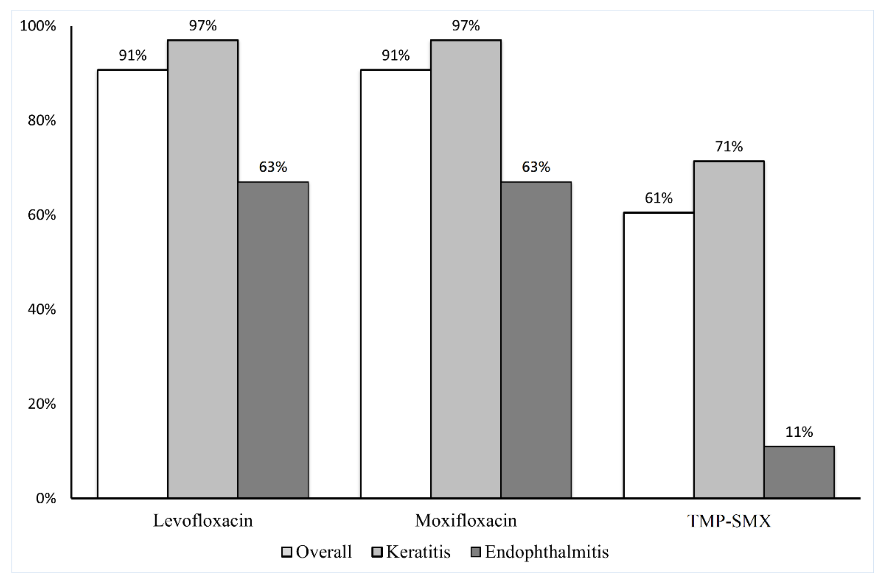
Descriptive statistics were used to evaluate the MIC data in this study. MIC50, representing the concentration of antibiotics that would inhibit the growth of 50% of isolates, and MIC90, representing the concentration of antibiotics that would inhibit the growth of 90% of isolates, were used for the evaluation. The mode was defined as the greatest frequency of MIC distribution for each antibiotic.
Since the MIC data for all antibiotic agents did not follow a normal distribution, the Mann–Whitney U test was used to compare the geometric mean (GM) of the MICs (hereafter denoted as GM-MICs) for keratitis isolates with those for endophthalmitis isolates. If the MIC values were more than 256, it was presumed to be 256 for the calculation of GM-MIC; if the MIC values were more than 32, it was presumed to be 32 for the calculation of GM-MIC. The significance level was set at p = 0.05. All statistical analyses were performed using MedCalc Statistical Software version 20.009 (MedCalc Software Ltd., Ostend, Belgium, 2021).

3. Results

A total of 43 S. maltophilia isolates were included in this study. Among our study group, thirty-five isolates were collected from patients with keratitis, and eight were collected from patients with endophthalmitis. The MICs of the antibiotics for S. maltophilia are listed in Table 1. Relatively low MIC50 values were observed for tigecycline (1 µg/mL), TMP–SMX (0.19 µg/mL), levofloxacin (0.5 µg/mL), gatifloxacin (0.38 µg/mL), and moxifloxacin (0.125 µg/mL). Moreover, relatively low MIC90 values were observed for tigecycline (3 µg/mL), levofloxacin (1 µg/mL), gatifloxacin (2 µg/mL), and moxifloxacin (0.75 µg/mL). Accordingly, based on the CLSI guidelines, S. maltophilia was determined to exhibit 100% susceptibility to tigecycline, 72% susceptibility to TMP–SMX, 91% susceptibility to levofloxacin and gatifloxacin, 93% susceptibility to moxifloxacin, and 40% susceptibility to ceftazidime.
Table 2 presents a comparison of the antibiotic MIC50, MIC90, and GM-MIC values between keratitis isolates and endophthalmitis isolates. The results revealed that the GM-MIC values of ceftazidime, TMP–SMX, levofloxacin, gatifloxacin, and moxifloxacin were significantly lower for the keratitis isolates than for the endophthalmitis isolates (p = 0.0047, 0.003, 0.0029, 0.0003, and 0.0004, respectively).
Figure 1 demonstrates the susceptibility of the isolates as determined through the Kirby–Bauer disc diffusion method. Of the 43 isolates, 39 (91%) were susceptible to levofloxacin and moxifloxacin and 26 (61%) were susceptible to TMP–SMX. Among the isolates collected from patients with keratitis, 34 (97%) were susceptible to levofloxacin and moxifloxacin and 25 (71%) were susceptible to TMP–SMX. Among the isolates collected from the patients with endophthalmitis, five (63%) were susceptible to both levofloxacin and moxifloxacin, and one (11%) was susceptible to TMP–SMX. The comparisons of the susceptibility of S. maltophilia isolates for each antibiotics using the disc diffusion method and E-test are presented in Figure 2.

4. Discussion

This study demonstrated the antibiotic susceptibility determined using MICs of S. maltophilia isolates obtained from patients with ocular infections. The S. maltophilia isolates exhibited a higher susceptibility and lower MICs to fluoroquinolones than to other antibiotics. By comparing the MICs of the antibiotics for S. maltophilia isolates collected from various ocular sites, we observed that the GM-MICs of fluoroquinolones, TMP–SMX, and ceftazidime were significantly lower for keratitis isolates than for endophthalmitis isolates. To the best of our knowledge, this study is the first to determine the MICs of antibiotics for S. maltophilia keratitis and to compare antibiotic susceptibility between keratitis isolates and endophthalmitis isolates.
Several methods are available for testing the susceptibility of S. maltophilia isolates to antibiotic agents [21,22]. The Kirby–Bauer disc diffusion method is among the most commonly used tests for determining antibiotic susceptibility in clinical laboratory settings [23]. This method provides adequate results and is simple and reproducible without the need for any expensive equipment [24]. The E-test is another method for testing the susceptibility of S. maltophilia isolates to antimicrobial agents. This test is less labor intensive and more convenient for daily laboratory routines [21]. Yao et al. [21] compared the performance of the agar dilution method and the E-test in determining S. maltophilia antibiotic susceptibility and reported that the E-test exhibited an overall agreement of 94% with the agar dilution method. Gülmez et al. [22] compared the performance of the E-test, Phoenix system, and disc diffusion method and discovered that the E-test exhibited acceptable agreement with the Phoenix system and disc diffusion method regarding susceptibility to TMP–SMX and tigecycline. Furthermore, Nicodemo et al. [25] compared the disc diffusion method, E-test, and agar dilution method and discovered that they exhibited good agreement in determining susceptibility to gatifloxacin, TMP–SMX, and tigecycline. Khan et al. [26] also reported similar results for TMP–SMX and levofloxacin, indicating that both were the most active antimicrobials agents for S. maltophilia isolates. In the present study, the E-test and disc diffusion method yielded similar susceptibility levels to levofloxacin and moxifloxacin, but the disc diffusion method revealed a lower susceptibility level to TMP–SMX than did the E-test.
According to the worldwide SENTRY program (1997–2016) which obtained 6467 S. maltophilia isolates from different body sites, the S. maltophilia isolates were 81.5% and 80.9% susceptible to levofloxacin worldwide and in the Asia-Pacific area based on the CLSI guideline, respectively [27]. Previous studies have reported that S. maltophilia isolates obtained from patients with ocular infections exhibited high susceptibility to fluoroquinolones [28,29]. Palioura et al. [28] reported that 96% of isolates collected from 26 patients with infectious keratitis were susceptible to fluoroquinolones. Wu et al. [29] collected S. maltophilia isolates from 21 patients with culture-proven keratitis; all collected isolates were susceptible to levofloxacin and moxifloxacin. Our previous study [18] revealed that levofloxacin as well as moxifloxacin were susceptible to S. maltophilia in 67% of isolates, and the MIC90 values were lowest for the S. maltophilia isolates in comparison with other antimicrobial agents (1, 8, and 12 µg/mL for levofloxacin, moxifloxacin, and gatifloxacin, respectively) from isolates from endophthalmitis. In the present study, the fluoroquinolones were over 90% susceptible for S. maltophilia isolates in both the disk diffusion test and the E-test. These results were consistent with previous studies and showed that the fluoroquinolones had presented high in vitro activity for S. maltophilia isolates. The possible explanation for the decrease in MICs for S. maltophilia isolates is related to the MDR efflux pumps. The SmeDEF, one of the efflux pumps in the MDR efflux system, is related to a two-to-eight-fold decrease of the MIC values of fluoroquinolones [30].
We also discovered that the S. maltophilia isolates exhibited 100% susceptibility to tigecycline at a low MIC90 value (3 µg/mL), indicating that tigecycline could be a clinical candidate for the treatment of S. maltophilia ocular infections. A multicentered study [31], including 1586 S. maltophilia isolates collected from the bloodstream and respiratory tract, demonstrated that tigecycline (to which 95.5% of the isolates susceptible at an MIC of ≤2 μg/mL) and TMP–SMX (to which 96.0% of the isolates were susceptible at an MIC of ≤2 μg/mL for TMP and 38 μg/mL for SMX) were the only tested antibiotics associated with more than 94% susceptibility. In a 10-year long multi-center study in Taiwan [32], S. maltophilia isolates were most susceptible to minocycline (99.7%) and tigecycline (96%) and then to TMP–SMX (82.5%) and levofloxacin (79.6%); the MIC90 values of both minocycline and tigecycline were 1 μg/mL, lower than those of TMP–SMX (8 μg/mL) and levofloxacin (4 μg/mL). The use of contact lenses may constitute a risk factor for S. maltophilia keratitis. Watanabe et al. [19] tested 40 S. maltophilia strains collected from contact lens cases, contact lenses, and eye swabs of contact lens wearers, including 27 asymptomatic wearers and 13 patients with keratitis; they reported that all isolates in their study were susceptible to tigecycline. In our previous study [29], 7 of 21 patients with S. maltophilia keratitis had a history of soft contact lens or therapeutic contact lens use. All isolates collected from these seven patients were susceptible to levofloxacin and moxifloxacin. Isolates from five patients were tested for tigecycline and were determined to be susceptible to tigecycline. Therefore, tigecycline could be a potential therapeutic option for the treatment of S. maltophilia ocular infections, but additional clinical evidence is required to confirm its efficacy.
TMP–SMX exhibited a lower MIC90 value for S. maltophilia isolates collected from patients with endophthalmitis (12 μg/mL) than for those collected from patients with keratitis (>32 μg/mL); by contrast, it exhibited lower MIC50 (0.19 μg/mL) and GM-MIC (0.350 μg/mL) values for isolates collected from patients with keratitis. TMP–SMX has been used to treat S. maltophilia systemic infections [33]. For S. maltophilia ocular infections, TMP–SMX has been used to reduce the risk of recurrent toxoplasma retinochoroiditis, but it has not been used routinely as a monotherapy [34]. Tatman-Otkun et al. [35] reported that S. maltophilia isolates exhibited 98.1% susceptibility to TMP–SMX, and the disc diffusion and E-test results regarding S. maltophilia isolate susceptibility to TMP–SMX were consistent. Khan et al. [36] reported that in blood steam isolates, TMP-SMX exhibited a more than 90% susceptibility in MicroScan, broth microdilution, and Phoenix testing using CLSI breakpoints. In the present study, the disc diffusion method revealed that the S. maltophilia isolates showed 61% susceptibility to TMP–SMX, especially for isolates collected from the patients with keratitis (71%) but not for those collected from the patients with endophthalmitis (11%). Similar results were obtained through the E-test, which revealed a 72% susceptibility level for the S. maltophilia isolates. Our results provide in vitro evidence that TMP–SMX may be effective in treating S. maltophilia keratitis.
In our study, amikacin, ceftazidime, and cefuroxime showed relatively high GM-MICs in both the keratitis and endophthalmitis groups. The relatively low susceptibility for these antimicrobials might be explained in several mechanisms. The 6′-N-aminoglycoside acetyltransferase as well as proteases, such as ClpA and HtpX, were related to the aminoglycoside resistance in previous studies [15,37]. On the other hand, the inactivation of L1 metallo-β-lactamases and class A L2 β-lactamases were related to the increase resistance of cephalosporins [30]. Future studies on the mechanism of resistance of amikacin, ceftazidime, and cefuroxime against S. maltophilia were needed to broaden the treatment choices of S. maltophilia infections.
This study has several limitations. For example, it applied a retrospective design and used different methods for clinical sample collection. For keratitis, isolates were collected through corneal scrapping, whereas for endophthalmitis, isolates were collected from the aqueous humor or vitreous through tapping and vitrectomy. Moreover, the number of endophthalmitis isolates was lower than that of keratitis isolates. Despite these limitations, this study presents in vitro evidence for the antibiotic susceptibility of S. maltophilia ocular infections, but further research is warranted to identify the most effective antibiotics against S. maltophilia ocular infections. To the best of our knowledge, this study is the first to compare the antibiotic susceptibility of S. maltophilia keratitis and endophthalmitis isolates and to determine the MICs of antibiotics for S. maltophilia keratitis.

5. Conclusions

Treating S. maltophilia ocular infections is a clinical challenge because of their resistance to multiple antibiotics. Fluoroquinolones are recommended for S. maltophilia ocular infections, especially for keratitis, because they are associated with higher susceptibility at lower MICs compared with other antibiotics. Furthermore, tigecycline and TMP–SMX can be used as alternative treatments for S. maltophilia ocular infections. Our findings regarding the antibiotic susceptibility and MICs of the S. maltophilia isolates may help ophthalmologists in treating patients with S. maltophilia ocular infections

Author Contributions

Conceptualization, K.-J.C.; data curation, M.M.-C.H., Y.-S.H. and K.-J.C.; formal analysis, M.M.-C.H. and M.-H.S.; funding acquisition, K.-J.C.; investigation, C.-H.H. and K.-J.C.; methodology, K.-J.C.; project administration, C.-H.H. and K.-J.C.; resources, C.-H.H., K.-J.C. and C.-C.L.; software, M.M.-C.H.; supervision, K.-J.C.; validation, M.-H.S. and K.-J.C.; writing—original draft, M.M.-C.H.; writing—review and editing, C.-H.H., M.-H.S., C.-C.L., W.-C.W., L.-K.Y., Y.-S.H. and K.-J.C. All authors have read and agreed to the published version of the manuscript.

Funding

This research was funded by a grant from the Taiwan Ministry of Science and Technology MOST 106-2314-B-182A-041 and Chang Gung Memorial Hospital CMRPG3 K1681.

Institutional Review Board Statement

The study was conducted in accordance with the Declaration of Helsinki and was approved by the Institutional Review Board (or Ethics Committee) of Chang Gung Memorial Hospital (CGMH 202000181B0, 2020/02/06v1).

Informed Consent Statement

Patient consent was waived by the Institutional Review Board of Chang Gung Memorial Hospital as it was a retrospective study.

Data Availability Statement

The data are not publicly available as they contain information that could compromise the privacy of the research participants.

Acknowledgments

The data analyzed during this study are available on request from the corresponding authors, Kuan-Jen Chen and Ching-Hsi Hsiao. The authors thanked Wen-Hsuan Chen, MS for technical assistance.

Conflicts of Interest

The authors declare that they have no conflict of interest.

References

  1. Jacob, A.; Iyadurai, R.; Punitha, J.V.; Chacko, B.; Jasmine, S.; Bharathy, M.; Mathew, D.; Veeraraghavan, B. Stenotrophomonas isolates in a tertiary care centre in South India. Indian J. Med. Microbiol. 2021, 40, 46–50. [Google Scholar] [CrossRef] [PubMed]
  2. Zöllner, S.K.; Kampmeier, S.; Froböse, N.J.; Herbrüggen, H.; Masjosthusmann, K.; van den Heuvel, A.; Reicherts, C.; Ranft, A.; Groll, A.H. Stenotrophomonas maltophilia Infections in Pediatric Patients-Experience at a European Center for Pediatric Hematology and Oncology. Front. Oncol. 2021, 11, 752037. [Google Scholar] [CrossRef] [PubMed]
  3. Kogler, W.; Davison, N.; Richardson, A.; Rollini, F.; Isache, C. Endocarditis caused by Stenotrophomonas maltophilia—A rare presentation of an emerging opportunistic pathogen. IDCases 2019, 17, e00556. [Google Scholar] [CrossRef] [PubMed]
  4. Brooke, J.S. Stenotrophomonas maltophilia: An emerging global opportunistic pathogen. Clin. Microbiol. Rev. 2012, 25, 2–41. [Google Scholar] [CrossRef] [PubMed] [Green Version]
  5. Park, B.C.; Lim, H.R.; Park, S.J.; Koh, J.W. Clinical Features and Management of Stenotrophomonas maltophilia Keratitis. Ophthalmol. Ther. 2021, 10, 525–533. [Google Scholar] [CrossRef]
  6. Penland, R.L.; Wilhelmus, K.R. Stenotrophomonas maltophilia ocular infections. Arch. Ophthalmol. 1996, 114, 433–436. [Google Scholar] [CrossRef]
  7. Ramos-Esteban, J.C.; Jeng, B.H. Posttraumatic Stenotrophomonas maltophilia infectious scleritis. Cornea 2008, 27, 232–235. [Google Scholar] [CrossRef]
  8. Ji, Y.; Jiang, C.; Ji, J.; Luo, Y.; Jiang, Y.; Lu, Y. Post-cataract endophthalmitis caused by multidrug-resistant Stenotrophomonas maltophilia: Clinical features and risk factors. BMC Ophthalmol. 2015, 15, 14. [Google Scholar] [CrossRef] [Green Version]
  9. Bostanghadiri, N.; Ardebili, A.; Ghalavand, Z.; Teymouri, S.; Mirzarazi, M.; Goudarzi, M.; Ghasemi, E.; Hashemi, A. Antibiotic resistance, biofilm formation, and biofilm-associated genes among Stenotrophomonas maltophilia clinical isolates. BMC Res. Notes 2021, 14, 151. [Google Scholar] [CrossRef]
  10. Govender, R.; Amoah, I.D.; Kumari, S.; Bux, F.; Stenström, T.A. Detection of multidrug resistant environmental isolates of acinetobacter and Stenotrophomonas maltophilia: A possible threat for community acquired infections? J. Environ. Sci. Health A Tox. Hazard. Subst. Environ. Eng. 2021, 56, 213–225. [Google Scholar] [CrossRef]
  11. Baseri, Z.; Dehghan, A.; Yaghoubi, S.; Razavi, S. Prevalence of resistance genes and antibiotic resistance profile among Stenotrophomonas maltophilia isolates from hospitalized patients in Iran. New Microbes New Infect. 2021, 44, 100943. [Google Scholar] [CrossRef] [PubMed]
  12. Çıkman, A.; Parlak, M.; Bayram, Y.; Güdücüoğlu, H.; Berktaş, M. Antibiotics resistance of Stenotrophomonas maltophilia strains isolated from various clinical specimens. Afr. Health Sci. 2016, 16, 149–152. [Google Scholar] [CrossRef] [PubMed] [Green Version]
  13. Karamanlıoğlu, D.; Dizbay, M. In vitro combination of tigecycline with other antibiotics in Stenotrophomonas maltophilia isolates. Turk. J. Med. Sci. 2019, 49, 683–686. [Google Scholar] [CrossRef]
  14. Gil-Gil, T.; Martínez, J.L.; Blanco, P. Mechanisms of antimicrobial resistance in Stenotrophomonas maltophilia: A review of current knowledge. Expert Rev. Anti-Infect. Ther. 2020, 18, 335–347. [Google Scholar] [CrossRef] [PubMed]
  15. Brooke, J.S. Advances in the Microbiology of Stenotrophomonas maltophilia. Clin. Microbiol. Rev. 2021, 34, e0003019. [Google Scholar] [CrossRef] [PubMed]
  16. Nicodemo, A.C.; Paez, J.I.G. Antimicrobial therapy for Stenotrophomonas maltophilia infections. Eur. J. Clin. Microbiol. Infect. Dis. 2007, 26, 229–237. [Google Scholar] [CrossRef]
  17. Andrews, J.M. Determination of minimum inhibitory concentrations. J. Antimicrob. Chemother. 2001, 48, 5–16. [Google Scholar] [CrossRef] [Green Version]
  18. Ho, M.C.; Hsiao, C.H.; Sun, M.H.; Hwang, Y.S.; Lai, C.C.; Wu, W.C.; Chen, K.J. Antimicrobial Susceptibility, Minimum Inhibitory Concentrations, and Clinical Profiles of Stenotrophomonas maltophilia Endophthalmitis. Microorganisms 2021, 9, 1840. [Google Scholar] [CrossRef]
  19. Watanabe, K.; Zhu, H.; Willcox, M. Susceptibility of Stenotrophomonas maltophilia clinical isolates to antibiotics and contact lens multipurpose disinfecting solutions. Investig. Ophthalmol. Vis. Sci. 2014, 55, 8475–8479. [Google Scholar] [CrossRef] [Green Version]
  20. CLSI. Performance Standards for Antimicrobial Susceptibility Testing, M100, 31st ed.; Clinical and Laboratory Standards Institute: Wayne, PA, USA, 2021. [Google Scholar]
  21. Yao, J.D.; Louie, M.; Louie, L.; Goodfellow, J.; Simor, A.E. Comparison of E test and agar dilution for antimicrobial susceptibility testing of Stenotrophomonas (Xanthomonas) maltophilia. J. Clin. Microbiol. 1995, 33, 1428–1430. [Google Scholar] [CrossRef]
  22. Gülmez, D.; Cakar, A.; Sener, B.; Karakaya, J.; Hasçelik, G. Comparison of different antimicrobial susceptibility testing methods for Stenotrophomonas maltophilia and results of synergy testing. J. Infect. Chemother. 2010, 16, 322–328. [Google Scholar] [CrossRef]
  23. Kiehlbauch, J.A.; Hannett, G.E.; Salfinger, M.; Archinal, W.; Monserrat, C.; Carlyn, C. Use of the National Committee for Clinical Laboratory Standards guidelines for disk diffusion susceptibility testing in New York state laboratories. J. Clin. Microbiol. 2000, 38, 3341–3348. [Google Scholar] [CrossRef] [PubMed] [Green Version]
  24. Jorgensen, J.H.; Ferraro, M.J. Antimicrobial susceptibility testing: General principles and contemporary practices. Clin. Infect. Dis. 1998, 26, 973–980. [Google Scholar] [CrossRef] [Green Version]
  25. Nicodemo, A.C.; Araujo, M.R.E.; Ruiz, A.S.; Gales, A.C. In vitro susceptibility of Stenotrophomonas maltophilia isolates: Comparison of disc diffusion, Etest and agar dilution methods. J. Antimicrob. Chemother. 2004, 53, 604–608. [Google Scholar] [CrossRef] [Green Version]
  26. Khan, A.; Pettaway, C.; Bard, J.D.; Arias, C.A.; Bhatti, M.M.; Humphries, R.M. Evaluation of the Performance of Manual Antimicrobial Susceptibility Testing Methods and Disk Breakpoints for Stenotrophomonas maltophilia. Antimicrob. Agents Chemother. 2021, 65, e02631-20. [Google Scholar] [CrossRef] [PubMed]
  27. Gales, A.C.; Seifert, H.; Gur, D.; Castanheira, M.; Jones, R.N.; Sader, H.S. Antimicrobial Susceptibility of Acinetobacter calcoaceticusAcinetobacter baumannii Complex and Stenotrophomonas maltophilia Clinical Isolates: Results from the SENTRY Antimicrobial Surveillance Program (1997–2016). Open Forum Infect. Dis. 2019, 6, S34–S46. [Google Scholar] [CrossRef] [PubMed] [Green Version]
  28. Palioura, S.; Gibbons, A.; Miller, D.; O’Brien, T.P.; Alfonso, E.C.; Spierer, O. Clinical Features, Antibiotic Susceptibility Profile, and Outcomes of Infectious Keratitis Caused by Stenotrophomonas maltophilia. Cornea 2018, 37, 326–330. [Google Scholar] [CrossRef]
  29. Wu, A.L.; Yeh, L.K.; Ma, D.H.; Chen, P.Y.; Lin, H.C.; Sun, C.C.; Tan, H.Y.; Chen, H.C.; Chen, S.Y.; Hsiao, C.H. Clinical Characteristics of Stenotrophomonas maltophilia Keratitis. Cornea 2016, 35, 795–800. [Google Scholar] [CrossRef] [PubMed]
  30. Li, X.Z.; Plésiat, P.; Nikaido, H. The challenge of efflux-mediated antibiotic resistance in Gram-negative bacteria. Clin. Microbiol. Rev. 2015, 28, 337–418. [Google Scholar] [CrossRef] [Green Version]
  31. Farrell, D.J.; Sader, H.S.; Jones, R.N. Antimicrobial Susceptibilities of a Worldwide Collection of Stenotrophomonas maltophilia Isolates Tested against Tigecycline and Agents Commonly Used for S. maltophilia Infections. Antimicrob. Agents Chemother. 2010, 54, 2735–2737. [Google Scholar] [CrossRef]
  32. Wu, H.; Wang, J.-T.; Shiau, Y.-R.; Wang, H.-Y.; Yang Lauderdale, T.-L.; Chang, S.-C. A multicenter surveillance of antimicrobial resistance on Stenotrophomonas maltophilia in Taiwan. J. Microbiol. Immunol. Infect. 2012, 45, 120–126. [Google Scholar] [CrossRef] [PubMed]
  33. Chang, Y.T.; Lin, C.Y.; Chen, Y.H.; Hsueh, P.R. Update on infections caused by Stenotrophomonas maltophilia with particular attention to resistance mechanisms and therapeutic options. Front. Microbiol. 2015, 6, 893. [Google Scholar] [CrossRef] [PubMed]
  34. Felix, J.P.F.; Lira, R.P.C.; Grupenmacher, A.T.; Filho, H.L.G.A.; Cosimo, A.B.; Nascimento, M.A.; Arieta, C.E.L. Long-term Results of Trimethoprim-Sulfamethoxazole Versus Placebo to Reduce the Risk of Recurrent Toxoplasma gondii Retinochoroiditis. Am. J. Ophthalmol. 2020, 213, 195–202. [Google Scholar] [CrossRef] [PubMed]
  35. Tatman-Otkun, M.; Gürcan, S.; Ozer, B.; Aydoslu, B.; Bukavaz, S. The antimicrobial susceptibility of Stenotrophomonas maltophilia isolates using three different methods and their genetic relatedness. BMC Microbiol. 2005, 5, 24. [Google Scholar] [CrossRef] [Green Version]
  36. Khan, A.; Arias, C.A.; Abbott, A.; Bard, J.D.; Bhatti, M.M.; Humphries, R.M. Evaluation of the Vitek 2, Phoenix, and MicroScan for Antimicrobial Susceptibility Testing of Stenotrophomonas maltophilia. J. Clin. Microbiol. 2021, 59, e0065421. [Google Scholar] [CrossRef]
  37. Huang, H.-H.; Lin, Y.-T.; Chen, P.-Y.; Li, L.-H.; Ning, H.-C.; Yang, T.-C. ClpA and HtpX Proteases Are Involved in Intrinsic Aminoglycoside Resistance of Stenotrophomonas maltophilia and Are Potential Aminoglycoside Adjuvant Targets. Antimicrob. Agents Chemother. 2018, 62, e00554-18. [Google Scholar] [CrossRef]
Figure 1. Susceptibility of S. maltophilia isolates determined through the Kirby–Bauer disc diffusion method. Of the 43 isolates, 39 (91%) were susceptible to levofloxacin and moxifloxacin, and 26 (61%) were susceptible to TMP–SMX.
Antibiotics 11 01457 g001
Figure 2. The comparisons of the susceptibility of S. maltophilia isolates for each antibiotic using the disc diffusion method and E-test. ND, no data.
Antibiotics 11 01457 g002
Table 1. Minimum Inhibitory Concentrations and Antibiotic Susceptibilities of Stenotrophomonas maltophilia Isolates.
AntibioticsnMIC (µg/mL)Susceptibility (%)
MIC50 aMIC90 bGM MICMode *MIC RangeSIR
Amikacin4312>25616.732>2562–>256---
Ceftazidime4348>25621.562>2560.064–>25640753
Cefuroxime43>256>256242.288>25624–>2560298
Tigecycline43131.24910.38–810000
TMP-SMX430.19>320.668>320.047–>3272028
Levofloxacin430.510.5750.50.064–>329172
Gatifloxacin430.3820.4240.380.094–129127
Moxifloxacin430.1250.750.1720.1250.032–89372
GM: geometric mean, MIC: minimum inhibitory concentration, a MIC50 values, b MIC90 values, I: intermediate, S: susceptible, TMP-SMX: trimethoprim–sulfamethoxazole, R: resistant; * the value among all observations that occurs at the greatest frequency. -: indicates absence of MIC breakpoint. The MIC breakpoints are cited from the 31st edition of the Clinical and Laboratory Standards Institute (CLSI) M100 guidelines (April, 2021) [20].
Table 2. Minimum Inhibitory Concentrations of S. maltophilia Isolates from Keratitis and Endophthalmitis.
AntibioticsKeratitis MIC (µg/mL)Endophthalmitis MIC (µg/mL)p Value
MIC50 aMIC90 bGM MIC MIC RangeMIC50MIC90GM MIC MIC Range
Amikacin16>25618.8172–>2564>25610.2122–>2560.0690
Ceftazidime24>25614.9310.064–>256>256>256174.1818–>2560.0047 *
Cefuroxime>256>256239.25924–>256>256>256256.000all > 2560.6121
Tigecycline121.1270.38–4282.4760.5–80.0535
TMP-SMX0.19>320.3500.047–>3212125.9920.5–120.0030 *
Levofloxacin0.510.4470.064–11121.9010.5–120.0029 *
Gatifloxacin0.250.750.3260.094–30.5121.9040.25–120.0003 *
Moxifloxacin0.1250.380.1280.032–1.50.2580.9390.125–80.0004 *
GM: geometric mean, MIC: minimum inhibitory concentration, a MIC50 values, b MIC90 values, TMP-SMX: trimethoprim–sulfamethoxazole; if the MIC value was >256, the MIC was presumed to be 256 for the calculation of GM MIC; if the MIC value was >32, the MIC was presumed to be 32 for the calculation of GM MIC. * p < 0.05.
Publisher’s Note: MDPI stays neutral with regard to jurisdictional claims in published maps and institutional affiliations.

