Article

Virulence Determinants and Antimicrobial Profiles of Pasteurella multocida Isolated from Cattle and Humans in Egypt

1
Department of Bacteriology, Mycology, and Immunology, Faculty of Veterinary Medicine, University of Sadat City, Sadat City, Minufiya 32897, Egypt
2
Department of Otolaryngology and Head and Neck Surgery, Faculty of Medicine, Benha University, Benha City, Qalyubia 13511, Egypt
3
Department of Molecular Biology, Genetic Engineering and Biotechnology Research Institute, University of Sadat City, Sadat City, Minufiya 32897, Egypt
4
Department of Clinical Pathology, Faculty of Veterinary Medicine, University of Sadat City, Sadat City, Minufiya 32897, Egypt
5
Department of Microbial Biotechnology, Genetic Engineering and Biotechnology Research Institute, University of Sadat City, Sadat City, Minufiya 32897, Egypt
6
Department of Molecular Diagnostics, Genetic Engineering and Biotechnology Research Institute, University of Sadat City, Sadat City, Minufiya 32897, Egypt
7
Faculty of Biotechnology, Badr University, Badr City, Cairo 19592, Egypt
8
Microbiology Department, Faculty of Agriculture, Zagazig University, Zagazig 44519, Egypt
9
Department of Medical Pharmacology, Faculty of Medicine, South Valley University, Qena 83523, Egypt
*
Author to whom correspondence should be addressed.
Antibiotics 2021, 10(5), 480; https://doi.org/10.3390/antibiotics10050480
Submission received: 17 February 2021 / Revised: 12 April 2021 / Accepted: 15 April 2021 / Published: 22 April 2021

Abstract

:
Pasteurella multocida is a Gram-negative bacterium that causes drastic infections in cattle and humans. In this study, 55 isolates were recovered from 115 nasal swabs from apparently healthy and diseased cattle and humans in Minufiya and Qalyubia, Egypt. These isolates were confirmed by kmt1 existence, and molecular classification of the capsular types showed that types B, D, and E represented 23/55 (41.8%), 21/55 (38.1%), and 11/55 (20.0%), respectively. The isolates were screened for five virulence genes with hgbA, hgbB, and ptfA detected in 28/55 (50.9%), 30/55 (54.5%), and 25/55 (45.5%), respectively. We detected 17 capsular and virulence gene combinations with a discriminatory power (DI) of 0.9286; the most prevalent profiles were dcbF type D and dcbF type D, hgbA, hgbB, and ptfA, which represented 8/55 (14.5%) each. These strains exhibited high ranges of multiple antimicrobial resistance indices; the lowest resistances were against chloramphenicol, ciprofloxacin, amoxicillin/clavulanic acid, and levofloxacin. The macrolide–lincosamide–streptogramin B methylase gene erm(Q), with erm(42) encoding MLSB monomethyltransferase, mph(E) encoding a macrolide efflux pump, and msr(E) encoding macrolide-inactivating phosphotransferase were present. The class 1 and 2 integrons and extended-spectrum β-lactamase genes intl1, intl2, blaCTX-M, blaCTX-M-1, and blaTEM were detected. It is obvious to state that co-occurrence of resistance genes resulted in multiple drug-resistant phenotypes. The identified isolates were virulent, genetically diverse, and resistant to antimicrobials, highlighting the potential risk to livestock and humans.

1. Introduction

Pasteurella multocida is a Gram-negative bacterial agent implicated in multi-host infections. These bacteria cause enzootic neonatal calf pneumonia, shipping fever in weaned calves, and hemorrhagic septicemia (HS) in bison, cattle, and buffalo in many regions of Africa, Asia, the Middle East, and southern Europe [1,2]. Human infection with P. multocida shows a wide variety of symptoms, from localized infections after scratches and bites of animals to generalized infections such as respiratory tract infections, sepsis, and meningitis [3]. The infection of cattle, causing bovine respiratory disease (BRD), leads to remarkable economic losses for the cattle industry due to increased deaths, high treatment costs, decreased growth rate, and inferior carcass quality [4]. BRD is a multifactorial disease distinguished by the intrusion of bacterial pathogens to the lower respiratory tract, with the predisposing factors being viral co-infection, weaning stress, poor management, diet changes, exaggerated weather changes, transportation, and commingling at auction markets [2]. P. multocida infection is characterized by acute respiratory disease accompanied by a high fever, respiratory distress, nasal discharge, polypnea, and death within a few days. Postmortem examination has elucidated extreme congestion of the trachea, lungs, small intestine, and liver [5]. The diagnosis of pasteurellosis in cattle is based on clinical manifestations, postmortem examinations, and traditional bacteriological techniques, which are considered time-consuming and lacking in reliability [6]. The typing of P. multocida is based on the capsule and cell envelope lipopolysaccharides. Serological typing has identified 5 capsular serogroups (A, B, D, E, and F) and/or 16 somatic serotypes [7]. Based on the capsular antigens, P. multocida strains are differentiated into the following five serogroups: type A, causing fowl cholera and bovine shipping fever; type B, causing hemorrhagic fever in ungulates; type D, causing atrophic rhinitis in swine; type E, an African serotype, infecting cattle and buffalo; and type F, also causing fowl cholera [8,9]. As traditional serological classification is complicated [10], molecular typing techniques have assigned P. multocida into five capsular genotypes (A, B, D, E, and F) [11], and eight LPS genotypes (L1–L8) [12]. The pathogenesis of P. multocida is based on several virulence factors, such as genes encoding the formation of the capsule, lipopolysaccharide, fimbriae, adhesins, outer-membrane protein toxins, iron acquisition and iron-regulated proteins, sialic acid metabolism, and hyaluronidase [13]. In many parts of the world, the intensive uncontrolled implementation of different types of antimicrobials for prevention and/or treatment of BRD has increased the rate of antimicrobial resistance [2,14,15,16]. The mechanisms of antimicrobial resistance are complex, and continuous bacterial exposure to antimicrobials can select for resistant strains [14], leading to multidrug resistance cassettes that can be shared among various bacterial pathogens by horizontal gene transfer [17]. Based on this, scrutiny of the utilization of antimicrobials has increased so as to lower the emergence of antimicrobial-resistant animal and human pathogens [2].
The methylation of adenine residue A2058 (Escherichia coli numbering) on the 23S rRNA is a fundamental mechanism of resistance to macrolides. This modification hinders the attachment of lincosamides, macrolides, and streptogramin B antimicrobial agents, and is controlled by the erm genes encoding RNA methylases [18]. Gram-negative bacteria possess many mechanisms for resistance to β-lactam antibiotics, including the production of β-lactamases. As β-lactam antimicrobials are vital for controlling animal and human bacterial infections, the extended-spectrum β-lactamase-producing strains (ESBLs) are of public health importance and cause serious economic losses in animal production [19]. The ESBLs confer their activity through hydrolysis of the β-lactam ring, causing resistance to such β-lactam antimicrobials as penicillins (amoxicillin and ampicillin), cephalosporins (including the third and fourth generations), and monobactams [20]. It has been reported that there are more than 500 types of β-lactamase produced by different bacterial species. β-lactamase-based resistance could be mediated by plasmid or chromosomal expression [21]. The most frequently observed ESBLs are CTX-M and TEM, which belong to ESBL class A; these are horizontally transferable and can be inhibited by clavulanic acid [22].
This study was designed to detect the prevalence of P. multocida in apparently healthy and diseased cattle and humans. We performed molecular classification of the capsular types of the gained isolates and detected different virulence genes so as to elucidate the most prevalent genotypic profiles, using a combination of molecular capsular types and virulence genes to detect the relatedness/diversity among the isolates. We also screened phenotypic antimicrobial resistance of the isolates to 14 antimicrobial agents. Moreover, we investigated the existence of macrolide–lincosamide–streptogramin B (MLSB) methylases and other macrolide resistance genes and extended-spectrum beta-lactamase genes as well. We then performed correlation matrix analyses and hierarchical clustering to identify associations between the phenotypic and genotypic features, and origins of these strains. Finally, we elucidated the co-occurrence of antimicrobial resistance, the existence of multiple and extensive drug-resistant isolates, and alternative effective antimicrobial agents.

2. Results

2.1. Origin, Types of Collected Samples, and P. multocida Isolation

A total of 100 nasal swabs were collected from cattle of variable age (10-day-old calves to 24-month-old mature cows) at Minufiya and Qalyubia localities in Egypt. From the screened cases, 48 were apparently healthy and 52 were diseased, showing signs of depression, fever ≥41.5 °C, loss of appetite, nasal discharge, moist cough, and rapid, shallow breathing. Bacterial isolation confirmed that 6/48 (12.5%) apparently healthy cases (five from Minufiya and one from Qalyubia) were infected, as colonies grew in media, while 44/52 (84.6%) diseased cases (40 from Minufiya and 4 from Qalyubia) were positive for bacterial infection. A total of 15 nasopharyngeal swabs were collected from 5 apparently healthy humans (2 from Minufiya and 3 from Qalyubia) and 10 diseased humans (5 from Minufiya and 5 from Qalyubia) suffering from signs of epiglottitis. The human cases were all animal owners or farm workers. Of the diseased cases, three from Qalyubia and two from Minufiya were positive for bacterial isolates, with a rate of 5/15 (33.3%) (Table 1).

2.2. Results of Isolation of P. multocida from Different Cases

The isolation results confirmed that 55 out of 115 samples collected (a total rate of 47.8%) were positive for P. multocida, distributed as follows: 49/55 (89.1%) isolates from the diseased cattle and human cases, and 6/55 (10.9%) from the apparently healthy cattle, while there were no isolates from the apparently healthy humans. There were significant differences between the isolates from both types of cases (p ≤ 0.05, Supplementary Table S1).

2.3. Molecular Confirmation of Isolates and Detection of Capsular Types and Virulence Genes

All 55 isolates were confirmed to be P. multocida using primers targeting kmt1, a gene for an integral component of the bacterial membrane. The molecular classification of the P. multocida capsular types demonstrated that 23/55 (41.8%) of the isolates belonged to type B and were distributed as follows: 18/55 (32.7%) from the diseased cattle, 5/55 (9.09%) from the apparently healthy cattle, and 0/55 (0.0%) from the diseased humans. Moreover, 21/55 (38.1%) of the isolates were type D and were distributed as follows: 15/55 (27.3%) from the diseased cattle, 1/55 (1.8%) from the apparently healthy cattle, and 5/55 (9.09%) from the diseased humans. Furthermore, 11/50 (20%) of the isolates from the diseased cattle were type E. The molecular capsular types A and F were not identified. There was a significant difference between the capsular types (p ≤ 0.05). For the screened virulence factors, the genes hgbA and hgbB, encoding the iron uptake process, were confirmed in 28/55 (50.9%) and 30/55 (54.5%) of the isolates, respectively. The distribution pattern of hgbA was as follows: 19/55 (34.5%) from the diseased cattle, 4/55 (7.3%) from the apparently healthy cattle, and 5/55 (9.09%) from the diseased humans. The distribution pattern of hgbB was as follows: 20/55 (36.4%) of the isolates from the diseased cattle, 5/55 (9.09%) from the apparently healthy cattle, and 5/55 (9.09%) from the diseased humans. The ptfA gene, encoding type IV fimbriae, was confirmed in 25/55 (45.5%) of the isolates and was distributed as follows: 16/55 (29.1%) of the isolates from the diseased cattle, 4/55 (7.3%) from the apparently healthy cattle, and 5/55 (9.09%) from the diseased humans. The genes sodA, encoding superoxide dismutase, and pfhA, encoding hemagglutinin, were not detected. There was a significant difference between the percentages of virulence genes expressed among groups (p ≤ 0.05). Of the isolates from the diseased cattle, 14/55 (25.5%) harbored none of the surveyed virulence genes, while 12/55 (21.8%) of the isolates from the diseased cattle and humans contained 1 virulence gene, 10/55 (18.2%) contained 2 virulence genes, and 8/55 (14.5%) carried 3 virulence genes. Of the isolates from the apparently healthy cases, 1/55 (1.8%) contained 1 virulence gene, 3/55 (5.5%) contained 2 virulence genes, and 2/55 (3.6%) harbored 3 virulence genes. All the isolates from the diseased humans harbored 3 virulence genes (Supplementary Table S2 and Figure 1).

2.4. The Obtained Genotypic Profiles Based on Capsular Types and Virulence Genes

Based on the molecular classification of capsular type and virulence factor combinations, 17 unique genetic profiles were obtained. The most prevalent profiles were dcbF type D and dcbF type D/hgbA/hgbB/ptfA, which represented 8/55 (14.5%) each, followed by bcbD type B/hgbA/hgbB/ptfA, which was observed in 7/55 (12.7%) of the isolates. Both the genetic profiles bcbD type B/ptfA and ecbJ type E/hgbA/hgbB were found in 4/55 (7.3%) of the isolates. The genetic profiles bcbD type B, bcbD typeB/hgbA, bcbD type B/hgbA/hgbB, and ecbJ type E were identified in 3/55 (5.5%) of the isolates each, while the genetic profiles bcbD type B/hgbB and ecbJ type E/ptfA were found in 2/55 (3.6%) of the isolates each. The genetic profiles bcbD type B/hgbB/ptfA, dcbF type D/hgbA/ptfA, dcbF type D/hgbB/ptfA, ecbJ type E/hgbA, and ecbJ type E/hgbB were only observed in 1/55 (1.8%) of the isolates each. There was a significant difference between the different genetic profiles (p ≤ 0.05, Supplementary Table S3 and Figure 2). The capsular type and virulence factor combinations in the 55 unrelated strains resulted in 17 combination types, which exhibited a high discriminatory power (discriminatory index (DI) of 0.9286; Figure 3).

2.5. Phenotypic Antimicrobial Resistance and Multiple Antimicrobial Resistance Index

All 55 P. multocida isolates were screened against 14 antimicrobials. The highest resistance was found for the following antimicrobials, in descending order: trimethoprim/sulfamethoxazole (55/55, 100%), oxytetracycline (54/55, 98.2%), danofloxacin (52/55, 94.5%), amikacin (50/55, 90.9%), cefotaxime (50/55, 90.9%), nalidixic acid (48/55, 87.5%), azithromycin (28/55, 50.9%), doxycycline (28/55, 50.9%), erythromycin (28/55, 50.9%), norfloxacin (23/55, 41.8%), ciprofloxacin (16/55, 29.09%), and chloramphenicol (15/55, 27.3%). The lowest resistance was found for the following antimicrobials: amoxicillin/clavulanic acid and levofloxacin, with resistance rates of 10/55 (18.2%) and 7/55 (12.7%), respectively. There was a significant difference between the resistance patterns of all the antimicrobials (p ≤ 0.05), and there was no significant difference between the resistance rates against amikacin and cefotaxime, or against chloramphenicol and ciprofloxacin. The multiple antimicrobial resistance (MAR) indices for all the screened isolates ranged from 0.23 to 1 (Supplementary Table S4), with a significant difference between dissimilar MAR indices (p ≤ 0.05).

2.6. The Existence of Macrolide–Lincosamide–Streptogramin B (MLSB) Methylases, Monomethyltransferase, Macrolide Efflux Pump, and Macrolide-Inactivating Phosphotransferase Genes among All the Gained Isolates

The distribution patterns of macrolide–lincosamide–streptogramin B methylase genes erm(A), (B), (C), (F), (G), and (Q), with erm(42) encoding MLSB monomethyltransferase, mph(E) encoding a macrolide efflux pump, and msr(E) encoding macrolide-inactivating phosphotransferase, were screened in all 55 P. multocida isolates. The erm(Q) gene was found in 3/55 (5.5%) of the isolates, two isolates coded numbers 14 and 48 from the diseased cattle cases with a rate of 3.6%, and one human isolate coded number 55 with a rate of 1.8%. Moreover, erm(42) frequency was 22/55 (40%), and 19/55 (34.5%) of the isolates harbored both mph(E) and msr(E). There was a significant difference between the distribution patterns of the tested genes, with the exception of only mph(E) and msr(E) (p ≤ 0.05). Based on these results, 24/55 (43.6%) of the isolates contained none of these resistance genes, 6/55 (10.9%) of the isolates were resistant to erythromycin, and 5/55 (9.1%) were resistant to azithromycin. Furthermore, 12/55 (21.8%) harbored only a single type of resistance gene, while 9/55 (16.4%) contained 2 different types, 7/55 (12.7%) contained 3 different types, and 3/55 (5.5%) possessed 4 different types of these resistance genes (Supplementary Table S5 and Figure 4).
As predicted for macrolide antimicrobials, there was a high matching rate between the genotype and phenotype as it represented 23/55 (41.8%) and 22/55 (40%) for azithromycin and erythromycin, respectively. For azithromycin, the number of isolates with inactive resistance genes represented 8/55 (14.5%), which was higher than the number of isolates with unexplained resistance phenotypes, which represented 4 (7.2%), and there was no significant difference between them p = 0.2205. While the situation for erythromycin was different as one isolate harbored inactive resistance genes, representing 1/55 (1.8%), and was lower than the number of isolates with unexplained resistance phenotypes, which represented 6/55 (10.9%), with a significant difference between them p < 0.05 (Supplementary Table S6).

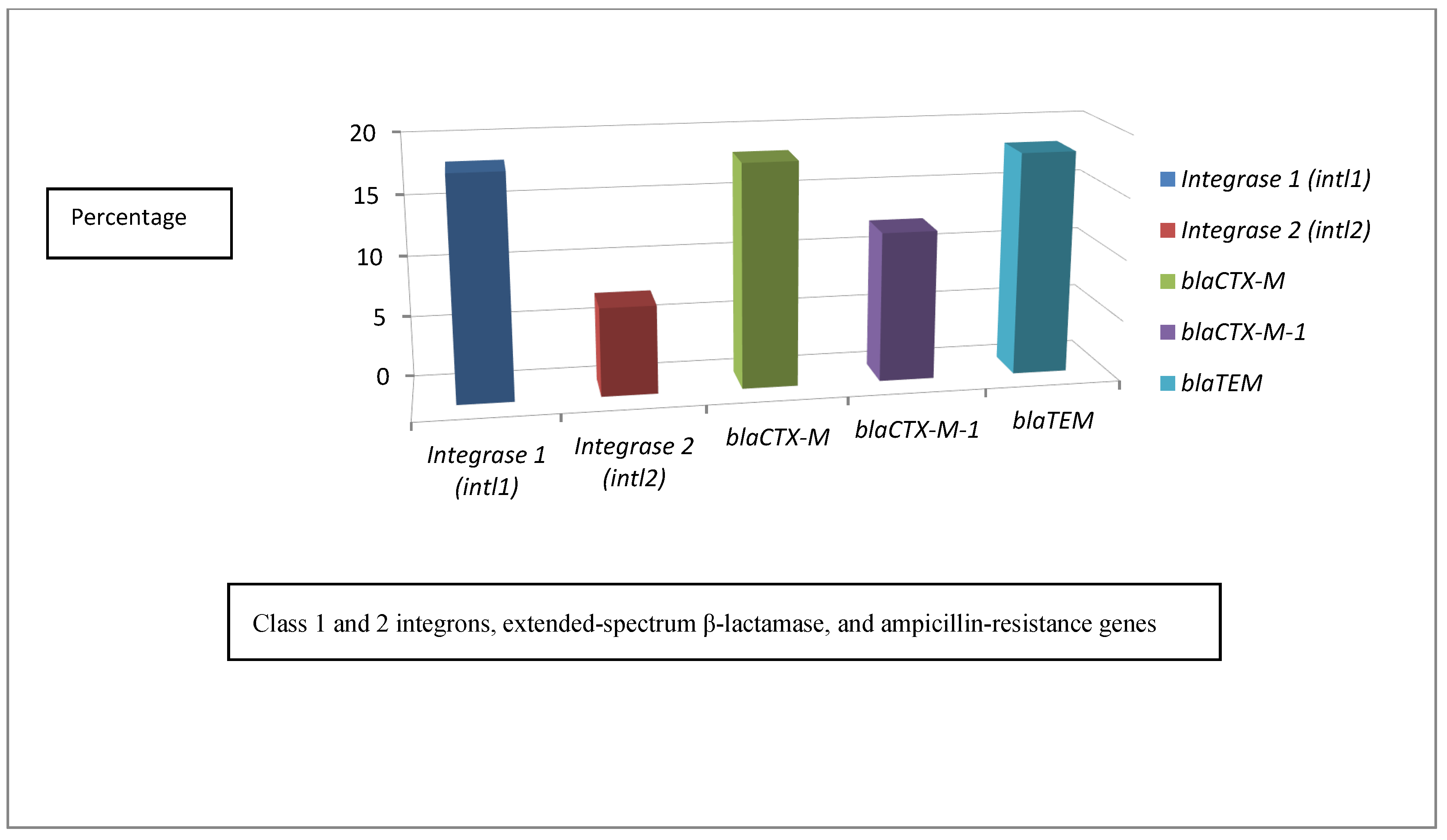
2.7. Distribution Patterns of Class 1 and 2 Integrons, Extended-Spectrum β-Lactamase, and Ampicillin-Resistance Genes

The presence of class 1 and 2 integrons, extended-spectrum β-lactamase, and ampicillin-resistance genes were investigated in all 55 P. multocida isolates. Class 1 integrons were present in 10/55 (18.2%) of the isolates, and were distributed as follows: 1/55 (1.8%) from the apparently healthy cattle, 8/55 (14.5%) from the diseased cattle, and 1/55 (1.8%) from the diseased humans. Class 2 integrons were present in 4/55 (7.3%) of the diseased cattle. The ESBL gene blaCTX-M was present in 10/55 (18.2%) of the isolates and was distributed as follows: 2/55 (3.6%) from the apparently healthy cattle, 7/55 (12.7%) from the diseased cattle, and 1/55 (1.8%) from the diseased humans. The blaCTX-M-1 gene was present in 7/55 (12.7%) of the isolates, and was distributed as follows: 2/55 (3.6%) from the apparently healthy cattle and 5/55 (9.1%) from the diseased cattle. The blaTEM gene was present in 10/55 (18.2%) of the isolates, including 2/55 (3.6%) from the apparently healthy cattle, 7/55 (12.7%) from the diseased cattle, and 1/55 (1.8%) from the diseased humans. There was a significant difference between the presence of blaCTX-M-1 and other ESBLs (p ≤ 0.05). A total of 39/55 (70.9%) harbored none of the targeted ESBL genes, including 37/55 (67.3%) sensitive isolates and 2/55 (3.6%) resistant isolates; 4/55 (7.3%) isolates contained a single type of ESBL gene, 5/55 (9.1%) showed 2 different types of ESBL genes, 3/55 (5.5%) possessed 3 different types of ESBL genes, 2/55 (3.6%) had 4 different types of ESBL genes, and 2/55 (3.6%) harbored 5 different types of ESBL genes (Supplementary Table S7 and Figure 5).

2.8. Associations between Isolation Source, Strain, and Phenotypic and Genotypic Characteristics

The relationships between virulence genes, antimicrobial resistance, resistance genes, strain, and sample sources were investigated to detect possible associations among the isolates. The phenotypic antimicrobial resistance profiles and associated genes confirmed in this study were changed to binary codes for statistical analysis. The sensitivity to a given antimicrobial agent was recorded as “0” and resistance as “1”. The presence or absence of a specific resistance gene was also scored as “1” or “0”, respectively. The Pearson correlation coefficient was calculated using the online tools at https://software.broadinstitute.org/morpheus/, accessed on 20 February 2019. Some strains exhibited high virulence, and phenotypic and genotypic antimicrobial resistance. There was a significant difference (p ≤ 0.05) in the impact of the strains on the virulence genes, phenotypic antimicrobial resistance, and resistance genes. Correlation matrix analyses and hierarchical clustering with heatmaps were used to detect the associations between the phenotypic and genotypic features, and the origins of the strains. There was a positive correlation between the presence of macrolide resistance genes erm(42), mph(E), and msr(E), and phenotypic resistance to azithromycin and erythromycin (p ≤ 0.05). The correlation analysis showed positive relationships between the resistance to β-lactams, especially to amoxicillin/clavulanic acid, and the presence of class 1 and 2 integrons (intl1 and intl2), extended-spectrum β-lactamases (blaCTX-M and blaCTX-M-1), and ampicillin-resistance genes (blaTEM) (Figure 6, p ≤ 0.05). Significant positive correlations between antibiotic resistances indicated that co-occurrence of resistance may be predominant (p ≤ 0.05), and it confirmed the presence of multiple drug-resistant (MDR) strains. For example, resistance to amoxicillin/clavulanic acid and cefotaxime were positively correlated with resistance to amikacin, azithromycin, danofloxacin, doxycycline, erythromycin, nalidixic acid, trimethoprim/sulfamethoxazole, and oxytetracycline. The presence of class 1 and 2 integrons (intl1 and intl2), extended-spectrum β-lactamases (blaCTX-M and blaCTX-M-1), and ampicillin-resistance gene blaTEM was positively correlated with resistance to trimethoprim/sulfamethoxazole and oxytetracycline (p ≤ 0.05). Moreover, resistance to azithromycin was positively correlated with resistance to cefotaxime, danofloxacin, nalidixic acid, trimethoprim/sulfamethoxazole, and oxytetracycline. The presence of the erm(42), mph(E), and msr(E) genes were positively correlated with resistance to danofloxacin, nalidixic acid, trimethoprim/sulfamethoxazole, and oxytetracycline (p ≤ 0.05).

3. Discussion

P. multocida is a zoonotic Gram-negative bacterium responsible for many infections in domestic animals, leading to serious economic losses [23]. Along with Mannheimia haemolytica, Mycoplasma bovis, and Haemophilus somnus pneumonia, it is involved in the pathogenesis of BRD, which causes significant morbidity and mortality among cattle all over the world [1,2,14]. The isolation and biochemical identification results confirmed that P. multocida could be isolated from diseased and apparently healthy cattle of variable ages, ranging from weaned calves to mature cattle [24]. The results of the isolation and identification confirmed the presence of P. multocida in 6/48 (12.5%) of the apparently healthy cases and 44/52 (84.6%) of the visibly diseased cases. This result surpassed that of Khamesipour et al. [24] from Iran, who recovered P. multocida from 11.4% of pneumonic cases and 4.4% of healthy cases. The isolation of P. multocida from cases of human epiglottitis was confirmed by Glickman and Klein [25] from the USA, who demonstrated its presence in adults without exposure to animals, and Moyko and Ali [26], who confirmed epiglottitis in a human who was exposed to animals. As the human samples were collected from animal owners and farm workers, this could suggest that exposure to animals can enhance infection transmission to humans. The composition and structure of the capsular material found in the P. multocida serotypes A, D, and F are very similar to mammalian glycosaminoglycans and mainly consist of hyaluronan, heparosan, and unsulfated chondroitin, respectively [27,28,29,30]. Crude capsular antigens from P. multocida serogroup B were shown to contain fructose, mannose, glucose, and glucosamine [31]. There are many functions attributed to the capsules, they are as follows: (a) they are highly hydrated and protect the bacterial cell from desiccation; (b) they impart anti-phagocytic activity due to the negative charge on their surface, repelling phagocytic cells; and (c) they may impart serum resistance and aid in the evasion of the bactericidal activity of complement against capsular type A [32]. As a swifter alternative to the traditional capsular serotyping method, a multiplex PCR assay was developed. Based on the P. multocida characterization, detection of sequences, and analysis of the biosynthetic loci of each capsular serogroup, specific primers were developed [11].
The most prevalent capsular type among the isolates was type B (46%), which is in agreement with the findings of Ataei et al. [33], who confirmed that in tropical countries P. multocida serotype B:2 causes the acute disease HS in cattle and buffalo. Although capsular type D is related to infections in pigs [11], it has also been recorded in cattle infections [24]. The isolation of capsular type E from cattle was confirmed by Townsend et al. [11], who recorded its presence in African cattle. Moreover, the absence of capsular type A and F within cattle isolates was also in agreement with Townsend et al. [9]. Virulence-associated genes described for the P. multocida isolates include adherence and colonization factors (ptfA, fimA, hsf-1, hsf-2, pfhA, and tadD), iron-regulated and acquisition proteins (exbB, exbD, tonB, hgbA, hgbB, and fur), extracellular enzymes such as neuraminidase (nanB and nanH), hyaluronidase (pmHAS), superoxide dismutases (sodA, sodC, and tbpA), toxins (toxA), lipopolysaccharides (LPS), capsule proteins, and a variety of outer-membrane proteins such as protectins (ompA, ompH, oma87, and plpB) [34]. The presence of the hemoglobin-binding proteins hgbA and hgbB, involved in the iron uptake process, were confirmed in 28/55 (50.9%) and 30/55 (54.5%) of the isolates, respectively, which was lower than the findings of Khamesipour et al. [24], who showed their existence in 86.7% and 93.3% of the isolates, respectively. The presence of hgbA and hgbB in this study showed high distribution among diseased isolates, as recorded by Khamesipour et al. [24].
The ptfA gene, encoding type IV fimbriae, was confirmed in 25/55 (45.5%) of the isolates, which was lower than that reported by Khamesipour et al. [24], who found it in 80.0% of the screened isolates and 92.0% of the isolates from diseased cattle with pneumonic lungs. The absence of the sodA gene for superoxide dismutase and the pfhA gene encoding hemagglutinin contradicts the findings of Khamesipour et al. [24], who found these genes in 83.3% and 60.0% of the isolates, respectively. The presence of multiple virulence genes is in agreement with Khamesipour et al. [24], who reported the existence of multiple virulence genes among 30 P. multocida isolates obtained from pneumonic and apparently healthy slaughter cattle. The genetic profiles obtained, based on capsular type and virulence factors, were different than those reported by Khamesipour et al. [24], as a different array of virulence factors were tested; both the genetic combinations dcbF type D and bcbD type B/hgbA/hgbB/ptfA were the highest, with rates of 16% and 14%, respectively, while bcbD type B/hgbB/ptfA, dcbF type D/hgbA/ptfA, dcbF type D/hgbB/ptfA, ecbJ type E/hgbA, and ecbJ type E/hgbB were the lowest, with rates of 2% each. The capsular type and virulence factor combinations in the 55 unrelated strains comprised 17 combinations, which exhibited a discriminatory power of 0.9286, which was high enough to suggest discrimination using various genetic profiles.
Antimicrobials are often used for prophylaxis to protect potentially susceptible animals, for metaphylaxis in herds receiving new animals, and for growth promotion to improve feed efficiency [35]. The uncontrolled implementation of antimicrobials leads to a powerful selective pressure that affects the microbial community, selecting for resistance genes and resistant bacteria in the bovine digestive flora [35]. The bovine microbiota includes many harmless bacteria and opportunistic pathogens that could acquire and transmit resistance genes within the microbial community through horizontal gene transfer. Likewise, the propagation of resistance genes could influence bovine-associated human pathogens, constituting a potential public health threat [35]. Based on the results of phenotypic antimicrobial resistance, it was clear that the P. multocida isolates exhibited resistance to oxytetracycline, amikacin, doxycycline, erythromycin, and chloramphenicol, which surpassed the findings of Khamesipour et al. [24] from Iran, who found that cattle strains exhibited lower resistance to these antimicrobials. The resistance to amoxicillin/clavulanic acid, trimethoprim/sulfamethoxazole, ciprofloxacin, and levofloxacin was higher than that reported by El-Seedy et al. [36] from Egypt. The presence of resistance in the P. multocida isolates to nalidixic acid was similar to that reported by Koike et al. [37] from Hungary, while resistance to norfloxacin was higher than reports from Anwar et al. [38] from Pakistan, who found that 75% of the isolates were sensitive to norfloxacin. The resistance to azithromycin was higher than that reported by Portis et al. [14], who found that 4.6% of the P. multocida isolates from cattle in the USA and Canada were resistant to tulathromycin. The resistance to danofloxacin contradicts Portis et al. [14], who confirmed that P. multocida from cattle in the USA and Canada exhibited high susceptibility results to danofloxacin, ranging from 88.2% to 91.5% from 2000 to 2009, respectively. The high resistance in the P. multocida isolates to cefotaxime contradicts the results of Elshemey et al. [39] from Egypt, who confirmed that cefotaxime was the most effective antimicrobial against P. multocida, accompanying the foot and mouth disease virus SAT 2 FMD on bovine farms in Alexandria province, Egypt. Scarce data were found concerning the antimicrobial susceptibility profiles of P. multocida isolated from human epiglottitis, although Glickman and Klein [25] confirmed that an isolate from an infected 44-year-old human patient from the USA was susceptible to ceftriaxone, and Moyko and Ali [26] showed that an isolate from a 49-year-old female from the USA was susceptible to ampicillin–sulbactam.
The MAR indices of all the screened isolates ranged from 0.23 to 1. The identification of isolates with resistance to most of the antimicrobial agents widely used in veterinary practice has been documented with increasing frequency in P. multocida [40]. The accumulation of mutations or resistance genes usually results in multidrug resistance [41]. In P. multocida, San Millan et al. [42] reported multiresistance linked to the coexistence of several small plasmids encoding resistance-conferring determinants. Multiresistant, but plasmid-free, P. multocida isolates have been mentioned in other studies, showing that resistance genes can be associated with integrative and conjugative elements, such as ICEPmu1 or ICEPmu2 [41,43]. These elements consist of resistance gene cassettes flanked by transposase or insertion sequences, suggesting that an integration or recombination mechanism mediated by an insertion sequence can insert these resistance genes into the genome [40]. Resistance to macrolides in the family Pasteurellaceae resulted from a combination of at least three genes, as follows: erm(42), which encodes an rRNA methylase gene; msr(E), which encodes for a macrolide efflux pump or transporter protein; and mph(E), which encodes a phosphotransferase that acts through macrolide inactivation [44]. Field isolates of P. multocida showed high resistance to lincosamides and moderate resistance to macrolides [45]. The high distribution patterns of erm(42) in 22/55 (40%) of the isolates, and the equally distributed mph(E) and msr(E) genes in 19/55 (34.5%) of the isolates each, were linked to increased resistance to the macrolides erythromycin and azithromycin. This result is in agreement with those of Klima et al. [17], Desmolaize et al. [45], and Kadlec et al. [46], who found dramatic increases in the minimum inhibitory concentrations of isolates containing erm(42) and msr(E)-mph(E) for erythromycin, tilmicosin, and tulathromycin. The erm(42) was found with a high frequency among the P. multocida isolates. From the results of the macrolide–lincosamide–streptogramin B methylase genes, erm(42) encoding MLSB monomethyltransferase, mph(E) encoding a macrolide efflux pump, and msr(E) encoding macrolide-inactivating phosphotransferase, 24/55 (43.6%) of the isolates harbored no resistance genes, 6/55 (10.9%) were resistant to erythromycin, 5/55 (9.1%) were resistant to azithromycin, 4/55 (7.3%) were intermediate to both antimicrobials, and 9/55 (16.4%) were sensitive to azithromycin and intermediate to erythromycin. This result of the resistant isolates harboring none of the targeted genes is in agreement with those of Ujvári et al. [40] and Rose et al. [44], who found an erythromycin-resistant P. multocida isolate that contained no chromosome-borne mobile genetic elements. This suggests that there is an alternative mechanism of antimicrobial resistance. The existence of many different antimicrobial resistance genes or combinations of erm(42) and/or msr(E)-mph(E) genes among the P. multocida isolates was elucidated by Rose et al. [44], who found combinations of erm(42) and/or msr(E)-mph(E) in 13 P. multocida strains. Moreover, the existence of msr(E) or mph(E) in tandem was previously confirmed by Desmolaize et al. [47]. From the results of macrolide methylase and other types of macrolide resistance genes, 12/55 (21.8%) of the isolates harbored a single resistance gene, 9/55 (16.4%) contained 2 different types, 7/55 (12.7%) harbored 3 different types, and 3/55 (5.5%) possessed 4 different types of these resistance genes. The rational interpretation of the similarity of the patterns among the isolates in harboring the same number of genes is that these antimicrobial resistance genes could be located next to transposases, and could show independent mobility within these cassettes. There is a possibility that some genes are acquired or lost in groups, while others could show individual mobility. This fact could account for the similarity and the overall diversity patterns of multidrug resistance profiles observed in the members of Pasteurellaceae [48,49].
The high matching between the genotype and phenotype, which constituted 23/55 (41.8%) and 22/55 (40%) for azithromycin and erythromycin, respectively, indicates the role of erm(42) and msr(E)-mph(E) in resistance [17,44,45]. While the existence of resistant isolates without any genes (genotype−ve/phenotype+ve) was previously reported by Ujvári et al. [40] and Rose et al. [44], suggesting alternative resistance mechanisms, the presence of sensitive strains harboring the resistance genes (genotype+ve/phenotype−ve) elucidates that these genes were inactive, as reported by Petrocchi-Rilo et al. [50].
Generally, class 1 and 2 integrons are defined by the existence of integrase genes intI1 and intI2 and the primary recombination sites attI1 and attI2, respectively [51]. As a basis for the division of integrons into “groups”, the amino acid sequences of intI1 integrases were used, with those carrying intI1 classified as “class 1”, intI2 as “class 2”, intI3 as “class 3”, etc. The integrase genes intI1, intI2, and intI3 were first detected in connection with mobile genetic elements, intI4, and others with chromosomal integrons. Both intI1 and intI2 encode integrases of the tyrosine recombinase family [51]. The mobility of integrons has been considered a major concern for clinical antibiotic resistance, as it has been recorded as widespread among clinical species, and is characterized as being associated with mobile DNA elements (transposons or plasmids) and genes of antibiotic resistance in addition to having a limited array size and significant heterogeneity in the attC site [51]. The presence of class 1 integrons among P. multocida from the healthy and diseased cattle was previously reported by Kong et al. [52]. Although there are no reports confirming the existence of class 2 integrons in P. multocida, Kehrenberg and Schwarz [53] confirmed the presence of a partially truncated class 2 integron in a P. aerogenes isolate.
The resistance of Pasteurellaceae to β-lactam antibiotics relies on the production of β-lactamase or the existence of low-affinity penicillin-binding proteins with β-lactams [54]. Other strategies include decreased permeability of the outer membrane and systems that can effectively export β-lactams from bacterial cells, or multidrug efflux systems. These mechanisms are only rarely recorded in Pasteurellaceae [54], although there are five of the following β-lactamase (bla) genes common among Pasteurellaceae: blaCMY-2, blaPSE-1, blaOXA-2, blaROB-1, and blaTEM-1. The complete blaOXA-2 gene was identified as part of ICEPmu1, which was shown to be nonfunctional in P. multocida, but functional in E. coli [54]. The enzymes encoded by blaCTX-M acquired cefotaximase resistance from a separate phylogenetic lineage to form a rapidly growing family of ESBLs with significant clinical effects [55]. Intrinsic cefotaximase chromosome-encoded genes in Kluyvera spp. are suggested to be the progenitors of the CTX-M family [55]. Most CTX-Ms show high activity against cefotaxime and ceftriaxone; however, some CTX-Ms exhibit enhanced catalytic efficiencies against ceftazidime, including CTX-M-15, CTX-M-16, and CTX-M-19 [55]. Records of blaCTX-M and blaCTX-M-1 in P. multocida isolates from cattle and humans, either healthy or diseased, are few in Egypt, so this may be the first report confirming the existence of these ESBL genes in P. multocida isolates from cattle and human sources in Egypt. However, the published results from Egypt by Awad et al. [56] and Elalamy et al. [57], confirmed the presence of blaROB-1 in 20% of the P. multocida isolates from diseased rabbits and 8.3% from diseased chicken, respectively. TEM-1 is able to hydrolyze penicillin and the early cephalosporins; its existence in the P. multocida isolates of human origin was reported by Naas et al. [58], and from cattle by Michael et al. [54], who confirmed that the presence of TEM-1 in P. multocida is of veterinary importance.
Several isolates demonstrated multiple drug-resistant (MDR) and extensive drug-resistant (XDR) phenotypes similar to Klima et al. [49]. Correlation analyses indicated the co-occurrence of resistance to various antimicrobials, illuminating a cause for significant concern for animal and human medicine alike. Moreover, resistance to some antimicrobials was linked with susceptibility to others; the high resistance to trimethoprim/sulfamethoxazole, oxytetracycline, danofloxacin, amikacin, cefotaxime, nalidixic acid, azithromycin, doxycycline, erythromycin, and norfloxacin were related to the low resistance to chloramphenicol, ciprofloxacin, amoxicillin/clavulanic acid, and levofloxacin. This finding is remarkable, because when discussing MDR or XDR of cattle- and human-origin P. multocida, it may promote the selection of alternative antimicrobials. Although there was a difference found in the screened antimicrobials against P. multocida by Kumar et al. [59] and our study, the interpretation of their findings proved that the susceptibility to chloramphenicol, ciprofloxacin, and amoxicillin correlated with increased resistance to amikacin, doxycycline, erythromycin, and sulfadiazine.
Correlation matrix analyses and hierarchical clustering with heatmaps were implemented to scrutinize the associations among the phenotypic and genotypic features and origin of these strains. There was a positive correlation between the macrolide resistance genes erm(42), mph(E), and msr(E), and the phenotypic resistance to azithromycin and erythromycin (p ≤ 0.05), as confirmed by Rose et al. [44] and Desmolaize et al. [45]. Furthermore, the correlation analysis showed positive relationships between the resistance to β-lactams, especially to amoxicillin/clavulanic acid, and the presence of class 1 and 2 integrons intl1 and intl2, extended-spectrum β-lactamases blaCTX-M and blaCTX-M-1, and the ampicillin-resistance gene blaTEM. Significant positive correlations of antibiotic resistances showed that the co-occurrence of resistance may be predominant (p ≤ 0.05), and it also confirmed the presence of MDR strains. For example, resistance to amoxicillin/clavulanic acid and cefotaxime was positively correlated with resistance to amikacin, azithromycin, danofloxacin, doxycycline, erythromycin, nalidixic acid, trimethoprim/sulfamethoxazole, and oxytetracycline. The presence of class 1 and 2 integrons intl1 and intl2, extended-spectrum β-lactamase blaCTX-M and blaCTX-M-1, and the ampicillin-resistance gene blaTEM showed positive correlation with trimethoprim/sulfamethoxazole and oxytetracycline resistance.

4. Materials and Methods

4.1. Sampling, Isolation, and Identification

A total of 100 nasal swabs were collected from cattle of variable age, from 10-day-old calves to 24-month-old adults. There were 85 samples collected from Minufiya and 15 from Qalyubia localities in Egypt. A total of 48 samples were taken from apparently healthy cases and 52 from diseased cases, showing signs of depression, fever ≥ 41.5 °C, loss of appetite, nasal discharge, moist cough, or rapid and shallow breathing. Furthermore, 15 nasopharyngeal swabs were collected from 5 apparently healthy and 10 diseased humans suffering from signs of epiglottitis, also from Minufiya and Qalyubia. The human cases were known to be animal owners and farm workers, and written informed consent was obtained from all human participants. This research was performed according to the recommendations of the US Government for the utilization and care of vertebrate animals used in testing, research, and training. The swabs from the screened cases were transported in 10 mL of brain–heart infusion broth (Difco, New Jersey, USA). The samples were labeled, packed, cooled in an icebox, and immediately transported to the Department of Bacteriology, Mycology, and Immunology laboratory at the Faculty of Veterinary Medicine, at the University of Sadat City, and stored at 4 °C. A volume of 1 mL was transferred from the transport broth to an enrichment broth using a sterile Pasteur pipette. The enrichment broth was composed of a base of brain–heart infusion broth. Then, gentamicin (Gibco, Grand Island, NY, USA) (0.5 µg/mL), potassium tellurite (Sigma Chemical Co., St. Louis, MO, USA) (0.0125 µg/mL), amphotericin B (Sigma) (5.0 µg/mL), and defibrinated sheep blood (Remel, San Diego, CA, USA) (5%) were added. The pH was adjusted to 10.0 ± 0.1 with sterile 1 N NaOH (about 40 mL/liter). All the tubes were incubated for 16 h at 37 °C in 5–10% CO2. From the tubes showing turbidity, a loopful was streaked onto the selective agar medium, composed of a base of brain–heart infusion agar. The additives, concentration, and pH of the medium were adjusted in a similar manner to the enrichment broth medium [60]. The presumptive P. multocida colonies (3–5) were subjected to microscopical examination and biochemical identification. Briefly, P. multocida showed high growth on the selective medium, without hemolysis. The colonies appeared smooth, with a slightly raised center, and were mucoid and sticky in nature. No growth was observed on MacConkey agar (Difco). After staining, the P. multocida appeared as Gram-negative, small, cocobacilli with rounded ends. The P. multocida also showed positive results to the H2S production test, indole production test, nitrate reduction test, and catalase test, and were able to ferment glucose, sucrose, mannose, and fructose sugars. The P. multocida showed negative results in the methyl red, gelatin liquification, and urease tests and could not ferment lactose, maltose, or salicin [61,62].
The positive colonies were subjected to further confirmation using the Biolog GN2 and GP2 micro-plates, using the Biolog Microlog 3.70 database and software (Biolog, Hayward, CA) [63]. P. multocida subsp. multocida ATCC 12947 and E. coli ATCC 11775 strains were included as positive and negative controls, respectively.
The study design and all the experimental methods were approved by the Committee for Animal Care and Use, the Faculty of Veterinary Medicine, at the University of Sadat City, Egypt; the approval number was 2018–34. Moreover, the committee approved the implementation of human samples in this study after revision of the informed consent form.

4.2. Phenotypic Antimicrobial Susceptibility Profiles of P. multocida Isolates

The antimicrobial susceptibility pattern of each P. multocida isolate was screened against a panel of 14 antimicrobials (Oxoid, San Diego, CA, USA) (Supplementary Table S8), selected for their medical importance [64]. A volume of 1 mL of calibrated bacterial suspension (0.5 McFarland) (1.5 × 10⁸ cells/mL) was inoculated on Mueller-Hinton agar (Oxoid). P. multocida subsp. multocida ATCC12947 was used as a quality control isolate. Kirby Bauer’s disk diffusion method was utilized, and the results were interpreted according to the Clinical and Laboratory Standards Institute (CLSI) criteria [65].

4.3. Molecular Techniques for Capsular Classification, Detection of Virulence Genes, and Antimicrobial Resistance Genes

The genomic DNA was extracted using a QIAamp kit according to the manufacturer’s instructions (Qiagen GmbH—Germany). All 55 isolates were confirmed to be P. multocida using primers against kmt1, as described by Townsend et al. [11], encoding an integral component of the membrane. All confirmed isolates were screened for virulence genes such as sodA, hgbA, hgbB, ptfA, and pfhA. The presence of macrolide–lincosamide–streptogramin B methylases erm(A), erm(B), erm(C), erm(F), erm(G), and erm(Q), with erm(42) encoding MLSB monomethyltransferase, mph(E) encoding a macrolide efflux pump, and msr(E) encoding macrolide-inactivating phosphotransferase, were also investigated in all the isolates, as was the presence of class 1 and 2 integrons intl1 and intl2, extended-spectrum β-lactamases blaCTX-M and blaCTX-M-1, and the ampicillin-resistance gene blaTEM (Table 2). The efficiency of PCR amplification for detecting molecular capsular types, virulence genes, and antimicrobial resistance genes was confirmed by the utilization of an internal positive control from the tested P. multocida isolates. The positive control isolates for the molecular capsular types were isolates coded 3, 1, and 2 for capsular types B, D, and E, respectively. While the P. multocida isolates ATCC 12945, ATCC 12948, and NCTC 10323, with our isolate coded 3, were the positive controls for the virulence genes. Furthermore, the isolate coded 14 was the positive control for the macrolide resistance genes erm(Q), erm(42), mph(E), and msr(E). Also, this isolate coded 14 was the positive control for class 1 and 2 integrons, extended-spectrum β-lactamase, and the ampicillin-resistance genes intl1, intl2, blaCTX-M, blaCTX-M-1, and blaTEM, respectively. Moreover, the PCR for each primer was repeated three times to confirm the positive or negative result.
The PCR protocols adopted for the confirmation, molecular capsular typing, and detection of the virulence factors of the P. multocida isolates were modified so as to use a simplex protocol instead of the multiplex technique described by Townsend et al. [11] and Furian et al. [64] as follows: the final volume was 10 µl, including 1 µL of 10X PCR Mg-free buffer (Invitrogen, Carlsbad, CA, USA), 2 mM MgCl2, 1U of Platinum Taq polymerase (Invitrogen), 0.2 mM of PCR nucleotide mix (Roche Applied Sciences, Basel, Switzerland), 1 µL of DNA template, and primers at a concentration of 3.2 µM. The PCR protocol for the amplification of resistance genes was performed as follows: the reaction volume was 25 μL, containing 12.5 μL of ready-to-use master mix, 2 μL of bacterial genomic DNA (100 ng/μL), 0.5 μL of each upstream and downstream primer (50 pmol/μL), and 9.5 μL of RNase-free water. The primers and master mix were supplied by Takara Holdings (Nojihigashi, Kusatsu, Shiga, Japan).

4.4. Statistical Analysis

The free online calculator for chi-square tests at https://www.socscistatistics.com/tests/chisquare2/default2.aspxaccessed, accessed on 9 February 2019 was used to detect significant differences between the recovery rates from P. multocida, the rates of virulence factors, virulence profile incidence, efficacies of antimicrobials, MAR indices, antimicrobial resistance gene incidence, and genotype+/phenotype and genotype/phenotype+ of azithromycin and erythromycin. The phenotypic antimicrobial resistance profiles and confirmed genes were changed to numerical codes for statistical analysis. The sensitivity to antimicrobials was defined as “0” and resistance as “1”. The existence or absence of a resistance gene was defined as “1” or “0”, respectively. Heatmaps, hierarchical clustering, and Pearson correlation coefficients were calculated using the online tools at https://software.broadinstitute.org/morpheus/, accessed on 1 February 2019. The free online tools at http://insilico.ehu.es/mini_tools/discriminatory_power/index.php, accessed on 1 February 2019 were used to detect the DI of the capsular and virulence gene combinations.

5. Conclusions

In conclusion, P. multocida are widely spread among cattle and humans. We conducted a detailed screening of the virulence factors, phenotypic and genotypic antimicrobial resistance, and capsular types. The obtained isolates showed decreased resistance to chloramphenicol, ciprofloxacin, amoxicillin/clavulanic acid, and levofloxacin. Moreover, the unrationalized implementation of antimicrobials for P. multocida infections in cattle and humans likely aggravated the situation, and led to the emergence of multiple and extensive drug-resistant phenotypes. There were significant differences among isolates in phenotypic antimicrobial resistance, virulence genes, and resistance genes. The capsular type and virulence gene combinations observed could serve as a suitable preliminary molecular epidemiological tool with high discriminatory power. Some human isolates shared the same genotypic profiles as animal isolates, suggesting that they originated from a common source. These results will be beneficial in instating control strategies to contain P. multocida, and in the evaluation of related clusters, cluster expansion, and transmission dynamics.

Supplementary Materials

The following are available online at https://www.mdpi.com/article/10.3390/antibiotics10050480/s1, Table S1. Detailed results of isolation of P. multocida on specific media in relation to origin, age, and signs of cases. Table S2. Results of molecular confirmation of P. multocida isolates, detection of capsular types, and virulence genes. Table S3. Results of the most prevalent genotypes. Table S4. Results of antimicrobial susceptibility testing and multiple antimicrobial resistance indices of tested P. multocida isolates. Table S5. Results of distribution patterns of macrolide resistance genes. Table S6. Numbers and percentages of Azithromycin and Erythromycin resistant P. multocida isolates with discrepancies between genotype-phenotype. Table S7. Results of class 1 and 2 integrons, extended-spectrum β-lactamase, ampicillin-resistance gene. Table S8. The list, categorization and prioritization of antimicrobials classified as critically important in human and veterinary medicine.

Author Contributions

Conceptualization, M.S.A.E.E., S.M.E., T.R., L.S., N.T., T.A., A.B.A.M., G.M.N., M.S.M.B., B.A.A., M.M.N., A.S.H. and A.S.; methodology, M.S.A.E.E., T.R. and A.S.; validation, M.S.A.E.E., S.M.E., T.R. and A.S.; formal analysis, M.S.A.E.E., S.M.E., T.R. and A.S. investigation, M.S.A.E.E., S.M.E., T.R. and A.S.; resources, M.S.A.E.E., S.M.E., T.R. and A.S.; data curation, M.S.A.E.E. and S.M.E.; writing—original draft preparation, M.S.A.E.E. and S.M.E.; writing—review and editing, M.S.A.E.E., S.M.E., T.R., A.B.A.M., G.M.N., M.S.M.B., B.A.A., M.M.N., A.S.H. and A.S.; visualization, M.S.A.E.E. and S.M.E.; supervision, M.S.A.E.E., S.M.E., T.R. and A.S.; project administration, M.S.A.E.E. All authors have read and agreed to the published version of the manuscript.

Funding

This research received no external funding.

Institutional Review Board Statement

The study design and all the experimental methods were approved by the Committee for Animal Care and Use, the Faculty of Veterinary Medicine, at the University of Sadat City, Egypt; the approval number was 2018–34. Moreover, the committee approved the implementation of human samples in this study after revision of the informed consent form.

Informed Consent Statement

Informed consent was obtained from all subjects involved in the study.

Data Availability Statement

The data represented in this study are available in the article or Supplementary Materials.

Conflicts of Interest

The authors declare no conflict of interest.

References

  1. Zhao, G.; He, H.; Wang, H. Use of a recombinase polymerase amplification commercial kit for rapid visual detection of Pasteurella multocida. BMC Vet. Res. 2019, 15, 154. [Google Scholar] [CrossRef] [PubMed] [Green Version]
  2. Stanford, K.; Zaheer, R.; Klima, C.; McAllister, T.; Peters, D.; Niu, Y.D.; Ralston, B. Antimicrobial resistance in members of the bacterial bovine respiratory disease complex isolated from lung tissue of cattle mortalities managed with or without the use of antimicrobials. Microorganisms 2020, 8, 288. [Google Scholar] [CrossRef] [PubMed] [Green Version]
  3. Arashima, Y.; Kubo, N.; Iwasaki, Y.; Okuyama, K.; Kumasaka, K.; Tsuchiya, T.; Kawano, K.; Ootsuka, M.; Saitoh, F.; Namikawa, K. Human respiratory tract infection by Pasteurella multocida subsp. multocida presumably derived from the cat. Kansenshogaku Zasshi. 1990, 64, 1200–1204. [Google Scholar] [CrossRef] [Green Version]
  4. Guo, Y.; McMullen, C.; Timsit, E.; Hallewell, J.; Orsel, K.; van der Meer, F.; Yan, S.; Alexander, T.W. Genetic relatedness and antimicrobial resistance in respiratory bacteria from beef calves sampled from spring processing to 40 days after feedlot entry. Vet. Microbiol. 2020, 240, 108478. [Google Scholar] [CrossRef]
  5. Annas, S.; Zamri-Saad, M.; Jesse, F.F.; Zunita, Z. New sites of localisation of Pasteurella multocida B:2 in buffalo surviving experimental haemorrhagic septicaemia. BMC Vet. Res. 2014, 10, 88. [Google Scholar] [CrossRef] [Green Version]
  6. Al-Maary, K.S.; Dawoud, T.M.; Mubarak, A.S.; Hessain, A.M.; Galal, H.M.; Kabli, S.A.; Mohamed, M.I. Molecular characterization of the capsular antigens of Pasteurella multocida isolates using multiplex PCR. Saudi J. Biol. Sci. 2017, 24, 367–370. [Google Scholar] [CrossRef] [Green Version]
  7. St Michael, F.; Harper, M.; Parnas, H.; John, M.; Stupak, J.; Vinogradov, E.; Adler, B.; Boyce, J.D.; Cox, A.D. Structural and genetic basis for the serological differentiation of Pasteurella multocida Heddleston serotypes 2 and 5. J. Bacteriol. 2009, 191, 6950–6959. [Google Scholar] [CrossRef] [Green Version]
  8. Carter, G.R. A new serological type of Pasteurella multocida from central Africa. Vet. Rec. 1961, 73, 1052. [Google Scholar]
  9. Rimler, R.B.; Rhoades, K.R. Serogroup F. A new capsule serogroup of Pasteurella multocida. J. Clin. Microbiol. 1987, 25, 615–618. [Google Scholar] [CrossRef] [Green Version]
  10. Peng, Z.; Liang, W.; Wu, B. Molecular typing methods for Pasteurella multocida—A review. Acta Microbiol. Sin. 2016, 56, 1521–1529. [Google Scholar] [CrossRef]
  11. Townsend, K.M.; Boyce, J.D.; Chung, J.Y.; Frost, A.J.; Adler, B. Genetic organization of Pasteurella multocida cap Loci and development of a multiplex capsular PCR typing system. J. Clin. Microbiol. 2001, 39, 924–929. [Google Scholar] [CrossRef] [Green Version]
  12. Harper, M.; John, M.; Turni, C.; Edmunds, M.; St Michael, F.; Adler, B.; Blackall, P.J.; Cox, A.D.; Boyce, J.D. Development of a rapid multiplex PCR assay to genotype Pasteurella multocida strains by use of the lipopolysaccharide outer core biosynthesis locus. J. Clin. Microbiol. 2015, 53, 477–485. [Google Scholar] [CrossRef] [Green Version]
  13. Peng, Z.; Liang, W.; Wang, F.; Xu, Z.; Xie, Z.; Lian, Z.; Hua, L.; Zhou, R.; Chen, H.; Wu, B. Genetic and phylogenetic characteristics of Pasteurella multocida isolates from different host species. Front. Microbiol. 2018, 9, 1408. [Google Scholar] [CrossRef] [PubMed]
  14. Portis, E.; Lindeman, C.; Johansen, L.; Stoltman, G. A ten-year (2000–2009) study of antimicrobial susceptibility of bacteria that cause bovine respiratory disease complex—Mannheimia haemolytica, Pasteurella multocida, and Histophilus somni—In the United States and Canada. J. Vet. Diagn. Investig. 2012, 24, 932–944. [Google Scholar] [CrossRef] [Green Version]
  15. DeDonder, K.D.; Apley, M.D. A literature review of antimicrobial resistance in pathogens associated with bovine respiratory disease. Anim. Health Res. Rev. 2015, 16, 125–134. [Google Scholar] [CrossRef] [PubMed]
  16. Anholt, R.M.; Klima, C.; Allan, N.; Matheson-Bird, H.; Schatz, C.; Ajilkumar, P.; Otto, S.J.G.; Peters, D.; Schmid, K.; Olson, M.; et al. Antimicrobial susceptibility of bacteria that cause bovine respiratory disease complex in Alberta Canada. Front. Vet. Sci. 2017, 4, 207. [Google Scholar] [CrossRef] [PubMed] [Green Version]
  17. Klima, C.L.; Zaheer, R.; Cook, S.R.; Booker, C.W.; Hendrick, S.; Alexander, T.W.; McAllister, T.A. Pathogens of bovine respiratory disease in north american feedlots conferring multidrug resistance via integrative conjugative elements. J. Clin. Microbiol. 2014, 52, 438–448. [Google Scholar] [CrossRef] [Green Version]
  18. Feng, Y.; Qi, W.; Wang, X.; Wang, L.; Li, X.; Luo, J.; Zhang, S.; Li, H. Genetic characterization of antimicrobial resistance in Staphylococcus aureus isolated from bovine mastitis cases in northwest China. J. Integr. Agric. 2016, 15, 60345–60347. [Google Scholar] [CrossRef] [Green Version]
  19. EFSA. Scientific Opinion on the public health risks of bacterial strains producing extendedspectrum β-lactamases and/or AmpC β-lactamases in food and food-producing animals. EFSA J. 2011, 9, 2322. [Google Scholar] [CrossRef] [Green Version]
  20. Michael, G.B.; Freitag, C.; Wendlandt, S.; Eidam, C.; Feßler, A.T.; Lopes, G.V.; Kadlec, K.; Schwarz, S. Emerging issues in antimicrobial resistance of bacteria from food-producing animals. Future Microbiol. 2015, 10, 427–443. [Google Scholar] [CrossRef]
  21. Bush, K.; Jacoby, G.A. Updated functional classification of beta-lactamases. Antimicrob. Agents Chemother. 2010, 54, 969–976. [Google Scholar] [CrossRef] [Green Version]
  22. Ur Rahman, S.; Ali, T.; Ali, I.; Khan, N.A.; Han, B.; Gao, J. The growing genetic and functional diversity of extended spectrum beta-lactamases. BioMed Res. Int. 2018, 2018, 9519718. [Google Scholar] [CrossRef]
  23. Steen, J.A.; Steen, J.A.; Harrison, P.; Seemann, T.; Wilkie, I.; Harper, M.; Adler, B.; Boyce, J.D. Fis is essential for capsule production in Pasteurella multocida and regulates expression of other important virulence factors. PLOS Pathog. 2010, 6, e1000750. [Google Scholar] [CrossRef]
  24. Khamesipour, F.; Momtaz, H.; Azhdary Mamoreh, M. Occurrence of virulence factors and antimicrobial resistance in Pasteurella multocida strains isolated from slaughter cattle in Iran. Front. Microbiol. 2014, 5, 536. [Google Scholar] [CrossRef] [PubMed] [Green Version]
  25. Glickman, M.; Klein, R.S. Acute Epiglottitis due to Pasteurella multocida in an Adult without Animal Exposure. Emerg. Infect. Dis. 1997, 3, 408–409. [Google Scholar] [CrossRef] [PubMed]
  26. Moyko, A.; Ali, N.J.; Dubosh, N.M.; Wong, M.L. Pasteurella multocida Epiglottitis. Clin. Pract. Cases Emerg. Med. 2017, 1, 22–24. [Google Scholar] [CrossRef] [PubMed] [Green Version]
  27. Pandit, K.K.; Smith, J.E. Capsular hyaluronic acid in Pasteurella multocida type A and its counterpart in type D. Res. Vet. Sci. 1993, 54, 20–24. [Google Scholar] [CrossRef]
  28. Rimler, R.B.; Rhoades, K.R. Hyaluronidase and chondroitinase activity of Pasteurella multocida serotype B:2 involved in hemorrhagic septicaemia. Vet. Rec. 1994, 134, 67–68. [Google Scholar] [CrossRef]
  29. DeAngelis, P.L. Enzymological characterization of the Pasteurella multocida hyaluronic acid synthase. Biochemistry 1996, 35, 9768–9771. [Google Scholar] [CrossRef] [PubMed]
  30. DeAngelis, P.L.; Padgett-McCue, A.J. Identification and molecular cloning of a chondroitin synthase from Pasteurella multocida type F. J. Biol. Chem. 2000, 275, 24124–24129. [Google Scholar] [CrossRef] [Green Version]
  31. Knox, K.W.; Bain, R.V.S. The antigens of Pasteurella multocida type I. Immunology 1960, 3, 352–362. [Google Scholar]
  32. Boyce, J.D.; Chung, J.Y.; Adler, B. Pasteurella multocida capsule: Composition, function and genetics. J. Biotechnol. 2000, 83, 153–160. [Google Scholar] [CrossRef]
  33. Ataei Kachooei, S.; Ranjbar, M.M.; Ataei Kachooei, S. Evaluation of Pasteurella multocida serotype B:2 resistance to immune serum and complement system. Vet. Res. Forum 2017, 8, 179–184. [Google Scholar]
  34. Katoch, S.; Sharma, M.; Patil, R.D.; Kumar, S.; Verma, S. In vitro and in vivo pathogenicity studies of Pasteurella multocida strains harbouring different ompA. Vet. Res. Commun. 2014, 38, 183–191. [Google Scholar] [CrossRef] [PubMed]
  35. Cameron, A.; McAllister, T.A. Antimicrobial usage and resistance in beef production. J. Anim. Sci. Biotechnol. 2016, 7, 68. [Google Scholar] [CrossRef] [PubMed] [Green Version]
  36. El-Seedy, F.R.; Abed, A.H.; Hassan, H.M.; Nabih, A.M.; Khalifa, E.; Salem, S.E. Antimicrobial and immunological studies on Pasteurella multocida and Mannheimia haemolytica recovered from calves affected with respiratory manifestations. JVMR 2019, 26, 55–63. [Google Scholar]
  37. Koike, S.; Inoue, K.; Yoneyama, S.; Ichikawa, Y.; Tajima, K. Antimicrobial susceptibility of respiratory bacterial pathogen isolated from cattle for the past 16 years in Tochigi Prefecture. JVMA 2009, 62, 533–537. [Google Scholar] [CrossRef]
  38. Anwar, M.A.H.; Rahman, S.U.; Ahmad, R. Antibiotic Sensitivity of Pasteurella multocida isolated from cattle and buffaloes. Pak. J. Biol. Sci. 2000, 3, 692–693. [Google Scholar] [CrossRef]
  39. Elshemey, T.M.; Abd-Elrahman, A.H. Hemorrhagic septicemia outbreak as a consequence to SAT2 FMD infection in buffalo and cattle in Alexandria province, Egypt. Life Sci. J. 2013, 10, 816–822. [Google Scholar]
  40. Ujvári, B.; Makrai, L.; Magyar, T. Characterisation of a multiresistant Pasteurella multocida strain isolated from cattle. Acta Vet. Hung. 2018, 66, 12–19. [Google Scholar] [CrossRef]
  41. Michael, G.B.; Kadlec, K.; Sweeney, M.T.; Brzuszkiewicz, E.; Liesegang, H.; Daniel, R.; Murray, R.W.; Watts, J.L.; Schwarz, S. ICEPmu1, an integrative conjugative element (ICE) of Pasteurella multocida: Analysis of the regions that comprise 12 antimicrobial resistance genes. J. Antimicrob. Chemother. 2012, 67, 84–90. [Google Scholar] [CrossRef]
  42. San Millan, A.; Escudero, J.A.; Gutierrez, B.; Hidalgo, L.; Garcia, N.; Llagostera, M.; Dominguez, L.; Gonzalez-Zorn, B. Multiresistance in Pasteurella multocida is mediated by coexistence of small plasmids. Antimicrob. Agents Chemother. 2009, 53, 3399–3404. [Google Scholar] [CrossRef] [PubMed] [Green Version]
  43. Moustafa, A.M.; Seemann, T.; Gladman, S.; Adler, B.; Harper, M.; Boyce, J.D.; Bennett, M.D. Comparative genomic analysis of Asian haemorrhagic septicaemia-associated strains of Pasteurella multocida identifies more than 90 haemorrhagic septicaemia-specific genes. PLoS ONE 2015, 10, e0130296. [Google Scholar] [CrossRef] [Green Version]
  44. Rose, S.; Desmolaize, B.; Jaju, P.; Wilhelm, C.; Warrass, R.; Douthwaite, S. Multiplex PCR to identify macrolide resistance determinants in Mannheimia haemolytica and Pasteurella multocida. Antimicrob. Agents Chemother. 2012, 56, 3664–3669. [Google Scholar] [CrossRef] [PubMed] [Green Version]
  45. Desmolaize, B.; Rose, S.; Warrass, R.; Douthwaite, S. A novel Erm monomethyltransferase in antibiotic-resistant isolates of Mannheimia haemolytica and Pasteurella multocida. Mol. Microbiol. 2011, 80, 184–194. [Google Scholar] [CrossRef]
  46. Kadlec, K.; Brenner Michael, G.; Sweeney, M.T.; Brzuszkiewicz, E.; Liesegang, H.; Daniel, R.; Watts, J.L.; Schwarz, S. Molecular basis of macrolide, triamilide, and lincosamide resistance in Pasteurella multocida from bovine respiratory disease. Antimicrob. Agents Chemother. 2011, 55, 2475–2477. [Google Scholar] [CrossRef] [PubMed] [Green Version]
  47. Desmolaize, B.; Rose, S.; Wilhelm, C.; Warrass, R.; Douthwaite, S. Combinations of macrolide resistance determinants in field isolates of Mannheimia haemolytica and Pasteurella multocida. Antimicrob. Agents Chemother. 2011, 55, 4128–4133. [Google Scholar] [CrossRef] [Green Version]
  48. Klima, C.L.; Cook, S.R.; Zaheer, R.; Laing, C.; Gannon, V.P.; Xu, Y.; Rasmussen, J.; Potter, A.; Hendrick, S.; Alexander, T.W.; et al. Comparative genomic analysis of Mannheimia haemolytica from bovine sources. PLoS ONE 2016, 11, e0149520. [Google Scholar] [CrossRef] [PubMed]
  49. Klima, C.L.; Holman, D.B.; Cook, S.R.; Conrad, C.C.; Ralston, B.J.; Allan, N.; Anholt, R.M.; Niu, Y.D.; Stanford, K.; Hannon, S.J.; et al. Multidrug resistance in Pasteurellaceae associated with bovine respiratory disease mortalities in north America from 2011 to 2016. Front. Microbiol. 2020, 11, 606438. [Google Scholar] [CrossRef]
  50. Petrocchi-Rilo, M.; Gutiérrez-Martín, C.B.; Pérez-Fernández, E.; Vilaró, A.; Fraile, L.; Martínez-Martínez, S. Antimicrobial resistance genes in porcine Pasteurella multocida are not associated with its antimicrobial susceptibility pattern. Antibiotics 2020, 9, 614. [Google Scholar] [CrossRef] [PubMed]
  51. Deng, Y.; Bao, X.; Ji, L.; Chen, L.; Liu, J.; Miao, J.; Chen, D.; Bian, H.; Li, Y.; Yu, G. Resistance integrons: Class 1, 2 and 3 integrons. Ann. Clin. Microbiol. Antimicrob. 2015, 14, 45. [Google Scholar] [CrossRef] [PubMed] [Green Version]
  52. Kong, L.C.; Wang, Z.; Wang, Y.M.; Dong, W.L.; Jia, B.Y.; Gao, D.; Jiang, X.Y.; Ma, H.X. Antimicrobial susceptibility and molecular typing of Pasteurella multocida isolated from six provinces in China. Trop. Anim. Health Prod. 2019, 51, 987–992. [Google Scholar] [CrossRef]
  53. Kehrenberg, C.; Schwarz, S. Trimethoprim resistance in a porcine Pasteurella aerogenes isolate is based on a dfrA1 gene cassette located in a partially truncated class 2 integron. J. Antimicrob. Chemother. 2011, 66, 450–452. [Google Scholar] [CrossRef] [PubMed] [Green Version]
  54. Michael, G.B.; Bossé, J.T.; Schwarz, S. Antimicrobial resistance in Pasteurellaceae of veterinary origin. Microbiol. Spectr. 2018, 6, 1–33. [Google Scholar] [CrossRef]
  55. Zhao, W.H.; Hu, Z.Q. Epidemiology and genetics of CTX-M extended-spectrum β-lactamases in Gram-negative bacteria. Crit. Rev. Microbiol. 2013, 39, 79–101. [Google Scholar] [CrossRef] [PubMed]
  56. Awad, N.F.S.; Abd El-Hamid, M.I. Coexistence of virulence and antibiotic resistance genes in Pasteurella multocida isolated from diseased rabbits. Zagazig Vet. J. 2019, 47, 91–102. [Google Scholar] [CrossRef]
  57. Elalamy, R.A.; Tartor, Y.H.; Ammar, A.M.; Eldesouky, I.E.; Esawy, A.E.I. Molecular characterization of extensively drug-resistant Pasteurella multocida isolated from apparently healthy and diseased chickens in Egypt. Pak. Vet. J. 2020, 40, 319–324. [Google Scholar] [CrossRef]
  58. Naas, T.; Benaoudia, F.; Lebrun, L.; Nordmann, P. Molecular identification of TEM-1 β-lactamase in a Pasteurella multocida isolate of human origin. Eur. J. Clin. Microbiol. Infect. Dis. 2001, 20, 210–213. [Google Scholar] [CrossRef] [PubMed]
  59. Kumar, P.; Singh, V.P.; Agrawal, R.K.; Singh, S. Identification of Pasteurella multocida isolates of ruminant origin using polymerase chain reaction and their antibiogram study. Trop. Anim. Health Prod. 2009, 41, 573–578. [Google Scholar] [CrossRef] [PubMed]
  60. Moore, M.K.; Cicnjak-Chubbs, L.; Gates, R.J. A new selective enrichment procedure for isolating Pasteurella multocida from avian and environmental samples. Avian Dis. 1994, 38, 317–324. [Google Scholar] [CrossRef]
  61. Kozarev, A.; Mamadudian, B.A. Biochemical properties of Pasteurella multocida strains from ruminants. Vet. Sibrika 1988, 86, 28–30. [Google Scholar]
  62. Ashraf, A.; Tariq, H.; Shah, S.; Nadeem, S.; Manzoor, I.; Ali, S.; Ijaz, A.; Gailani, S.; Mehboob, S. Characterization of Pasteurella multocida strains isolated from cattle and buffaloes in Karachi, Pakistan. Afr. J. Microbiol. Res. 2011, 5, 4673–4677. [Google Scholar] [CrossRef]
  63. Sellyei, B.; Wehmann, E.; Makrai, L.; Magyar, T. Evaluation of the Biolog system for the identification of certain closely related Pasteurella species. Diagn. Microbiol. Infect. Dis. 2011, 71, 6–11. [Google Scholar] [CrossRef] [PubMed]
  64. WHO (World Health Organization). Critically Important Antimicrobials for Human Medicine—5th Revision; World Health Organization: Geneva, Switzerland, 2017. [Google Scholar]
  65. CLSI. Performance Standards for Antimicrobial Susceptibility Testing; Twenty-Fourth Informational Supplement CLSI Document M100-S24; Clinical and Laboratory Standards Institute: Wayne, PA, USA, 2014. [Google Scholar]
  66. Furian, T.Q.; Borges, K.A.; Rocha, S.L.S.; Rodrigues, E.E.; Nascimento, V.P.; Salle, C.T.P.; Moraes, H.L.S. Detetion of virulence-associated genes of Pasteurella multocida isolated from cases of fowl cholera by multiplex-PCR. Pesq. Vet. Bras. 2013, 33, 177–182. [Google Scholar] [CrossRef] [Green Version]
  67. Elsayed, M.S.A.E.; Roshdey, T.; Salah, A.; Tarabees, R.; Younis, G.; Eldeep, D. Phenotypic and genotype methods for identification of slime layer production, efflux pump activity, and antimicrobial resistance genes as potential causes of the antimicrobial resistance of some mastitis pathogens from farms in Menoufia, Egypt. Mol. Biol. Rep. 2019, 46, 6533–6546. [Google Scholar] [CrossRef] [PubMed]
  68. Raphael, E.; Wong, L.K.; Riley, L.W. Extended-spectrum beta-lactamase gene sequences in Gram-negative saprophytes on retail organic and nonorganic spinach. Appl. Environ. Microbiol. 2011, 77, 1601–1607. [Google Scholar] [CrossRef] [Green Version]
Figure 1. The results of molecular capsular typing and virulence genes.
Antibiotics 10 00480 g001
Figure 2. The percentages of the obtained genotypes.
Antibiotics 10 00480 g002
Figure 3. Heatmap and hierarchical clustering of the P. multocida isolates to 17 clusters based on the molecular detection of capsular types and virulence genes. The numbers on the right of the heatmap refer to the isolate numbers from 1 to 55.
Antibiotics 10 00480 g003
Figure 4. The results of macrolide–lincosamide–streptogramin B (MLSB) methylases, monomethyltransferase, macrolide efflux pump, and macrolide-inactivating phosphotransferase genes.
Antibiotics 10 00480 g004
Figure 5. The results of class 1 and 2 integrons, extended-spectrum β-lactamase, and ampicillin-resistance genes.
Antibiotics 10 00480 g005
Figure 6. A heatmap and hierarchical clustering of the P. multocida isolates to 7 clusters based on their phenotypic (antimicrobial resistance), genotypic (antimicrobial resistance genes), and virulence genes expressing differences between isolates. Red represents presence and blue represents absence of phenotypic resistance, resistance genes, and virulence genes. Hierarchical clustering was performed using Wald’s method and a binary distance matrix. The numbers on the right of the heatmap refer to the isolate numbers from 1 to 55.
Antibiotics 10 00480 g006
Table 1. Results of collected samples, origin, and types of cases.
Origin of SamplesTypes of Cases
Apparently HealthyDiseased
No.%No.%
MinufiyaCattle (85)4040/100 (40)4545/100 (45)
Human (7)22/15 (13.3)55/15 (33.3)
QalyubiaCattle (15)88/100 (8)77/100 (7)
Human (8)33/15 (20)55/15 (33.3)
Total collected
samples
Cattle (100)4848/100 (48)5252/100 (52)
Human (15)55/15 (33.3)1010/15 (66.7)
Positive isolationCattle (50)6 (5 from Minufiya and
1 from Qalyubia)
6/48 (12.5)44
(40 from Minufiya and
4 from Qalyubia)
44/52 (84.6)
Human (5)00/15 (0.0)5 from Qalyubia5/15 (33.3)
Table 2. Primer sequences, anticipated amplicon size, and amplification conditions.
NameAmplicon
Size (bp)
Cycle
Number
Annealing
Temperature (°C) and Time (Seconds)
Primer SequencePurposeReference
kmt14603055 °C, 30 sKMT1T7-ATCCGCTATTTACCCAGTGG
KMT1SP6-GCTGTAAACGAACTCGCCAC
Confirmation of isolates using
primer targeting an integral component of membrane
[11]
hyaD-hyaC10443055 °C, 30 sCAPA-TGCCAAAATCGCAGTCAG
CAPA-TTGCCATCATTGTCAGTG
Molecular capsular typing
bcbD7603055 °C, 30 sCAPB-CATTTATCCAAGCTCCACC
CAPB-GCCCGAGAGTTTCAATCC
dcbF6573055 °C, 30 sCAPD-TTACAAAAGAAAGACTAGGAGCCC
CAPD-CATCTACCCACTCAACCATATCAG
ecbJ5113055 °C, 30 sCAPE-TCCGCAGAAAATTATTGACTC
CAPE-GCTTGCTGCTTGATTTTGTC
fcbD8513055 °C, 30 sCAPF-AATCGGAGAACGCAGAAATCAG
CAPF-TTCCGCCGTCAATTACTCTG
sodA3612555 °C, 30 sF:TACCAGAATTAGGCTACGC
R:GAAACGGGTTGCTGCCGCT
Superoxide dismutase[66]
hgbA4192555 °C, 30 sF:TGGCGGATAGTCATCAAG
R:CCAAAGAACCACTACCCA
Iron uptake
hgbB7882555 °C, 30 sF:ACCGCGTTGGAATTATGATTG
R:CATTGAGTACGGCTTGACAT
Iron uptake
ptfA4882555 °C, 30 sF:TGTGGAATTCAGCATTTTAGTGTGTC
R:TCATGAATTCTTATGCGCAAAATCCTGCTGG
Type IV fimbriae
pfhA2752555 °C, 30 sF:AGCTGATCAAGTGGTGAAC
RTGGTACATTGGTGAATGCTG
Hemagglutinin
erm(A)1574063 °C, 30 sF:AGTCAGGCTAAATATAGCTATC
R:CAAGAACAATCAATACAGAGTCTAC
Macrolide–lincosamide–streptogramin
B methylases
[67]
erm(B)1914065 °C, 30 sF:GGTTGCTCTTGCACACTCAAG
R:CAGTTGACGATATTCTCGATTG
erm(C)2934063 °C, 30 sF:AATCGTGGAATACGGGTTTGC
R:CGTCAATTCCTG CATGTTTTAAGG
erm(F)4244065 °C, 30 sF:TCTGGGAGGTTCCATTGTCC
R:TTCAGGGACAACTTCCAG C
erm(G)2554063 °C, 30 sF:GTGAGGTAACTCGTAATAAGCTG
R:CCTCTGCCATTAACAGCAATG
erm(Q)1543568 °C, 30 sF:CACCAACTGATATGTGGCTAG
R:CTAGGCATGGGATGGAAGTC
erm(42)1732568 °C, 30 sF:TGCACCATCTTACAAGGAGT
R:CATGCCTGTCTTCAAGGTTT
MLSB monomethyltransferase [43]
mph(E)2712568 °C, 30 sF:ATGCCCAGCATATAAATCGC
R:ATATGGACAAAGATAGCCCG
Macrolide efflux pump
msr(E)3952568 °C, 30 sF:TATAGCGACTTTAGCGCCAA
R:GCCGTAGAATATGAGCTGAT
Macrolide-inactivating phosphotransferase
intl12803364 °C, 30 sF:CCTCCCGCACGATGATC
R:TCCACGCATCGTCAGGC
Detect class 1 and 2 integrons[68]
intl23003364 °C, 30 sF:GCAAACGCAAGCATTCATTA
R:ACGGATATGCGACAAAAAGG
blaCTX-M5003555 °C, 1 minF:TTTGCGATGTGCAGTACCAGTAA
R:CTCCGCTGCCGGTTTTATC
Detect extended-spectrum
β-lactamase
blaCTX-M-14153555 °C, 1 minF:AAAAATCACTGCGCCAGTTC
R:AGCTTATTCATCGCCACGTT
blaTEM8003055 °C, 1 minF:CCGTGTCGCCCTTATTCC
R:AGGCACCTATCTCAGCGA
Ampicillin-resistance gene
Publisher’s Note: MDPI stays neutral with regard to jurisdictional claims in published maps and institutional affiliations.

