Article

Characterization of Fitness Cost Caused by Tigecycline-Resistance Gene tet(X6) in Different Host Bacteria

1
College of Veterinary Medicine, Yangzhou University, Yangzhou 225009, China
2
Jiangsu Co-Innovation Center for Prevention and Control of Important Animal Infectious Diseases and Zoonoses, Joint International Research Laboratory of Agriculture and Agri-Product Safety, The Ministry of Education of China, Yangzhou University, Yangzhou 225009, China
3
Institute of Comparative Medicine, Yangzhou University, Yangzhou 225009, China
*
Authors to whom correspondence should be addressed.
Antibiotics 2021, 10(10), 1172; https://doi.org/10.3390/antibiotics10101172
Submission received: 31 August 2021 / Revised: 23 September 2021 / Accepted: 25 September 2021 / Published: 27 September 2021
(This article belongs to the Section Mechanism and Evolution of Antibiotic Resistance)

Abstract

:
The emergence and prevalence of the tet(X) gene and its variants in the environment and in clinical settings constitute a growing concern for public health worldwide. Accordingly, the tigecycline resistance gene variant tet(X6) is widely detected in Proteus spp. and Acinetobacter spp. rather than Enterobacteriaceae, while the underpinning behind this phenomenon is still unclear. To investigate the mechanisms underlying this distinct phenomenon, we assessed the fitness of the engineered plasmid pBAD-tet(X6) in different host bacteria by monitoring their growth curves, relative fitness and the ability of biofilm formation, as well as virulence in a Galleria mellonella model. MIC and qRT-PCR analysis indicated the successful expression of the tet(X6) gene in these strains in the presence of l-arabinose. Furthermore, we found that pBAD-tet(X6) displayed the lowest fitness cost in P. mirabilis compared with that in E. coli or S. Enteritidis, suggesting the fitness difference of tet(X6)-bearing plasmids in different host bacteria. Consistently, the carriage of pBAD-tet(X6) remarkably reduced the biofilm production and virulence of E. coli or S. Enteritidis. These findings not only indicate that the fitness cost difference elicited by the tet(X6) gene may be responsible for its selectivity in host bacteria but also sheds new insight into the dissemination of antibiotic resistance genes (ARGs) in clinical and environmental isolates.

1. Introduction

Antimicrobial resistance is a growing global threat to public health and human safety [1]. Tigecycline is a broad-spectrum glycosyl cyclopeptide antibiotic, belonging to the third generation of tetracycline antibiotics [2,3]. It is recognized as the last line of defense for the treatment of multidrug-resistant (MDR) bacterial infections, particularly for carbapenem-resistant Enterobacteriaceae (CRE). However, with the emergence of plasmid-mediated high-level tigecycline resistance genes tet(X3/X4), the risk of treatment failure for MDR bacteria is increasing dramatically [4,5]. Moreover, the tet(X) gene can also be transferred horizontally by conjugative plasmids between intra- and inter-species. Enterobacteriaceae were revealed as one of the important hosts for the tet(X) gene, particularly for the tet(X4) gene [6,7]. Recently, a novel tet(X) variant named tet(X6) was frequently detected from Proteus spp. [8] and Acinetobacter spp. [9], rather than from Escherichia coli, Salmonella spp. and other commonly occurring bacteria. However, the mechanisms by which these tet(X6) plasmids have better adaptability in Proteus spp. remain unclear.
Previous studies suggest that antibiotic resistance is commonly accompanied by a biological fitness cost for resistant bacteria, expressed as, e.g., reduced growth rates, lower virulence or decreased transmission [10,11]. For example, the increased expression of the mobile colistin resistance gene (mcr-1) has led to a decreased growth rate, cell viability and lower production of IL-6 and TNF, as well as alleviated virulence in the Galleria mellonella infection model [12]. Nevertheless, processes such as compensatory evolution, cost-free resistance and co-selection between the resistance mechanism and other selected markers can affect the dynamics of bacterial resistance spread within the population, both in the presence and absence of antibiotic pressure, and sometimes allow the resistance mechanism to establish itself in a bacterial population [13,14]. Recently, Yang et al. identified a putative ProQ/FinO family protein named PcnR, which regulated the expression of the mcr-1 gene and bacterial fitness by inhibiting the copy number of IncI2 plasmids [15]. A comprehensive investigation of fitness costs in antibiotic-resistant bacteria would be helpful to forecast the development of antibiotic resistance.
Given that the tet(X6) gene is rarely found in Enterobacteriaceae, we hypothesized that the existence of tet(X6) in Proteus spp. may be accompanied by a lower fitness cost compared with Enterobacteriaceae such as E. coli or S. Enteritidis. To test this hypothesis, we evaluated the fitness of the engineered plasmid pBAD-tet(X6) in different host bacteria by monitoring their growth curve, relative fitness, the ability of biofilm formation and virulence in a Galleria mellonella model. Deciphering the underlying mechanisms of action would be conducive to understanding the dissemination of tet(X6) genes in clinical and environmental samples and developing effective countermeasures.

2. Materials and Methods

2.1. Bacterial Strains and Plasmids

Three bacterial strains, including Escherichia coli TOP10, Salmonella Enteritidis ATCC 13076 and Proteus mirabilis HS1-T, were used in this study. Unless otherwise stated, bacteria were grown in LB broth or LB agar at 37 °C. To test the effect of tet(X6) expression on bacterial growth, the gene encoding the Tet(X6) resistance protein was cloned into a pBAD-HisA vector using the optimized primers (Table 1). All resultant plasmids were transformed into E. coli TOP10, S. Enteritidis ATCC 13076 and Proteus mirabilis HS1-T. The positive transformants were screened by agar plate containing tigecycline (2 μg/mL) and verified by PCR analysis.

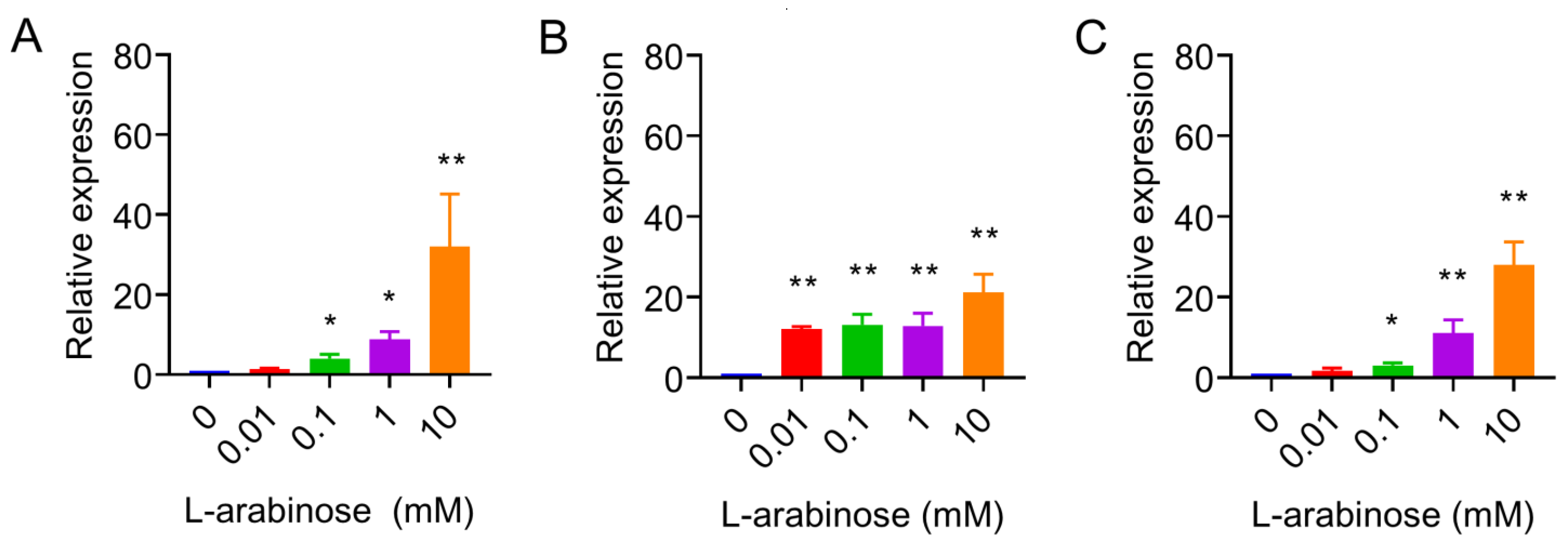
2.2. Quantitative Real-Time PCR

Relative expression of tet(X6) gene in different host bacteria was determined by a two-step qRT-PCR analysis using primers qtet(X6)-F, qtet(X6)-R and 16S rRNA as the reference gene (Table 1). Briefly, total RNA was extracted from bacteria using the RNA-easy Isolation Reagent (Vazyme, Nanjing, China), followed by complementary DNA synthesis with DNA integrated genomic DNA (gDNA) removal using reverse transcription kit. Relative expression results were calculated using the 2−ΔΔCt method.

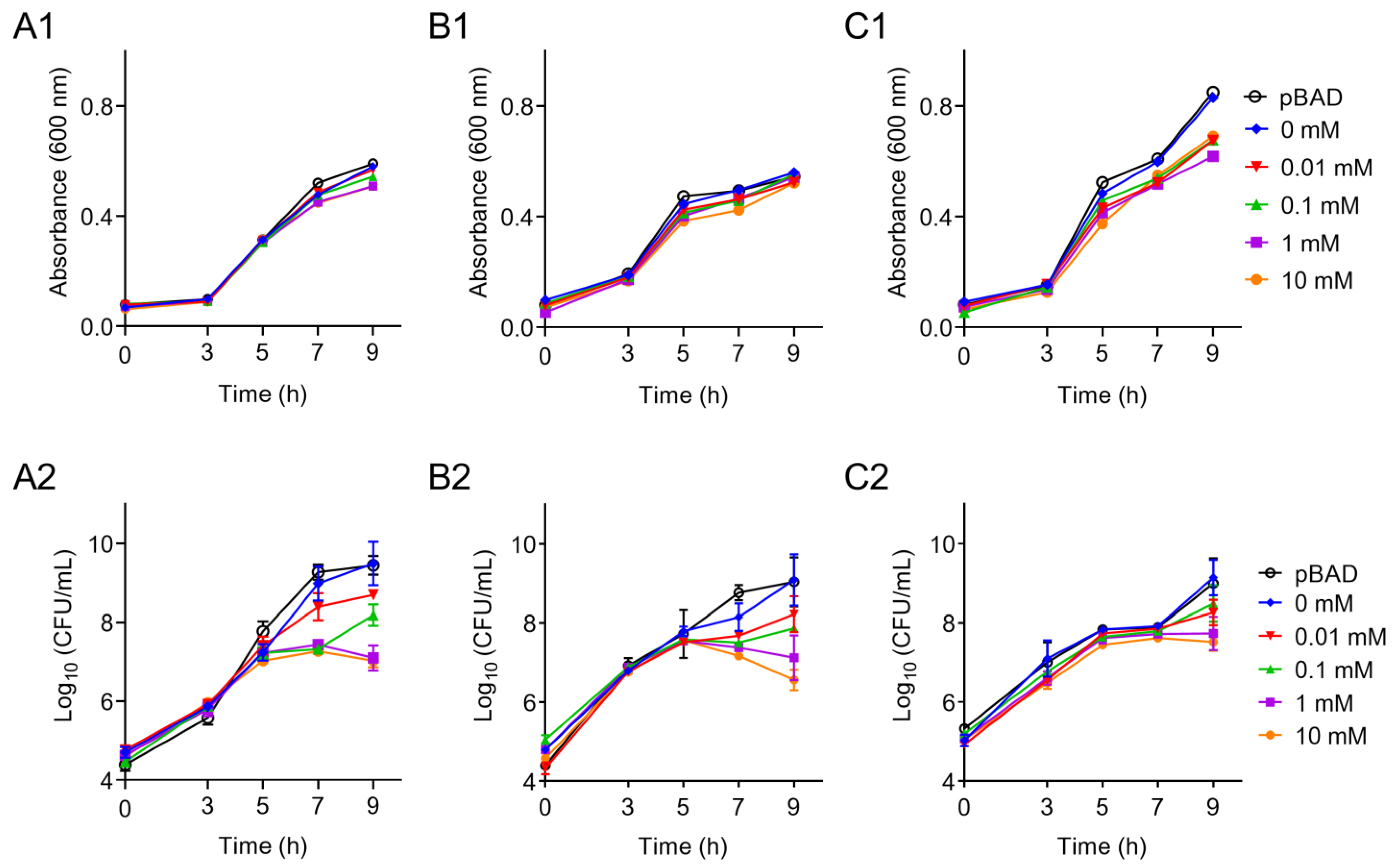
2.3. Effects of tet(X6) Overexpression on Bacterial Growth

An overnight culture of above strains was diluted 10-fold appropriately, and these strains were exposed to different l-arabinose concentrations from 0 to 10 mM and incubated at 37 °C. Bacteria counts were determined at 0, 3, 5, 7 and 9 h by subjecting to 10-fold serial dilution and then plating onto MH agar for visible counts calculation. Meanwhile, the absorbance of culture at 600 nm was determined using an Infinite M200 Microplate reader (Tecan, Männedorf, Switzerland) [16].

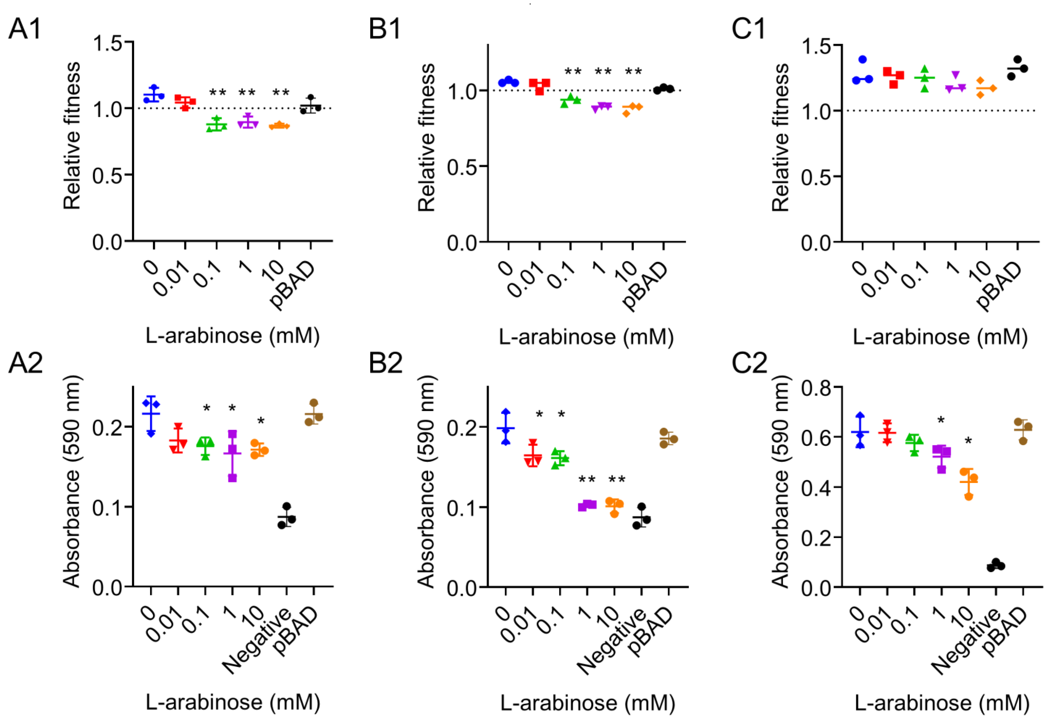
2.4. In Vitro Competition and Biofilm Formation

In vitro competition experiment [12] was used to measure the relative fitness of the E. coli TOP10 (tet(X6)/pBAD) and S. Enteritidis ATCC13076 (tet(X6)/pBAD) and P. mirabilis HS1-T (tet(X6)/pBAD). These strains were competed against their parental strains E. coli TOP10, S. Enteritidis ATCC13076 and P. mirabilis HS1-T, respectively. In biofilm formation test, bacterial cells were stained with crystal violet and the absorbance at 590 nm was measured based on previous study [17]. Blank LB broth was used as negative control.

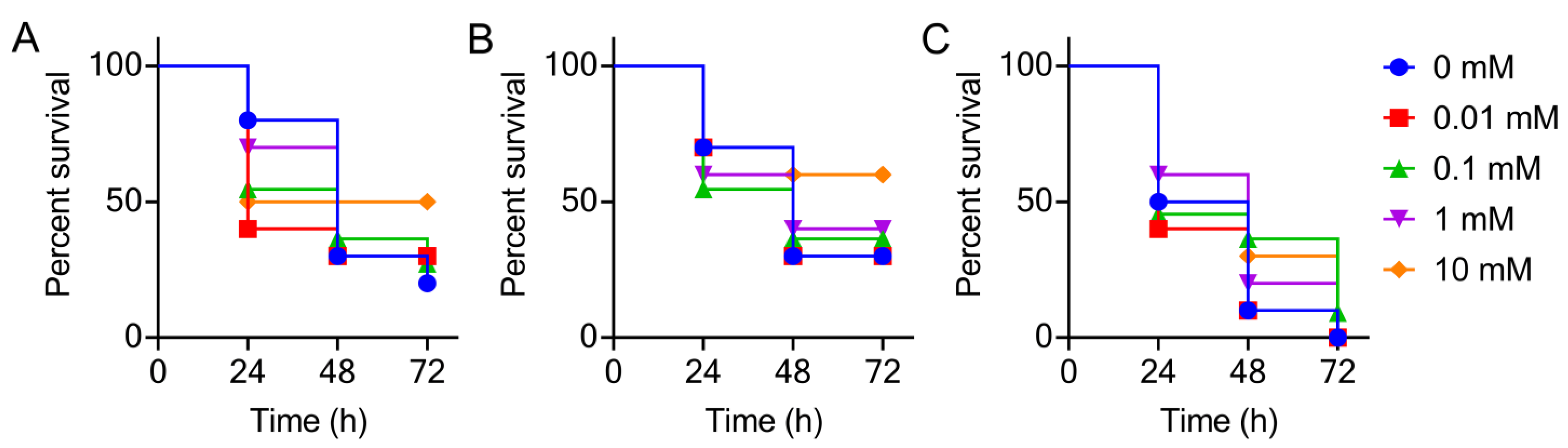
2.5. Galleria mellonella Infection Model

In vivo virulence was evaluated using a G. mellonella infection model [18]. The wax moth G. mellonella in larval stage was stored in dark and used within 3 days. Prior to inoculation into larvae, bacterial pellets were washed with sterile saline and then diluted into an appropriate cell density. Then, the larvae (n = 10 per group) were infected with either bacterial suspension (10 μL, 107 CFUs per larvae) pre-incubated with different concentrations of l-arabinose or vehicle at the left posterior gastropoda using a 50 μL Hamilton syringe.

2.6. Statistical Analysis

Statistical analysis was performed using GraphPad Prism, version 8.3.0. All data were presented as the mean ± SD from three biological replicates. p values were determined using an unpaired, two-tailed Student’s t-test.

3. Results and Discussion

In this study, three kinds of strains were used as host cells, including E. coli TOP10, S. Enteritidis ATCC13076 and P. mirabilis HS1-T. The E. coli TOP10 and S. Enteritidis ATCC13076 are standard strains and have been widely used in previous studies [19,20]. The wild-type P. mirabilis strains were isolated and identified by our laboratory [21], which were naturally resistant to tigecycline. In order to ensure that the genetic background of the strains was clear, P. mirabilis were sequenced and those without the tet(X6) gene were chosen. Strain HS1-T was sensitive to ampicillin, which meets the requirement for subsequent transformants screening. Considering these points, P. mirabilis HS1-T was selected as the host bacteria.
To validate the expression of tet(X6) gene in these strains, MIC values were determined by a broth micro-dilution method in accordance with CLSI guidelines [22]. All media were freshly prepared in order to minimize the oxidative degradation of tigecycline. The MICs for tigecycline ranged from 0.125 to 4 μg/mL in the uninduced group and 1 to 8 μg/mL in the presence of 10 mM l-arabinose (Table 2). Specifically, the MIC values of E. coli TOP10 (tet(X6)/pBAD) and S. Enteritidis ATCC13076 (tet(X6)/pBAD) were significantly increased 8-fold after being incubated with l-arabinose (10 mM), implying the successful expression of tet(X6) gene in these strains. Interestingly, only two-fold MIC changes in P. mirabilis HS1-T (tet(X6)/pBAD) were observed. We reasoned that the intrinsic resistance of P. mirabilis to tigecycline affects the appearance of the tet(X6)-mediated resistance phenotype.
Next, the relative expression of the tet(X6) gene under the exposure of increasing concentrations of l-arabinose from 0 to 10 mM was monitored using qRT-PCR analysis. The mRNA expression of the tet(X6) gene was significantly increased in these three tested strains in a dose-dependent manner (Figure 1), indicating that the expression vector in three strains, particularly for P. mirabilis HS1-T, was successfully constructed. Interestingly, when the concentration of l-arabinose was lower than 1 mM, the tet(X6) expression level in E. coli TOP10 and P. mirabilis HS1-T was low, similar to the uninduced group. In contrast, 1 mM l-arabinose resulted in a significantly increased expression of the tet(X6) gene in S. Enteritidis ATCC13076, indicating that S. Enteritidis is more sensitive to the stimulation of l-arabinose at low levels.
According to the bacterial growth curves, we found that the OD600 of all three host strains increased and reached the peak value at 9 h (Figure 2A1–C1). However, no significant difference between the control and l-arabinose treated groups was observed. Considering that the absorbance of culture could not distinguish the live and dead bacteria, we next determined the bacterial CFUs using a plate counting method. Consequently, we found that the addition of l-arabinose remarkably decreased the colony quantity of E. coli TOP10 (tet(X6)/pBAD) and S. Enteritidis ATCC13076 (tet(X6)/pBAD) in a dose-dependent manner after 7 h of culture (Figure 2A2–B2). However, the overexpression of the tet(X6) gene even under the stimulation of high levels of l-arabinose showed little effect on HS1-T (tet(X6)/pBAD) (Figure 2C2). These data indicated that the increased expression of the tet(X6) gene will pose an extra burden on the bacteria growth of Enterobacteriaceae rather than the Proteus species. These results were consistent with previous findings that the overexpression of antibiotic resistance genes (ARGs) would inhibit the growth of certain bacteria and even lead to the death of bacteria [23,24]. We supposed that there are some regulation mechanisms or compensatory evolution that exist in P. mirabilis. These unknown mechanisms lower the fitness cost induced by the tet(X6) gene and may promote the epidemic of antibiotic resistance [25].
In vitro competition experiments showed that the relative fitness of P. mirabilis HS1-T (tet(X6)/pBAD) is more than one, regardless of the addition of l-arabinose, suggesting that the tet(X6) expression imposed no fitness cost for P. mirabilis (Figure 3C1). By contrast, a decreased competitive ability was found in E. coli TOP10 (tet(X6)/pBAD) and S. Enteritidis ATCC13076 (tet(X6)/pBAD) when the l-arabinose concentration was above 0.1 mM (Figure 3A1,B1). Previous studies have reported that bacterial biofilm production is related to its survival status, thus, the ability of biofilm formation can also reflect bacteria fitness [26,27]. A crystal violet staining method was applied to evaluate the ability of biofilm formation in different host bacteria carrying the tet(X6) gene. Consistent with the relative fitness results, P. mirabilis HS1-T (tet(X6)/pBAD) displayed the highest biofilm formation ability among them by more than two-fold than the other two strains (Figure 3A2–C2). These results support our idea that the expression of the tet(X6) gene in Enterobacteriaceae such as E. coli and S. Enteritidis rather than P. mirabilis would affect bacterial biofilm formation and pathogenicity. Combined with the growth characteristics of strains, we concluded that the overexpression of the tet(X6) gene resulted in the remarkable fitness cost in Enterobacteriaceae. This also means that drug-resistant Enterobacteriaceae will lose their competitive advantage over sensitive strains in the absence of antibiotics [28]. By contrast, a balance was achieved in P. mirabilis between the fitness cost and tet(X6) gene expression. Further studies are warranted to reveal the mechanisms underlying how P. mirabilis maintains the tet(X6) resistance gene without an obvious fitness cost.
It is suggested that the fitness cost caused by antibiotic resistance may lead to the reduction of virulence in bacteria, such as in P. aeruginosa [29]. In our study, a significant increase in the survival of G. mellonella larvae was also observed in E. coli TOP10- and S. Enteritidis ATCC13076 transformants-infected groups as increased concentrations of l-arabinose (Figure 4). However, the P. mirabilis HS1-T transformant retained its pathogenicity on larvae despite the expression of the tet(X6) gene. This could also be attributed to a difference in the growth rates of these strains in vivo; the relatively fast growth of P. mirabilis HS1-T transformant resulted in a more rapid increase in the bacterial burden in the animal infection model compared with E. coli TOP10 and S. Enteritidis ATCC13076 transformants.

4. Conclusions

In summary, our findings reveal that the carrying and expression of tet(X6) gene would result in significant fitness costs in E. coli and S. Enteritidis rather than in P. mirabilis, which may explain the clinical phenomenon that most tet(X6) genes were isolated from Proteus spp. but not from Enterobacteriaceae. The possible reasons may be attributed to some specific regulator mechanisms in Proteus spp., which balances the fitness cost caused by the expression of tet(X6) gene and deserves further studies.

Author Contributions

Data curation, W.C.; Funding acquisition, Z.W. and Y.L.; Methodology, L.J., W.C. and F.T.; Software, L.J.; Supervision, Z.W. and Y.L.; Validation, F.T.; Writing–original draft, L.J.; Writing–review & editing, Y.L. All authors have read and agreed to the published version of the manuscript.

Funding

This work was supported by the National Natural Science Foundation of China (31872526 and 32002331), Natural Science Foundation of Jiangsu Province of China (BK20190893), Agricultural Science and Technology Independent Innovation Fund of Jiangsu Province (CX(20)3091 and CX(21)2010), A Project Funded by the Priority Academic Program Development of Jiangsu Higher Education Institutions (PAPD) and Young Elite Scientists Sponsorship Program by CAST (2020QNRC001).

Data Availability Statement

All data in this study have been included in this manuscript.

Conflicts of Interest

The authors declare no conflict of interest.

References

  1. Yelin, I.; Kishony, R. Antibiotic resistance. Cell 2018, 172, 1136. [Google Scholar] [CrossRef] [PubMed]
  2. Niebel, M.; Quick, J.; Prieto, A.M.; Hill, R.L.; Pike, R.; Huber, D.; David, M.; Hornsey, M.; Wareham, D.; Oppenheim, B.; et al. Deletions in a ribosomal protein-coding gene are associated with tigecycline resistance in Enterococcus faecium. Int. J. Antimicrob. Agents 2015, 46, 572–575. [Google Scholar] [CrossRef] [PubMed]
  3. Sun, Y.; Cai, Y.; Liu, X.; Bai, N.; Liang, B.; Wang, R. The emergence of clinical resistance to tigecycline. Int. J. Antimicrob. Agents 2013, 41, 110–116. [Google Scholar] [CrossRef]
  4. Zeng, Y.; Dong, N.; Zhang, R.; Liu, C.; Sun, Q.; Lu, J.; Shu, L.; Cheng, Q.; Chan, E.W.; Chen, S. Emergence of an Empedobacter falsenii strain harbouring a tet(X)-variant-bearing novel plasmid conferring resistance to tigecycline. J. Antimicrob. Chemother. 2020, 75, 531–536. [Google Scholar] [CrossRef]
  5. Fang, L.X.; Chen, C.; Cui, C.Y.; Li, X.P.; Zhang, Y.; Liao, X.P.; Sun, J.; Liu, Y.H. Emerging high-level tigecycline resistance: Novel tetracycline destructases spread via the mobile Tet(X). BioEssays 2020, 42, e2000014. [Google Scholar] [CrossRef] [PubMed]
  6. Ding, Y.; Saw, W.-Y.; Tan, L.W.L.; Moong, D.K.N.; Nagarajan, N.; Teo, Y.Y.; Seedorf, H. Emergence of tigecycline- and eravacycline-resistant Tet(X4)-producing Enterobacteriaceae in the gut microbiota of healthy Singaporeans. J. Antimicrob. Chemother. 2020, 75, 3480–3484. [Google Scholar] [CrossRef]
  7. Sun, C.; Cui, M.; Zhang, S.; Wang, H.; Song, L.; Zhang, C.; Zhao, Q.; Liu, D.; Wang, Y.; Shen, J.; et al. Plasmid-mediated tigecycline-resistant gene tet(X4) in from food-producing animals, China, 2008–2018. Emerg. Microbes Infect. 2019, 8, 1524–1527. [Google Scholar] [CrossRef] [Green Version]
  8. He, D.; Wang, L.; Zhao, S.; Liu, L.; Liu, J.; Hu, G.; Pan, Y. A novel tigecycline resistance gene, tet(X6), on an SXT/R391 integrative and conjugative element in a Proteus genomospecies 6 isolate of retail meat origin. J. Antimicrob. Chemother. 2020, 75, 1159–1164. [Google Scholar] [CrossRef]
  9. Zheng, X.R.; Zhu, J.H.; Zhang, J.; Cai, P.; Sun, Y.H.; Chang, M.X.; Fang, L.X.; Sun, J.; Jiang, H.X. A novel plasmid-borne tet(X6) variant co-existing with blaNDM-1 and blaOXA-58 in a chicken Acinetobacter baumannii isolate. J. Antimicrob. Chemother. 2020, 75, 3397–3399. [Google Scholar] [CrossRef] [PubMed]
  10. Andersson, D.I. The biological cost of mutational antibiotic resistance: Any practical conclusions? Curr. Opin. Microbiol. 2006, 9, 461–465. [Google Scholar] [CrossRef] [PubMed]
  11. Andersson, D.I.; Levin, B.R. The biological cost of antibiotic resistance. Curr. Opin. Microbiol. 1999, 2, 489–493. [Google Scholar] [CrossRef]
  12. Yang, Q.; Li, M.; Spiller, O.B.; Andrey, D.O.; Hinchliffe, P.; Li, H.; MacLean, C.; Niumsup, P.; Powell, L.; Pritchard, M. Balancing mcr-1 expression and bacterial survival is a delicate equilibrium between essential cellular defence mechanisms. Nat. Commun. 2017, 8, 2054. [Google Scholar] [CrossRef] [Green Version]
  13. San Millan, A.; MacLean, R.C. Fitness costs of plasmids: A limit to plasmid transmission. Microbiol. Spectr. 2017, 5, MTBP-0016-2017. [Google Scholar] [CrossRef] [Green Version]
  14. San Millan, A. Evolution of plasmid-mediated antibiotic resistance in the clinical context. Trends Microbiol. 2018, 26, 978–985. [Google Scholar] [CrossRef] [Green Version]
  15. Yang, J.; Wang, H.-H.; Lu, Y.; Yi, L.-X.; Deng, Y.; Lv, L.; Burrus, V.; Liu, J.-H. A ProQ/FinO family protein involved in plasmid copy number control favours fitness of bacteria carrying mcr-1-bearing IncI2 plasmids. Nucleic Acids Res. 2021, 49, 3981–3996. [Google Scholar] [CrossRef]
  16. Durante-Mangoni, E.; Del Franco, M.; Andini, R.; Bernardo, M.; Giannouli, M.; Zarrilli, R. Emergence of colistin resistance without loss of fitness and virulence after prolonged colistin administration in a patient with extensively drug-resistant Acinetobacter baumannii. Diagn. Microbiol. Infect. Dis. 2015, 82, 222–226. [Google Scholar] [CrossRef]
  17. Ommen, P.; Zobek, N.; Meyer, R.L. Quantification of biofilm biomass by staining: Non-toxic safranin can replace the popular crystal violet. J. Microbiol. Methods 2017, 141, 87–89. [Google Scholar] [CrossRef]
  18. Liu, Y.; Jia, Y.; Yang, K.; Tong, Z.; Shi, J.; Li, R.; Xiao, X.; Ren, W.; Hardeland, R.; Reiter, R.J.; et al. Melatonin overcomes MCR-mediated colistin resistance in Gram-negative pathogens. Theranostics 2020, 10, 10697–10711. [Google Scholar] [CrossRef]
  19. Jousset, A.B.; Rosinski-Chupin, I.; Takissian, J.; Glaser, P.; Bonnin, R.A.; Naas, T. Transcriptional landscape of a plasmid and response to imipenem exposure in TOP10. Front. Microbiol. 2018, 9, 2929. [Google Scholar] [CrossRef] [PubMed]
  20. Bao, H.; Zhang, P.; Zhang, H.; Zhou, Y.; Zhang, L.; Wang, R. Bio-control of Salmonella Enteritidis in foods using bacteriophages. Viruses 2015, 7, 4836–4853. [Google Scholar] [CrossRef] [PubMed]
  21. Li, R.; Peng, K.; Li, Y.; Liu, Y.; Wang, Z. Exploring tet(X)-bearing tigecycline-resistant bacteria of swine farming environments. Sci. Total Environ. 2020, 733, 139306. [Google Scholar] [CrossRef] [PubMed]
  22. CLSI. Methods for Dilution Antimicrobial Susceptibility Tests for Bacteria that Grow Aerobically, 11th ed.; CLSI Standard M07; Clinical and Laboratory Standards Institute: Wayne, PA, USA, 2018. [Google Scholar]
  23. Olaitan, A.O.; Morand, S.; Rolain, J.M. Mechanisms of polymyxin resistance: Acquired and intrinsic resistance in bacteria. Front. Microbiol. 2014, 5, 643. [Google Scholar] [CrossRef] [PubMed] [Green Version]
  24. Li, Y.; Wang, Q.; Peng, K.; Liu, Y.; Li, R.; Wang, Z. Emergence of carbapenem- and tigecycline-resistant Proteus cibarius of animal origin. Front. Microbiol. 2020, 11, 1940. [Google Scholar] [CrossRef]
  25. Hall, J.P.J.; Brockhurst, M.A.; Dytham, C.; Harrison, E. The evolution of plasmid stability: Are infectious transmission and compensatory evolution competing evolutionary trajectories? Plasmid 2017, 91, 90–95. [Google Scholar] [CrossRef] [PubMed]
  26. Deschaine, B.M.; Heysel, A.R.; Lenhart, B.A.; Murphy, H.A. Biofilm formation and toxin production provide a fitness advantage in mixed colonies of environmental yeast isolates. Ecol. Evol. 2018, 8, 5541–5550. [Google Scholar] [CrossRef]
  27. Kowalski, C.H.; Kerkaert, J.D.; Liu, K.W.; Bond, M.C.; Hartmann, R.; Nadell, C.D.; Stajich, J.E.; Cramer, R.A. Fungal biofilm morphology impacts hypoxia fitness and disease progression. Nat. Microbiol. 2019, 4, 2430–2441. [Google Scholar] [CrossRef]
  28. Sun, J.; Zhu, D.; Xu, J.; Jia, R.; Chen, S.; Liu, M.; Zhao, X.; Yang, Q.; Wu, Y.; Zhang, S.; et al. Rifampin resistance and its fitness cost in Riemerella anatipestifer. BMC Microbiol. 2019, 19, 107. [Google Scholar] [CrossRef]
  29. Abdelraouf, K.; Kabbara, S.; Ledesma, K.R.; Poole, K.; Tam, V.H. Effect of multidrug resistance-conferring mutations on the fitness and virulence of Pseudomonas aeruginosa. J. Antimicrob. Chemother. 2011, 66, 1311–1317. [Google Scholar] [CrossRef]
Figure 1. Relative expression level of tet(X6) gene in E. coli TOP10 (A), S. Enteritidis ATCC13076 (B) and P. mirabilis HS1-T (C) in the presence of l-arabinose ranging from 0 to 10 mM. Data are representative of three biological replicates and expressed as mean ± SD. The differences in tet(X6) expression were determined using two-tailed Student’s t-test (* p < 0.05 and ** p < 0.01).
Antibiotics 10 01172 g001
Figure 2. Effect of tet(X6) overexpression on bacterial growth in different host bacteria. (A1,A2): effect of tet(X6) expression on bacterial growth induced with different concentrations of l-arabinose in E. coli TOP10. (B1,B2): effect of tet(X6) expression on bacterial growth induced with different concentrations of l-arabinose in S. Enteritidis ATCC13076. (C1,C2): effect of tet(X6) expression on bacterial growth induced with different concentrations of l-arabinose in P. mirabilis HS1-T. (A1C1), bacterial growth was represented by absorbance at 600 nm; (A2C2), bacterial growth was represented by bacterial CFUs. Host bacteria carrying pBAD plasmid without tet(X6) gene were used as a control. Data are representative of three biological replicates and expressed as mean ± SD.
Antibiotics 10 01172 g002
Figure 3. Bacterial fitness cost (A1C1) and the ability of biofilm formation (A2C2) in different host bacteria after exposure to different concentrations of l-arabinose. (A1,A2): effect of tet(X6) expression on bacterial fitness cost and biofilm formation in E. coli TOP10. (B1,B2): effect of tet(X6) expression on bacterial fitness cost and biofilm formation in S. Enteritidis ATCC13076. (C1,C2): effect of tet(X6) expression on bacterial fitness cost and biofilm formation in P. mirabilis HS1-T. Blank LB broth was used as negative control. Host bacteria carrying pBAD plasmid without tet(X6) gene were also determined. Data are representative of three biological replicates and expressed as mean ± SD. The p values (* p < 0.05 and ** p < 0.01) were calculated by two-tailed Student’s t-test.
Antibiotics 10 01172 g003
Figure 4. Kaplan–Meier plots showing the percentage of survival of G. mellonella over 72 h, infected with different host bacteria induced by different concentrations of l-arabinose. Survival rate of G. mellonella larvae (n = 10 per group) infected with E. coli TOP10 (tet(X6)/pBAD) (A) and S. Enteritidis ATCC13076 (tet(X6)/pBAD) (B) and P. mirabilis HS1-T (tet(X6)/pBAD) (C) (107 CFUs per larvae) over 72 h.
Antibiotics 10 01172 g004
Table 1. PCR primers used in this study.
GenesSequences (5′ to 3′)Amplicon Size (bp)
tet(X6)F: CGAGCTCATGACTTTACTAAAACATAAAAAAATTAC
R: CCCAAGCTTTTATAGATTCATTAGTTTTTGGAAAGAA
1149
qPCR-tet(X6)F: TGTCGTTGATTTTCTCCTG
R: TTGATTCTGCCTGTGCTT
332
16S rRNAF: TTCGGGAACCGTGAGA
R: CTGGCAACAAAGGATAAGG
103
The underlined sequence represents restriction enzyme cutting site.
Table 2. MIC values of tigecycline for three engineered strains in the absence and presence of l-arabinose.
StrainsMIC (μg/mL)MIC (μg/mL) with l-Arabinose (10 mM)
E. coli TOP10 (tet(X6)/pBAD)0.1251
S. Enteritidis ATCC13076 (tet(X6)/pBAD)18
P. mirabilis HS1-T (tet(X6)/pBAD)48
Publisher’s Note: MDPI stays neutral with regard to jurisdictional claims in published maps and institutional affiliations.

