Review

Scope of Onsite, Portable Prevention Diagnostic Strategies for Alternaria Infections in Medicinal Plants

1
Manav Rachna Centre for Medicinal Plant Pathology, Manav Rachna International Institute of Research and Studies, Faridabad 121004, India
2
TERI-Deakin Nanobiotechnology Centre, The Energy and Resources Institute, Gurgaon 122003, India
3
Department of Biotechnology, Yeungnam University, Gyeongsan 38541, Republic of Korea
*
Authors to whom correspondence should be addressed.
Biosensors 2023, 13(7), 701; https://doi.org/10.3390/bios13070701
Submission received: 25 April 2023 / Revised: 18 June 2023 / Accepted: 27 June 2023 / Published: 1 July 2023

Abstract

:
Medicinal plants are constantly challenged by different biotic inconveniences, which not only cause yield and economic losses but also affect the quality of products derived from them. Among them, Alternaria pathogens are one of the harmful fungal pathogens in medicinal plants across the globe. Therefore, a fast and accurate detection method in the early stage is needed to avoid significant economic losses. Although traditional methods are available to detect Alternaria, they are more time-consuming and costly and need good expertise. Nevertheless, numerous biochemical- and molecular-based techniques are available for the detection of plant diseases, but their efficacy is constrained by differences in their accuracy, specificity, sensitivity, dependability, and speed in addition to being unsuitable for direct on-field studies. Considering the effect of Alternaria on medicinal plants, the development of novel and early detection measures is required to detect causal Alternaria species accurately, sensitively, and rapidly that can be further applied in fields to speed up the advancement process in detection strategies. In this regard, nanotechnology can be employed to develop portable biosensors suitable for early and correct pathogenic disease detection on the field. It also provides an efficient future scope to convert innovative nanoparticle-derived fabricated biomolecules and biosensor approaches in the diagnostics of disease-causing pathogens in important medicinal plants. In this review, we summarize the traditional methods, including immunological and molecular methods, utilized in plant-disease diagnostics. We also brief advanced automobile and efficient sensing technologies for diagnostics. Here we are proposing an idea with a focus on the development of electrochemical and/or colorimetric properties–based nano-biosensors that could be useful in the early detection of Alternaria and other plant pathogens in important medicinal plants. In addition, we discuss challenges faced during the fabrication of biosensors and new capabilities of the technology that provide information regarding disease management strategies.

1. Introduction

Medicinal plants have always been important and a matter of concern for people as they had therapeutic-based usage in the past [1]. In general, medicinal plants have been a healing resource in communities around the world for thousands of years. Nevertheless, they remain important today as primary health care for about 85% of the world’s population [2] and as a resource for pharmaceutical research, from which 80% of all synthetic drugs are derived [3]. Medicinal plants were used as traditional remedies to treat a number of lethal diseases from ancient times and produce diverse chemical scaffolds that can interact with biological targets. India is the place of origin of a wide variety of medicinal plants and precious traditional medicine knowledge [4]. It is reported that 80% of the population in developing countries benefit from herbal products for therapeutic uses and purposes.
Like other crop plants, medicinal plants are constantly challenged by an array of biotic inconveniences that pose a serious threat to their survival. Among them, fungal pathogens such as Alternaria spp. cause severe economic losses in medicinal plants and affect their product quality [4]. The co-occurrence of abiotic and biotic stresses in medicinal plants has increased dramatically as a result of global climate change, exposing the risk of their existence. Commercial cultivation has brought the unavoidable problem of more pests and diseases, ultimately leading to crop loss at various levels. Additionally, overharvesting has placed many medicinal species at risk of extinction [5]. Therefore, Alternaria diseases could further increase the risk of their extinction. Hence, there is a need to develop accurate, sensitive, and rapid detection tools to detect Alternaria diseases at early stages under both lab and field conditions. Several researchers have published data on medicinal plants in terms of therapeutic values, value chain losses, and commercial aspects; however, there is still a lack of big data values of medicinal plants related to their loss due to limited disease diagnostics. Figure 1a illustrates 32 years of data on the number of published articles on medicinal-plant-based evolutionary research. Similarly, Figure 1b represents data on the number of studies published on plant diseases with the keyword “plant disease” in different database systems (ScienceDirect, Nature) pertaining to medicinal plants, which gives clear indications of the need for advanced research on medicinal plants’ diagnostic systems.
Alternaria is a cosmopolitan genus of fungi that are saprophytic, endophytic, and pathogenic to plants and animals and is ever present in many favorable and anthropogenic environments [7]. Several edible medicinal herbs have been utilized for more than thousands of years to prevent and treat human disorders. In addition, these herbs are consumed as spices, additives, and/or edible foods. Unfortunately, these medicinal herbs can become contaminated with Alternaria toxins (ATs) before harvest, raising safety concerns [8]. Among Alternaria pathogens, Alternaria alternata has been reported to cause leaf spot and leaf blight diseases in various medicinal plants, and its toxins are of great concern [4]. Therefore, pathogens in medicinal plants and their negative impact should be identified as soon as possible to have more surety and take cost-effective measures, which play a crucial role in ensuring the security of food products and reducing yield loss [9].
Nowadays, pathogen-related losses of medicinal plants are responsible for the reduction in the country’s financial returns [10]. Therefore, the earliest diagnostics with the properties of rapidness, inexpensiveness, and accuracy have become the best combinations in plant pathological studies. Furthermore, in a global market, innovative disease-sensitive and field-ready tools represent a new frontier from a diagnostic laboratory perspective, ensuring that the instruments and tools used are much more suitable in case of operational situations [11]. The commonly established methods, such as polymerase chain reaction [12], fluorescence in situ hybridization [13], enzyme-linked immunosorbent assay [14], immunofluorescence [15], flow cytometry [16], and gas chromatography–mass spectrometry [17], are already existing and broadly used for disease identification in plants. However, they are comparatively tough to use, require experts, are tedious, and are unsuitable for on-field diagnostic and early management strategies because they cannot provide real-time detection data [18]. Meanwhile, some imaging techniques, for example, thermography and fluorescence imaging, are capable of on-field disease detection and analysis, but they are sensitive to variable changes in the environment and lack specificity for types of diseases [19].
A previous study reported the role of various biosensors in pathogenic disease detection in plants, which opens new directions for the development of a new pathogen-specific detection tool kit [17]. Recently, advancements in nanotechnology have paved the way for the better development of nanoparticle-based easy assays for the detection of bioanalytes, which emerged in the improvement of much-sensitive biosensors because of new nanofabrication methods. Nanoparticles (NPs) with uncommon optical properties and more surface area are widely used for the easy detection of bioanalytes of interest in multiple samples [20]. The great physicochemical properties of NPs have proved them to be much more suitable over conventional detection techniques for diagnostic purposes; emerged as a potential way to improve the sensitivity, accuracy rate, and fastness of plant pathogen identification tools; and paved the way for high-throughput analysis [21], which might be suitable for the detection of Alternaria diseases.
The specificity of nanomaterial-based biosensors could be efficiently improved due to the use of enzymes, antibodies, DNA, and bacteriophage [17]. The present review is an update on the current global scenario of medicinal plant losses due to Alternaria infections and further elaborates on the possible diagnostics for early, rapid, and cost-effective detection via nanomaterial-based biosensing strategies.

2. Global Status of Disease Burden on Medicinal Plants

Contamination of food and crops by environmental contaminants, synthetic drugs, residues of chemicals, and pathogens, including various other anthropogenic and naturally present contaminants, is a serious problem in public health worldwide [22,23]. In particular, fungal disease has been a major constraint in medicinal plants across the globe [7]. In this review, we summarize different fungal diseases in medicinal plants with the main focus on Alternaria pathogens (Table 1).
Alternaria toxins (ATs) are a serious concern to humans and animals owing to their wide range of side effects. Data on the occurrence of ATs in medicinal herbs are relatively limited, as previous studies did not routinely examine the amounts of these toxins in herbs and edible products [43,44]. ATs have recently been called “emerging mycotoxins” of medicinal plants. Despite this, there are recently no monitoring guidelines and limitations for ATs in food and/or medicinal plant products. Therefore, there is an urgent need to find reliable analytical methods to monitor ATs that are also applicable to herbs [18,37].

Alternaria Toxins in Medicinal Plants and Their Effects on Humans

Alternaria is capable of producing several secondary metabolites with toxicity under specific conditions, also called ATs [45,46]. A. alternata and A. tenuissima were found to be associated with muskmelon foliar diseases in China in the area of Beijing [47]. These diseases significantly reduced the production and quality of important crops, vegetables, and fruits, resulting in negative impacts on the agricultural economy [22]. Alternaria species range from saprophytic to pathogenic fungi, and therefore, they are able to cause fungal diseases in medicinal plants, such as withering, leaf spot, plant decay, and other visible symptoms [48,49].
Alternaria species produce various host-specific (HSTs) and non-host-specific (nHSTs) phytotoxins, which affect different plant cellular organelles. Reported non-host-specific toxins include “tentoxin (TEN), alternaric acid, alternariol (AOH), alternariol 9-monomethyl ether (AME), brefeldin A (dehydro-), alternuene (ALT), altertoxin-I, Altertoxin-II, altertoxin-III, zinniol, tenuazonic acid [TeA], curvularin, and alterotoxin (ATX) I, II, III” [28,50]. On the other hand, HST toxins are “Japanese-pear-pathotype (A. kikuchiana) (AK), Strawberry-pathotype (AF), Tangerine-pathotype (A. citri) (ACT), and Apple pathotype (A. mali) (AM), including, Alternaria alternata lycopersici toxins (AAL) and ACR-toxins, such as maculosin and destruxin A and B, etc.” [22]. Further, we have shown the effect of different ATs on plant cellular organelles, as shown in Figure 2a. Non-host-specific toxins act as contaminants in fruits and vegetables [51]. Figure 2b deals with different enzymatic metabolisms in Alternaria for developing Alternaria toxicants, including mycotoxins, namely AOH, AME, and ATXs, produced by Alternaria, which are hazardous to humans with different cytotoxic and genotoxic effects. With their pathogenic properties, they affect many agricultural products and may then get into the food chain effectively [52,53]. In 2011, the Food Safety Authority (EFSA) worked on assessing the risk of Alternaria toxins, and found that these toxins could induce genotoxicity and cytotoxicity, including reproductive and developmental toxicity [23].
Cytotoxicity could inhibit cellular growth [54], restrain protein synthesis, and induce apoptosis [55]. It is mentioned in previous reports that the high incidence of esophageal cancer might be due to the ingestion of Alternaria-mycotoxin-contaminated food, recorded in the Linxian area of Henan Province, China [56]. Recent studies have shown that Alternaria mycotoxins exhibit genotoxic effects, and a few of them could induce gene locus mutation. DNA damage or synthesis disorder, chromosome aberration, and other effects of Alternaria mycotoxins have also been reported through in vitro studies [22].

3. Preventive Measures for Alternaria Infections in Medicinal Plants

Alternaria diseases in medicinal plants are a major concern as they can considerably impact the economy by drastically reducing their yield and quality. The conventional approaches to controlling and preventing Alternaria infections may include the spraying of chemically synthesized fungicides; for example, the fungicides pyraclostrobin and azoxystrobin in combination with difenoconazole were generally effective in cases of leaf blight disease [57,58]. In addition, a few medicinal plants have been protected from diseases by integrating resistant genotypes of Jerusalem artichoke with Trichoderma harzianum isolate to prevent Alternaria leaf spot [42,59]. As a research advancement, researchers have used various other biocontrol strategies for fungal disease control, including liquid copper fungicides, powdered sulfur fungicides, and potential bacterial strains of Bacillus subtilis, as well as other physical control methods, such as removing dead plant matter, drip irrigation, planting disease-free seeds and resistant cultivars, sterilized tools, and rotation of crops [60,61]. Although chemical treatment remains the most important method for reducing the incidence of fungal disease, frequent use can make fungal populations less sensitive. Moreover, excessive spraying can lead to environmental pollution and impose a large economic burden on producers [62]. However, a few methods, such as mulching, crop rotation, cleaning up fallen leaves, and elimination of water on leaves, can help the slow spread of Alternaria spores in the soil and control Alternaria diseases in plants [63]. Due to the above limitations, it is suggested that optical sensing techniques can help to identify the primary diseases and areas of varying disease severities in the field, especially in the case of Alternaria spp. [64]. To prevent and control the spread of pathogenic disease, producers need to be able to identify causative strains accurately, sensitively, and quickly. This allows you to select and implement the best possible disease management strategies [58].

4. Common Available Diagnostic Strategies

Many fungal pathogens during disease development can impose similar effects on medicinal plants, so it becomes crucial to differentiate between causal fungal pathogenic species to ensure well-informed management [65]. In the field, biosensors are preferred for detecting lethal pathogens in plants because they are inexpensive, require much less expertise, and identify target pathogens very quickly with high specificity and sensitivity [66,67].

4.1. Conventional Methods

Conventional methods are feasible [68] for a person without any strong science background as the steps are easy to understand. They are reliable and have easy steps to develop the protocol but are very tedious and unsuitable for the onsite early detection of Alternaria infections and viruses or bacteria that colonize inside the plant tissues [69].
Some of the conventional disease diagnostic methods in developing countries are mentioned below.

4.1.1. Visual Observation

Depending on the symptoms, visual monitoring can be performed in the plant samples; it is frequently feasible to detect the primary pathogenesis of a worm, fungal spp., bacterial spp., or viruses [70]. Plant diseases can be identified by examining leaf curling, color change, and spotting of either brown or white color [71]. The entire procedure occasionally takes so long that the disease cannot be completely eradicated. Large areas also make this process exceedingly laborious [72].

4.1.2. Microscopy

Highly precise microscopes are needed and are often used tools for precise diagnostics. The contaminated plant portions are collected for examination via a microscope after being observed visually in the lab [73]. It is frequently feasible to make a diagnosis by examining the hyphae, microsclerosis, conidiophores, conidia, and bacterial cell clusters of a pathogenic fungus on the diseased plant components under a microscope [74]. However, in unfavorable weather, a fungus might not even form spores or have fungal mycelium on the affected surface [75].

4.1.3. Mycological Diagnosis

Researchers can record the proliferation and pattern of pathogens using the moist chamber method [76]. The “wet chamber” technique includes incubating a portion of the ill plant under a high-humidity maintained chamber (Petri dishes, etc.). In this method, fungi in the diseased tissues proliferate and start to manifest as a result of favorable conditions [77]. Under a commonly used microscope, it is possible to identify the genera and species of pathogenic fungal organisms through the observation of their hyphae, macro conidia, and microconidia [78]. Certain phytopathogens may be difficult to identify using moist chamber techniques [79].

4.1.4. Biological Assays or Indicator Plant Tests

This methodology is commonly used for the diagnosis of phytoviruses and phytoplasmas, in addition to other diagnostic methodologies. Biologically based diagnostics are implied on indicator trees and herbaceous plants, as a testing [80]. Infection is transmitted to indicator plants by mechanical inoculation or injection. Symptomatic leaves rubbed onto healthy indicator plants obtain and are injected with standard buffer suspensions [80]. The artificial infection method by micrografting is also used for indicator trees and shrubs, and such tests are performed under strictly isolated and controlled conditions. Its main drawbacks are long latency and the need for culture chambers, isolated from others [81].

4.2. Rapid Lab-Based Diagnostic Methods

4.2.1. Antigen-/Antibody-Based Diagnostics

Another molecular approach for diagnosing plant diseases with the use of antibodies and color-changing assays is the enzyme-linked immunosorbent assay (ELISA) [14]. With this technique, antibodies attached to enzymes are used to precisely bind the specific epitopes (antigens) from viruses, bacteria, and fungi. ELISA is the most advanced serology-derived diagnostic technique for fungi [77]. Based on color changes brought on by the reaction of the substrate and the immobilized enzyme, the detection can be seen [49]. The techniques of monoclonal antibodies and recombinant antibodies can significantly increase the performance of ELISA and are capable of identifying tiny molecules, viruses, and their quantitative markers [79]. Although there are many methods for immunodiagnostics, among them, ELISA is considered sensitive and accurate, allowing a definitive diagnosis [75]. The drawback of the ELISA technique is that it has low sensitivity for complex bacteria [82].

4.2.2. Molecular Genetic Identification

Numerous techniques, including “nucleic-acid-hybridization, polymerase-chain-reaction (PCR), reverse-transcription PCR (RT-PCR), real-time PCR, and DNA-derived-microchips”, are available for the molecular genetic characterization of harmful microorganisms in plants [83]. Plant pathogen detection now makes extensive use of PCR techniques as they are greatly sensitive and advanced, such as RT-PCR, which has been used for lethal plant pathogen identification [12]. The idea behind multiplex PCR is to perform a single reaction while simultaneously detecting many types of DNA or RNA particles and a speedy identification of plant diseases using real-time PCR technologies [84]. Pathogen diagnostic applications have adopted a variety of PCR amplification assay types to enhance the assay specificity, for example, nested PCR; signal identification, for example, magnetic capture hybridization and ELISA PCR; and multiplexing ability as multiplex PCR techniques [85]. A single picogram of pure fungal DNA can be identified using simple, dependable, and scalable PCR techniques. Nielsen and colleagues (2002) found the increasing detection sensitivity of quantitative PCR (qPCR), using real-time monitoring of amplification, depending on fluorescence to precisely establish the starting concentrations [62].
Although the PCR approach has been utilized for multiplexed pathogen detection, still it is constrained by the lack of operational flexibility even though it enhances the sensitivity of detection due to the fidelity of DNA amplification [46]. For PCR-based identification, nucleic acid isolation, genetic marker selection, specific primer designing, PCR machine, electrophoresis, DNA purification, and result determination are all required [84]. Inhibitors in the sample, polymerase activity, PCR buffer, and deoxynucleoside triphosphate concentration all effect PCR performance. This is the limitation of this technique in practical applicability for disease field sampling [62,86].

4.2.3. Flow Cytometry (FCM)

Flow cytometry (FCM) is a commonly used laser-beam-based optical technology for cell number counting and sorting, finding biomarkers, and creating proteins [87]. FCM is used to quickly identify cells as they go fast on an electronic detection device in a liquid stream. The capacity to test multiple parameters simultaneously is a benefit of this technology. The method [88] makes use of an incident laser beam and evaluates the scattering and fluorescence of the beam reflected off the sample. Although FCM has mostly been used to research cell cycle kinetics and antibiotic sensitivity, it has also been used to count live and dead bacteria, and it characterizes bacterial DNA and fungal spores [16]. Detection with this technique is based on nonspecific staining of DNA or characterization of scattered patterns and autofluorescence [89].

4.2.4. Gas Chromatography

It is a non-optical-based indirect-type method for detecting lethal plant disease, including volatile chemical character profiling of infected plants. Volatile organic compounds (VOCs) are produced in green-leafed plants [90] and emitted differently from plants infected by diseases and mechanical damage [91]. The type and form of infection might be determined through the profiling of such VOCs, which could then be used for illness diagnosis and confirmation [92]. The particular VOC that is irrelated to disease particularly could be determined by gas chromatography [GC] analysis of the volatile signature of plants [93].
Gas chromatography and mass spectrometry are frequently coupled (GC–MS) to improve the performance of compound differentiation and to identify unidentified compounds in volatile samples [94]. However, the requirement for a sample precollected VOC for a longer length of time prior to data processing substantially restricts the application of GC/GC–MS on the field, unlike the imaging technology, by which the data can be obtained onsite directly [17].
Therefore, generally used traditional lab strategies for plant pathogen detection could not be applied for the detection of Alternaria infections because they are time-consuming and have multiple drawbacks, in addition to the fact that they are unsuitable for the variables and can cause severe loss due to time limitations and incorrect diagnostics.

5. Portable Diagnosis Techniques

The early detection of plant pathogens is an appropriate preventive strategy for the management of crop yield and quality. For this reason, effective diagnostic techniques and tools, which are simple, specific, rapid, and economical, are needed to be developed [95].

5.1. PCR-Based Systems

Whatman FTA cards developed by GE Healthcare are a fast, cost-effective, and simple method for extracting DNA from different plant tissues and can store DNA at room temperature for years, which can be used later for PCR purposes [96]. To diagnose a disease directly in the field easily, portable devices, such as “Palm PCR”, were developed by the “Ahram Biosystems” Korea, which has helped to overcome barriers of electrical supply [97]. Additionally, a “Twista recombinase polymerase amplification (RPA) fluorometer” developed by TwistDx was utilized for immediate and fast diagnosis, allowing an on-time suitable treatment [98]. This technology was used for the detection of Phytophthora spp. in plant tissue [99]. Despite these advantages, portable PCR devices still face challenges, such as sample preparation, DNA extraction, accurate temperature control systems, and sample evaporation in open devices [100].

5.2. Lateral Flow Assays (LFAs)

LFAs based on immunodetection and immunoprinting kits are considered among the first grower-friendly pathogen monitoring methods [101]. For the detection of plant diseases, tissue-printing ELISA and lateral-flow devices have been manufactured to allow onsite detection [17]. To date, LFA devices are available in the market for the detection of several plant pathogens, such as Phytophthora spp., Ralstonia solanacearum, and potato virus, with easy handling and steps (e.g., pocketdiagnostic.com); hence, they are very handy for farmers. However, LFAs have some disadvantages, such as limited potential for multiplex detection of target analytes. Additionally, low sensitivity has been reported for some LFA-based biosensors [102].

5.3. Microsphere Immunoassays (MIA)

MIA techniques have emerged as a promising alternative for microbial detection. This technique is based on microspheres coded with different fluorophores. Briefly, the microspheres are conjugated with specific antibodies to capture the pathogen antigen, and a magnet is used to separate the bound and nonbound microspheres [103,104]. After the separation, specific antibodies linked with a reporter fluorophore are added to the microsphere sample mix. A Luminex xMAP analyzer excites the internal dyes of the microspheres with a red laser and the reporter fluorochrome, captured by the antigen, with a green laser, allowing the specific detection of multiple analytes at the same time [100].

5.4. Hyperspectral Imaging Techniques

These techniques can be utilized between 350 and 2500 nm hyperspectral imaging to gather important data regarding the health of plants [105,106]. The hyperspectral examination combines optical spectroscopy and picture investigation strategies, permitting both physiological and morphological parameters to be assessed simultaneously [107]. The hyperspectral imaging technique (HIS) has been applied for the early detection of citrus and apple fruit diseases caused by pathogens in the field and has also been used to estimate the ripeness of strawberries in the field [108]. In any case, each interaction between a plant and a pathogen has a certain spatial and worldly flow, and these forms influence a diverse range of the electromagnetic parameters [106]. However, HIS has still not been utilized for the early detection of black spots [109].

5.5. Fluorescence Imaging

This method involves measuring the fluorescence that came off from the chlorophyll on the leaves by calculating the reaction of the incident light. Based on changes in the photosynthetic instrument and electron transport reactions [110], the fluorescence patterns can be used to analyze the level of pathogen infections. Efficient sensors along with LED [111] are frequently used in chlorophyll fluorescence imaging equipment to measure photosynthetic electron transport [112]. The variations in photosynthetic activity brought on by biotic and abiotic inconvenience over the leaf area have been studied using this method [19]. The distinction and measurement of fungi infections have been demonstrated to benefit from the integration of fluorescence imaging and image analysis tools [113]. It is challenging to use existing chlorophyll fluorescence imaging technologies in typical agricultural greenhouses or field conditions because the preparation of the plants must adhere to a tight routine [64]. In order to assess plant disease at the field level, research has focused on observing fluorescence aspects from sunlight-induced reflectance at the field level and using chlorophyll-fluorescence-based imaging and thermography to understand and compare infections by a fungus and a virus in plant leaves [82].

5.6. Loop-Mediated Isothermal Amplification (LAMP)

LAMP assay is a single-step method in which 4 to 6 primers bind the target sites laterally using a strand displacement Bst DNA polymerase, allowing amplification under isothermal conditions [114,115], as well as gel electrophoresis, turbidimeter, lateral flow dipstick, or the naked eye used for detecting amplified products [116]. LAMP assay was used for the rapid detection of Alternaria spp., including A. alternata, on infected pears [3,117]. LAMP can be applied in the field and in laboratories. LAMP assay has some limitations and defects, as it contaminates easily, needs a source of light, and requires some skills for assessing the results [62].

6. Smartphone-Based Portable Device Diagnoses

Every plant releases volatile organic compounds (VOCs) as they breathe through the pores in their leaves. However, when a plant is diseased, the type and concentration of these VOCs change. Smartphone-based portable technologies can detect VOCs released by almost every plant within a fraction of a minute. Scientists at North Carolina State University developed a handheld device that is designed to be attached to the farmer’s smartphone, sitting over the top of its camera, and it works by sampling the airborne VOCs that plants release [118].
A WeChat applet-based mobile solution was developed for citrus disease diagnosis with a simplified DenseNet deep learning architecture, which was further connected to a server where web requests can be made and help to recognize diseases from the captured leaf or fruit image samples [109]. On the same basis, as a proof of concept, server and mobile client cloud-based applications using a ResNet50 deep learning architecture were reported for tomato disease classification [119]. Similarly, an in-field, automatic wheat disease diagnosis system along with a server for the disease identification steps and a mobile client for in-field image capturing was built [120]. However, these methods require internet connectivity and high computational power to process the images in a central server. Conversely, a mobile-based solution that can operate directly on mobile devices without any need of an internet connection was proposed and developed [121,122].

7. Nanomaterial-Based Biosensing Strategies for Plant Pathogen Detection

Nanomaterials are ideal candidates for the analysis of plant pathogens as their dimensions typically fall in the range of 1 to 100 nm, which can provide enhanced surface-to-volume ratio and unique chemical, optical, and electrical properties, which are not observed in the bulk counterparts [123]. Nanomaterials are normally synthesized via specific self-assembly procedures at the nanoscale level [124].
Nanoscale materials can also interact with biomolecular targets more efficiently due to their small sizes and fast diffusion rates [123]. Nanomaterials, such as carbon-based materials, such as graphene and carbon nanotubes, including graphite, silver (Ag), gold (Au), silica (Si), and copper (Cu) nanoparticles [125], can be prepared in many different morphologies ranging from spherical particles, cubes, rods, wires, plates, prisms, and core–shell structures to more complicated 3D architectures. In addition, new advanced nanomaterials, such as nanotubes, nanorods, nanospheres, nanoreefs, nanoclusters, tetrapods, nanopores, and nanochannels, have been created as a result of the growth of nanotechnology as sensing tools [126], which have been widely applied in the areas of in vivo imaging [127,128,129]. These could undergo shape transformation or agglomeration, which alters their chemical or physical properties in response to various external stimuli [130].
Biosensor devices are derived by their bioinspired receptor units with differently efficient specificities to their targeted analytes [131]. For example, enzymes can react with certain molecules rapidly and selectively, and nucleic acids can bind to their complementary sequences delicately on the nanoscale [132]. The biorecognition components may consist of enzymes, nucleic acids, entire cells, aptamers, or polymers, whereas the analyte may be an ion, molecule, cell, nucleic acid, or protein [133]. The addition of nanomaterials (NMs) into a biosensor can detect the signals and provide a more accurate and specific identification of an analyte [134,135]. For example, carbon nanomaterials are suitable for the conjugation of biomolecules (enzymes, antibodies, DNA, cell, etc.) [132]. In addition, biomolecules can be immobilized and conjugated with other materials by surface modification through the recombination or introduction of chemical linkers [136]. In biotechnology-derived sensing techniques, gold nanoparticles (AuNPs) are commonly used; they can be used as carriers in the case of biorecognition molecules, antibodies, or aptamers, and can also be the labels for signal transduction and amplification [137]. For example, absorption-based colorimetric sensing involves aggregation-induced interparticle surface plasmon coupling of AuNPs, which results in a visible color change from red to blue. This concept has provided a practical platform for the detection of any target analyte that triggers the AuNPs’ aggregation or redispersion [138]. AuNPs are widely utilized in electrochemical-based biosensors as they can increase the surface area and conductivity and have been reported in the detection of microbial food pathogens [139].
These advanced nanomaterials have recently begun to play a crucial role in the creation of biosensors, tissue engineering, battery materials, and multifunctional materials [127,140]. The three main parts of a biosensor are (i) a biorecognition element for detecting the presence and concentration of an analyte; (ii) an optical, electrochemical, piezoelectric, or thermally based transducer surface; and (iii) a detector to measure the signals produced [141].
The sensing applications would be a fruitful strategy for developing a novel tool of diagnostics, especially for Alternaria.

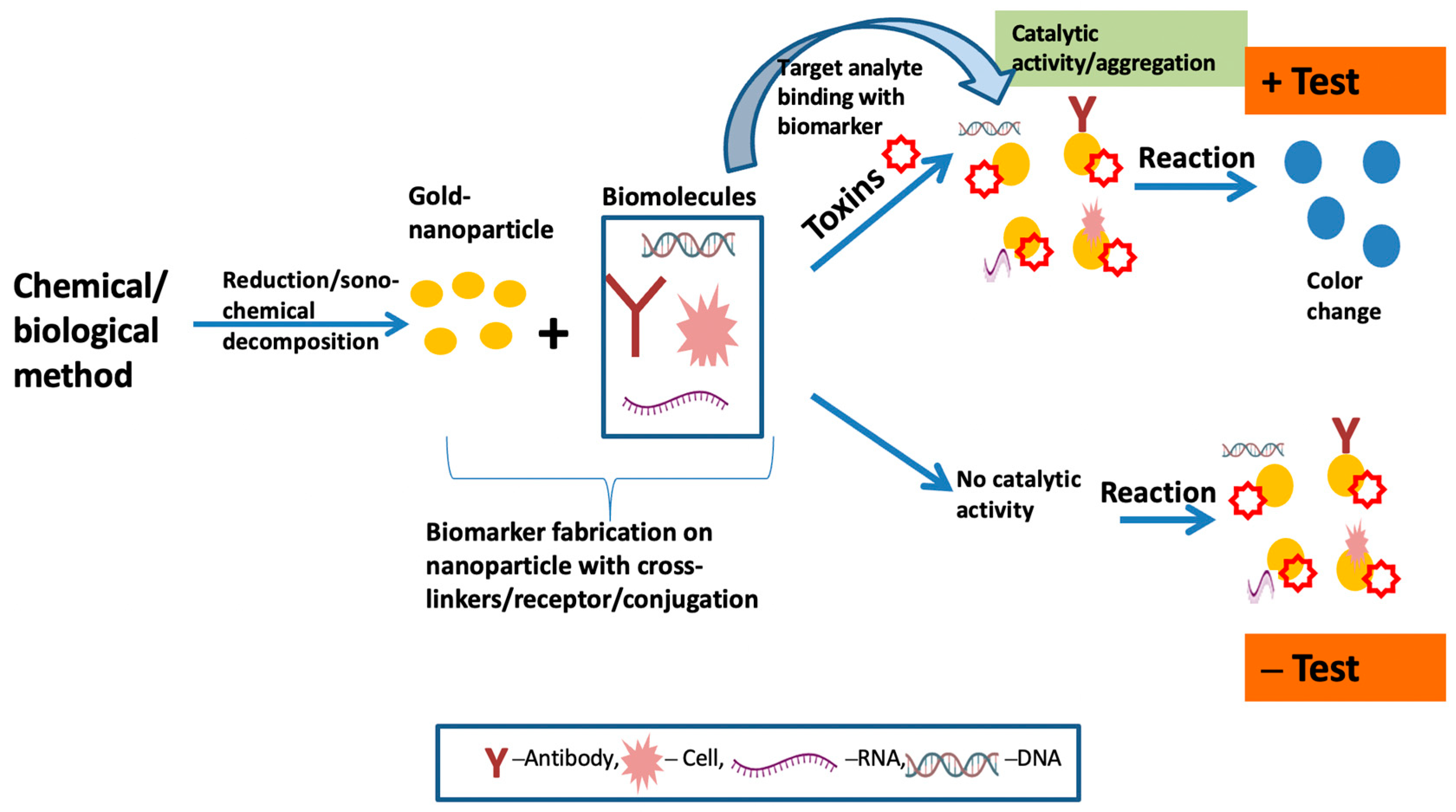
7.1. Colorimetric Sensing

Colorimetric sensors are based on simple chemical reaction principles and have become more usable and widespread due to their vast availability, simple use, inexpensiveness, and ability to respond sensitively and selectively to a variety of analytes. Colorimetric sensors are one of the types of optical sensors; they change color in response to external inputs if any modification to the physical or chemical surroundings qualifies as such a stimulus [142]. Four examples of colorimetric techniques include “indirect target-mediated aggregation, chromogenic substrate-mediated catalytic activity, point-of-care testing (POCT) devices, and machine learning-assisted colorimetric sensor arrays”, which can be utilized for pathogen analysis [143,144]. Figure 3 depicts a pictorial general working overview of colorimetric-based biosensors with the fabrication of AuNPs to identify the target analytes in samples. To attain mobility and intellectualization in clinical applications, colorimetric-sensor-based gold nanoparticles can be further integrated with newly developed portable devices, such as paper-based chips. This can be applied to smartphone readout applications and can help with the visualization and in situ detection of germs [69].

7.2. Paper-Based Sensing

Paper is a versatile material and gaining attention as a substrate for developing paper-based analytical devices (PADs) in point-of-care (POC) format, being an alternative to the detection of conventional biomarkers [145]. Due to its porous nature, the paper has an inherent hydrophilic feature that is particularly useful since it may promote the flowing of fluid via capillary reaction without external pumping [146]. PADs are less expensive and simpler to make than sensors composed of materials such as glass, silicon, and plastic [147]. Additionally, PADs can be disposed of in an environmentally friendly manner and are simple to store, transport, and store. Three types of PADs include dipstick assay, lateral flow assay (LFA), and microfluidic paper-based analytical device [148]. Membranes made of cellulose and nitrocellulose are the two main forms of paper substrates.
Paper-derived colorimetric sensors are able to use enzyme-based reactions and have the ability for real-field diagnosis. The sample is put onto a test strip, and the result is displayed within 5 to 10 minutes, using the paper-based lateral flow immunochromatographic analysis test, which analyzes the quality and quantity of the material in a complicated mixture [75]. A paper-based colorimetric sensor was used to predict plant diseases in Allium crops for the easy and simple measurement of PG activity with the help of the principle of the ruthenium red (RR) dye method [149]; however, the drawback of hindrance can be developed by blurry signaling at the detection point due to capillary reaction forces on the liquid and its corresponding membrane [150]. Figure 4 illustrates the fundamental functioning of a microfluid paper-based biosensor, implemented with the colorimetric methodology for the observation of color change in case of any targeted toxins detection.

7.3. Surface Plasmon Resonance (SPR)-Based Biosensing

SPR-derived sensors are suitable for the measurement of biological molecular associations in real time, and are helpful analytical tools for calculating the adsorption rate of target analytes onto molecularly derived probes connected with a metallic surface [151]. The electromagnetic (EM) resonance of the group oscillations of free electrons connected to a plasmonic metal (silver or gold for visible spectrum) and a dielectric semi-infinite interface is known as surface plasmon resonance (SPR) [152]. This resonance produces a linked, exponentially decaying surface EM field along the metal dielectric contact. This field can be employed as a sensing layer to implement SPR-based sensors since it is particularly sensitive to changes in the refractive index (RI) of the dielectric layer [153]. The SPR-based biosensor was utilized for the detection of the fungus P. fijiensis in samples of banana leaf extracts [154]. However, plasmonic sensors also have many drawbacks, such as nonspecific surface binding, mass transportation restrictions, steric hindrance during the binding event, and data misinterpretation during common events [88]. Localized surface plasmon resonance (LSPR) is another promising method related to SPR biosensing with high sensitivity, a type of SPR phenomenon in which the resonant EM field is constrained to the metallic nanostructure and sensitive to RI change of the medium surrounding it only within a few tens of nm [155]. For colloidal and randomly oriented nanoparticles, scattering and absorption effects are dominant. The advantages of LSPR over SPR include a high aspect ratio that permits a larger interaction surface area for immobilizing the sensing elements, a miniaturized probe to create compact devices, and broad applicability and compatibility with a variety of phenomena, including fluorescence, Raman, and IR spectroscopy, among others less perceptive than traditional SPR [156,157].

7.4. Chip-Based Rapid Sensing

In chip-based biosensors, the sensing layer is created over a thin metal (50 nm)–coated glass substrate, and the analyte to be recognized flows through a microfluidic channel near the sensing layer [153]. As mentioned, these chip-based sensors have many benefits, including label-free detection, dynamic measurement of binding–unbinding kinetics to watch the reaction mechanism occurring over the sensing surface, and high sensitivity [158]. Microfluidics, for example, can be integrated with chip-based substrates that are highly sensitive, repeatable, and miniaturized [155]. Chip-based biosensors are appropriate for microfluidics-based methods, which allow mixing, filtering, and detection to be carried out on the same chip by adjusting tiny amounts of liquid. The DNA of Ganoderma boninense, a pathogenic fungus that infects oil palm trees in Malaysia, was extracted using a microfluidic chip device [159]. The increasing research and development in this area have demonstrated its potential for real-time evaluation and mobility, as well as prospective future works in terms of the improvement of multiple biosensors and multiple pathogen detection in a single-chip platform [159,160].

7.5. Electrochemical Sensing

In electrochemical-based biosensors, a molecular recognition coating and an electrochemical transducer, which transforms biological data obtained from a connecting event into an electrical signal and then displays them on a readout device, are two main components [161]. Different types and forms of nanomaterials and nanocomposites could be used in electrochemical biosensors to improve the sensitivity of the detection mechanisms and to give better detection limits through various tactics [126,162]. Microfluidic systems and electrochemical biosensors can be integrated to provide miniaturized components on a single platform [163]. The urediniospores of soybean rust fungi were detected with aptamer-based sensitive electrochemical sensing. For this, spore-bound aptamer units were incubated with a streptavidin–alkaline phosphatase agent, leading to the enzymatic formation of p-nitrophenol, which is characterized by its unique electrochemical properties [164]. Regarding the electrochemical nano-biosensor of bacterial species, recently, an electrochemical-based immune sensor was manufactured for the detection of Escherichia coli O157:H7 (E. coli O157:H7) using cyclic voltammetry (CV) with the combination of nanomaterials [143,165]. An electrochemical-derived impedance immune sensor for the detection of strained Staphylococcus aureus was also introduced [166].
Electrochemical methods, such as electrochemical impedance spectroscopy (EIS), differential pulse voltammetry (DPV), cyclic voltammetry (CV), square wave voltammetry (SWV), and amperometry, have been implemented for pathogen detection and identification [167]. Nanomaterials or polymers have been normally applied with these methods to improve signals. Figure 5 defines the fundamental principle of the electrochemical-based nanosensor techniques for any kind of desired analyte.
Electrochemical-based platforms are the most successful biosensors, and they have been used for the detection and identification of huge numbers of biomarkers and the diagnosis of infectious diseases, including cancers [168]. Despite the advantages of electrochemical-based biosensors, there are a few problems associated, such as analyte detection and analyte loss during the transporting of the sample to the electrode surface [169,170]. By assessing the different applications and utility of these sensing techniques with plant pathogens, it was discovered that they are fast, sensitive, accurate, specific, and suitable for their portability. This can be applied with slight modifications to manufacture an efficient tool for the detection of Alternaria.

8. Idea for Suitable Future Strategies in the Detection of Alternaria Species

In several research disciplines, such as environmental tracking, diagnostic of various airborne lethal pathogens, real-time diagnostic of contents in human blood and disease-causing microorganisms, and detection of pesticide substances in food products and beverages, biosensors have emerged as cutting-edge detection techniques [72,171,172]. To date, no suitable strategies have been reported for the detection of Alternaria in the field. There are only a few developed strategies that are advanced and capable of detecting Alternaria. For example, in pomegranate, Alternaria spp. cause quality issues in postharvested fruits. They infect pomegranate at an early stage of ripeness, grow, and spread within the fruit, and degrade the cell wall by developing alternariol (AOH), alternariol methyl ether (AME), and tenuazonic acid (TA) toxins and detoxifying resistance compounds in the fruit [173]. To resolve this, most analytical methods, such as nuclear magnetic resonance (NMR) and magnetic resonance imaging (MRI), have been utilized to characterize and detect the black heart but are often time-consuming and require a complicated sample pretreatment [174].
For the identification of Alternaria, researchers have used a technique such as the electronic nose (E-nose) technique [175] due to it being a fast and nondestructive technique that imitates the human olfactory system [176]. Compared with other methods [177], it presents precise and stable performance, a simple sample pretreatment procedure, a short measurement time, low cost, and ease in handling [11]. Composed of metal oxide semiconductor (MOS) biosensors, the E-nose has been utilized in detecting the changes in different food products during storage time and is helpful in the assessment of shelf life and spoilage [175,178].
A DNA-HRP functionalized silver nanoparticle probe was developed for the detection of Alternaria in citrus [179]. Therefore, there is a requirement of eco-friendly materials and biodegradable printable polymers with high conductivity in electrochemical biosensors and colorimetric biosensors, which leads to the identification of Alternaria toxins in desired samples [180]. Developing alternative methods that overcome the drawbacks of conventional methods and a lab-based method in detecting Alternaria spp. fungus in medicinal plants is necessary.
Current developments in micro- and nanotechnology have made it possible to create biosensor-based tests that deliver incredibly accurate, sensitive, and quick results. For general adoption and implementation in agricultural and horticultural areas, there are numerous additional factors and potential difficulties that must be taken into account [181]. Additionally, the in-situ sampling technique has a significant impact on the precision of biosensor-based detection [67]. To control and possibly prevent the transmission of lethal plant diseases and plant pathogens, such as Alternaria, to new areas in medicinal plants, it is imperative to develop a rapid, efficient, and cost-effective methodology for the early detection of disease-causing pathogens. To stop the spread of disease with the least amount of damage to crop productivity and food safety, rapid detection systems with high sensitivity and selectivity for plant pathogens are crucial. For the quick and precise identification of Alternaria, nanotechnology systems provide an option in which all detection procedures are carried out by a portable, miniaturized instrument [133,182]. Additionally, pathogen detection requires several desired qualities, in which functionalized metal nanoparticles as a sensing component of nanomaterials give advanced sensing capabilities for use in the field [62,66]. Over the past few decades, biosensors have been used for the detection and monitoring of a wide variety of bacterial species. These gadgets are highly sensitive detection platforms with small detection limits. Electrochemical biosensors and colorimetric biosensing among other forms of biosensors are particularly adept at avoiding many issues with the use of traditional and common laboratory methods [183].
This chapter gives a general overview of how nanotechnology is being used in the field of microbiological diagnostics, with an emphasis on medicinal plant pathogens, in particular, Alternaria.

9. Conclusions

This paper briefs and offers insights on technologies that are frequently used for detecting various diseases and pathogenic microbes in plants in the past few years and their associated problems. Advancement in the technologies is required to overcome various challenges that arise in these techniques, including the sensitivity, selectivity, and specificity in the detection system using nanotechnology. Diagnostic instruments that are conventional/traditional are quickly being replaced by nano-biosensing-based technologies and devices. These low-cost, quick, portable, very sensitive, and specialized tools for the field detection of pathogens associated with significant medicinal plants will soon find widespread adoption with further experimentation for application in a variety of environments. With the use of these portable nano-biosensors, the frequency and volume of chemically derived materials and their applications to herbs both before and after harvesting, the costs associated with on-farm production, and the loss of herbs’ quality and yield due to plant pathogen diseases would all be significantly decreased. However, more research is needed to improve these nano-biosensors so they can simultaneously monitor and detect many disease-causing fungi/bacteria on the field. Electrochemical-based wearable biosensors with nanofabrication of biomolecules on suitable nanomaterials have the ability to sense wirelessly, detect, and give signals of the presence and abundance of pathogenic organisms and, therefore, can be used for the early detection of Alternaria strains in medicinal plants that will also improve their production and quality. In this review, the applications of a variety of sensors as a diagnostic tool were highlighted. These tools typically combine a biological sensing component with a physicochemical transducer, electrochemical properties, and colorimetric-based technology. These tools can provide the desired identification strategies for the Alternaria disease detection in medicinal plants and help to improve early detection and enable the most effective prevention for farmers and the economy.

Author Contributions

Conceptualization, P.S., S.S. (Shruti Shukla) and N.D.; writing—original draft preparation, S.S. (Sadhana Shukla); writing—review and editing, S.S. (Sadhana Shukla), S.A. and N.D.; visualization, S.A. and S.S. (Sadhana Shukla); supervision, N.D. All authors have read and agreed to the published version of the manuscript.

Funding

This research received no external funding.

Institutional Review Board Statement

Not applicable.

Informed Consent Statement

Not applicable.

Data Availability Statement

Not applicable.

Conflicts of Interest

The authors declare no conflict of interest.

References

  1. Mahmoudi, R. Application of Medicinal Plants: From Past to Present. MOJ Biol. Med. 2017, 1, 80. [Google Scholar] [CrossRef] [Green Version]
  2. Pešić, M. Development of Natural Product Drugs in a Sustainable Manner. Brief for United Nations Global Sustainable Development Report 2015. 2015. Available online: https://sustainabledevelopment.un.org/content/documents/6544118_Pesic_Development%20of%20natural%20product%20drugs%20in%20a%20%20sustainable%20manner.pdf (accessed on 16 November 2022).
  3. Fitzgerald, M.; Heinrich, M.; Booker, A. Medicinal Plant Analysis: A Historical and Regional Discussion of Emergent Complex Techniques. Front. Pharmacol. 2019, 10, 1480. [Google Scholar] [CrossRef] [PubMed]
  4. Swetha, P.; Sundararaj, R. Diseases of Medicinal Plants Cultivated in Karnataka and Their Management. In Medicinal Plants; Intechopen: London, UK, 2022. [Google Scholar] [CrossRef]
  5. Roberson, E.B.; Frances, A.; Havens, K.; Maschinski, J.; Meyer, A.; Ott, L. Fund plant conservation to solve biodiversity crisis. Science 2020, 367, 258. [Google Scholar] [CrossRef] [PubMed]
  6. Salmeron-Manzano, E.; Garrido-Cardenas, J.A.; Manzano-Agugliaro, F. Worldwide Research Trends on Medicinal Plants. Int. J. Environ. Res. Public Health 2020, 17, 3376. [Google Scholar] [CrossRef]
  7. Zhao, X.; Liu, D.; Yang, X.; Zhang, L.; Yang, M. Detection of seven Alternaria toxins in edible and medicinal herbs using ultra-high performance liquid chromatography-tandem mass spectrometry. Food Chem. X 2022, 13, 100186. [Google Scholar] [CrossRef]
  8. De Berardis, S.; De Paola, E.L.; Montevecchi, G.; Garbini, D.; Masino, F.; Antonelli, A.; Melucci, D. Determination of four Alternaria alternata mycotoxins by QuEChERS approach coupled with liquid chromatography-tandem mass spectrometry in tomato-based and fruit-based products. Food Res. Int. 2018, 106, 677–685. [Google Scholar] [CrossRef] [PubMed]
  9. FAO Launches 2020 as the UN’s International Year of Plant Health 2020. 2 December 2019. Available online: http://www.fao.org/news/story/en/item/1253551/icode/ (accessed on 3 December 2022).
  10. Savary, S.; Willocquet, L.; Pethybridge, S.J.; Esker, P.; McRoberts, N.; Nelson, A. The global burden of pathogens and pests on major food crops. Nat. Ecol. Evol. 2019, 3, 430–439. [Google Scholar] [CrossRef]
  11. Buja, I.; Sabella, E.; Monteduro, A.G.; Chiriacò, M.S.; De Bellis, L.; Luvisi, A.; Maruccio, G. Advances in Plant Disease Detection and Monitoring: From Traditional Assays to In-Field Diagnostics. Sensors 2021, 21, 2129. [Google Scholar] [CrossRef]
  12. Cai, H.Y.; Caswell, J.; Prescott, J.F. Nonculture Molecular Techniques for Diagnosis of Bacterial Disease in Animals: A Diagnostic Laboratory Perspective. Veter. Pathol. 2014, 51, 341–350. [Google Scholar] [CrossRef] [Green Version]
  13. Kliot, A.; Kontsedalov, S.; Lebedev, G.; Brumin, M.; Cathrin, P.B.; Marubayashi, J.M.; Škaljac, M.; Belausov, E.; Czosnek, H.; Ghanim, M. Fluorescence in situ Hybridizations (FISH) for the Localization of Viruses and Endosymbiotic Bacteria in Plant and Insect Tissues. J. Vis. Exp. 2014, 84, e51030. [Google Scholar] [CrossRef] [Green Version]
  14. López, M.M.; Llop, P.; Cubero, J.; Penyalver, R.; Caruso, P.; Bertolini, E.; Peñalver, J.; Gorris, M.T.; Cambra, M. Strategies for Improving Serological and Molecular Detection of Plant Pathogenic Bacteria. In Plant Pathogenic Bacteria; Springer: Berlin/Heidelberg, Germany, 2001; pp. 83–86. [Google Scholar] [CrossRef]
  15. Ward, E.; Foster, S.J.; A Fraaije, B.; Mccartney, H.A. Plant pathogen diagnostics: Immunological and nucleic acid-based approaches. Ann. Appl. Biol. 2004, 145, 1–16. [Google Scholar] [CrossRef]
  16. Chitarra, L.G.; Bulk, R.W.V.D. The Application of Flow Cytometry and Fluorescent Probe Technology for Detection and Assessment of Viability of Plant Pathogenic Bacteria. Eur. J. Plant Pathol. 2003, 109, 407–417. [Google Scholar] [CrossRef]
  17. Fang, Y.; Ramasamy, R.P. Current and Prospective Methods for Plant Disease Detection. Biosensors 2015, 5, 537–561. [Google Scholar] [CrossRef] [Green Version]
  18. Sinha, P.; Govil, J.N.; Singh, V.K. (Eds.) Recent Progress in Medicinal Plants. Diseases and Their Management; SCI Tech Publishing LLC.: Bradford, PA, USA, 2002; pp. 1–6. [Google Scholar]
  19. Scholes, J.D.; Rolfe, S.A. Chlorophyll fluorescence imaging as tool for understanding the impact of fungal diseases on plant performance: A phenomics perspective. Funct. Plant Biol. 2009, 36, 880–892. [Google Scholar] [CrossRef]
  20. Sankar, M.; He, Q.; Engel, R.V.; Sainna, M.A.; Logsdail, A.J.; Roldan, A.; Willock, D.J.; Agarwal, N.; Kiely, C.J.; Hutchings, G.J. Role of the Support in Gold-Containing Nanoparticles as Heterogeneous Catalysts. Chem. Rev. 2020, 120, 3890–3938. [Google Scholar] [CrossRef] [PubMed] [Green Version]
  21. Shivashankarappa, A.; Sanjay, K.R. Escherichia coli-based synthesis of cadmium sulfide nanoparticles, characterization, antimicrobial and cytotoxicity studies. Braz. J. Microbiol. 2020, 51, 939–948. [Google Scholar] [CrossRef]
  22. Chen, A.; Mao, X.; Sun, Q.; Wei, Z.; Li, J.; You, Y.; Zhao, J.; Jiang, G.; Wu, Y.; Wang, L.; et al. Alternaria Mycotoxins: An Overview of Toxicity, Metabolism, and Analysis in Food. J. Agric. Food Chem. 2021, 69, 7817–7830. [Google Scholar] [CrossRef]
  23. EFSA Panel on Contaminants in the Food Chain (CONTAM). Scientific Opinion on the risks for animal and public health related to the presence of Alternaria toxins in feed and food. EFSA J. 2011, 9, 2407. [Google Scholar] [CrossRef]
  24. Sun, C.; Li, F.; Wei, M.; Xiang, Z.; Chen, C.; Xu, D. Detection and Biological Characteristics of Alternaria alternata Resistant to Difenoconazole from Paris polyphylla var. chinensis, an Indigenous Medicinal Herb. Plant Dis. 2021, 105, 1546–1554. [Google Scholar] [CrossRef]
  25. Apnikheti. SafedMusli. 2022. Available online: https://www.apnikheti.com/en/pn/agriculture/horticulture/medicinal-plants/safed-musli (accessed on 12 December 2022).
  26. Manjunath, H.; Nakkeeran, S.; Raguchander, T. First Report of Anthracnose on noni Caused by Colletotrichum gloeosporioides in India. Arch. Phytopathol. Plant Prot. 2012, 45, 276–279. [Google Scholar]
  27. Barman, S.; Ghosh, R.; Dalal, D.; Mandal, N.C. Suppression of Leaf Blight of Ocimum sanctum L. Using Lactic Acid Bacteria as Novel Bio-control Agent. Proc. Natl. Acad. Sci. USA India Sect. B Boil. Sci. 2018, 88, 1389–1397. [Google Scholar] [CrossRef]
  28. Ghosh, S.K.; Banerjee, S. First Report of Alternaria brassicae Leaf Spot Disease of Aloe vera and It’s Disease Intensity in West Benga. Eur. J. Biotechnol. Biosci. 2014, 2, 37–43. [Google Scholar]
  29. Shivanna, M.B.; Parashurama, T.R.; Achar, K.G.S.; Vasanthakumari, M.M. Fungal foliar diseases in Withania somnifera and its effect on secondary metabolites. Plant Biosyst. Int. J. Deal. all Asp. Plant Biol. 2014, 148, 907–916. [Google Scholar] [CrossRef]
  30. Kulkarni, M.S.; Prashanthi, S.K. Diseases Medicinal Plants Vlll- Holybasil, Mint and lpecac. In Becent Progress in Medicinal Plants. Diseasesand Their Management; Sinha, P., Govil, J.N., Singh, V.K., Eds.; SCI Tech Publishing LLC.: Bradford, PA, USA, 2002; pp. 127–136. [Google Scholar]
  31. Kalra, A.; Singh, H.B.; Pandey, R.; Samad, A.; Patra, N.K.; Kumar, S. Diseases in Mint: Causal Organisms, Distribution, and Control Measures. J. Herbs, Spices Med. Plants 2005, 11, 71–91. [Google Scholar] [CrossRef]
  32. Prajapati, P.; Sharma, K. A Hand Book of Medicinal Plants; Agrobios: Jodhpur, India, 2003; p. 81. (In Indian) [Google Scholar]
  33. Gupta, M.L.; Misra, H.O.; Kalra, A.; Khanuja, S.P.S. Root-Rot and Wilt: A New Disease of Ashwagandha (Withania Somnifera) Caused by Fusarium solani. J. Med. Aromat. Plant Sci. 2004, 26, 285–287. [Google Scholar]
  34. Chandel, S.; Dubey, K.; Kaushal, P. Major Diseases of Medicinal and Aromatic Plants Recorded in Himachal Pradesh-India. J. Plant Dis. Sci. 2014, 9, 145–153. [Google Scholar]
  35. Sarkar, S.; Dasgupta, B. First Report of Leaf Spot of Aswagandha (Withania Somnifera Dunal) caused by Colletotrichum gloeosporioides from West Bengal, India. J. Mycopathol. Res. 2017, 55, 257–259. [Google Scholar]
  36. Achar, K.G.S.; Parashurama, T.R.; Shivanna, M.B. A New Record of Leaf Spot Caused by Xanthomonas campestris in Tinospora cordifolia in India. Int. J. Curr. Microbiol. Appl. Sci. 2014, 1, 269–273. [Google Scholar]
  37. Kawuri, R.; Suprapta, D.N.; Nitta, Y.; Homma, T. Destructive Leaf Rot Disease Caused by Fusarium oxysporum on Aloe barbadensis Miller in Bali. Agril. Sci. Res. J. 2012, 2, 295–301. [Google Scholar]
  38. Reddy, P.P. Fungal Diseases and Their Management in Horticultural Crops; Scientific Publishing: New Delhi, India, 2010; Volume 342001, 359p. [Google Scholar]
  39. Morrison, W.R.; Linderman, S.; Hausbeck, M.K.; Werling, B.P.; Szendrei, Z. Disease and Insect Pests of Asparagus. Extension Bulletin e3219, Michigan State University. 2014. Available online: http://msue.anr.msu.edu/uploads/resources/pdfs/Disease_and_Insect_Pests_of_Asparagus_(E3219) (accessed on 12 December 2022).
  40. Mondal, G. Occurrence of Synchytrium Inducing Gall on Punarnava (Boerhavia diffusa) from West Bengal, India. In Proceedings of the 6th International Conference on “Plant, Pathogens and People” with the Mission “Challenges in Plant Pathologyto Benefit Humankind”, New Delhi, India, 23–27 February 2016; Volume 27, p. 110012. [Google Scholar]
  41. Jetawat, R.P.S.; Mathur, K.; Singh, K.; Singh, A. Effect of Fungicides against Fusarium solani and Rhizoctonia solani Infecting Ashwagandha. Ecoscan 2015, 7, 75–78. [Google Scholar]
  42. de Figueirêdo, G.S.; de Figueirêdo, L.C.; Cavalcanti, F.C.N.; dos Santos, A.C.; da Costa, A.F.; de Oliveira, N.T. Biological and chemical control of Sclerotinia sclerotiorum using Trichoderma spp. and Ulocladium atrum and pathogenicity to bean plants. Braz. Arch. Biol. Technol. 2010, 53, 1–9. [Google Scholar] [CrossRef] [Green Version]
  43. Wu, Y.; Ye, J.; Xuan, Z.; Li, L.; Wang, H.; Wang, S.; Liu, H.; Wang, S. Development and validation of a rapid and efficient method for simultaneous determination of mycotoxins in coix seed using one-step extraction and UHPLC-HRMS. Food Addit. Contam. Part A Chem. Anal. Control Expo. Risk Assess. 2021, 38, 148–159. [Google Scholar] [CrossRef] [PubMed]
  44. Xing, L.; Zou, L.; Luo, R.; Wang, Y. Determination of five Alternaria toxins in wolfberry using modified QuEChERS and ultra-high performance liquid chromatography-tandem mass spectrometry. Food Chem. 2020, 311, 125975. [Google Scholar] [CrossRef] [PubMed]
  45. Dall’asta, C.; Cirlini, M.; Falavigna, C. Chapter Three—Mycotoxins from Alternaria: Toxicological Implications. In Advances in Molecular Toxicology; Elsevier: Amsterdam, The Netherlands, 2014; Volume 8, pp. 107–121. [Google Scholar] [CrossRef]
  46. Dai, J.; Peng, H.; Chen, W.; Cheng, J.; Wu, Y. Development of multiplex real-time PCR for simultaneous detection of three Potyviruses in tobacco plants. J. Appl. Microbiol. 2013, 114, 502–508. [Google Scholar] [CrossRef]
  47. Zhao, J.; Bao, S.; Ma, G.; Wu, X. Characterization of Alternaria species associated with muskmelon foliar diseases in Beijing municipality of China. J. Gen. Plant Pathol. 2016, 82, 29–32. [Google Scholar] [CrossRef]
  48. Blagojević, J.; Vukojević, J.; Ivanović, B.; Ivanović, Ž. Characterization of Alternaria Species Associated with Leaf Spot Disease of Armoracia rusticana in Serbia. Plant Dis. 2020, 104, 1378–1389. [Google Scholar] [CrossRef] [PubMed]
  49. Matić, S.; Tabone, G.; Garibaldi, A.; Gullino, M.L. Alternaria Leaf Spot Caused by Alternaria Species: An Emerging Problem on Ornamental Plants in Italy. Plant Dis. 2020, 104, 2275–2287. [Google Scholar] [CrossRef]
  50. Ghosh, R.; Barman, S.; Khatun, J.; Mandal, N.C. Biological control of Alternaria alternata causing leaf spot disease of Aloe vera using two strains of rhizobacteria. Biol. Control 2016, 97, 102–108. [Google Scholar] [CrossRef]
  51. Pinto, V.E.F.; Patriarca, A. Alternaria Species and Their Associated Mycotoxins. Methods Mol. Biol. 2017, 1542, 13–32. [Google Scholar] [CrossRef]
  52. Goessens, T.; De Baere, S.; De Troyer, N.; Deknock, A.; Goethals, P.; Lens, L.; Pasmans, F.; Croubels, S. Multi-residue analysis of 20 mycotoxins including major metabolites and emerging mycotoxins in freshwater using UHPLC-MS/MS and application to freshwater ponds in flanders, Belgium. Environ. Res. 2021, 196, 110366. [Google Scholar] [CrossRef]
  53. Mujahid, C.; Savoy, M.-C.; Baslé, Q.; Woo, P.M.; Ee, E.C.Y.; Mottier, P.; Bessaire, T. Levels of Alternaria Toxins in Selected Food Commodities Including Green Coffee. Toxins 2020, 12, 595. [Google Scholar] [CrossRef]
  54. Lehmann, L.; Esch, H.; Wagner, J.; Rohnstock, L.; Metzler, M. Estrogenic and genotoxic potential of equol and two hydroxylated metabolites of Daidzein in cultured human Ishikawa cells. Toxicol. Lett. 2005, 158, 72–86. [Google Scholar] [CrossRef]
  55. Bensassi, F.; Gallerne, C.; El Dein, O.S.; Hajlaoui, M.R.; Bacha, H.; Lemaire, C. Cell death induced by the Alternaria mycotoxin Alternariol. Toxicol. In Vitro 2012, 26, 915–923. [Google Scholar] [CrossRef]
  56. Pfeiffer, E.; Eschbach, S.; Metzler, M. Alternaria toxins: DNA strand-breaking activity in mammalian cellsin vitro. Mycotoxin Res. 2007, 23, 152–157. [Google Scholar] [CrossRef] [PubMed]
  57. Neils, A.L.; Brisco-McCann, E.I.; Harlan, B.R.; Hausbeck, M.K. Management strategies for Alternaria leaf blight on American ginseng. Crop Prot. 2021, 139, 105302. [Google Scholar] [CrossRef]
  58. Waterworth, K. Treatment for Alternaria—Recognizing and Preventing Alternaria Symptoms. Gardening Know How. 20 July 2022. Available online: https://www.gardeningknowhow.com/plant-problems/disease/alternaria-leaf-spot.htm (accessed on 31 January 2023).
  59. Viriyasuthee, W.; Jogloy, S.; Saksirirat, W.; Saepaisan, S.; Gleason, M.L.; Chen, R.S. Biological Control of Alternaria Leaf Spot Caused by Alternaria spp. in Jerusalem Artichoke (Helianthus tuberosus L.) under Two Fertilization Regimes. Plants 2019, 8, 463. [Google Scholar] [CrossRef] [Green Version]
  60. Kgatle, M.; Flett, B.; Truter, M.; Aveling, T. Control of Alternaria leaf blight caused by Alternaria alternata on sunflower using fungicides and Bacillus amyloliquefaciens. Crop Prot. 2020, 132, 105146. [Google Scholar] [CrossRef]
  61. Nielsen, L. Alternaria Leaf Spot Prevention and Treatment, Epic Gardening. 2021. Available online: https://www.epicgardening.com/Alternaria-leaf-spot/ (accessed on 28 December 2022).
  62. Dyussembayev, K.; Sambasivam, P.; Bar, I.; Brownlie, J.C.; Shiddiky, M.J.A.; Ford, R. Biosensor Technologies for Early Detection and Quantification of Plant Pathogens. Front. Chem. 2021, 9, 636245. [Google Scholar] [CrossRef] [PubMed]
  63. Chase, A.R. Advanced Treatment of Alternaria. Greenhouse Product News. 11 February 2021. Available online: https://gpnmag.com/article/advanced-treatment-Alternaria/ (accessed on 28 December 2022).
  64. Mahlein, A.-K. Plant Disease Detection by Imaging Sensors—Parallels and Specific Demands for Precision Agriculture and Plant Phenotyping. Plant Dis. 2016, 100, 241–251. [Google Scholar] [CrossRef] [PubMed] [Green Version]
  65. Oerke, E.-C. Remote Sensing of Diseases. Annu. Rev. Phytopathol. 2020, 58, 225–252. [Google Scholar] [CrossRef]
  66. Şensoy, K.G.; Muti, M. The Novel Nanomaterials Based Biosensors and Their Applications. In Novel Nanomaterials; Intechopen: London, UK, 2021. [Google Scholar] [CrossRef]
  67. Ray, M.; Ray, A.; Dash, S.; Mishra, A.; Achary, K.G.; Nayak, S.; Singh, S. Fungal disease detection in plants: Traditional assays, novel diagnostic techniques and biosensors. Biosens. Bioelectron. 2017, 87, 708–723. [Google Scholar] [CrossRef]
  68. Lau, H.Y.; Wu, H.; Wee, E.J.H.; Trau, M.; Wang, Y.; Botella, J.R. Specific and Sensitive Isothermal Electrochemical Biosensor for Plant Pathogen DNA Detection with Colloidal Gold Nanoparticles as Probes. Sci. Rep. 2017, 7, 38896. [Google Scholar] [CrossRef] [Green Version]
  69. Yang, J.; Wang, X.; Sun, Y.; Chen, B.; Hu, F.; Guo, C.; Yang, T. Recent Advances in Colorimetric Sensors Based on Gold Nanoparticles for Pathogen Detection. Biosensors 2022, 13, 29. [Google Scholar] [CrossRef]
  70. Chane, T.B.; Boyraz, N. Biotechnological Tools for Detection, Identification and Management of Plant Diseases. Afr. J. Biotechnol. 2019, 18, 798–805. [Google Scholar]
  71. Kaur, L.; Sharma, S.G. Identification of plant diseases and distinct approaches for their management. Bull. Natl. Res. Cent. 2021, 45, 169. [Google Scholar] [CrossRef]
  72. Liu, L.; Dong, Y.; Huang, W.; Du, X.; Ren, B.; Huang, L.; Zheng, Q.; Ma, H. A Disease Index for Efficiently Detecting Wheat Fusarium Head Blight Using Sentinel-2 Multispectral Imagery. IEEE Access 2020, 8, 52181–52191. [Google Scholar] [CrossRef]
  73. Patil, P.; Narayankar, P.; Mulimani, D.; Patil, M. Using Microscopic Images to Predict Plant Diseases in a Deep Learning Environment. In ICT for Competitive Strategies; CRC Press: Boca Raton, FL, USA, 2020; pp. 807–813. [Google Scholar] [CrossRef]
  74. Nezhad, A.S. Future of portable devices for plant pathogen diagnosis. Lab Chip 2014, 14, 2887–2904. [Google Scholar] [CrossRef]
  75. Khakimov, A.; Salakhutdinov, I.; Omolikov, A.; Utaganov, S. Traditional and current-prospective methods of agricultural plant diseases detection: A review. IOP Conf. Ser. Earth Environ. Sci. 2022, 951, 012002. [Google Scholar] [CrossRef]
  76. Krug, J.C.; Müller, G.M.; Bills, G.F.; Foster, M.S. Moist chambers for the development of fungi. In Biodiversity of Fungi: Inventory and Monitoring Methods; Elsevier: Amsterdam, The Netherlands, 2004; pp. 589–593. [Google Scholar]
  77. Khasanov, B.A. Diseases of Agricultural Crops and Measures to Combat Them. Textbook for Graduate Students; TashSAU Publishing Department: Tashkent, Uzbekistan, 2011. [Google Scholar]
  78. Sharma, R. Diseases of Medicinal & Aromatic Plants and Their Management. Recent Approaches Manag. Plant Dis. 2018, 251, 283. [Google Scholar]
  79. Dyakov, Y.T.; Elansky, S.N. General Phytopathology; Yurayt Publishing House: Moscow, Russia, 2019. [Google Scholar]
  80. Bunsevich, L.L.; Kostyuk, M.A.; Danilyuk, Y.P. Fruit Growing and Viticulture of the South of Russia [Electronic Resource]; NCZRIHV: Krasnodar, Russia, 2012; Volume 5, p. 6. [Google Scholar]
  81. Upadyshev, M.T. Heat resistance of pear plants during recovery from latent viruses using dry-air thermotherapy. Hortic. Vitic. 2022, 1, 44–51. (In Russian) [Google Scholar] [CrossRef]
  82. Singh, A. Techniques, to Identify Plant Pathogens (Part-1). Plant Cell Technology. Your Partner in Plant Tissue Culture. Available online: https://www.plantcelltechnology.com/blog/techniques-to-identify-plant-pathogens-part1-2239da (accessed on 23 March 2022).
  83. Narayanasamy, P. Microbial Plant Pathogens-Detection and Disease Diagnosis: Fungal Pathogens; Springer: Berlin/Heidelberg, Germany, 2011. [Google Scholar] [CrossRef]
  84. Schaad, N.W.; Opgenorth, D.; Gaush, P. Real-Time Polymerase Chain Reaction for One-Hour On-Site Diagnosis of Pierce’s Disease of Grape in Early Season Asymptomatic Vines. Phytopathology 2002, 92, 721–728. [Google Scholar] [CrossRef] [PubMed] [Green Version]
  85. Capote, N.; Pastrana, A.M.; Aguado, A.; Sánchez-Torres, P. Molecular Tools for Detection of Plant Pathogenic Fungi and Fungicide Resistance. Plant Pathol. 2012, 59, 151–202. [Google Scholar]
  86. Egamberdiev, S.S.; Salakhutdinov, I.B.; Radjabov, F.S.; Kurbanov, A.Y.; Abdurakhmonov, I.Y. Methodical Instruction on the Detection of Pathogen of Fusarium Genera, Identifying More Aggressive Forms and Their Control Measures; Center of Genomics and Bioinformatics of SA of RUz “Fan va talim poligraf” LLC.: Tashkent, Uzbekistan, 2017. [Google Scholar]
  87. Shapiro, H.M. Practical Flow Cytometry; John Wiley & Sons Inc.: Hoboken, NJ, USA, 2003. [Google Scholar]
  88. Greilhuber, J.; Temsch, E.M.; Loureiro, J.C.M. Nuclear DNA Content Measurement. In Flow Cytometry with Plant Cells; Doležel, J., Greilhuber, J., Suda, J., Eds.; Wiley-VCH Press: Hoboken, NJ, USA, 2007; pp. 67–101. [Google Scholar]
  89. D’Hondt, L.; Höfte, M.; VAN Bockstaele, E.; Leus, L. Applications of flow cytometry in plant pathology for genome size determination, detection and physiological status. Mol. Plant Pathol. 2011, 12, 815–828. [Google Scholar] [CrossRef]
  90. Nicolì, F.; Negro, C.; Nutricati, E.; Vergine, M.; Aprile, A.; Sabella, E.; Damiano, G.; De Bellis, L.; Luvisi, A. Accumulation of Azelaic Acid in Xylella fastidiosa-Infected Olive Trees: A Mobile Metabolite for Health Screening. Phytopathology 2019, 109, 318–325. [Google Scholar] [CrossRef] [PubMed] [Green Version]
  91. Dorokhov, Y.; Komarova, T.; Sheshukova, E. Volatile organic compounds and plant virus–host interaction. In Plant Virus–Host Interactio; Academic Press: Cambridge, MA, USA, 2014; pp. 241–262. [Google Scholar] [CrossRef]
  92. Agustika, D.K.; Mercuriani, I.S.; Ariyanti, N.A.; Purnomo, C.W.; Triyana, K.; Iliescu, D.D.; Leeson, M.S. Gas Chromatography-Mass Spectrometry Analysis of Compounds Emitted by Pepper Yellow Leaf Curl Virus-Infected Chili Plants: A Preliminary Study. Separations 2021, 8, 136. [Google Scholar] [CrossRef]
  93. Eid, K.; El-Sayed, A.-N.; Shoala, T. Gas Chromatography-Mass Spectrometry (GC-MS) Analysis of Sugar Beet Leaf Extracts in Response to Exogenous Application of Resistance Inducers to Manage Sugar Beet Powdery Mildew. Egypt. J. Phytopathol. 2018, 46, 257–277. [Google Scholar] [CrossRef]
  94. Tholl, D.; Hossain, O.; Weinhold, A.; Röse, U.S.R.; Wei, Q. Trends and applications in plant volatile sampling and analysis. Plant J. 2021, 106, 314–325. [Google Scholar] [CrossRef]
  95. Luna-Moreno, D.; Sánchez-Álvarez, A.; Islas-Flores, I.; Canto-Canche, B.; Carrillo-Pech, M.; Villarreal-Chiu, J.F.; Rodríguez-Delgado, M. Early Detection of the Fungal Banana Black Sigatoka Pathogen Pseudocercospora fijiensis by an SPR Immunosensor Method. Sensors 2019, 19, 465. [Google Scholar] [CrossRef] [Green Version]
  96. Gueidan, C.; Aptroot, A.; Cáceres, M.E.D.S.; Binh, N.Q. Erratum to: Molecular phylogeny of the tropical lichen family Pyrenulaceae: Contribution from dried herbarium specimens and FTA card samples. Mycol. Prog. 2016, 15, 14. [Google Scholar] [CrossRef]
  97. Monis, P.T.; Giglio, S. Nucleic acid amplification-based techniques for pathogen detection and identification. Infect. Genet. Evol. 2006, 6, 2–12. [Google Scholar] [CrossRef]
  98. Khiyami, M.A.; Almoammar, H.; Awad, Y.; Alghuthaymi, M.A.; Abd-Elsalam, K.A. Plant pathogen nafnodiagnostic techniques: Forthcoming changes? Biotechnol. Biotechnol. Equip. 2014, 28, 775–785. [Google Scholar] [CrossRef]
  99. Miles, T.D.; Martin, F.N.; Coffey, M.D. Development of Rapid Isothermal Amplification Assays for Detection of Phytophthora spp. in Plant Tissue. Phytopathology 2015, 105, 265–278. [Google Scholar] [CrossRef] [Green Version]
  100. Donoso, A.; Valenzuela, S. In-field molecular diagnosis of plant pathogens: Recent trends and future perspectives. Plant Pathol. 2018, 67, 1451–1461. [Google Scholar] [CrossRef]
  101. De Boer, S.H.; López, M.M. New Grower-Friendly Methods for Plant Pathogen Monitoring. Annu. Rev. Phytopathol. 2012, 50, 197–218. [Google Scholar] [CrossRef] [Green Version]
  102. Pöhlmann, C.; Dieser, I.; Sprinzl, M. A lateral flow assay for identification of Escherichia coli by ribosomal RNA hybridisation. Analyst 2014, 139, 1063–1071. [Google Scholar] [CrossRef] [PubMed]
  103. Charlermroj, R.; Himananto, O.; Seepiban, C.; Kumpoosiri, M.; Warin, N.; Oplatowska, M.; Gajanandana, O.; Grant, I.R.; Karoonuthaisiri, N.; Elliott, C.T. Multiplex Detection of Plant Pathogens Using a Microsphere Immunoassay Technology. PLoS ONE 2013, 8, e62344. [Google Scholar] [CrossRef] [PubMed] [Green Version]
  104. Vignali, D.A. Multiplexed particle-based flow cytometric assays. J. Immunol. Methods 2000, 243, 243–255. [Google Scholar] [CrossRef] [PubMed]
  105. Mahlein, A.-K.; Steiner, U.; Hillnhütter, C.; Dehne, H.-W.; Oerke, E.-C. Hyperspectral imaging for small-scale analysis of symptoms caused by different sugar beet diseases. Plant Methods 2012, 8, 3. [Google Scholar] [CrossRef] [Green Version]
  106. Zhang, N.; Yang, G.; Pan, Y.; Yang, X.; Chen, L.; Zhao, C. A Review of Advanced Technologies and Development for Hyperspectral-Based Plant Disease Detection in the Past Three Decades. Remote Sens. 2020, 12, 3188. [Google Scholar] [CrossRef]
  107. Cheshkova, A.F. A review of hyperspectral image analysis techniques for plant disease detection and identif ication. Vavilovskii Zhurnal Genet. Selektsii 2022, 26, 202–213. [Google Scholar] [CrossRef]
  108. Gao, Z.; Shao, Y.; Xuan, G.; Wang, Y.; Liu, Y.; Han, X. Real-time hyperspectral imaging for the in-field estimation of strawberry ripeness with deep learning. Artif. Intell. Agric. 2020, 4, 31–38. [Google Scholar] [CrossRef]
  109. Pan, T.-T.; Chyngyz, E.; Sun, D.-W.; Paliwal, J.; Pu, H. Pathogenetic process monitoring and early detection of pear black spot disease caused by Alternaria alternata using hyperspectral imaging. Postharvest Biol. Technol. 2019, 154, 96–104. [Google Scholar] [CrossRef]
  110. Bauriegel, E.; Brabandt, H.; Gärber, U.; Herppich, W. Chlorophyll fluorescence imaging to facilitate breeding of Bremia lactucae-resistant lettuce cultivars. Comput. Electron. Agric. 2014, 105, 74–82. [Google Scholar] [CrossRef]
  111. Chaerle, L.; Hagenbeek, D.; De Bruyne, E.; Valcke, R.; Van Der Straeten, D. Thermal and Chlorophyll-Fluorescence Imaging Distinguish Plant-Pathogen Interactions at an Early Stage. Plant Cell Physiol. 2004, 45, 887–896. [Google Scholar] [CrossRef] [PubMed] [Green Version]
  112. Murchie, E.; Lawson, T. Chlorophyll fluorescence analysis: A guide to good practice and understanding some new applications. J. Exp. Bot. 2013, 64, 3983–3998. [Google Scholar] [CrossRef] [Green Version]
  113. Konanz, J.; Herrle, F.; Weiss, C.; Post, S.; Kienle, P. Quality of life of patients after low anterior, intersphincteric, and abdominoperineal resection for rectal cancer—A matched-pair analysis. Int. J. Color. Dis. 2013, 28, 679–688. [Google Scholar] [CrossRef]
  114. Notomi, T.; Okayama, H.; Masubuchi, H.; Yonekawa, T.; Watanabe, K.; Amino, N.; Hase, T. Loop-mediated isothermal amplification of DNA. Nucleic Acids Res. 2000, 28, E63. [Google Scholar] [CrossRef] [Green Version]
  115. Tomita, N.; Mori, Y.; Kanda, H.; Notomi, T. Loop-mediated isothermal amplification (LAMP) of gene sequences and simple visual detection of products. Nat. Protoc. 2008, 3, 877–882. [Google Scholar] [CrossRef]
  116. Meng, X. Determination of Porosity of Lignocellulosic Biomass before and after Pretreatment by Using Simons’ Stain and NMR Techniques Bioresource Technology. Bioresour. Technol. 2013, 144, 467–476. [Google Scholar] [CrossRef]
  117. Castaldi, S.; Zorrilla, J.G.; Petrillo, C.; Russo, M.T.; Ambrosino, P.; Masi, M.; Cimmino, A.; Isticato, R. Alternaria alternata Isolated from Infected Pears (Pyrus communis) in Italy Produces Non-Host Toxins and Hydrolytic Enzymes as Infection Mechanisms and Exhibits Competitive Exclusion against Botrytis cinerea in Co-Infected Host Fruits. J. Fungi 2023, 9, 326. [Google Scholar] [CrossRef]
  118. Malewar, A. Smartphone-Based Portable Device Diagnoses Plant Diseases in the Field. Tech Explorist—Latest Science News. 2019. Available online: https://www.techexplorist.com (accessed on 11 June 2023).
  119. Verma, S.; Chug, A.; Singh, A.P.; Sharma, S.; Rajvanshi, P. Deep Learning-Based Mobile Application for Plant Disease Diagnosis: A Proof of Concept with a Case Study on Tomato Plant. In Advances in Environmental Engineering and Green Technologies; IGI Global: Hershey, PA, USA, 2019; pp. 242–271. [Google Scholar]
  120. Lu, J.; Hu, J.; Zhao, G.; Mei, F.; Zhang, C. An in-field automatic wheat disease diagnosis system. Comput. Electron. Agric. 2017, 142, 369–379. [Google Scholar] [CrossRef] [Green Version]
  121. Ramcharan, A.; McCloskey, P.; Baranowski, K.; Mbilinyi, N.; Mrisho, L.; Ndalahwa, M.; Legg, J.; Hughes, D.P. A Mobile-Based Deep Learning Model for Cassava Disease Diagnosis. Front. Plant Sci. 2019, 10, 272. [Google Scholar] [CrossRef] [Green Version]
  122. Janarthan, S.; Thuseethan, S.; Rajasegarar, S.; Yearwood, J. P2OP—Plant Pathology on Palms: A deep learning-based mobile solution for in-field plant disease detection. Comput. Electron. Agric. 2022, 202, 107371. [Google Scholar] [CrossRef]
  123. Khan, I.; Saeed, K.; Khan, I. Nanoparticles: Properties, applications and toxicities. Arab. J. Chem. 2019, 12, 908–931. [Google Scholar] [CrossRef]
  124. Chen, M.; Hou, C.; Huo, D.; Fa, H.; Zhao, Y.; Shen, C. A sensitive electrochemical DNA biosensor based on three-dimensional nitrogen-doped graphene and Fe3O4 nanoparticles. Sens. Actuators B Chem. 2017, 239, 421–429. [Google Scholar] [CrossRef]
  125. Mohamad, N.R.; Che Marzuki, N.H.; Buang, N.A.; Huyop, F.; Wahab, R.A. An overview of technologies for immobilization of enzymes and surface analysis techniques for immobilized enzymes. Biotechnol. Biotechnol. Equip. 2015, 29, 205–220. [Google Scholar] [CrossRef] [PubMed]
  126. Wu, L.; Xiong, E.; Zhang, X.; Zhang, X.; Chen, J. Nanomaterials as signal amplification elements in DNA-based electrochemical sensing. Nano Today 2014, 9, 197–211. [Google Scholar] [CrossRef]
  127. Chauhan, N.; Saxena, K.; Jain, U. Single molecule detection; from microscopy to sensors. Int. J. Biol. Macromol. 2022, 209, 1389–1401. [Google Scholar] [CrossRef]
  128. Michalet, X.; Pinaud, F.F.; Bentolila, L.A.; Tsay, J.M.; Doose, S.; Li, J.J.; Sundaresan, G.; Wu, A.M.; Gambhir, S.S.; Weiss, S. Quantum Dots for Live Cells, in Vivo Imaging, and Diagnostics. Science 2005, 307, 538–544. [Google Scholar] [CrossRef] [PubMed] [Green Version]
  129. Maghsoudi, A.S.; Hassani, S.; Mirnia, K.; Abdollahi, M. Recent Advances in Nanotechnology-Based Biosensors Development for Detection of Arsenic, Lead, Mercury, and Cadmium. Int. J. Nanomed. 2021, 16, 803–832. [Google Scholar] [CrossRef]
  130. Zheng, K.; Zhou, D.; Wu, L.; Li, J.; Zhao, B.; Zhang, S.; He, R.; Xiao, L.; Zoya, I.; Yu, L.; et al. Gold-Nanoparticle-Based Multistage Drug Delivery System for Antitumor Therapy. Drug Deliv. 2022, 29, 3186–3196. [Google Scholar] [CrossRef]
  131. Goyal, R.N.; Gupta, V.K.; Chatterjee, S. Voltammetric biosensors for the determination of paracetamol at carbon nanotube modified pyrolytic graphite electrode. Sens. Actuators B Chem. 2010, 149, 252–258. [Google Scholar] [CrossRef]
  132. Malhotra, B.D.; Ali, M.A. Nanomaterials in Biosensors. In Nanomaterials for Biosensors; Elsevier: Amsterdam, The Netherlands, 2018; pp. 1–74. [Google Scholar] [CrossRef]
  133. Wu, L.; Zhou, M.; Wang, Y.; Liu, J. Nanozyme and aptamer- based immunosorbent assay for aflatoxin B1. J. Hazard. Mater. 2020, 399, 123154. [Google Scholar] [CrossRef]
  134. Jain, S.; Nehra, M.; Kumar, R.; Dilbaghi, N.; Hu, T.; Kumar, S.; Kaushik, A.; Li, C.-Z. Internet of medical things (IoMT)-integrated biosensors for point-of-care testing of infectious diseases. Biosens. Bioelectron. 2021, 179, 113074. [Google Scholar] [CrossRef]
  135. Reich, M.; Bosshard, P.P.; Stark, M.; Beyser, K.; Borgmann, S. Species Identification of Bacteria and Fungi from Solid and Liquid Culture Media by MALDI-TOF Mass Spectrometry. J. Bacteriol. Parasitol. 2013, 10. [Google Scholar] [CrossRef] [Green Version]
  136. Yoon, J.; Shin, M.; Lee, T.; Choi, J.-W. Highly Sensitive Biosensors Based on Biomolecules and Functional Nanomaterials Depending on the Types of Nanomaterials: A Perspective Review. Materials 2020, 13, 299. [Google Scholar] [CrossRef] [PubMed] [Green Version]
  137. Jiang, P.; Wang, Y.; Zhao, L.; Ji, C.; Chen, D.; Nie, L. Applications of Gold Nanoparticles in Non-Optical Biosensors. Nanomaterials 2018, 8, 977. [Google Scholar] [CrossRef] [PubMed] [Green Version]
  138. Chang, C.-C.; Chen, C.-P.; Wu, T.-H.; Yang, C.-H.; Lin, C.-W.; Chen, C.-Y. Gold Nanoparticle-Based Colorimetric Strategies for Chemical and Biological Sensing Applications. Nanomaterials 2019, 9, 861. [Google Scholar] [CrossRef] [PubMed] [Green Version]
  139. Bülbül, G.; Hayat, A.; Andreescu, S. Portable Nanoparticle-Based Sensors for Food Safety Assessment. Sensors 2015, 15, 30736–30758. [Google Scholar] [CrossRef] [PubMed]
  140. Malik, P.; Gupta, R.; Malik, V.; Ameta, R.K. Emerging nanomaterials for improved biosensing. Meas. Sens. 2021, 16, 100050. [Google Scholar] [CrossRef]
  141. Turner, A.P.F. Biosensors: Sense and sensibility. Chem. Soc. Rev. 2013, 42, 3184–3196. [Google Scholar] [CrossRef] [Green Version]
  142. Ebralidze, I.I.; Laschuk, N.O.; Poisson, J.; Zenkina, O.V. Colorimetric Sensors and Sensor Arrays. In Nanomaterials Design for Sensing Applications; Elsevier: Amsterdam, The Netherlands, 2019; pp. 1–39. [Google Scholar]
  143. Dester, E.; Kao, K.; Alocilja, E.C. Detection of Unamplified E. coli O157 DNA Extracted from Large Food Samples Using a Gold Nanoparticle Colorimetric Biosensor. Biosensors 2022, 12, 274. [Google Scholar] [CrossRef]
  144. Lau, H.Y.; Botella, J.R. Advanced DNA-Based Point-of-Care Diagnostic Methods for Plant Diseases Detection. Front. Plant Sci. 2017, 8, 2016. [Google Scholar] [CrossRef] [PubMed] [Green Version]
  145. Mahmoudi, T.; de la Guardia, M.; Baradaran, B. Lateral flow assays towards point-of-care cancer detection: A review of current progress and future trends. TrAC Trends Anal. Chem. 2020, 125, 115842. [Google Scholar] [CrossRef]
  146. Hristov, D.R.; Rodriguez-Quijada, C.; Gomez-Marquez, J.; Hamad-Schifferli, K. Designing Paper-Based Immunoassays for Biomedical Applications. Sensors 2019, 19, 554. [Google Scholar] [CrossRef] [Green Version]
  147. Nguyen, Q.H.; Kim, M.I. Nanomaterial-mediated paper-based biosensors for colorimetric pathogen detection. TrAC Trends Anal. Chem. 2020, 132, 116038. [Google Scholar] [CrossRef]
  148. Hu, J.; Wang, S.; Wang, L.; Li, F.; Pingguan-Murphy, B.; Lu, T.J.; Xu, F. Advances in paper-based point-of-care diagnostics. Biosens. Bioelectron. 2014, 54, 585–597. [Google Scholar] [CrossRef] [PubMed]
  149. Choi, Y.-S.; Lee, M.R.; Yang, K.-Y.; Kim, C.S.; Lee, K.-H. Paper-based colorimetric sensor for easy and simple detection of polygalacturonase activity aiming for diagnosis of Allium white rot disease. Anal. Chim. Acta 2020, 1113, 1–8. [Google Scholar] [CrossRef]
  150. Jang, H.; Park, J.-H.; Oh, J.; Kim, K.; Kim, M.-G. Advanced Colorimetric Paper Sensors Using Color Focusing Effect Based on Asymmetric Flow of Fluid. ACS Sens. 2019, 4, 1103–1108. [Google Scholar] [CrossRef]
  151. Florschütz, K.; Schröter, A.; Schmieder, S.; Chen, W.; Schweizer, P.; Sonntag, F.; Danz, N.; Baronian, K.; Kunze, G. ‘Phytochip’: On-chip detection of phytopathogenic RNA viruses by a new surface plasmon resonance platform. J. Virol. Methods 2013, 189, 80–86. [Google Scholar] [CrossRef]
  152. Li, Z.; Yu, T.; Paul, R.; Fan, J.; Yang, Y.; Wei, Q. Agricultural nanodiagnostics for plant diseases: Recent advances and challenges. Nanoscale Adv. 2020, 2, 3083–3094. [Google Scholar] [CrossRef]
  153. Abdulhalim, I.; Zourob, M.; Lakhtakia, A. Surface Plasmon Resonance for Biosensing: A Mini-Review. Electromagnetics 2008, 28, 214–242. [Google Scholar] [CrossRef]
  154. Shpacovitch, V.; Hergenröder, R. Surface Plasmon Resonance (SPR)-Based Biosensors as Instruments with High Versatility and Sensitivity. Sensors 2020, 20, 3010. [Google Scholar] [CrossRef]
  155. Li, M.; Cushing, S.K.; Wu, N. Plasmon-enhanced optical sensors: A review. Analyst 2015, 140, 386–406. [Google Scholar] [CrossRef] [PubMed] [Green Version]
  156. Helmerhorst, E.; Chandler, D.J.; Nussio, M.; Mamotte, C.D. Real-time and Label-free Bio-sensing of Molecular Interactions by Surface Plasmon Resonance: A Laboratory Medicine Perspective. Clin. Biochem. Rev. 2012, 33, 161–173. [Google Scholar] [PubMed]
  157. Shrivastav, A.M.; Cvelbar, U.; Abdulhalim, I. A comprehensive review on plasmonic-based biosensors used in viral diagnostics. Commun. Biol. 2021, 4, 70. [Google Scholar] [CrossRef] [PubMed]
  158. Jahanshahi, P.; Zalnezhad, E.; Sekaran, S.D.; Adikan, F.R.M. Rapid Immunoglobulin M-Based Dengue Diagnostic Test Using Surface Plasmon Resonance Biosensor. Sci. Rep. 2014, 4, 3851. [Google Scholar] [CrossRef] [Green Version]
  159. Patel, R.; Mitra, B.; Vinchurkar, M.; Adami, A.; Patkar, R.; Giacomozzi, F.; Lorenzelli, L.; Baghini, M.S. A review of recent advances in plant-pathogen detection systems. Heliyon 2022, 8, e11855. [Google Scholar] [CrossRef] [PubMed]
  160. Elmer, W.; White, J.C. The Future of Nanotechnology in Plant Pathology. Annu. Rev. Phytopathol. 2018, 56, 111–133. [Google Scholar] [CrossRef]
  161. Ronkainen, N.J.; Halsall, H.B.; Heineman, W.R. Electrochemical biosensors. Chem. Soc. Rev. 2010, 39, 1747–1763. [Google Scholar] [CrossRef]
  162. Topkaya, S.N.; Azimzadeh, M.; Ozsoz, M. Electrochemical Biosensors for Cancer Biomarkers Detection: Recent Advances and Challenges. Electroanalysis 2016, 28, 1402–1419. [Google Scholar] [CrossRef]
  163. Yazdanparast, S.; Benvidi, A.; Banaei, M.; Nikukar, H.; Tezerjani, M.D.; Azimzadeh, M. Dual-aptamer based electrochemical sandwich biosensor for MCF-7 human breast cancer cells using silver nanoparticle labels and a poly(glutamic acid)/MWNT nanocomposite. Microchim. Acta 2018, 185, 405. [Google Scholar] [CrossRef] [PubMed]
  164. Krivitsky, V.; Granot, E.; Avidor, Y.; Borberg, E.; Voegele, R.T.; Patolsky, F. Rapid Collection and Aptamer-Based Sensitive Electrochemical Detection of Soybean Rust Fungi Airborne Urediniospores. ACS Sens. 2021, 6, 1187–1198. [Google Scholar] [CrossRef] [PubMed]
  165. Güner, A.; Çevik, E.; Şenel, M.; Alpsoy, L. An electrochemical immunosensor for sensitive detection of Escherichia coli O157:H7 by using chitosan, MWCNT, polypyrrole with gold nanoparticles hybrid sensing platform. Food Chem. 2017, 229, 358–365. [Google Scholar] [CrossRef] [PubMed]
  166. Bekir, K.; Barhoumi, H.; Braiek, M.; Chrouda, A.; Zine, N.; Abid, N.; Maaref, A.; Bakhrouf, A.; Ouada, H.B.; Jaffrezic-Renault, N.; et al. Electrochemical impedance immunosensor for rapid detection of stressed pathogenic Staphylococcus aureus bacteria. Environ. Sci. Pollut. Res. 2015, 22, 15796–15803. [Google Scholar] [CrossRef] [PubMed]
  167. Wang, X.; Wei, X.; Dai, H.; Wu, Q. State Estimation of Lithium Ion Battery Based on Electrochemical Impedance Spectroscopy with On-Board Impedance Measurement System. In Proceedings of the 2015 IEEE Vehicle Power and Propulsion Conference, Montreal, QC, Canada, 19–22 October 2015; pp. 1–5. [Google Scholar] [CrossRef]
  168. Alatraktchi, F.A.; Svendsen, W.E.; Molin, S. Electrochemical Detection of Pyocyanin as a Biomarker for Pseudomonas aeruginosa: A Focused Review. Sensors 2020, 20, 5218. [Google Scholar] [CrossRef] [PubMed]
  169. Ensafi, A.A. An introduction to sensors and biosensors. In Electrochemical Biosensors; Elsevier: Amsterdam, The Netherlands, 2019; pp. 1–10. [Google Scholar]
  170. Kaya, H.O.; Cetin, A.E.; Azimzadeh, M.; Topkaya, S.N. Pathogen detection with electrochemical biosensors: Advantages, challenges and future perspectives. J. Electroanal. Chem. 2021, 882, 114989. [Google Scholar] [CrossRef]
  171. Liu, C.; Xu, C.; Xue, N.; Sun, J.H.; Cai, H.; Li, T.; Liu, Y.; Wang, J. Enzyme Biosensors for Point-of-Care Testing. In MEMS Sensors—Design and Application; Intechopen: London, UK, 2018. [Google Scholar] [CrossRef] [Green Version]
  172. Sawant, S.N. Development of Biosensors From Biopolymer Composites. In Biopolymer Composites in Electronics; Elsevier: Amsterdam, The Netherlands, 2017; pp. 353–383. [Google Scholar]
  173. Zarei, A.; Zamani, Z.; Fattahi, R.; Salami, A.; Mousavi, A. Analysis of the Phenylpropanoid Enzyme Activities and Products in the Soft- and Hard-Seeded Pomegranate Genotypes during Fruit Development. Int. J. Fruit Sci. 2015, 16, 242–258. [Google Scholar] [CrossRef]
  174. Nouri, B.; Mohtasebi, S.S.; Rafiee, S. Quality detection of pomegranate fruit infected with fungal disease. Int. J. Food Prop. 2020, 23, 9–21. [Google Scholar] [CrossRef] [Green Version]
  175. MacDougall, S.; Bayansal, F.; Ahmadi, A. Emerging Methods of Monitoring Volatile Organic Compounds for Detection of Plant Pests and Disease. Biosensors 2022, 12, 239. [Google Scholar] [CrossRef]
  176. Allison, J.D.; Marcotte, M.; Noseworthy, M.; Ramsfield, T. Forest Biosecurity in Canad-An Integrated Multi-Agency Approach. Front. For. Glob. Chang. 2021, 144. [Google Scholar] [CrossRef]
  177. Azfar, S.; Nadeem, A.; Basit, A. Pest detection and control techniques using wireless sensor network: A review. J. Entomol. Zool. Stud. JEZS 2015, 3, 92–99. [Google Scholar]
  178. Effah, E.; Holopainen, J.K.; McCormick, A.C. Potential roles of volatile organic compounds in plant competition. Perspect. Plant Ecol. Evol. Syst. 2019, 38, 58–63. [Google Scholar] [CrossRef]
  179. He, Y.; Zhou, J.; Fu, R.; Liu, Y.; Wang, Y.; Liu, H.; Zhao, J.; Cui, Y.; Jiao, B. The application of DNA-HRP functionalized AuNP probes in colorimetric detection of citrus-associated Alternaria genes. Talanta 2022, 237, 122917. [Google Scholar] [CrossRef]
  180. Fethi, A. Novel materials for electrochemical sensing platforms. Sens. Int. 2020, 1, 100035. [Google Scholar] [CrossRef]
  181. Yoo, S.M.; Lee, S.Y. Optical Biosensor for the Detection of Pathogenic Micro-organisms. Trends Biotechnol. 2016, 34, 7–25. [Google Scholar] [CrossRef]
  182. Crudo, F.; Barilli, A.; Mena, P.; Rotoli, B.M.; Del Rio, D.; Dall’asta, C.; Dellafiora, L. An in vitro study on the transport and phase II metabolism of the mycotoxin alternariol in combination with the structurally related gut microbial metabolite urolithin C. Toxicol. Lett. 2021, 340, 15–22. [Google Scholar] [CrossRef]
  183. Mehrannia, L.; Khalilzadeh, B.; Rahbarghazi, R.; Milani, M.; Kanberoglu, G.S.; Yousefi, H.; Erk, N. Electrochemical Biosensors as a Novel Platform in the Identification of Listeriosis Infection. Biosensors 2023, 13, 216. [Google Scholar] [CrossRef]
Figure 1. (a) Number of papers published in the past 32 years on medicinal plants and related to their evolutionary research in these published articles till the years 2020 referred from Salmerón-Manzano et al. (2020) [6]. (b) Highlights of data on the number of studies published on diseases in plants with the keyword “plant disease” in different databases (ScienceDirect, Nature).
Biosensors 13 00701 g001
Figure 2. Alternaria toxins and their targets: (a) presenting the target sites of HSTs produced by Alternaria species in the plant cells; (b) representing enzymatic metabolism for the development of Alternaria-developed toxicants and Alternaria major mycotoxins.
Biosensors 13 00701 g002
Figure 3. Schematic diagram of colorimetric-based biosensor for analyte detection.
Biosensors 13 00701 g003
Figure 4. Representation of a basic paper-based biosensor via microfluid mechanism for the target.
Biosensors 13 00701 g004
Figure 5. Pictorial representation of the fundamental process of the electrochemical-based nanosensor.
Biosensors 13 00701 g005
Table 1. Different fungal diseases in medicinal plants.
DiseaseStrainFamilyInfection in Medicinal PlantsRegionReferences
Black spotA. alternataPleosporaceaeParis polyphylla var. chinensisChina[24]
Leaf spotA. alternataPleosporaceaeAdosa (L.) NeesBangalore[4]
Leaf spotA. alternataPleosporaceaeSafed musli (Chlorophytum borivilianum)Karnataka[25]
Leaf blightA. alternataPleosporaceaeNoni (Morinda citrifolia L.)Tamil Nadu and Karnataka[26]
Leaf blightA. alternataPleosporaceaeTulsi (Ocimum sanctum L.)West Bengal[27]
Leaf spotA. brassicaePleosporaceaeAloe vera (Aloe barbadensis Mill.)Parganas, West Bengal[28]
Leaf blightA. tenuisPleosporaceaeSarpagandha (Rauwolfia serpentina (L.) Benth. Ex Kurz)Bengaluru[29]
Leaf blightA. alternataPleosporaceaeAfrican basil (Ocimum gratissimum L.)Karnataka[30]
Leaf spotA. alternataPleosporaceaeMint (Mentha arvensis L.)Jammu and Kashmir[31]
Leaf blightA. alternataPleosporaceaeCatharanthus roseus L. G. Don.Haryana[32]
Leaf spotA. tenuisPleosporaceaeAshwagandha (Withania somnifera)West Bengal[33]
Leaf blightA. alternataPleosporaceaeSenna (Cassia angustifolia)Himachal Pradesh[34]
Leaf spotColletotrichum gloeosporioidesGlomerellaceaeAshwagandha (Withania somnifera)West Bengal[35]
Leaf spotXanthomonas campestrisXanthomonadaceaeGiloy (Tinospora cordifolia (Thunb.) Miers)Karnataka[36]
Basal stem rotFusarium oxysporumNectriaceaeAloe vera (Aloe barbadensis Mill.)Bali[37]
Damping offPhytophthora parasitica and Rhizoctinia solaniPeronosporaceae and CeratobasidiaceaeBelladonna (Atropa belladonna L.)Jodhpur[38]
RustPuccinia asparagiPucciniaceaeAsparagus (Asparagus officinalis L.)Michigan State[39]
WartSynchytrium lepidagathidisSynchytriaceaeKalmegh (Andrographis paniculata (Burm. f.) Wall. ex Nees)West Bengal[40]
Damping offRhizoctonia solaniCeratobasidiaceaeAshwagandha (Withania somnifera)Udaipur[41]
Sclerotinia blightSclerotinia sclerotiorumSclerotiniaceaeMint (Mentha arvensis L.)Jammu and Kashmir[31,42]
WiltFusarium solaniNectriaceaeAshwagandha (Withania somnifera)Lucknow[33]
Disclaimer/Publisher’s Note: The statements, opinions and data contained in all publications are solely those of the individual author(s) and contributor(s) and not of MDPI and/or the editor(s). MDPI and/or the editor(s) disclaim responsibility for any injury to people or property resulting from any ideas, methods, instructions or products referred to in the content.

