Assessment of Brain Functional Activity Using a Miniaturized Head-Mounted Scanning Photoacoustic Imaging System in Awake and Freely Moving Rats


 ,
, 
Abstract
:1. Introduction
2. Materials and Methods
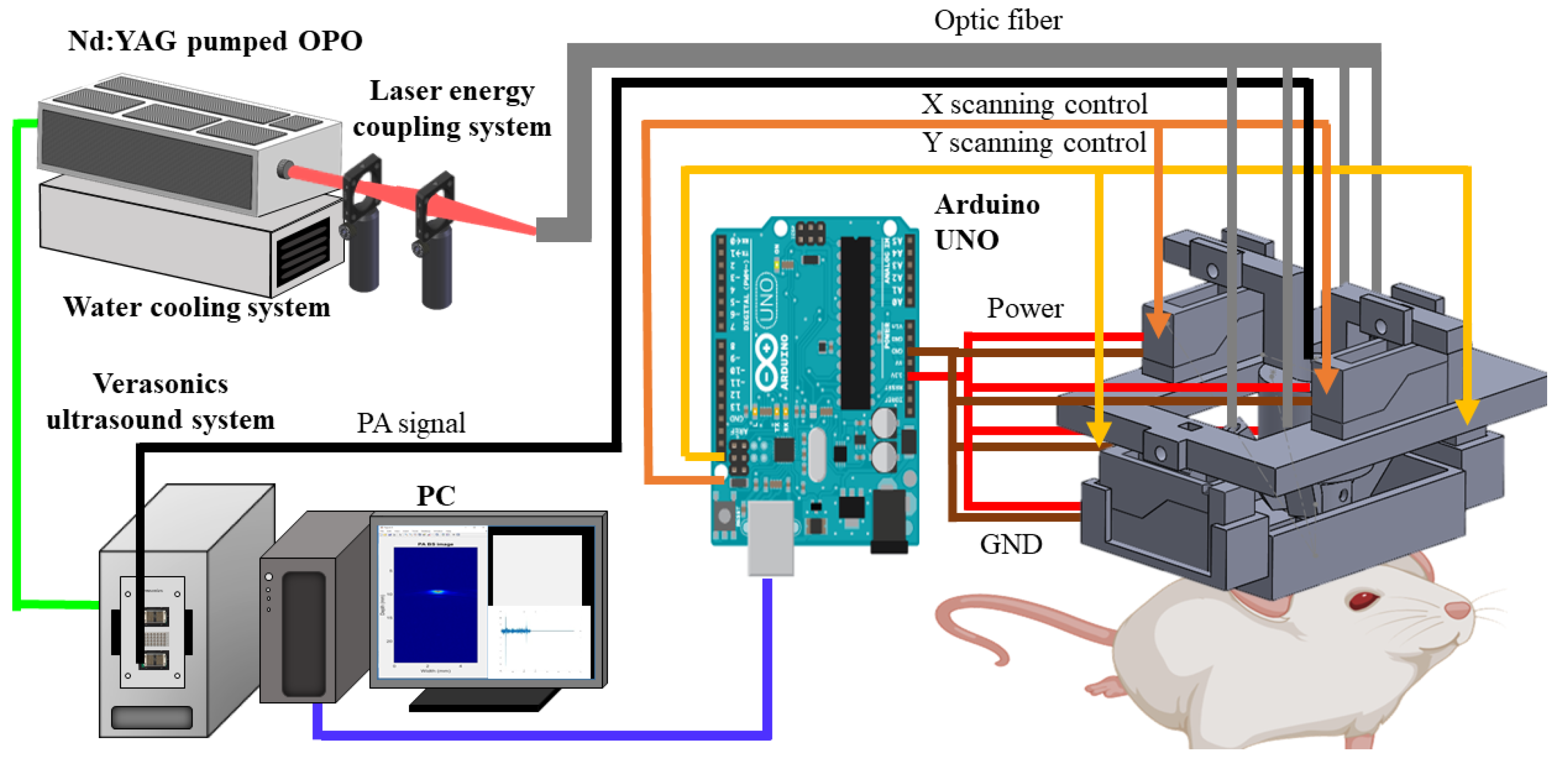
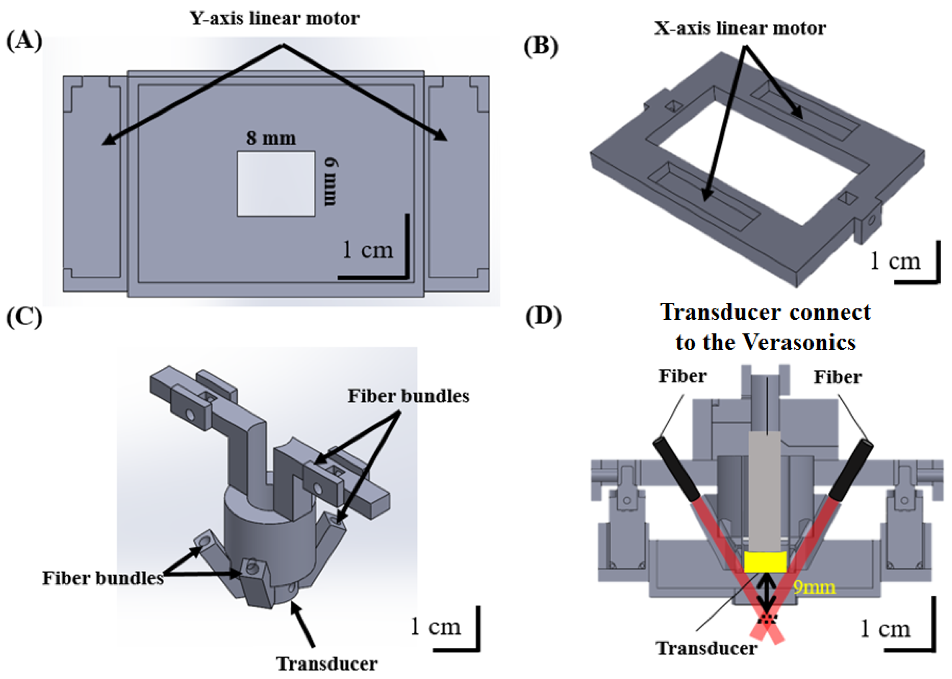
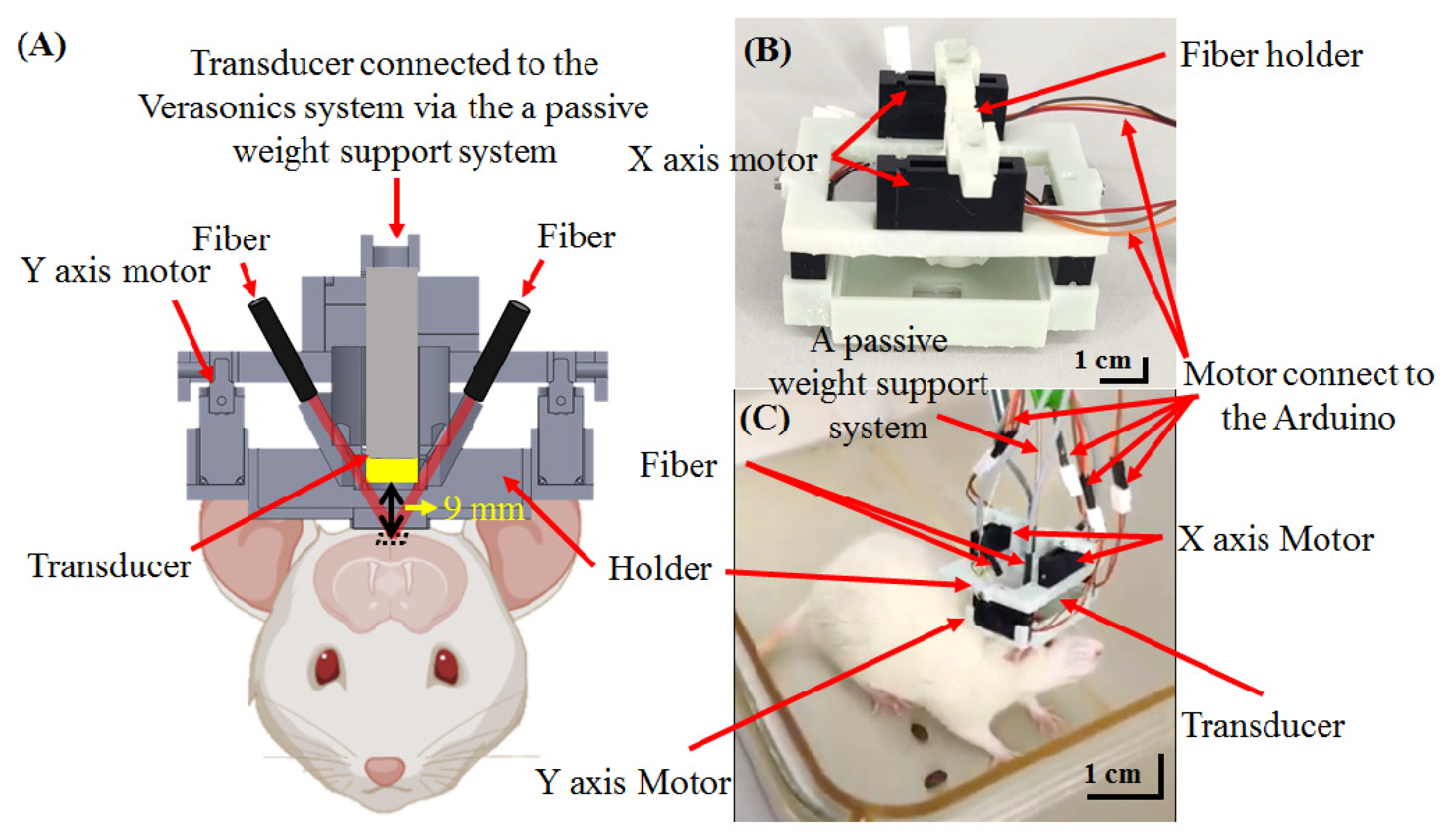
2.1. Dark-Field Miniature hmPAI System with Fiber-Bundle-Based Illumination
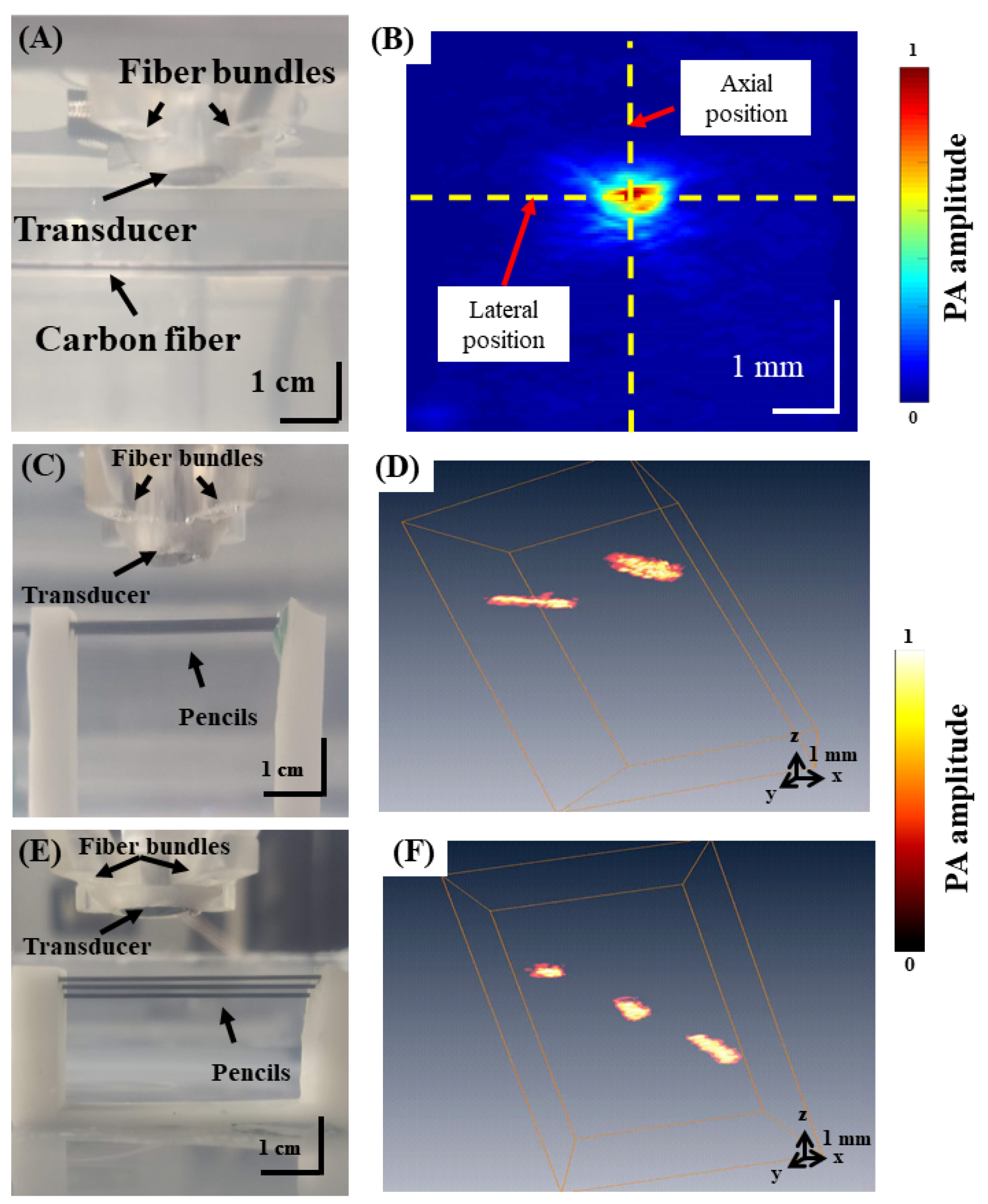
2.2. Assessing the Spatial Resolution of the Developed hmPAI System
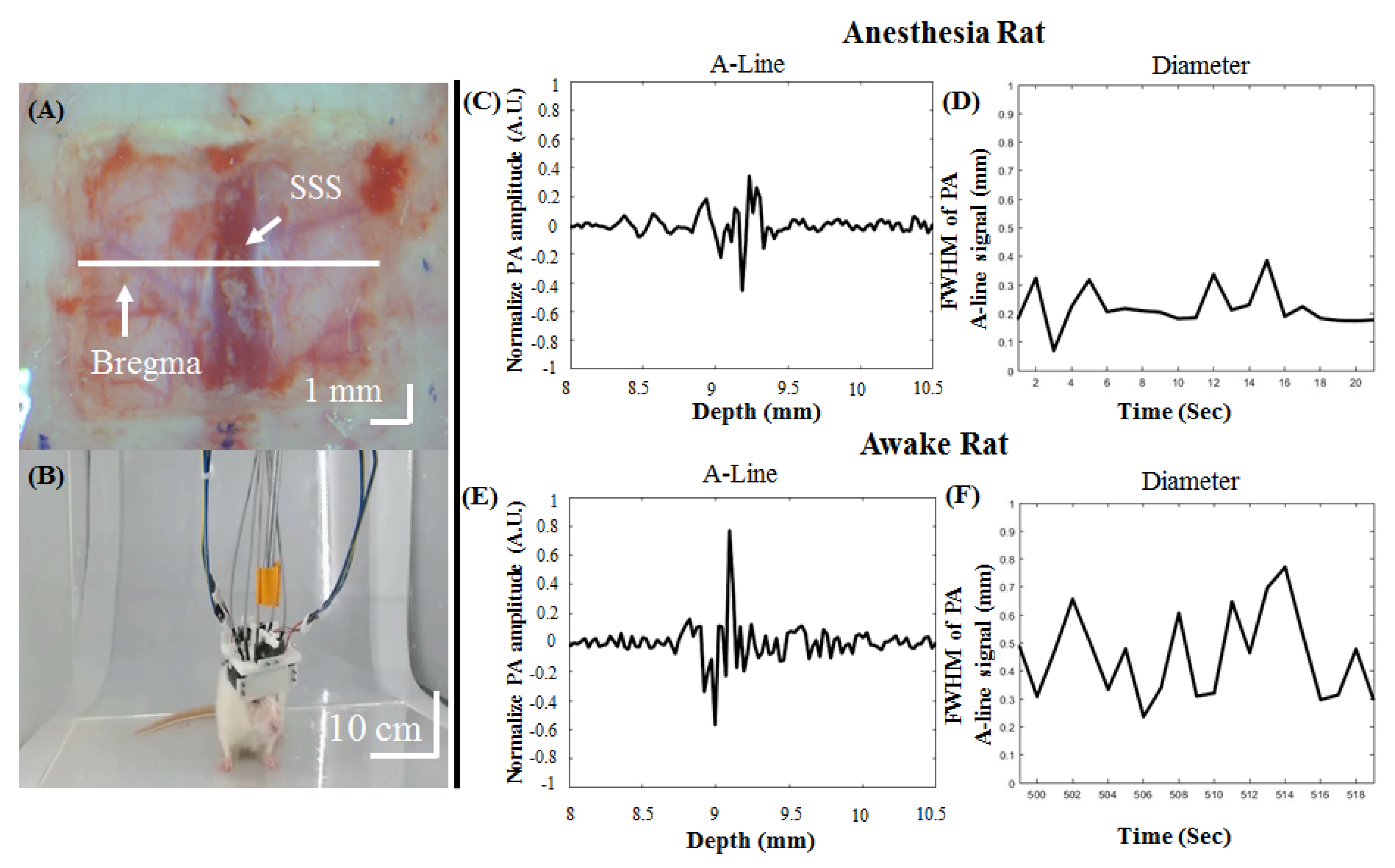
2.3. Craniotomy for Imaging Cortical Blood Vessels in Awake Animals
3. Results
3.1. Imaging Performance of the Developed hmPAI System
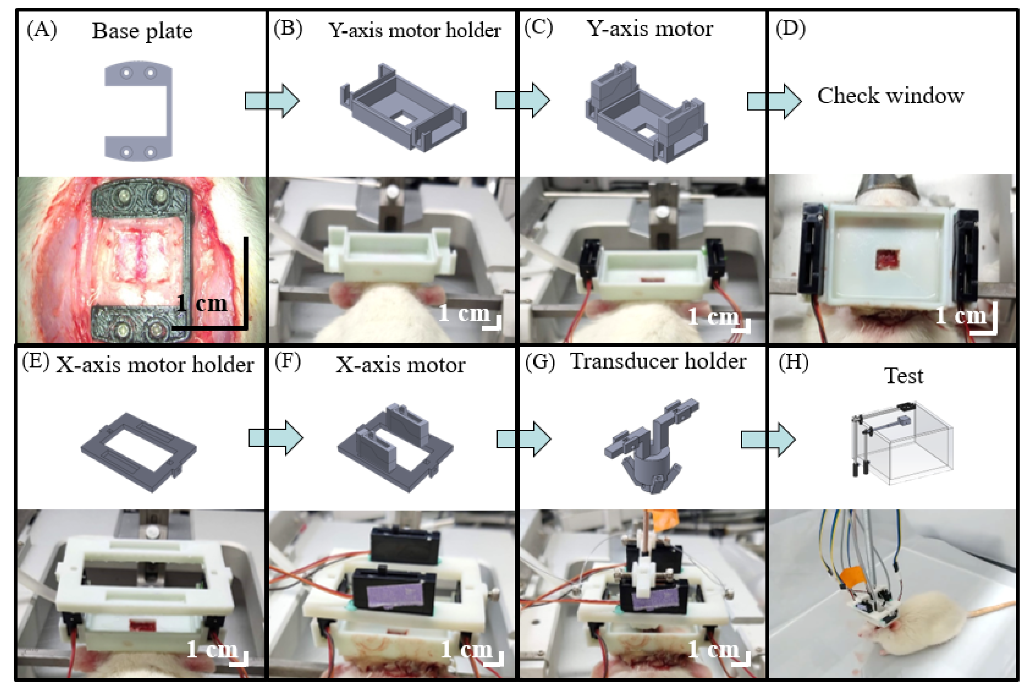
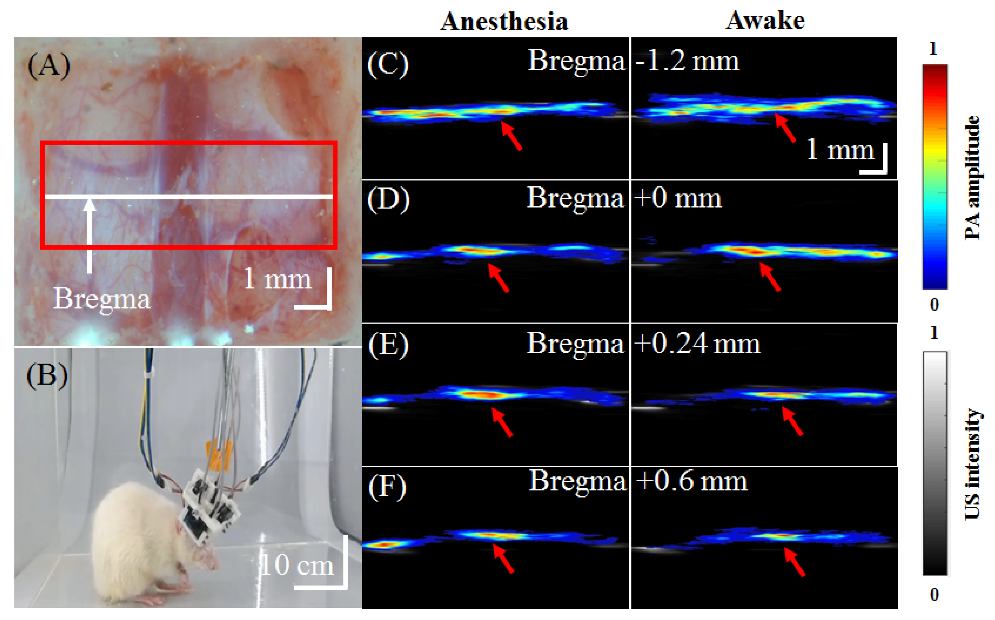
3.2. Setup Details of In Vivo Experiments Using the Developed hmPAI System
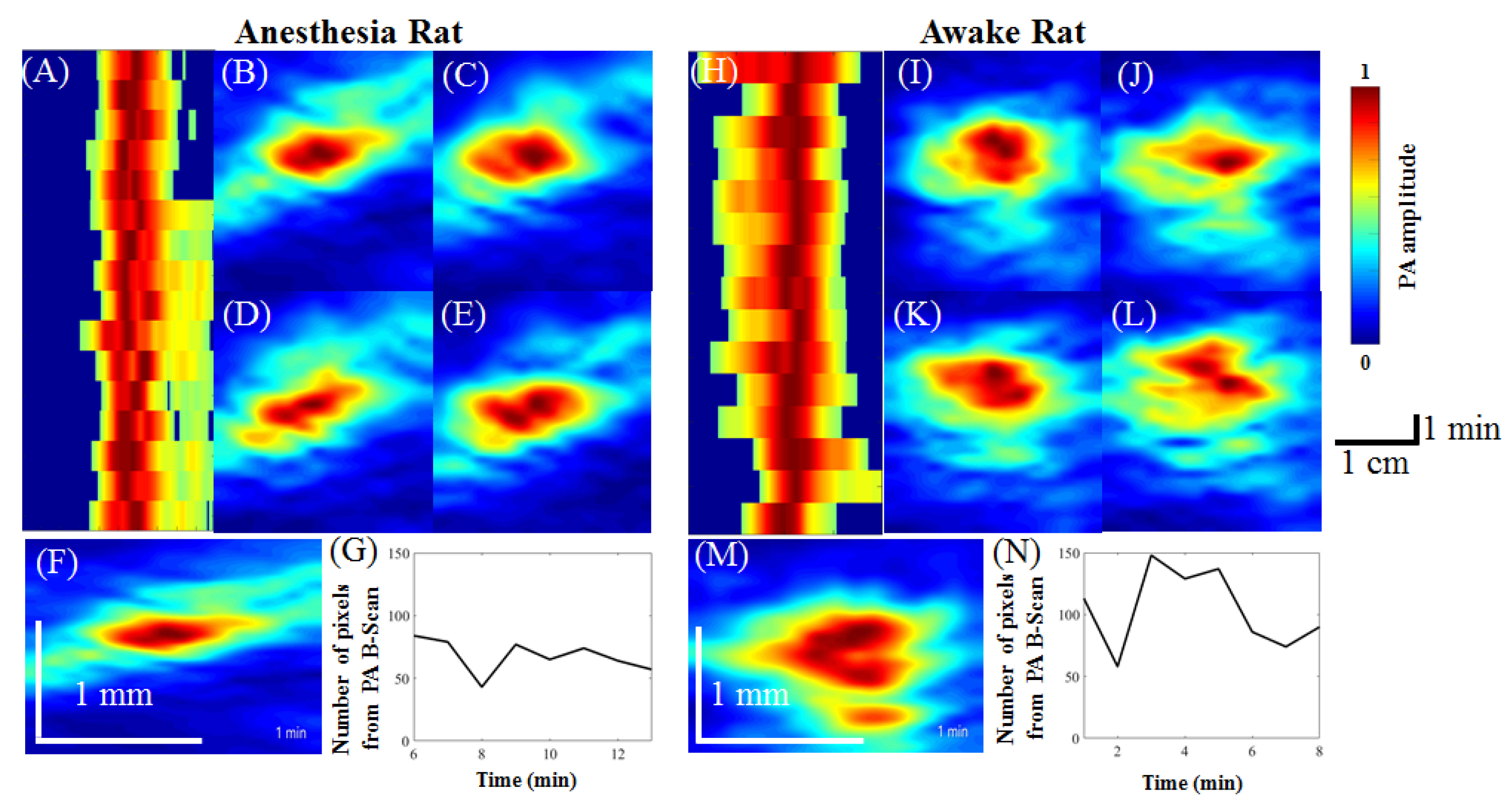
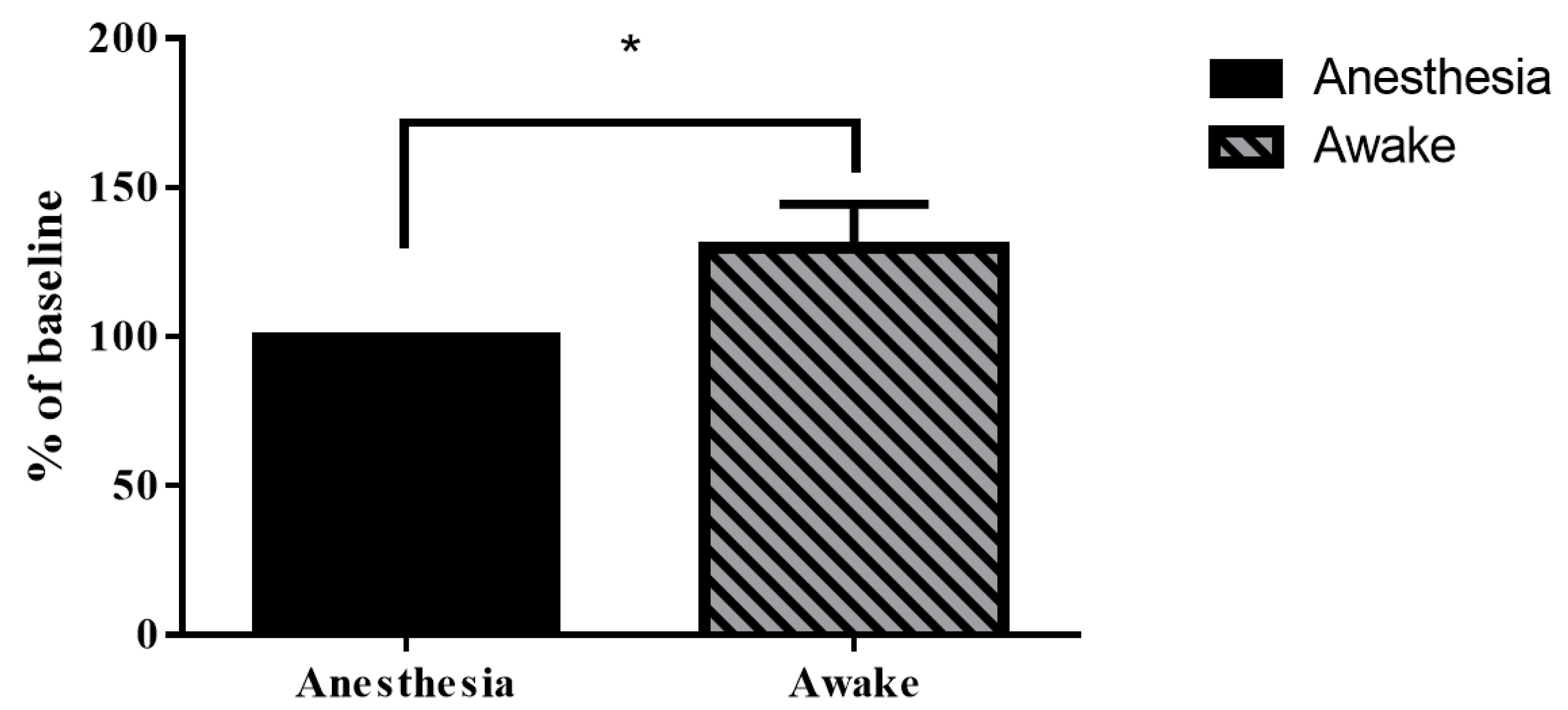
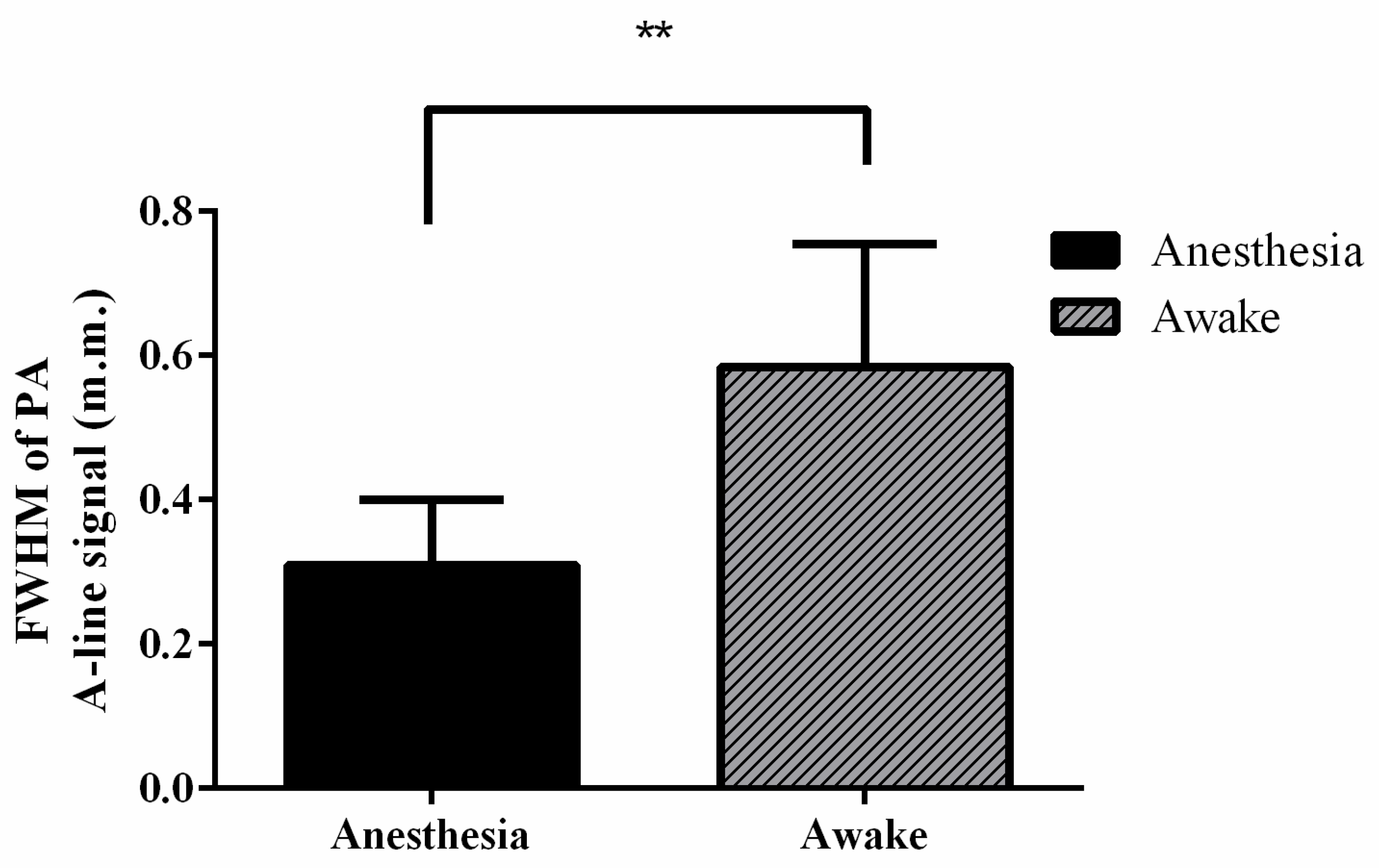
3.3. In Vivo Functional Imaging of Changes in Cortical Hemodynamics in SSS Blood Vessels
4. Discussion
4.1. Measurement of Cerebral Hemodynamics in Anesthetized and Awake Rats Using the Developed hmPAI System
4.2. Prospects of the Developed hmPAI System
5. Conclusions
Supplementary Materials
Author Contributions
Funding
Institutional Review Board Statement
Informed Consent Statement
Data Availability Statement
Conflicts of Interest
References
- Makeig, S.; Kothe, C.; Mullen, T.; Bigdely-Shamlo, N.; Zhang, Z.; Kreutz-Delgado, K. Evolving Signal Processing for Brain-Computer Interfaces. Proc. IEEE 2012, 100, 1567–1584. [Google Scholar] [CrossRef]
- Lance, B.J.; Kerick, S.E.; Ries, A.J.; Oie, K.S.; McDowell, K. Brain-Computer Interface Technologies in the Coming Decades. Proc. IEEE 2012, 100, 1585–1599. [Google Scholar] [CrossRef]
- Costecalde, T.; Aksenova, T.; Torres-Martinez, N.; Eliseyev, A.; Mestais, C.; Moro, C.; Benabid, A.L. A Long-Term BCI Study with ECoG Recordings in Freely Moving Rats. Neuromodulation 2018, 21, 149–159. [Google Scholar] [CrossRef]
- Insanally, M.; Trumpis, M.; Wang, C.; Chiang, C.H.; Woods, V.; Palopoli-Trojani, K.; Bossi, S.; Froemke, R.C.; Viventi, J. A low-cost, multiplexed muECoG system for high-density recordings in freely moving rodents. J. Neural. Eng. 2016, 13, 026030. [Google Scholar] [CrossRef] [PubMed] [Green Version]
- Garcia-Cortadella, R.; Schwesig, G.; Jeschke, C.; Illa, X.; Gray, A.L.; Savage, S.; Stamatidou, E.; Schiessl, I.; Masvidal-Codina, E.; Kostarelos, K.; et al. Graphene active sensor arrays for long-term and wireless mapping of wide frequency band epicortical brain activity. Nat. Commun. 2021, 12, 211. [Google Scholar] [CrossRef] [PubMed]
- Liao, L.D.; Tsytsarev, V.; Delgado-Martínez, I.; Li, M.L.; Erzurumlu, R.; Vipin, A.; Orellana, J.; Lin, Y.R.; Lai, H.Y.; Chen, Y.Y.; et al. Neurovascular coupling: In vivo optical techniques for functional brain imaging. Biomed. Eng. Online 2013, 12, 38. [Google Scholar] [CrossRef] [Green Version]
- Denk, W.; Strickler, J.H.; Webb, W.W. Two-photon laser scanning fluorescence microscopy. Science 1990, 248, 73–76. [Google Scholar] [CrossRef] [Green Version]
- Karagyozov, D.; Skanata, M.M.; Lesar, A.; Gershow, M. Recording Neural Activity in Unrestrained Animals with Three-Dimensional Tracking Two-Photon Microscopy. Cell Rep. 2018, 25, 1371. [Google Scholar] [CrossRef] [Green Version]
- Price, P. PET as a potential tool for imaging molecular mechanisms of oncology in man. Trends Mol. Med. 2001, 7, 442–446. [Google Scholar] [CrossRef]
- Cosgrove, D.; Lassau, N. Imaging of perfusion using ultrasound. Eur. J. Nucl. Med. Mol. Imaging 2010, 37 (Suppl. 1), S65–S85. [Google Scholar] [CrossRef] [PubMed]
- Xie, W.; Liu, S.; Su, H.; Wang, Z.; Zheng, Y.; Fu, Y. Ultrasound microbubbles enhance recombinant adeno-associated virus vector delivery to retinal ganglion cells in vivo. Acad. Radiol. 2010, 17, 1242–1248. [Google Scholar] [CrossRef]
- Weng, J.C.; Wu, S.K.; Lin, W.L.; Tseng, W.Y. Detecting blood-brain barrier disruption within minimal hemorrhage following transcranial focused ultrasound: A correlation study with contrast-enhanced MRI. Magn. Reson. Med. 2011, 65, 802–811. [Google Scholar] [CrossRef]
- Xie, J.; Liao, Y.; Yang, L.; Wu, J.; Liu, C.; Xuan, W.; Li, M.; Zhang, L.; Liu, Y.; Wu, P.; et al. Ultrasound molecular imaging of angiogenesis induced by mutant forms of hypoxia-inducible factor-1alpha. Cardiovasc. Res. 2011, 92, 256–266. [Google Scholar] [CrossRef] [Green Version]
- Beard, P. Biomedical photoacoustic imaging. Interface Focus 2011, 1, 602–631. [Google Scholar] [CrossRef]
- Wang, L.V. Tutorial on photoacoustic microscopy and computed tomography. IEEE J. Sel. Top Quantum Electron. 2008, 14, 171–179. [Google Scholar] [CrossRef] [Green Version]
- Bandla, A.; Liao, L.D.; Chan, S.J.; Ling, J.M.; Liu, Y.H.; Shih, Y.Y.I.; Pan, H.C.; Wong, P.T.H.; Lai, H.Y.; King, N.K.K.; et al. Simultaneous functional photoacoustic microscopy and electrocorticography reveal the impact of rtPA on dynamic neurovascular functions after cerebral ischemia. J. Cereb. Blood Flow Metab. 2018, 38, 980–995. [Google Scholar] [CrossRef] [PubMed]
- Liu, Y.H.; Liao, L.D.; Tan, S.S.; Kwon, K.Y.; Ling, J.M.; Bandla, A. Assessment of neurovascular dynamics during transient ischemic attack by the novel integration of micro-electrocorticography electrode array with functional photoacoustic microscopy. Neurobiol. Dis. 2015, 82, 455–465. [Google Scholar] [CrossRef]
- Liao, L.D.; Liu, Y.H.; Lai, H.Y.; Bandla, A.; Shih, Y.Y.I.; Chen, Y.Y.; Thakor, N.V. Rescue of cortical neurovascular functions during the hyperacute phase of ischemia by peripheral sensory stimulation. Neurobiol. Dis. 2015, 75, 53–63. [Google Scholar] [CrossRef] [PubMed]
- Leng, H.; Wang, Y.; Jhang, D.F.; Chu, T.S.; Tsao, C.H.; Tsai, C.H.; Giamundo, S.; Chen, Y.Y.; Liao, K.W.; Chuang, C.C.; et al. Characterization of a Fiber Bundle-Based Real-Time Ultrasound/Photoacoustic Imaging System and Its In Vivo Functional Imaging Applications. Micromachines 2019, 10, 820. [Google Scholar] [CrossRef] [Green Version]
- Liao, L.D.; Li, M.L.; Lai, H.Y.; Shih, Y.Y.I.; Lo, Y.C.; Tsang, S.; Chao, P.C.P.; Lin, C.T.; Jaw, F.S.; Chen, Y.Y. Imaging brain hemodynamic changes during rat forepaw electrical stimulation using functional photoacoustic microscopy. Neuroimage 2010, 52, 562–570. [Google Scholar] [CrossRef] [PubMed]
- Paxinos, G.; Watson, C. The Rat Brain in Stereotaxic Coordinates; Academic Press: San Diego, CA, USA, 2007. [Google Scholar]
- Helmchen, F.; Fee, M.S.; Tank, D.W.; Denk, W. A miniature head-mounted two-photon microscope. high-resolution brain imaging in freely moving animals. Neuron 2001, 31, 903–912. [Google Scholar] [CrossRef] [Green Version]
- Kim, J.; Park, S.; Jung, Y.; Chang, S.; Park, J.; Zhang, Y.; Lovell, J.F.; Kim, C. Programmable Real-time Clinical Photoacoustic and Ultrasound Imaging System. Sci. Rep. 2016, 6, 35137. [Google Scholar] [CrossRef] [Green Version]
- Yao, J.; Wang, L.V. Perspective on fast-evolving photoacoustic tomography. J. Biomed. Opt. 2021, 26, 060602. [Google Scholar] [CrossRef] [PubMed]
- Wang, X.D.; Xie, X.Y.; Ku, G.N.; Wang, L.H.V. Noninvasive imaging of hemoglobin concentration and oxygenation in the rat brain using high-resolution photoacoustic tomography. J. Biomed. Opt. 2006, 11, 024015. [Google Scholar] [CrossRef] [Green Version]
- Tsygan, N.V.; Trashkov, A.P.; Litvinenko, I.V.; Yakovleva, V.A.; Ryabtsev, A.V.; Vasiliev, A.G.; Churilov, L.P. Autoimmunity in acute ischemic stroke and the role of blood-brain barrier: The dark side or the light one? Front. Med. 2019, 13, 420–426. [Google Scholar] [CrossRef] [PubMed]
- Gao, Y.R.; Ma, Y.; Zhang, Q.; Winder, A.T.; Liang, Z.; Antinori, L.; Drew, P.J.; Zhang, N. Time to wake up: Studying neurovascular coupling and brain-wide circuit function in the un-anesthetized animal. Neuroimage 2017, 153, 382–398. [Google Scholar] [CrossRef]
- Cao, R.; Li, J.; Ning, B.; Sun, N.; Wang, T.; Zuo, Z.; Hu, S. Functional and oxygen-metabolic photoacoustic microscopy of the awake mouse brain. Neuroimage 2017, 150, 77–87. [Google Scholar] [CrossRef] [Green Version]
- Minhas, J.S.; Rook, W.; Panerai, R.B.; Hoiland, R.L.; Ainslie, P.N.; Thompson, J.P.; Mistri, A.K.; Robinson, T.G. Pathophysiological and clinical considerations in the perioperative care of patients with a previous ischaemic stroke: A multidisciplinary narrative review. Br. J. Anaesth. 2020, 124, 183–196. [Google Scholar] [CrossRef] [Green Version]
- Madsen, P.L.; Vorstrup, S. Cerebral blood flow and metabolism during sleep. Cerebrovasc. Brain Metab. Rev. 1991, 3, 281–296. [Google Scholar]
- Wang, L.V.; Hu, S. Photoacoustic tomography: In vivo imaging from organelles to organs. Science 2012, 335, 1458–1462. [Google Scholar] [CrossRef] [Green Version]
- Najafzadeh, E.; Ghadiri, H.; Alimohamadi, M.; Farnia, P.; Mehrmohammadi, M.; Ahmadian, A. Evaluation of multi-wavelengths LED-based photoacoustic imaging for maximum safe resection of glioma: A proof of concept study. Int. J. Comput. Assist Radiol. Surg. 2020, 15, 1053–1062. [Google Scholar] [CrossRef] [PubMed]
- Liu, C.; Liang, Y.; Wang, L. Single-shot photoacoustic microscopy of hemoglobin concentration, oxygen saturation, and blood flow in sub-microseconds. Photoacoustics 2020, 17, 100156. [Google Scholar] [CrossRef] [PubMed]
- Fang, H.; Maslov, K.; Wang, L.V. Photoacoustic Doppler effect from flowing small light-absorbing particles. Phys. Rev. Lett. 2007, 99, 184501. [Google Scholar] [CrossRef] [Green Version]
- Heo, C.; Park, H.; Kim, Y.T.; Baeg, E.; Kim, Y.H.; Kim, S.G.; Suh, M. A soft, transparent, freely accessible cranial window for chronic imaging and electrophysiology. Sci. Rep. 2016, 6, 27818. [Google Scholar] [CrossRef] [PubMed] [Green Version]
- Kang, J.; Zhang, H.K.; Kadam, S.D.; Fedorko, J.; Valentine, H.; Malla, A.P.; Yan, P.; Harraz, M.M.; Kang, J.U.; Rahmim, A.; et al. Transcranial Recording of Electrophysiological Neural Activity in the Rodent Brain in vivo Using Functional Photoacoustic Imaging of Near-Infrared Voltage-Sensitive Dye. Front. Neurosci. 2019, 13, 597. [Google Scholar] [CrossRef] [Green Version]
- Zhang, H.F.; Maslov, K.; Stoica, G.; Wang, L.V. Functional photoacoustic microscopy for high-resolution and noninvasive in vivo imaging. Nat. Biotechnol. 2006, 24, 848–851. [Google Scholar] [CrossRef]
- Wen, C.; Zhao, L.; Han, T.; Li, W.; Zhang, G.; Li, C. A versatile dark-field acoustic-resolution photoacoustic microscopy system aided by 3D printing. Rev. Sci. Instrum. 2019, 90, 083704. [Google Scholar] [CrossRef]
- Tang, J.; Xi, L.; Zhou, J.; Huang, H.; Zhang, T.; Carney, P.R.; Jiang, H. Noninvasive high-speed photoacoustic tomography of cerebral hemodynamics in awake-moving rats. J. Cereb. Blood Flow Metab. 2015, 35, 1224–1232. [Google Scholar] [CrossRef] [Green Version]
- Zhang, W.C.; Li, J.D.; Yang, S.H. Real-time interleaved photoacoustic and ultrasound imaging for guiding interventional procedures. Appl. Acoust. 2019, 156, 1–6. [Google Scholar] [CrossRef]
- Senarathna, J.; Murari, K.; Etienne-Cummings, R.; Thakor, N.V. A miniaturized platform for laser speckle contrast imaging. IEEE Trans. Biomed. Circuits Syst. 2012, 6, 437–445. [Google Scholar] [CrossRef]
- Bergonzi, K.M.; Bauer, A.Q.; Wright, P.W.; Culver, J.P. Mapping functional connectivity using cerebral blood flow in the mouse brain. J. Cereb. Blood Flow Metab. 2015, 35, 367–370. [Google Scholar] [CrossRef] [PubMed] [Green Version]
- Silva, A.C.; Koretsky, A.P. Laminar specificity of functional MRI onset times during somatosensory stimulation in rat. Proc. Natl. Acad. Sci. USA 2002, 99, 15182–15187. [Google Scholar] [CrossRef] [PubMed] [Green Version]
- Kim, J.Y.; Lee, C.; Park, K.; Lim, G.; Kim, C. Fast optical-resolution photoacoustic microscopy using a 2-axis water-proofing MEMS scanner. Sci. Rep. 2015, 5, 1–5. [Google Scholar] [CrossRef] [PubMed]
- Dean-Ben, X.L.; Ozbek, A.; Razansky, D. Volumetric Real-Time Tracking of Peripheral Human Vasculature with GPU-Accelerated Three-Dimensional Optoacoustic Tomography. IEEE Trans. Med. Imaging 2013, 32, 2050–2055. [Google Scholar] [CrossRef] [PubMed]


Publisher’s Note: MDPI stays neutral with regard to jurisdictional claims in published maps and institutional affiliations. |
© 2021 by the authors. Licensee MDPI, Basel, Switzerland. This article is an open access article distributed under the terms and conditions of the Creative Commons Attribution (CC BY) license (https://creativecommons.org/licenses/by/4.0/).
Share and Cite
Wang, Y.; Chu, T.-S.; Lin, Y.-R.; Tsao, C.-H.; Tsai, C.-H.; Ger, T.-R.; Chen, L.-T.; Chang, W.-S.W.; Liao, L.-D. Assessment of Brain Functional Activity Using a Miniaturized Head-Mounted Scanning Photoacoustic Imaging System in Awake and Freely Moving Rats. Biosensors 2021, 11, 429. https://doi.org/10.3390/bios11110429
Wang Y, Chu T-S, Lin Y-R, Tsao C-H, Tsai C-H, Ger T-R, Chen L-T, Chang W-SW, Liao L-D. Assessment of Brain Functional Activity Using a Miniaturized Head-Mounted Scanning Photoacoustic Imaging System in Awake and Freely Moving Rats. Biosensors. 2021; 11(11):429. https://doi.org/10.3390/bios11110429
Chicago/Turabian StyleWang, Yuhling, Tsung-Sheng Chu, Yan-Ren Lin, Chia-Hui Tsao, Chia-Hua Tsai, Tzong-Rong Ger, Li-Tzong Chen, Wun-Shaing Wayne Chang, and Lun-De Liao. 2021. "Assessment of Brain Functional Activity Using a Miniaturized Head-Mounted Scanning Photoacoustic Imaging System in Awake and Freely Moving Rats" Biosensors 11, no. 11: 429. https://doi.org/10.3390/bios11110429
APA StyleWang, Y., Chu, T.-S., Lin, Y.-R., Tsao, C.-H., Tsai, C.-H., Ger, T.-R., Chen, L.-T., Chang, W.-S. W., & Liao, L.-D. (2021). Assessment of Brain Functional Activity Using a Miniaturized Head-Mounted Scanning Photoacoustic Imaging System in Awake and Freely Moving Rats. Biosensors, 11(11), 429. https://doi.org/10.3390/bios11110429