Review

Contrast Agents Delivery: An Up-to-Date Review of Nanodiagnostics in Neuroimaging

by
Daniel Mihai Teleanu
1,
Cristina Chircov
2,3,
Alexandru Mihai Grumezescu
3,4,
Adrian Volceanov
3,* and
Raluca Ioana Teleanu
5
1
Emergency University Hospital, “Carol Davila” University of Medicine and Pharmacy, 050474 Bucharest, Romania
2
Faculty of Engineering in Foreign Languages, Politehnica University of Bucharest, 060042 Bucharest, Romania
3
Faculty of Applied Chemistry and Materials Science, Politehnica University of Bucharest, 011061 Bucharest, Romania
4
ICUB—Research Institute of University of Bucharest, University of Bucharest, 36-46 M. Kogalniceanu Blvd., Bucharest 050107, Romania
5
“Victor Gomoiu” Clinical Children’s Hospital, “Carol Davila” University of Medicine and Pharmacy, 050474 Bucharest, Romania
*
Author to whom correspondence should be addressed.
Nanomaterials 2019, 9(4), 542; https://doi.org/10.3390/nano9040542
Submission received: 1 March 2019 / Revised: 28 March 2019 / Accepted: 29 March 2019 / Published: 3 April 2019

Abstract

:
Neuroimaging is a highly important field of neuroscience, with direct implications for the early diagnosis and progression monitoring of brain-associated diseases. Neuroimaging techniques are categorized into structural, functional and molecular neuroimaging, each possessing advantages and disadvantages in terms of resolution, invasiveness, toxicity of contrast agents and costs. Nanotechnology-based approaches for neuroimaging mostly involve the development of nanocarriers for incorporating contrast agents or the use of nanomaterials as imaging agents. Inorganic and organic nanoparticles, liposomes, micelles, nanobodies and quantum dots are some of the most studied candidates for the delivery of contrast agents for neuroimaging. This paper focuses on describing the conventional modalities used for imaging and the applications of nanotechnology for developing novel strategies for neuroimaging. The aim is to highlight the roles of nanocarriers for enhancing and/or overcome the limitations associated with the most commonly utilized neuroimaging modalities. For future directions, several techniques that could benefit from the increased contrast induced by using imaging probes are presented.

1. Introduction

Affecting approximately a billion individuals worldwide, neurological diseases are one of the world’s leading causes of death. Their prevalence is expected to further increase in the following decade due to the rapid growth in elderly population. Considering the complexity of the brain, the pathogenesis of the central nervous system diseases is not fully understood and therefore the diagnosis and treatment pose serious challenges [1,2].
The clinical manifestations of the most prevalent brain disorders—including neurodegenerative conditions, brain tumours, stroke, traumatic brain injuries, epilepsy and infections—commonly involve dementia, hypokinetic and hyperkinetic movement disorders [3], headaches, seizures, cognitive changes, incontinence, gait disorders [4], depression, cognitive impairments, physical disability [5], fatigue, sleep disorders [6], anxiety [7], nausea, vomiting, photophobia, fever and loss of consciousness [8]. However, to appropriately diagnose brain diseases, the basic neurological examination which includes patient’s history and physical examination is not sufficient. Therefore, identification and usage of proper imaging techniques based on clinical symptoms are crucial for neurologic diagnosis and initial and subsequent management [9,10]. Apart from brain diseases diagnosis, neuroimaging techniques are extensively applied in neuroscience to visualize neural activity, understand brain mechanisms and identify biomarkers [11]. Furthermore, proper biomarkers and neuroimaging techniques are essentially necessary in diagnosis owing to the possibility of early detection of pre-symptomatic pathological changes, efficient differentiation between related neurological diseases, identification of disease progression and treatment effects, non-invasive confirmation of the underlying pathology and screening large populations for risk assessments [12]. Additionally, through the integration of genomic and neuroimaging data, the investigation of genetic risk factors shaping variations in brain phenotypes and mechanisms underlying neurological and neuropsychiatric disorders is possible [13].
The recent advances in neuroimaging have enabled an improved and detailed morphological and functional evaluation of the brain [14]. Depending on the requirements, neuroimaging techniques can be structural, including computed tomography (CT) and magnetic resonance imaging (MRI), which are used for the diagnosis of gross intracranial diseases, such as tumours, strokes, injuries [15] and neurodegenerative diseases [16], functional, including optical imaging, functional magnetic resonance imaging (fMRI), electroencephalography (EEG), magnetoencephalography (MEG), functional near-infrared spectroscopic imaging (fNIRS) and functional ultrasound (fUS) and molecular, including positron emission tomography (PET), single photon emission computed tomography (SPECT) and molecular magnetic resonance imaging (mMRI), each technique providing different information [17,18].
Conventional neuroimaging techniques are characterized by different advantages and disadvantages. Several advantages include high sensitivity for PET and non-invasiveness and cost-effectiveness for optical imaging. However, disadvantages such as low sensitivity, soft tissue contrast and signal-to-noise ratio for CT [19], long acquisition time for MRI, low resolution and short half-life of isotopes for PET and strong scattering of light in biological tissues for optical imaging must be addressed. Considering the limitations of current neuroimaging techniques, there is an emerging necessity for developing novel approaches [20]. Nanotechnology, the continuously evolving field that encompasses knowledge from multiple disciplines including chemistry, physics, engineering and biology, represents a potential strategy to overcome these limitations [21,22]. Nanotechnology implicates the development and modification of materials within the size range of 1–100 nm in at least one dimension, offering the possibility to understand, manipulate and control the matter at the level of individual atoms and molecules [23,24]. Therefore, the implication of nanotechnology to design nanostructures as contrast agents might facilitate imaging for the accurate characterization and resection of tumours, the delivery of therapeutic stem cells and the detection of neurodegenerative diseases in early stages. Furthermore, the nanotechnology-based contrast agents offer high chemical and biological stability and possibilities of multi-functionalization for targeted delivery, administration through various routes and blood-brain barrier penetration [20]. The development of nanostructured contrast agents has also offered the prospect of simultaneously acquiring images using several imaging techniques or capturing images at different moments. Thus, the platform for multimodal imaging allows for benefiting from the advantages of each neuroimaging technique while overcoming their specific limitations [25].
The present article focuses on reviewing the common nanotechnology-based neuroimaging techniques, along with the most recent advancements in the applications of nanotechnology to design novel contrast agents to improve the examination and diagnosis of brain-associated disorders.

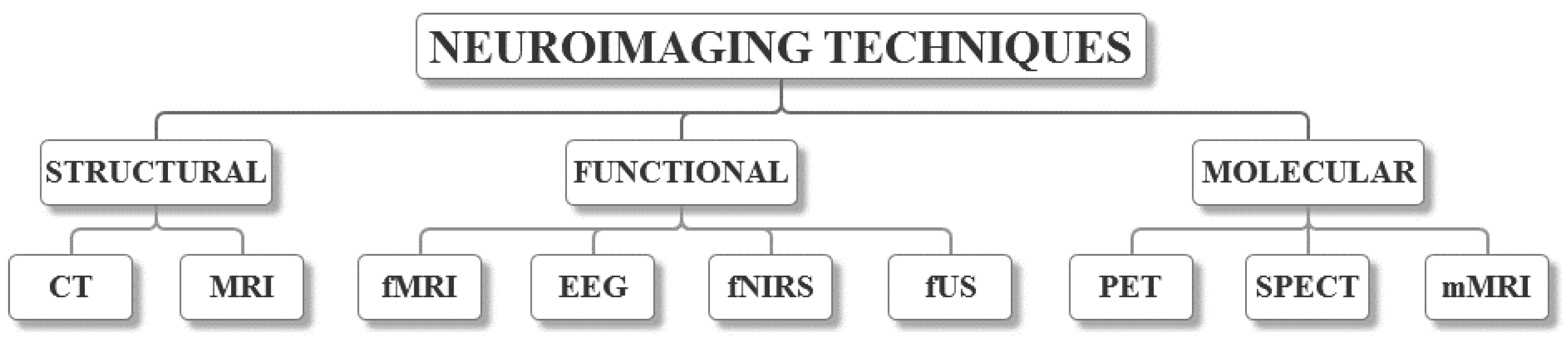
2. Neuroimaging Techniques

Depending on the requirements for the brain images, neuroimaging techniques can be categorized into structural, functional and molecular neuroimaging, each method described, as follows (Figure 1). The characteristics of each method that were found in the literature [26,27,28,29] are summarized in Table 1. However, there have been some issues associated with collecting the information due to the inconsistency between different papers.

2.1. Structural Neuroimaging

Structural neuroimaging has almost entirely replaced the conventional studies of the anatomy and morphology of the brain. The clinical applications of structural neuroimaging target the exact diagnosis followed by the individualized treatment plan, whereas in research, it has led to the understanding of the neuroanatomy at the individual and group level. Furthermore, the structural imaging-based measurements of specific lesions has allowed for the association between the symptom severity, lesion load and lesion location. The most common methods involve CT and MRI [30].

2.1.1. Computed Tomography (CT)

The use of CT for the diagnosis of intracranial pathology has replaced common techniques, including radiography, encephalography or even angiography [31]. X-ray beam technologies are responsible for creating tomographic slices of the brain, which result in superior contrast resolution that allows for the distinction between fluids and soft tissues. Additionally, the problem of superimposition that radiographs pose is eliminated [32]. Modern scanners spin or surround the patient, thus imaging the entire volume of tissue. The acquired information is reconstructed into two-dimensional greyscale images of the slices, which represent the maps of tissue density [31]. The main challenge of CT is represented by the compromise of using a low radiation dose that results in lower signal-to-noise and contrast-to-noise ratios and limited discrimination between tissue with slightly different x-ray opacities [33].
Contrast agents might be injected in the body for perfusion CT imaging, which will provide high-contrast perfusion maps for the diagnosis of ischemic stroke [34]. Moreover, this technique is characterized by several benefits, including the increased sensitivity and specificity towards an exact diagnosis and the provision of information regarding prognosis and treatment decisions. However, the increased radiation exposure and imaging time and the administration of toxic contrast agents which could lead to kidney failure are limitations that must be overcome [35]. Additionally, as common contrast agents cannot pass through the blood-brain barrier, novel strategies for the formulation of nanoagents with limited toxicity are necessary [34].

2.1.2. Magnetic Resonance Imaging (MRI)

The standard tool in clinical diagnosis, disease follow up, treatment evaluation and brain development monitoring is the MRI technique. It has been extensively used owing to its non-invasiveness, high-resolution imaging, enhanced contrast between tissues and multiplanar imaging capabilities [36,37]. MRI acquisition is based on the interaction between an external magnetic field and the magnetic moment of the water protons, which causes alterations in the orientation of the spinning nucleus [38]. Structural MRI techniques, including T1- and T2-weighted imaging, diffusion tensor imaging, magnetization transfer imaging and iron sensitive MRI, such as susceptibility weighted imaging and quantitative susceptibility mapping, are used to highlight specific markers for brain diseases [39].
However, this technique is associated with long imaging times as time plays a key role in the quality of the acquired images. Specifically, the longer the acquisition time, the higher the signal-to-noise and contrast-to-noise ratios, as they are dependent on the number of pulse sequences performed [33,40]. Another option to improve the sensitivity and the signal-to-noise ratio is represented by the increase of the magnetic field strength using stronger magnets. However, this strategy involves higher costs and special requirements for installation, which limit the use of magnets with field strengths between 7 T and 21 T [41]. Therefore, to overcome the long imaging time and the high costs associated, the quality of the images must be reduced [33].

2.2. Functional Neuroimaging

Functional neuroimaging is applied in brain function evaluation for an improved understanding of the correlations between the activity in specific brain areas and the mental functions [42]. The invention of the blood oxygen level-dependent (BOLD) fMRI has subsequently led to the tremendous advances in the field of functional neuroimaging [43]. Furthermore, the development of multichannel EEG and intracranial electroencephalography (iEEG) and fNIRS has allowed for the understanding of complex anatomical correlations and functional relationships between the lesion and the adjacent cortical areas and white matter structures [42].

2.2.1. Functional Magnetic Resonance Imaging (fMRI)

fMRI has been developed for the study of vascular or metabolic reactions of the brain in response to different stimuli [44]. BOLD fMRI is the leading technique for functional neuroimaging [45]. The acquired signals represent the relative concentrations of diamagnetic oxyhaemoglobin and paramagnetic deoxyhaemoglobin, which rely on the modifications in the vasculature blood oxygenation correlated to neuronal activity, a process called neurovascular coupling [46]. Furthermore, BOLD fMRI reflects the increase in cerebral metabolic rate of oxygen consumption, cerebral blood flow and cerebral blood volume caused by neural activity [45,46]. Specifically, this technique images the dynamic changes caused by neural metabolism changes due to the variations in the oxygenation levels of brain tissue as a consequence to neural responses to specific cognitive processes or spontaneous fluctuations in resting states [47]. Therefore, the signal is different in patients at rest and during cognitive and sensation activity [46,48,49].
Functional magnetic resonance has greatly contributed to the understanding of the impact of brain disorders on the cognitive functions of patients. Therefore, the differentiation between the vegetative state and the minimally conscious state is a very practical clinical application of this technique [50]. However, the low temporal resolution and signal-to-noise ratio are major issues that pose challenges in fMRI measurements [51]. To improve the specificity and sensitivity of the acquired signals, the administration of contrast agents that typically contain gadolinium might be a solution. However, the associated instability, blood-brain barrier permeation difficulties and the toxicity of these agents must be addressed [52,53].

2.2.2. Electroencephalography (EEG)

EEG is a non-invasive neuroimaging technique that records the electrical activity of the brain [54]. Owing to its simplicity and cost effectiveness, EEG is an extensively used technique in neuroscience, providing high temporal resolution and understanding of the underlying mechanisms that generate the spontaneous electrical activity [55]. The signals originate from the ionic movement through the extracellular space of the neurons and therefore the electroencephalogram represents the sum of all synchronous activity of the neurons that have the same approximate vertical orientation to the scalp [54,56]. The signal is measured by placing a sensor or a headset provided with several electrodes on the scalp [54].
Novel strategies involve the implantation of electrodes in the subdural or deep areas. The iEEG, including electrocorticography and stereo-EEG, provides a neuroelectrophysiological signal that offers more accurate information regarding epileptic discharge patterns and diagnostic information that could aid surgical interventions [57,58]. Moreover, it is a neuroimaging technique that could offer an insight into brain activities as it provides high spatial and temporal resolution for the affected brain areas [57]. However, there are several limitations that must be considered, including the accessibility, as it requires specially trained teams of clinicians and investigators and special equipment [59]. Nonetheless, a sufficient number of electrodes must be placed in order to appropriately define the brain network [58]. These electrodes are usually 5–10 mm apart and consequently global coverage of the brain is difficult to achieve. Additionally, as the electrodes used are usually either cylindrical with a contact length of 2 mm, a diameter of 1 mm and a total surface area of 10 mm2 or circular plates with a diameter of 2 mm and a surface area of 4 mm2, they capture the signal from a large population of cells [59]. However, this approach does not record from sulcal depths, thus it only provides information from the surface of the cerebral crests [60].
Therefore, nanotechnology might offer the solution to improve the accuracy of iEEG method. Nanotechnology has been applied for the development of nanoelectrodes that could be invasively implanted in the brain for a high-quality neural recording and stimulation. Hence, the potential of the next generation of neuroprostheses could lead to the ability to both control the output of the prosthesis by executing a certain motor function and to record relevant sensory information [61]. Several strategies have been employed to design neural probes, including polypyrrole/graphene oxide composite films [62] and polyimide-based probes [63] with platinum and gold electrodes, respectively. However, the available information is limited and further studies are required for designing reliable nanotechnology-based approaches for neuroimaging through iEEG.

2.2.3. Functional Near-Infrared Spectroscopic Imaging (fNIRS)

fNIRS is a promising neuroimaging method which allows for non-invasive and long-term brain function mapping through the measurement and imaging of local changes in haemoglobin concentrations [64]. The emitted near-infrared light onto the scalp is partly absorbed by haemoglobin and partly scattered and collected by specific sensors. The changes in oxygenated haemoglobin and deoxygenated haemoglobin are regarded as an indicator for variations in the regional cerebral blood volume [65]. The study of brain function through fNIRS requires a good acquaintance with how the diffuse optical neuroimage encodes the information related to it [66].
fNIRS presents a series of advantages, including portability, easy to use, reduced costs, robustness to head movement, high temporal resolution and spatial resolution higher than EEG but lower than fMRI [67]. Nevertheless, the disadvantages of this method, including the limited brain regions that can be reached, specifically the cortical regions beneath the scalp and the interference of superficial veins and arteries and extracortical components that limit signal specificity [68].

2.2.4. Ultrasound-Based Functional Imaging Techniques

fUS is a recently developed functional neuroimaging technique which relies on ultrafast imaging scanners. fUS has brought novel insights in neurodiagnostic imaging and blood flow imaging as it can capture over 20,000 frames per second, contrary to the 50 frames per second in conventional ultrasound scanners [69,70]. The principle of fUS is based on the neurovascular coupling, which correlates local neural activity and relative changes in the cerebral blood volume [71]. While BOLD fMRI depends on blood oxygenation, cerebral blood volume is related to the number of red blood cells within a pixel. Furthermore, fUS allows for brain mapping in response to sensory, motor and odour-evoked stimuli [70]. Similarly, the functional transcranial Doppler ultrasound is a non-invasive neuroimaging technique based on blood-flow velocities within cerebral arteries measurements. It is a low cost, simple and safe imaging modality with high temporal resolution and accuracy, that measures brain activity and functional lateralization [72].
Another ultrasound-based functional neuroimaging technique is photoacoustic tomography, which is used for functional, metabolic and histologic imaging through endogenous contrast and for molecular and cellular imaging through exogenous contrast [73]. Photoacoustic computed tomography (PACT) uses diffused high energy pulsed laser light to illuminate the tissue, which will subsequently generate photoacoustic waves. Deeper imaging depths are usually targeted with PACT but with lower spatial resolution [74]. Based on the detection geometry, PACT systems can be categorized into circular-view PACT, which provides cross-sectional brain images, planar-view PACT, which detects photoacoustic signals along a two-dimensional plane and spherical-view PACT, which is ideal for volumetric imaging, as it provides nearly isotropic spatial resolution [75].

2.3. Molecular Neuroimaging

Visualizations, measurements and understanding of biochemical processes and mechanisms at the molecular and cellular levels of the brain have been possible owing to the advances in the non-invasive molecular neuroimaging techniques. Common molecular imaging modalities involve PET, SPECT [18] and mMRI.

2.3.1. Positron Emission Tomography (PET)

PET is a minimally invasive imaging procedure, extensively applied in the evaluation of the neurophysiology of the normal brain and the pathophysiology of various brain disorders [76]. Images are acquired through the internal administration of nanomolar quantities of target-specific radiopharmaceuticals. A camera is used to detect two coincident high energy gamma-rays resulting from the annihilation of the emitted positron with a nearby electron [77]. Although the most common clinical radiopharmaceutical is 18F-fluorodeoxyglucose, which quantifies the rate of glucose metabolism, there are countless other tracers which study different molecular processes, including amino acid metabolism, blood flow and neurotransmitter systems [78]. Moreover, by assessing the activity at the serotonin and dopamine receptors, the mechanisms underlying anxiety, depression and addiction can be studied [79].
PET is characterized by a spatial resolution similar to fNIRS, specifically lower than fMRI and EEG and a low temporal resolution. Furthermore, as it requires the injection of a radioactive tracer, the number of measurements performed on the patient is limited [79].

2.3.2. Single Photon Emission Computed Tomography (SPECT)

SPECT is a molecular imaging technique that is commonly used for the diagnosis and therapy monitoring of brain diseases [80]. Neuroimages are acquired through the detection of a photon emitted by a single photon-emitting radionuclide during its radioactive decay. The gamma-ray energy is converted into light in the dedicated gamma-camera crystal and subsequently converted into an electric pulse. These interactions are recorded over a circular orbit around the patient, followed by digitization and reconstruction into tomographic images [81,82]. The radiopharmaceuticals utilized in SPECT are regional cerebral blood flow compounds, cationic compounds, labelled amino acids, labelled antibodies, labelled somatostatin analogues and apoptosis compounds [83].
Besides the need for radioactive materials, SPECT is characterized by several disadvantages including a low spatial resolution in the case of traditional collimators, which is insufficient for several applications in the human brain. Moreover, the acquisition time of a complete set of projections is up to several minutes [84]. Additionally, the associated infrastructure and instrumentation for this method implies a high cost. Compared to PET, the molecular sensitivity of SPECT is significantly lower. However, considering that the costs are lower, the half-life of the radionuclides used is longer and it uses dual-labelled compounds, this technique is widely applied in clinical practice and preclinical research [85].

2.3.3. Molecular Magnetic Resonance Imaging (mMRI)

The use of contrast agents in MRI has led to the development of a non-invasive method to visualize biological processes at cellular and molecular level. mMRI is used for the detection and localization of disease biomarkers, cells or therapeutic agents [86]. The development of contrast agents is continuously under research, due to the potential to be encapsulated into macromolecular vehicles, such as liposomes or nanoparticles, for pharmacokinetic transport or cells tracking. Additionally, properties related to vascular permeability and perfusion and blood-brain barrier integrity can be determined. Furthermore, the use of mMRI might improve the differentiation between normal and pathological tissues [87]. By attaching or introducing imaging probes into the cells, mMRI can be used for instant assessments of cell-based therapies and as biomarkers for tumour response [88]. Studies have reported the use of various contrast agents for mMRI, especially in cancer research, including perfluoropolyether, iron oxide nanoparticles and microparticles [89], ferumoxide [88], caspase-3-sensitive nanoaggregation MRI [90] and manganese, iron [91] and gadolinium complexes [91,92].

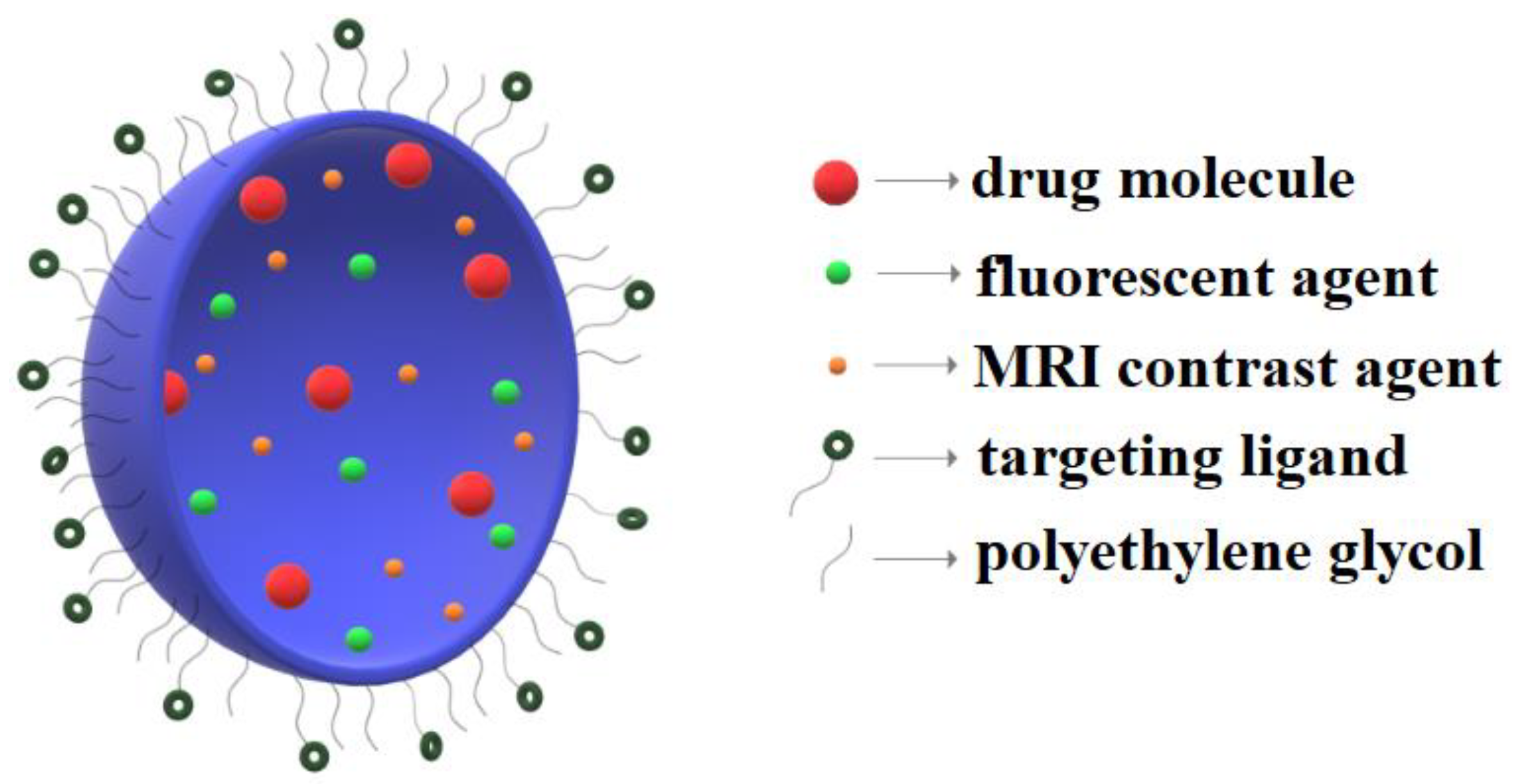
3. Nanotechnology-Based Approaches for Neuroimaging

Whereas early detection is highly involved in the efficient treatment of many brain diseases, such as brain cancer [93] and neurodegenerative disorders [94], the development of novel strategies for neuroimaging is crucial [93]. The interlink between nanotechnology and neuroscience, particularly neuroimaging, has shown a great potential in the field of nanomedicine [95], providing new possibilities for designing contrast agents and nanocarriers that target the brain [96]. Nanotechnology-based materials, devices and electronic biosensors allow for enhanced visualization of brain tissue, resulting in greater spatial and temporal resolution and accuracy [95]. Furthermore, nanotechnology approaches improve the identification of biomarkers, the indicators for the biological state of disease, which is of key importance in early diagnosis [93]. Moreover, the blood-brain barrier restricts the permeation of conventional contrast agents that are used for neuroimaging as they are usually hydrophobic in nature and have a reduced half-time circulation. Additionally, large hydrophobic molecules can reach the brain parenchyma mostly through active pathways, such as carrier- and receptor-mediated transcytosis or by disrupting the blood-brain barrier which could lead to serious consequences [94,97]. Therefore, nanotechnology strategies mostly involve the development of nanocarriers that can efficiently reach and permeate the blood-brain barrier after oral or intravenous administration.
Additionally, these nanoprobes for neuroimaging offer the possibility of attaching targeting molecules on their surface which could enhance the accumulation at specific sites, such as tumour tissue accumulation (Figure 2).
The main strategies based on nanotechnology and the current associated tests for neuroimaging applications are summarized in Table 2. It should be mentioned that the studies chosen to be included in this review are from the last 5 years, found in databases including but not limited to Scopus and PubMed.

3.1. Nanoparticles

The application of nanoparticles has led to the development of potentially novel imaging and diagnostic agents for brain disorders. Nanoparticles can either be used as nanosized imaging agents or as nanocarriers functionalized with contrast agents. Furthermore, they can be designed as theranostic agents through functionalization with therapeutic agents [98]. Thus, the drawbacks of conventional nanoparticles, such as patient compliance and safety, could be overcome by employing theranostic nanoparticles in disease management. [99] Commonly studied imaging agents are iron oxide, gold, manganese oxide and carbon-based nanoparticles.
Iron oxide-based nanoparticles have received outstanding attention as contrast agents owing to their unique physicochemical and superparamagnetic properties. Thus, medical applications, including cell labelling and sorting, cell transfection, diagnostic imaging based on MRI, PET or multimodal imaging could be improved by the use of iron-oxide magnetic nanoparticles [100]. It should be mentioned that ferumoxytol (Feraheme) is a type of magnetic iron oxide nanoparticles which has been approved by the US Food and Drug Administration and it is intensively used in bioimaging [101,102]. In vitro studies reported the use of theranostic iron oxide nanoparticles functionalized with caffeic acid for glioblastoma MRI and reactive oxygen species generation as a therapeutic strategy [103]. Moreover, the theranostic application of iron oxide nanoparticles conjugated with a highly potent vascular disrupting agent and an MMP-14 (matrix metalloproteinase 14) peptide was reported. Results demonstrated their potential for inducing glioblastoma initiating cells apoptosis and impairing tumour growth, as well as the in vivo tracking through MRI [104]. Additionally, iron oxide nanoparticles functionalized with phosphonate polyethylene glycol chains and covalently coupled to cyclic RGD have been applied for in vivo MRI of glioblastoma in mice [105]. The efficiency of these strategies was proved by the preferential accumulation of nanoparticles at the tumour site owing to the passive targeting through the enhanced permeability and retention effect and active targeting induced by cyclic RGD peptides, respectively. The use of superparamagnetic iron oxide nanoparticles has been reported for in vitro and in vivo studies using a gel brain phantom and New Zealand rabbits and a middle-aged human male to rapidly diagnose the emergent stroke through microwave imaging. Injection of the nanoparticles has led to the approximation of an area of reduced attenuation difference associated with ischemic hypo-perfusion of the left carotid circulation [106]. Neuroinflammation could also be diagnosed through a multimodal imaging strategy based on PET and MRI probe as sulphated dextran-coated iron oxide nanoparticles are highly taken by activated microglia [107]. Furthermore, by labelling mesenchymal stem cells with iron oxide nanoparticles, the in vitro process of differentiation into neural-like cells has been visualized through MRI [108].
Gold nanoparticles have attracted a great scientific interest as contrast agents for preoperative, intraoperative and postoperative neuroimaging [109]. Additionally, by attaching chemical moieties and targeting molecules to the surface, gold nanoparticles can be used as multimodal contrast agents with prolonged circulation time, allowing for wider imaging windows [110]. The development of a target-specific imaging system based on peptide-coated gold nanoparticles for specifically detecting glioma cell biomarkers has confirmed the fluorescence signal-based property of the imaging agent [111]. Moreover, gold nanoparticles could permit the visualization of transplanted stem cells inside the brain. One study proposed the use of gadolinium labelled DNA gold nanoparticles for tracking neural stem cells through MRI [112]. In a similar way, CT has been applied for tracking mesenchymal stem cell-derived exosomes through glucose-coated gold nanoparticles [113].
Although all the commercially available intravenous agents contain gadolinium, which is widely studied for MRI, gadolinium-based contrast agents are highly toxic, associated with nephrogenic systemic fibrosis [114,115]. Thus, research scientists have been focusing on developing gadolinium-free contrast agents. Manganese represents the most viable alternative to gadolinium, with the strong paramagnetic properties of high spin Mn2+ ion and the long electronic T1 [115]. Manganese oxide nanoparticles were also used for in vivo glioblastoma MRI. Nanoparticles were first synthesized as oleic acid capped nanoparticles and further transformed by replacing the oleic acid with N-(trimethoxysilylpropyl) ethylene diamine triacetic acid silane, which allowed for the subsequent conjugation of folic acid, a glioma-specific moiety [116]. One study focused on the evaluation of the evolution of hypoxic-ischemic brain injury using hollow manganese nanoparticles as positive T1 contrast agents for MRI [117]. The purpose for using hollow nanoparticles is to increase the specific surface area and subsequently the water-surface interactions, which will further enhance the signal contrast [114]. Therefore, the in vivo imaging of the apoptotic brain areas was possible for up to 21 days, which proves the potential of these nanoparticles for monitoring brain injuries [117].
There has been considerable progress made in research works regarding diagnosis and medical imaging based on carbon nanoparticles strategies. For an effective contrast agent development, coupling of carbon nanoparticles with superparamagnetic iron oxide nanoparticles and gadolinium-functionalized carbon nanoparticles represent potential strategies [118]. The conjugation of the Pittsburgh Compound B with carbon nanotubes for imaging Aβ plaque deposition through various techniques might lead to the efficient early diagnosis of Alzheimer’s disease and therapy and disease progression monitoring [119].
Other theranostic approaches for glioblastoma diagnosis and treatment focused on the development of polysiloxane-based nanoparticles, combining a gadolinium-based contrast agent (AGuIX), 5-(4-carboxyphenyl)-10,15,20-triphenylporphyrin as a photosensitizer and the KDKPPR ligand peptide motif targeting neuropilin-1, a receptor overexpressed by angiogenic endothelial cells of the tumour vasculature [120].

3.2. Liposomes

Similar to natural cell membranes, liposomes are self-assembled lipid-based bilayer vesicles of varying size and structural complexity [121,122]. Liposomes can incorporate a wide range of polar, non-polar and amphiphilic imaging agents or drugs, both within the aqueous core and the lipid bilayers and target specific sites in the body [121,123].
The application of liposomes for the diagnosis of neurological diseases has been intensively studied and strategies involving the covalent bonding of peptides, antibodies and RNA aptamers and the formulation of external stimuli-responsive liposomes have shown great potential for neuroimaging [124].
Research has reported the application of heptamethine cyanine dye IR780 incorporated into liposomes for in vitro and in vivo near-infrared fluorescence imaging of brain tumours using the human glioblastoma multiforme xenograft model and the spontaneous glioblastoma multiforme mouse model respectively [125]. Furthermore, functionalized polyethylene glycol liposomal formulations with antibodies specific for brain tumours have been studied. Aiming to apply in vivo MRI and optical imaging for non-invasive studies, iron oxide nanoparticles and a near-infrared fluorescence dye were encapsulated in the liposomes. Results showed the efficient uptake of the nanoplatforms at the tumour site, with enhanced spatial and temporal resolution in MRI [126]. The formulation of gadolinium-loaded liposomes functionalized with the GBI-10 aptamer to specifically target tumour cells with the overexpressed Tenascin-C glycoprotein. The in vitro results showed an increased endocytosis of the aptamer-functionalized liposomes and a higher relaxivity compared to commercially available MRI contrast agents [127]. A multi-functional hybrid system containing biocompatible liposomes and magneto-plasmonic nanoparticles for image-guided delivery of anti-HIV drugs to the brain has also been developed. The in vitro distribution of the nanocarriers was assessed through MRI, magnetic particle imaging and micro-CT. Results demonstrated the potential of the multimodal nanosystems for the future development of efficient therapy strategies against HIV infected cells [128].
Another research work reported the administration of liposomes incorporating indocyanine green as cargo and two liposomal markers, 1,2-dipalmitoyl-sn-glycero-3-phosphoglycerol and 1,2-distearoyl-sn-glycero-3-phosphoethanolamine conjugated with monodisperse polyethylene glycol into the tail vein of male C57BL/6JRj mice [129]. Additionally, paramagnetic chelate gadolinium-diethylenetriaminepentaacetic acid-loaded pH-sensitive liposomes coated with polyethylene glycol were administered for glioma cell targeting and treatment. The targeting and accumulation efficiency of the liposomes was monitored through in vivo MRI [130]. Moreover, a theranostic approach focused on the formulation of liposomes incorporating an anti-cancer drug, doxorubicin and quantum dots for MRI. Results proved that the application of focused ultrasounds improved the glioma-targeted accumulation, as they can reversibly disrupt the blood-brain barrier without damaging the tissues. Thus, this study offers a promising alternative for chemotherapy with minimal side effects in future clinical application [131]. Similarly, RGD-TPGS-functionalized theranostic liposomes containing docetaxel, an anti-cancer drug and quantum dots were developed for brain cancer imaging and treatment. The RGD peptide allows for the active targeting of tumours and for the internalization through receptor-mediated endocytosis. A higher delivery efficiency compared to the commercially available formulation of docetaxel and to the non-targeting theranostic liposomes was proved in the in vivo studies performed on Charles Foster rats [132].

3.3. Micelles

Micelles are vesical nanoconstructs formed in aqueous solutions by the self-assembly of amphiphilic molecules, with the hydrophilic/polar region on the external surface and the hydrophobic/non-polar region on the inner surface, forming the core [133]. The usual size of pharmaceutical micellar formulations is less than 80 nanometres and the value of the critical micelle concentration, the concentration at which the monomeric amphiphile forms micelles, should be in the millimolar region [134]. Similar to liposomes, micelles can be conjugated or functionalized with polymers, oligonucleotides, peptides or carbohydrates for improving the specificity and the pharmacological behaviours. However, compared to liposomes, micelles are more rapidly accumulated at the tumour sites owing to their smaller size [135].
Various micellar formulations have been designed as imaging agents for different biomedical imaging techniques, including MRI and CT, for the purposes of brain diseases diagnosis or drug delivery monitoring using micelles as nanocarriers [136].
Hyper-permeable blood-brain barrier areas that could be related to solid tumour tissues have been visualized by using gadolinium-incorporated micelles for MRI. This system represents a novel strategy for quantitative haemorrhage-risk evaluation due to the correlation between the extravasation of micelles and the haemorrhagic oedema site [137]. The early detection of neuroinflammation for investigating the triggering and the progression of neurodegenerative diseases might be possible by using MRI-detectable micelles targeted towards the vascular cell adhesion molecule, which is overexpressed in neuroinflammation. Thus, in vivo injected paramagnetic gadolinium-loaded targeting micelles have the potential to indicate neuroinflammation, making tremendous progress in neuroscience [138].
One study focused on developing a theranostic approach using a novel formulation of gold and superparamagnetic iron oxide-loaded micelles coated with polyethylene glycol and polycaprolactone polymers [139].

3.4. Nanobodies

Nanobodies are the antigen-binding or variable heavy chain domain, comprised of four conserved sequence stretches surrounding three hypervariable complementarity-determining regions. Nanobodies are small molecules, possessing unique physical and chemical properties [140]. They are recognized for their solubility, specificity, cost-effective production [141] and remarkable stability under extreme conditions, including high temperature and pressure, low pH and presence of proteases. Furthermore, subsequent to in vivo administration, nanobodies can rapidly diffuse and penetrate body tissues [140]. Therefore, nanobodies represent great candidates for molecular imaging modalities, allowing for fast tumour visualizations owing to homogenous tumour accumulation and fast blood clearance [141].
Moreover, nanobodies can be engineered to detect neuropathological lesions in Alzheimer’s disease, specifically amyloid plaques and neurofibrillary tangles. The in vivo imaging of the nanobodies through two-photon fluorescence imaging (2PFI) represents a fundamental step for unravelling the mechanisms underlying neurodegenerative diseases [142].

3.5. Quantum Dots

Defined as ‘small crystals containing a variable number of electrons that occupy well-defined, discrete quantum states and have electronic properties intermediate between bulk and discrete fundamental particle,’ quantum dots represent the new generation of nanosized semiconductor inorganic crystals [143]. Quantum dots possess unique physical and optical properties, including high quantum yield, narrow and tuneable emitting spectra, which make them great candidates as tools in molecular biology, chemical analysis and materials science [144,145].
Furthermore, quantum dots represent a method for unravelling the mechanisms of molecules and cells behaviour inside the body. Marking them with quantum dots could improve in vivo visualization, allowing for tracking them during a specific period of time. Owing to their longer photostability, quantum dots are superior to conventional fluorescence and organic dyes [146].
Carbon quantum dots are widely used in bioimaging applications owing to their excellent biocompatibility, low cost and easy preparation. By functionalization for tissue imaging and brain gliomas targeting, the tumour uptake could be visualized through fluorescence imaging. However, further studies should be developed for the in vivo imaging of the tumour [147]. Recent studies focused on the imaging of polyethylene glycol quantum dots systems at the tumour site through IVIS imaging system. Although targeted accumulations were observed in tumour tissues, the skulls of the mice were removed in order to acquire the images [148].
Similarly, semiconducting polymer dots with donor-acceptor structure were synthesized for the in vivo mouse brain imaging through 2PFI. The in vivo angiography of the brain indicated large penetration depth, demonstrating the potential of 2PFI of polymer dots for deep-tissue in vivo imaging [149].
Another application of quantum dots for neuroimaging is the single-cell in vivo imaging which has evolved with the development of near-infrared fluorophores and nanotechnology strategies for targeted delivery. This technique has the capacity to offer more detailed information for diagnosis purposes [150]. One study reported the use of quantum dots-antibodies conjugates for in vivo cytometry of cells in their unperturbed microenvironment, which could offer information regarding single cell movement and cell-cell and cell-extracellular matrix interactions. These nanosystems are advantageous as they can be easily and efficiently decorated with targeting molecules or secondary reporters, have a low tendency to non-specifically bind to other cells or serum components, exhibit longer blood circulation times, diffuse through dense in vivo environments and have a narrow emission for multiplexed imaging. Moreover, the use of quantum dots as probes for in vivo imaging allow for long-term single cell tracking in healthy or diseased tissues in vivo and for functional analyses, such as oxygen level, glucose concentration or local mechanical stress measurements [151].
Table 2. A summary of the nanotechnology-based applications for the diagnosis of brain diseases.
Nanotechnology-Based StrategyImaging ProbeTargeting StrategyNeuroimaging TechniqueTargeted Brain DiseaseExperimental StageRef.
Iron-oxide nanoparticlesiron oxide nanoparticles functionalized with caffeic acidpassive—enhanced permeability and retention effectMRIglioblastomain vivo—orthotopic U87-MG tumour implanted in nude mouse brain[103]
iron oxide nanoparticles functionalized with phosphonate polyethylene glycol chains and covalently coupled to cyclic RGDactive—cyclic RGD peptidesMRIglioblastomain vivo—orthotopic U87-MG tumour implanted in nude mouse brain[105]
superparamagnetic iron oxide nanoparticlespassivemicrowave imagingemergent strokein vivo—New Zealand white rabbits; in vivo—middle aged human male volunteer[106]
sulphated dextran-coated iron oxide nanoparticles conjugated with the macrocyclic chelator 1,4,7,10-tetraazacyclododecane-1,4,7-triacetic acidactive—sulphated dextran-coatingPET and MRIneuroinflammationin vivo—BALB/c mice[107]
iron oxide nanoparticlesactive—MMP-14 peptideMRIglioblastomain vivo—NOD scid gamma mice
Gold nanoparticlespolyethylene glycol coated gold nanoparticles conjugated with CBP4 peptideactive—CD133 glioma biomarkerlaser scanning confocal microscopeglioblastomain vivo—U373 glioma cell culture[111]
Manganese oxide nanoparticleshollow manganese oxide nanoparticlespassiveMRIhypoxic-ischemic brain injuryin vivo—Sprague Dawley rat pups [117]
N-(trimethoxysilylpropyl) ethylene diamine triacetic acid silane and folic acid-conjugated manganese oxide nanoparticlesactive—folic acid, a glioma-specific moietyMRIglioblastomain vivo—male nude mice (BALB/C)[116]
Carbon-based nanoparticlesmulti-walled carbon nanotubes conjugated with Pittsburgh Compound B and gadolinium complexesactive—Pittsburgh Compound B for binding to Aβ plaquesSPECT and CTAlzheimer’s diseasein vivo—female C57BL/6 mice[119]
Polysiloxane-based nanoparticlesAGuIXactive—KDKPPR ligand peptide motifMRIglioblastomain vivo—dorsal skinfold chamber using female nude mice[120]
Liposomesheptamethine cyanine dye IR780 incorporated into liposomesactive—IR780 dye for tumour targetingnear-infrared fluorescence imagingglioblastomain vivo—T98G and U87MG cells; in vivo—nude mice bearing U87M2/luc tumours[125]
iron oxide nanoparticles and near-infrared fluorescence dye DiR incorporated into polyethylene glycol liposomes functionalized with the F(ab’)2 fragments of PGN635active—phosphatidylserine targetingMRI and near-infrared optical imagingglioblastomain vivo—human glioma U87MG cells; in vivo—BALB/c mice[126]
paramagnetic chelate gadolinium-diethylenetriaminepentaacetic acid-loaded liposomes coated with polyethylene glycolpassiveMRIglioblastomain vivo—tumour bearing C57BL6 adult male mice[130]
gadolinium-loaded liposomesactive—GBI-10 aptamerMRIglioblastomain vitro—MDA-MB-435s human breast duct cell line[127]
quantum dots and doxorubicin-loaded liposomesactive—focused ultrasoundMRIglioblastomain vivo—Adult male Sprague–Dawley rats[131]
quantum dots and docetaxel-loaded liposomesactive—RGD-TPGS peptideglioblastomain vivo—Charles Foster rats[132]
Micellespolyethylene glycol-b-poly(l-lysine-DOTA-gadolinium) micellespassive—enhanced permeability and retention effectMRIischemia-reperfusion injuryin vivo—Wistar male rats[137]
paramagnetic gadolinium-loaded targeting micellesactive—targeting the vascular cell adhesion molecule MRIneuroinflammationin vivo—C57BL/6J female mice[138]
gold and superparamagnetic iron oxide-loaded micelles coated with polyethylene glycol and polycaprolactonePassiveMRI and CTglioblastomain vivo—female athymic nude mice[139]
Nanobodiesanti-Aβ and anti-pTau VHHsactive—amyloid plaques and neurofibrillary tangles2PFIAlzheimer’s diseasein vivo—PS2APP mice overexpressing hAPP Swedish mutation combined with PS2 N141I mutation and Tg4510 mice with the hMAPT P301L gene mutation[142]
Quantum dotssemiconducting polymer dots encapsulated into poly(styrene-co-maleic anhydride) and conjugated with poly(ethylene glycol)active—donor-acceptor structure2PFIin vivo—ICR female mice [149]

4. Conclusions and Perspectives

Conventional neuroimaging techniques possess several disadvantages regarding the spatial and temporal resolution, the invasiveness of the imaging modality, the toxicity of the contrast agents and the costs implicated in image acquiring. As neuroimaging is of key importance in early diagnosis and therapy efficacy monitoring of brain diseases, overcoming the associated limitations is crucial. Nanotechnology represents a potential strategy for improving neuroimaging techniques by developing novel imaging agents or nanocarriers for conventional contrast agents. Although there is a great research performed for designing nanotechnology approaches for brain imaging, further studies are necessary for both in vivo imaging improvement and long-term impact establishment. As there is a limited number of commercially-approved nanocarriers for the targeted delivery of contrast agents to the brain, thorough investigations regarding their toxicity and their associated side effects, as well as the ways to overcome these limitations must be performed.
As future perspectives, specialists in the field of neuroimaging should consider the application of nanotechnology for single-neuron detection [152,153] and phase-contrast X-ray imaging techniques [154] which could offer the possibility of visualizing brain areas and activities that are currently obscure or undetectable for conventional methods. In the case of single-neuron detection, current studies focus on the use of diamond chips with quantum defects as sensors that can detect time-varying magnetic fields generated by action potentials of neurons [155]. Similarly, magnetic induction tomography performed with optical atomic magnetometry allows for the detection across a large frequency range in low-conductivity targets, such as the biological tissues [156]. Moreover, the phase-contrast X-ray imaging offers the possibility of 3D visualization of soft tissue-organs, such as the lung and brain. The currently available techniques that use phase-contrast are propagation- and analyser-based, crystal and grafting interferometry and non-interferometry methods [157]. This technique might overcome the poor soft tissue contrast of conventional CT and the poor spatial resolution of MRI [158].

Author Contributions

D.M.T., C.C., A.M.G., A.V. and R.I.T. designed and wrote the paper.

Funding

This research received no external funding.

Conflicts of Interest

The authors declare no conflict of interest.

References

  1. Gürsoy-Özdemir, Y.; Bozdağ-Pehlivan, S.; Sekerdag, E. (Eds.) Preface. In Nanotechnology Methods for Neurological Diseases and Brain Tumors; Academic Press: Cambridge, MA, USA, 2017; pp. xv–xvi. [Google Scholar]
  2. Mollaamin, F. The effect of biointerface of chemicals and inhibitors in the cerebral cortex of brain on language cognition. Biointerface Res. Appl. Chem. 2018, 8, 3628–3634. [Google Scholar]
  3. Kovacs, G.G. Chapter 21—Concepts and classification of neurodegenerative diseases. In Handbook of Clinical Neurology; Kovacs, G.G., Alafuzoff, I., Eds.; Elsevier: Amsterdam, The Netherlands, 2018; Volume 145, pp. 301–307. [Google Scholar]
  4. McFaline-Figueroa, J.R.; Lee, E.Q. Brain Tumors. Am. J. Med. 2018, 131, 874–882. [Google Scholar] [CrossRef]
  5. Das, J.; G K, R. Post stroke depression: The sequelae of cerebral stroke. Neurosci. Biobehav. Rev. 2018, 90, 104–114. [Google Scholar] [CrossRef] [PubMed]
  6. Khoury, S.; Benavides, R. Pain with traumatic brain injury and psychological disorders. Prog. Neuro-Psychopharmacol. Biol. Psychiatry 2018, 87, 224–233. [Google Scholar] [CrossRef] [PubMed]
  7. Johnson, A.L.; McLeish, A.C.; Shear, P.K.; Privitera, M. Panic and epilepsy in adults: A systematic review. Epilepsy Behav. 2018, 85, 115–119. [Google Scholar] [CrossRef] [PubMed]
  8. Giovane, R.A.; Lavender, P.D. Central Nervous System Infections. Prim. Care Clin. Off. Pract. 2018, 45, 505–518. [Google Scholar] [CrossRef] [PubMed]
  9. George, E.; Guenette, J.P.; Lee, T.C. Introduction to Neuroimaging. Am. J. Med. 2018, 131, 346–356. [Google Scholar] [CrossRef]
  10. Dash, H.H.; Chavali, S. Management of traumatic brain injury patients. Korean J. Anesthesiol. 2018, 71, 12–21. [Google Scholar] [CrossRef]
  11. Tulay, E.E.; Metin, B.; Tarhan, N.; Arikan, M.K. Multimodal Neuroimaging: Basic Concepts and Classification of Neuropsychiatric Diseases. Clin. EEG Neurosci. 2019, 50, 20–33. [Google Scholar] [CrossRef]
  12. Furukawa, K.; Ishiki, A.; Tomita, N.; Onaka, Y.; Saito, H.; Nakamichi, T.; Hara, K.; Kusano, Y.; Ebara, M.; Arata, Y.; et al. Introduction and overview of the special issue “Brain imaging and aging”: The new era of neuroimaging in aging research. Ageing Res. Rev. 2016, 30, 1–3. [Google Scholar] [CrossRef]
  13. Mufford, M.S.; Stein, D.J.; Dalvie, S.; Groenewold, N.A.; Thompson, P.M.; Jahanshad, N. Neuroimaging genomics in psychiatry—A translational approach. Genome Med. 2017, 9, 102. [Google Scholar] [CrossRef]
  14. Bulut, E.; Akgoz, A.; Oguz, K.K. Chapter 11—Neuroimaging: Techniques and General Applications. In Nanotechnology Methods for Neurological Diseases and Brain Tumors; Gürsoy-Özdemir, Y., Bozdağ-Pehlivan, S., Sekerdag, E., Eds.; Academic Press: Cambridge, MA, USA, 2017; pp. 209–224. [Google Scholar]
  15. Krishnan, K.R.R. Chapter 6—Structural imaging in psychiatric disorders. In Handbook of Clinical Neurology; Aminoff, M.J., Boller, F., Swaab, D.F., Eds.; Elsevier: Amsterdam, The Netherlands, 2012; Volume 106, pp. 89–95. [Google Scholar]
  16. Hanganu, A.; Monchi, O. Structural Neuroimaging Markers of Cognitive Decline in Parkinson’s Disease. Parkinson’s Dis. 2016, 2016, 8. [Google Scholar] [CrossRef] [PubMed]
  17. Boly, M.; Gosseries, O.; Massimini, M.; Rosanova, M. Chapter 2—Functional Neuroimaging Techniques. In The Neurology of Conciousness (Second Edition); Laureys, S., Gosseries, O., Tononi, G., Eds.; Academic Press: San Diego, CA, USA, 2016; pp. 31–47. [Google Scholar]
  18. Im, J.J.; Namgung, E.; Choi, Y.; Kim, J.Y.; Rhie, S.J.; Yoon, S. Molecular Neuroimaging in Posttraumatic Stress Disorder. Exp. Neurobiol. 2016, 25, 277–295. [Google Scholar] [CrossRef] [PubMed]
  19. Sanders, J.W.; Song, H.; Frank, S.J.; Bathala, T.; Venkatesan, A.M.; Anscher, M.; Tang, C.; Bruno, TL.; Wei, W.; Ma, J. Parallel imaging compressed sensing for accelerated imaging and improved signal-to-noise ratio in MRI-based postimplant dosimetry of prostate brachytherapy. Brachytherapy 2018, 17, 816–824. [Google Scholar] [CrossRef]
  20. Malik, S.; Sachan, M.; Nara, S. Nano-Strategies for Neuro-Imaging and Diagnostics. In Advances in Neurotherapeutic Delivery Technologies; Kumar, P., Pillay, V., Choonara, Y.E., Eds.; OMICS International: Hyderabad, India, 2015. [Google Scholar]
  21. Sabry, N.M.; Tolba, S.T.M.; Abdel-Gawad, F.K.; Bassem, S.M.; Nassar, H.; El-Taweel, G.E.; Ibrahim, M.A. On the molecular modeling analyses of the interaction between nano zinc oxide and bacteria. Biointerface Res. Appl. Chem. 2018, 8, 3294–3297. [Google Scholar]
  22. Faisal, N.; Kumar, K. Polymer and metal nanocomposites in biomedical applications. Biointerface Res. Appl. Chem. 2017, 7, 2286–2294. [Google Scholar]
  23. Kaur, M.; Singh, G.; Khanna, K.; Kaur, N. Nanotechnology: A Review. In Proceedings of the Second National Conference on Advances in Manufacturing Systems, S B S State Technical Campus, Ferozepur, India, 23–24 December 2015. [Google Scholar]
  24. Sabry, N.M.; Tolba, S.; Abdel-Gawad, F.K.; Bassem, S.M.; Nassar, H.F.; El-Taweel, G.E.; Medhat, A.I. Interaction between nano silver and bacteria: Modeling approach. Biointerface Res. Appl. Chem. 2018, 8, 3570–3574. [Google Scholar]
  25. Sridhar, S.; Mishra, S.; Gulyas, M.; Padmanabhan, P.; Gulyas, B. An Overview of Multimodal Neuroimaging Using Nanoprobes. Int. J. Mol. Sci. 2017, 18, 311. [Google Scholar] [CrossRef] [PubMed]
  26. Lafortuna, C.; Tabozzi, S.; Rizzo, G. Functional brain imaging and its application to uncover mechanisms driving food intake in humans. J. Biomed. Graph. Comput. 2014, 4, 10–27. [Google Scholar] [CrossRef]
  27. Zippo, A.; Castiglioni, I. Integration of 18FDG-PET Metabolic and Functional Connectomes in the Early Diagnosis and Prognosis of the Alzheimer’s Disease. Curr. Alzheimer Res. 2016, 13, 487–497. [Google Scholar] [CrossRef]
  28. Beckmann, N. In Vivo magnetic resonance techniques and drug discovery. Braz. J. Phys. 2006, 36, 16–22. [Google Scholar] [CrossRef]
  29. Morton, D.L.; Sandhu, J.S.; Jones, A.K. Brain imaging of pain: State of the art. J. Pain Res. 2016, 9, 613–624. [Google Scholar] [CrossRef]
  30. Forkel, S.J.; Catani, M. Structural Neuroimaging. In Research Methods in Psycholinguistics and the Neurobiology of Language: A Practical Guide, 1st ed.; John Wiley & Sons, Inc.: Hoboken, NJ, USA, 2018; pp. 288–308. [Google Scholar]
  31. Skipper, N.T.; Igra, M.S.; Davidson, A.J. Brain imaging for anaesthetists and intensivists: Part 1—Computed tomography. BJA Educ. 2018, 18, 300–309. [Google Scholar] [CrossRef]
  32. Pease, A.P.; Nelson, N.C. Chapter 71—Computed Tomography. In Equine Surgery (fifth Edition); Auer, J.A., Stick, J.A., Kümmerle, J.M., Prange, T., Eds.; W.B. Saunders: Philadelphia, PA, USA, 2019; pp. 1193–1201. [Google Scholar]
  33. Edelstein, W.A.; Mahesh, M.; Carrino, J.A. MRI: Time is dose—And money and versatility. J. Am. Coll. Radiol. JACR 2010, 7, 650–652. [Google Scholar] [CrossRef]
  34. Copen, W.A.; Lev, M.H.; Rapalino, O. Chapter 6—Brain perfusion: Computed tomography and magnetic resonance techniques. In Handbook of Clinical Neurology; Masdeu, J.C., González, R.G., Eds.; Elsevier: Amsterdam, The Netherlands, 2016; Volume 135, pp. 117–135. [Google Scholar]
  35. Heit, J.J.; Wintermark, M. Perfusion Computed Tomography for the Evaluation of Acute Ischemic Stroke. Stroke 2016, 47, 1153–1158. [Google Scholar] [CrossRef]
  36. Chou, E.T.; Carrino, J.A. Chapter 10—Magnetic Resonance Imaging. In Pain Management; Waldman, S.D., Bloch, J.I., Eds.; W.B. Saunders: Philadelphia, PA, USA, 2007; pp. 106–117. [Google Scholar]
  37. González-Villà, S.; Oliver, A.; Valverde, S.; Wang, L.; Zwiggelaar, R.; Lladó, X. A review on brain structures segmentation in magnetic resonance imaging. Artif. Intell. Med. 2016, 73, 45–69. [Google Scholar] [CrossRef]
  38. Rabai, F.; Ramani, R. Chapter 31—Magnetic Resonance Imaging: Anesthetic Implications. In Essentials of Neuroanesthesia; Prabhakar, H., Ed.; Academic Press: Cambridge, MA, USA, 2017; pp. 519–532. [Google Scholar]
  39. Wilson, H.; Dervenoulas, G.; Politis, M. Chapter Nine—Structural Magnetic Resonance Imaging in Huntington’s Disease. In International Review of Neurobiology; Politis, M., Ed.; Academic Press: Cambridge, MA, USA, 2018; Volume 142, pp. 335–380. [Google Scholar]
  40. Taghizadeh, S.; Labuda, C.; Yang, C.C.; Morris, B.; Kanakamedala, M.R.; Vijayakumar, S.; Rey-Dios, R.; Duggar, W.N.; Florez, E.; Fatemi, A. Optimizing MRI sequences and images for MRI-based stereotactic radiosurgery treatment planning. Rep. Pract. Oncol. Radiother. 2019, 24, 12–19. [Google Scholar] [CrossRef]
  41. Moser, E.; Laistler, E.; Schmitt, F.; Kontaxis, G. Ultra-High Field NMR and MRI—The Role of Magnet Technology to Increase Sensitivity and Specificity. Front. Phys. 2017, 5, 33. [Google Scholar] [CrossRef]
  42. Kasliwal, M.K. Functional Neuroimaging: Current Status. OMICS J. Radiol. 2012, 1, e111. [Google Scholar] [CrossRef]
  43. Medaglia, J.D. Functional Neuroimaging in Traumatic Brain Injury: From Nodes to Networks. Front. Neurol. 2017, 8, 407. [Google Scholar] [CrossRef] [PubMed]
  44. Kropotov, J.D. (Ed.) Chapter 1.3—Functional Magnetic Resonance Imaging. In Functional Neuromarkers for Psychiatry; Academic Press: San Diego, CA, USA, 2016; pp. 17–25. [Google Scholar]
  45. Buchbinder, B.R. Chapter 4—Functional magnetic resonance imaging. In Handbook of Clinical Neurology; Masdeu, J.C., González, R.G., Eds.; Elsevier: Amsterdam, The Netherlands, 2016; Volume 135, pp. 61–92. [Google Scholar]
  46. Fisicaro, R.; Brennan, N.P.; Holodny, A. Chapter 29—Functional Magnetic Resonance Imaging. In Handbook of Neuro-Oncology Neuroimaging (Second Edition); Newton, H.B., Ed.; Academic Press: San Diego, CA, USA, 2016; pp. 317–325. [Google Scholar]
  47. Chen, J.E.; Glover, G.H. Functional Magnetic Resonance Imaging Methods. Neuropsychol. Rev. 2015, 25, 289–313. [Google Scholar] [CrossRef] [PubMed]
  48. Glasser, M.F.; Coalson, T.S.; Robinson, E.C.; Hacker, C.D.; Harwell, J.; Yacoub, E.; Ugurbil, K.; Andersson, J.; Beckmann, C.F.; Jenkinson, M.; et al. A multi-modal parcellation of human cerebral cortex. Nature 2016, 536, 171. [Google Scholar] [CrossRef] [PubMed]
  49. Glasser, M.F.; Coalson, T.S.; Bijsterbosch, J.D.; Harrison, S.J.; Harms, M.P.; Anticevic, A.; Van Essen, D.C.; Smith, S.M. Using temporal ICA to selectively remove global noise while preserving global signal in functional MRI data. NeuroImage 2018, 181, 692–717. [Google Scholar] [CrossRef] [PubMed]
  50. Sharma, R. Preface. In Radiology Diagnosis. Functional Magnetic Resonance Imaging—Advanced Neuroimaging Applications; Sharma, R., Ed.; Intech Open: London, UK, 2012. [Google Scholar]
  51. Fröhlich, F. Chapter 13—Imaging Functional Networks with MRI. In Network Neuroscience; Fröhlich, F., Ed.; Academic Press: San Diego, CA, USA, 2016; pp. 177–185. [Google Scholar]
  52. Fukuda, M.; Poplawsky, A.J.; Kim, S.G. Chapter 6—Submillimeter-resolution fMRI: Toward understanding local neural processing. In Progress in Brain Research; Masamoto, K., Hirase, H., Yamada, K., Eds.; Elsevier: Amsterdam, The Netherlands, 2016; Volume 225, pp. 123–152. [Google Scholar]
  53. Huettel, S.A. Functional MRI (fMRI). In Encyclopedia of Spectroscopy and Spectrometry (Third Edition); Lindon, J.C., Tranter, G.E., Koppenaal, D.W., Eds.; Academic Press: Oxford, UK, 2017; pp. 778–784. [Google Scholar]
  54. Abhang, P.A.; Gawali, B.W.; Mehrotra, S.C. Chapter 1—Introduction to Emotion, Electroencephalography and Speech Processing. In Introduction to EEG- and Speech-Based Emotion Recognition; Abhang, P.A., Gawali, B.W., Mehrotra, S.C., Eds.; Academic Press: Cambridge, MA, USA, 2016; pp. 1–17. [Google Scholar]
  55. Kulkarni, N.; Bairagi, V. (Eds.) Chapter Two—Electroencephalogram and Its Use in Clinical Neuroscience. In EEG-Based Diagnosis of Alzheimer Disease; Academic Press: Cambridge, MA, USA, 2018; pp. 25–35. [Google Scholar]
  56. Proekt, A. Chapter Fifteen—Brief Introduction to Electroencephalography. In Methods in Enzymology; Eckenhoff, R.G., Dmochowski, I.J., Eds.; Academic Press: Cambridge, MA, USA, 2018; Volume 603, pp. 257–277. [Google Scholar]
  57. Wang, Y.; Yan, J.; Wen, J.; Yu, T.; Li, X. An Intracranial Electroencephalography [iEEG] Brain Function Mapping Tool with an Application to Epilepsy Surgery Evaluation. Front. Neuroinform. 2016, 10, 15. [Google Scholar] [CrossRef]
  58. Nagahama, Y.; Schmitt, A.J.; Nakagawa, D.; Vesole, A.S.; Kamm, J.; Kovach, C.K.; Hasan, D.; Granner, M.; Dlouhy, B.J.; Howard, M.A.; et al. Intracranial EEG for seizure focus localization: Evolving techniques, outcomes, complications and utility of combining surface and depth electrodes. J. Neurosurg. 2018, 1, 1–13. [Google Scholar] [CrossRef]
  59. Parvizi, J.; Kastner, S. Promises and limitations of human intracranial electroencephalography. Nat. Neurosci. 2018, 21, 474–483. [Google Scholar] [CrossRef]
  60. Binder, J.R. Chapter 37—Phoneme Perception. In Neurobiology of Language; Hickok, G., Small, S.L., Eds.; Academic Press: San Diego, CA, USA, 2016; pp. 447–461. [Google Scholar]
  61. Adewole, D.O.; Serruya, M.D.; Harris, J.P.; Burrell, J.C.; Petrov, D.; Chen, H.I.; Wolf, J.A.; Cullen, D.K. The Evolution of Neuroprosthetic Interfaces. Crit. Rev. Biomed. Eng. 2016, 44, 123–152. [Google Scholar] [CrossRef]
  62. Deng, M.; Yang, X.; Silke, M.; Qiu, W.; Xu, M.; Borghs, G.; Wolf, J.A.; Cullen, D.K. Electrochemical deposition of polypyrrole/graphene oxide composite on microelectrodes towards tuning the electrochemical properties of neural probes. Sens. Actuators B Chem. 2011, 158, 176–184. [Google Scholar] [CrossRef]
  63. Chung, T.; Wang, J.Q.; Wang, J.-Y.; Cao, B.; Li, Y.; Pang, S.W. Electrode modifications to lower electrode impedance and improve neural signal recording sensitivity. J. Neural Eng. 2015, 12, 056018. [Google Scholar] [CrossRef]
  64. Yücel, M.A.; Selb, J.J.; Huppert, T.J.; Franceschini, M.A.; Boas, D.A. Functional Near Infrared Spectroscopy: Enabling routine functional brain imaging. Curr. Opin. Biomed. Eng. 2017, 4, 78–86. [Google Scholar] [CrossRef]
  65. Peng, K.; Pouliot, P.; Lesage, F.; Nguyen, D.K. Multichannel continuous electroencephalography-functional near-infrared spectroscopy recording of focal seizures and interictal epileptiform discharges in human epilepsy: A review. Neurophotonics 2016, 3, 031402. [Google Scholar] [CrossRef]
  66. Herrera-Vega, J.; Treviño-Palacios, C.G.; Orihuela-Espina, F. Neuroimaging with functional near infrared spectroscopy: From formation to interpretation. Infrared Phys. Technol. 2017, 85, 225–237. [Google Scholar] [CrossRef]
  67. Pfeifer, M.D.; Scholkmann, F.; Labruyère, R. Signal Processing in Functional Near-Infrared Spectroscopy (fNIRS): Methodological Differences Lead to Different Statistical Results. Front. Hum. Neurosci. 2018, 11, 641. [Google Scholar] [CrossRef] [PubMed]
  68. Balardin, J.B.; Zimeo Morais, G.A.; Furucho, R.A.; Trambaiolli, L.; Vanzella, P.; Biazoli, C.; Sato, J.R. Imaging Brain Function with Functional Near-Infrared Spectroscopy in Unconstrained Environments. Front. Hum. Neurosci. 2017, 11, 258. [Google Scholar] [CrossRef]
  69. Deffieux, T.; Demene, C.; Pernot, M.; Tanter, M. Functional ultrasound neuroimaging: A review of the preclinical and clinical state of the art. Curr. Opin. Neurobiol. 2018, 50, 128–135. [Google Scholar] [CrossRef] [PubMed]
  70. Gesnik, M.; Blaize, K.; Deffieux, T.; Gennisson, J.-L.; Sahel, J.-A.; Fink, M.; Picaud, S.; Tanter, M. 3D functional ultrasound imaging of the cerebral visual system in rodents. NeuroImage 2017, 149, 267–274. [Google Scholar] [CrossRef]
  71. Demene, C.; Baranger, J.; Bernal, M.; Delanoe, C.; Auvin, S.; Biran, V.; Alison, M.; Mairesse, J.; Harribaud, E.; Pernot, M.; et al. Functional ultrasound imaging of brain activity in human newborns. Sci. Transl. Med. 2017, 9, eaah6756. [Google Scholar] [CrossRef]
  72. Hage, B.; Alwatban, M.R.; Barney, E.; Mills, M.; Dodd, M.D.; Truemper, E.J.; Bashford, G.R. Functional Transcranial Doppler Ultrasound for Measurement of Hemispheric Lateralization During Visual Memory and Visual Search Cognitive Tasks. IEEE Trans. Ultrason. Ferroelectr. Freq. Control 2016, 63, 2001–2007. [Google Scholar] [CrossRef]
  73. Wang, L.V.; Yao, J. A practical guide to photoacoustic tomography in the life sciences. Nat. Methods 2016, 13, 627–638. [Google Scholar] [CrossRef]
  74. Zafar, M.; Kratkiewicz, K.; Manwar, R.; Avanaki, M. Development of Low-Cost Fast Photoacoustic Computed Tomography: System Characterization and Phantom Study. Appl. Sci. 2019, 9, 374. [Google Scholar] [CrossRef]
  75. Wang, L.V.; Xia, J.; Yao, J. Photoacoustic Neuroimaging. In Neurophotonics and Brain Mapping; Chen, Y., Kateb, B., Eds.; CRC Press: Boca Raton, FL, USA, 2017. [Google Scholar]
  76. Lameka, K.; Farwell, M.D.; Ichise, M. Chapter 11—Positron Emission Tomography. In Handbook of Clinical Neurology; Masdeu, J.C., González, R.G., Eds.; Elsevier: Amsterdam, The Netherlands, 2016; Volume 135, pp. 209–227. [Google Scholar]
  77. Goel, S.; England, C.G.; Chen, F.; Cai, W. Positron emission tomography and nanotechnology: A dynamic duo for cancer theranostics. Adv. Drug Deliv. Rev. 2017, 113, 157–176. [Google Scholar] [CrossRef] [PubMed]
  78. Moghbel, M.; Newberg, A.; Alavi, A. Chapter 12—Positron emission tomography: Ligand imaging. In Handbook of Clinical Neurology; Masdeu, J.C., González, R.G., Eds.; Elsevier: Amsterdam, The Netherlands, 2016; Volume 135, pp. 229–240. [Google Scholar]
  79. Penny, W.D.; Friston, K.J. Functional imaging. Scholarpedia 2007, 2, 1478. [Google Scholar] [CrossRef]
  80. Du, Y.; Zaidi, H. Single-Photon Emission Computed Tomography: Principles and Applications. In Encyclopedia of Biomedical Engineering; Narayan, R., Ed.; Elsevier: Oxford, UK, 2019; pp. 493–506. [Google Scholar]
  81. Goffin, K.; van Laere, K. Chapter 13—Single-photon emission tomography. In Handbook of Clinical Neurology; Masdeu., J.C., González, R.G., Eds.; Elsevier: Amsterdam, The Netherlands, 2016; Volume 135, pp. 241–250. [Google Scholar]
  82. Kijewski, M.F. Chapter 32—Positron Emission Tomography (PET) and Single-Photon Emission Computed Tomography (SPECT) Physics. In Handbook of Neuro-Oncology Neuroimaging (Second Edition); Newton, H.B., Ed.; Academic Press: San Diego, CA, USA, 2016; pp. 353–358. [Google Scholar]
  83. Yeh, R.; Miloushev, V.Z.; Ichise, M. Chapter 33—Positron Emission Tomography (PET) and Single Photon Emission Computed Tomography (SPECT) Imaging. In Handbook of Neuro-Oncology Neuroimaging (Second Edition); Newton, H.B., Ed.; Academic Press: San Diego, CA, USA, 2016; pp. 359–370. [Google Scholar]
  84. Cherry, S.R.; Sorenson, J.A.; Phelps, M.E. Chapter 17—Single Photon Emission Computed Tomography. In Physics in Nuclear Medicine (Fourth Edition); Cherry, S.R., Sorenson, J.A., Phelps, M.E., Eds.; W.B. Saunders: Philadelphia, PA, USA, 2012; pp. 279–306. [Google Scholar]
  85. Wang, Z.J.; Chang, T.T.A.; Slauter, R. Chapter 35—Use of Imaging for Preclinical Evaluation. In A Comprehensive Guide to Toxicology in Nonclinical Drug Development (Second Edition); Faqi, A.S., Ed.; Academic Press: Boston, MA, USA, 2017; pp. 921–938. [Google Scholar]
  86. Hengerer, A.; Grimm, J. Molecular magnetic resonance imaging. Biomed. Imaging Interv. J. 2006, 2, e8. [Google Scholar] [CrossRef] [PubMed]
  87. Sinharay, S.; Pagel, M.D. Advances in Magnetic Resonance Imaging Contrast Agents for Biomarker Detection. Annu. Rev. Anal. Chem. [Palo Alto, Calif.] 2016, 9, 95–115. [Google Scholar] [CrossRef]
  88. Haris, M.; Yadav, S.K.; Rizwan, A.; Singh, A.; Wang, E.; Hariharan, H.; Ravinder, R.; Marincola, F.M. Molecular magnetic resonance imaging in cancer. J. Transl. Med. 2015, 13, 313. [Google Scholar] [CrossRef]
  89. Gauberti, M.; Fournier, A.P.; Docagne, F.; Vivien, D.; Martinez de Lizarrondo, S. Molecular Magnetic Resonance Imaging of Endothelial Activation in the Central Nervous System. Theranostics 2018, 8, 1195–1212. [Google Scholar] [CrossRef]
  90. Shuhendler, A.J.; Ye, D.; Brewer, K.D.; Bazalova-Carter, M.; Lee, K.-H.; Kempen, P.; Wittrup, K.D.; Graves, E.E.; Rutt, B.; Rao, J. Molecular Magnetic Resonance Imaging of Tumor Response to Therapy. Sci. Rep. 2015, 5, 14759. [Google Scholar] [CrossRef]
  91. Li, J.; You, J.; Wu, C.; Dai, Y.; Shi, M.; Dong, L.; Xu, K. T1-T2 molecular magnetic resonance imaging of renal carcinoma cells based on nano-contrast agents. Int. J. Nanomed. 2018, 13, 4607–4625. [Google Scholar] [CrossRef]
  92. Bonnet, C.S.; Tóth, É. Molecular Magnetic Resonance Imaging Probes Based on Ln3+ Complexes. Adv. Org. Chem. 2016, 68, 43–96. [Google Scholar]
  93. Bhargava, A.; Cheung, J.; Eshaghian-Wilner, M.M.; Lee, W.; Ravicz, K.; Schlesinger, M.; Shah, Y.; Uppal, A. An introduction to nanomedicine. In Wireless Computing in Medicine; John Wiley & Sons, Inc.: Hoboken, NJ, USA, 2016. [Google Scholar]
  94. Zhang, W.; Wang, W.; Yu, D.X.; Xiao, Z.; He, Z. Application of nanodiagnostics and nanotherapy to CNS diseases. Nanomedicine 2018, 13, 2341–2371. [Google Scholar] [CrossRef]
  95. Kumar, A.; Tan, A.; Wong, J.; Spagnoli, J.C.; Lam, J.; Blevins, B.D.; G, N.; Thorne, L.; Ashkan, K.; Xie, J.; et al. Nanotechnology for Neuroscience: Promising Approaches for Diagnostics, Therapeutics and Brain Activity Mapping. Adv. Funct. Mater. 2017, 27, 1700489. [Google Scholar] [CrossRef] [PubMed]
  96. Mandal, A.; Bisht, R.; Pal, D.; Mitra, A.K. Chapter 4—Diagnosis and Drug Delivery to the Brain: Novel Strategies. In Emerging Nanotechnologies for Diagnostics, Drug Delivery and Medical Devices; Mitra, A.K., Cholkar, K., Mandal, A., Eds.; Elsevier: Boston, MA, USA, 2017; pp. 59–83. [Google Scholar]
  97. Teleanu, D.M.; Chircov, C.; Grumezescu, A.M.; Volceanov, A.; Teleanu, R.I. Blood-Brain Delivery Methods Using Nanotechnology. Pharmaceutics 2018, 10, 269. [Google Scholar] [CrossRef]
  98. Posadas, I.; Monteagudo, S.; Ceña, V. Nanoparticles for brain-specific drug and genetic material delivery, imaging and diagnosis. Nanomedicine 2016, 11, 833–849. [Google Scholar] [CrossRef] [PubMed]
  99. Ramanathan, S.; Archunan, G.; Sivakumar, M.; Tamil Selvan, S.; Fred, A.L.; Kumar, S.; Gulyás, B.; Padmanabhan, P. Theranostic applications of nanoparticles in neurodegenerative disorders. Int. J. Nanomed. 2018, 13, 5561–5576. [Google Scholar] [CrossRef] [PubMed]
  100. Nedyalkova, M.; Donkova, B.; Romanova, J.; Tzvetkov, G.; Madurga, S.; Simeonov, V. Iron oxide nanoparticles—In vivo/in vitro biomedical applications and in silico studies. Adv. Colloid Interface Sci. 2017, 249, 192–212. [Google Scholar] [CrossRef]
  101. Vallabani, N.V.S.; Singh, S. Recent advances and future prospects of iron oxide nanoparticles in biomedicine and diagnostics. 3 Biotech 2018, 8, 279. [Google Scholar] [CrossRef] [PubMed]
  102. Daldrup-Link, H.E. Ten Things You Might Not Know about Iron Oxide Nanoparticles. Radiology 2017, 284, 616–629. [Google Scholar] [CrossRef]
  103. Richard, S.; Saric, A.; Boucher, M.; Slomianny, C.; Geffroy, F.; Mériaux, S.; Lalatonne, Y.; Petit, P.X.; Motte, L. Antioxidative Theranostic Iron Oxide Nanoparticles toward Brain Tumors Imaging and ROS Production. ACS Chem. Biol. 2016, 11, 2812–2819. [Google Scholar] [CrossRef] [PubMed]
  104. Mohanty, S.; Chen, Z.; Li, K.; Morais, G.R.; Klockow, J.; Yerneni, K.; Pisani, L.; Chin, F.T.; Mitra, S.; Cheshier, S.; et al. A Novel Theranostic Strategy for MMP-14-Expressing Glioblastomas Impacts Survival. Mol. Cancer Ther. 2017, 16, 1909–1921. [Google Scholar] [CrossRef]
  105. Richard, S.; Boucher, M.; Lalatonne, Y.; Mériaux, S.; Motte, L. Iron oxide nanoparticle surface decorated with cRGD peptides for magnetic resonance imaging of brain tumours. Biochim. Biophys. Acta (BBA)—Gen. Subj. 2017, 1861, 1515–1520. [Google Scholar] [CrossRef] [PubMed]
  106. Hudson, J.S.; Chung, T.K.; Prout, B.S.; Nagahama, Y.; Raghavan, M.L.; Hasan, D.M. Iron nanoparticle contrast enhanced microwave imaging for emergent stroke: A pilot study. J. Clin. Neurosci. 2019, 59, 284–290. [Google Scholar] [CrossRef] [PubMed]
  107. Tang, T.; Valenzuela, A.; Petit, F.; Chow, S.; Leung, K.; Gorin, F.; Louie, A.Y.; Dhenain, M. In Vivo MRI of Functionalized Iron Oxide Nanoparticles for Brain Inflammation. Contrast Media Mol. Imaging 2018, 2018, 10. [Google Scholar] [CrossRef] [PubMed]
  108. Lu, C.-W.; Hsiao, J.-K.; Liu, H.-M.; Wu, C.-H. Characterization of an iron oxide nanoparticle labelling and MRI-based protocol for inducing human mesenchymal stem cells into neural-like cells. Sci. Rep. 2017, 7, 3587. [Google Scholar] [CrossRef]
  109. Meola, A.; Rao, J.; Chaudhary, N.; Sharma, M.; Chang, S.D. Gold Nanoparticles for Brain Tumor Imaging: A Systematic Review. Front. Neurol. 2018, 9, 328. [Google Scholar] [CrossRef]
  110. Mahan, M.M.; Doiron, A.L. Gold Nanoparticles as X-ray, CT and Multimodal Imaging Contrast Agents: Formulation, Targeting and Methodology. J. Nanomater. 2018, 2018, 15. [Google Scholar] [CrossRef]
  111. Cho, J.-H.; Kim, A.R.; Kim, S.-H.; Lee, S.-J.; Chung, H.; Yoon, M.-Y. Development of a novel imaging agent using peptide-coated gold nanoparticles toward brain glioma stem cell marker CD133. Acta Biomater. 2017, 47, 182–192. [Google Scholar] [CrossRef]
  112. Nicholls, F.J.; Rotz, M.W.; Ghuman, H.; MacRenaris, K.W.; Meade, T.J.; Modo, M. DNA–gadolinium–gold nanoparticles for in vivo T1 MR imaging of transplanted human neural stem cells. Biomaterials 2016, 77, 291–306. [Google Scholar] [CrossRef]
  113. Betzer, O.; Perets, N.; Barnoy, E.; Offen, D.; Popovtzer, R. Labeling and tracking exosomes within the brain using gold nanoparticles. Proc. SPIE 2018, 10506, 1050618. [Google Scholar]
  114. Azria, D.; Blanquer, S.; Verdier, J.-M.; Belamie, E. Nanoparticles as contrast agents for brain nuclear magnetic resonance imaging in Alzheimer’s disease diagnosis. J. Mater. Chem. B 2017, 5, 7216–7237. [Google Scholar] [CrossRef]
  115. Gale, E.M.; Caravan, P. Gadolinium-Free Contrast Agents for Magnetic Resonance Imaging of the Central Nervous System. ACS Chem. Neurosci. 2018, 9, 395–397. [Google Scholar] [CrossRef]
  116. Chen, N.; Shao, C.; Qu, Y.; Li, S.; Gu, W.; Zheng, T.; Ye, L.; Yu, C. Folic Acid-Conjugated MnO Nanoparticles as a T1 Contrast Agent for Magnetic Resonance Imaging of Tiny Brain Gliomas. ACS Appl. Mater. Interfaces 2014, 6, 19850–19857. [Google Scholar] [CrossRef]
  117. Jeon, T.Y.; Kim, J.H.; Im, G.H.; Kim, J.H.; Yang, J.; Yoo, S.Y.; Lee, J.H. Hollow manganese oxide nanoparticle-enhanced MRI of hypoxic-ischaemic brain injury in the neonatal rat. Br. J. Radiol. 2016, 89, 20150806. [Google Scholar] [CrossRef]
  118. Servant, A.; Jacobs, I.; Bussy, C.; Fabbro, C.; da Ros, T.; Pach, E.; Ballesteros, B.; Prato, M.; Nicolay, K.; Kostarelos, K. Gadolinium-functionalised multi-walled carbon nanotubes as a T1 contrast agent for MRI cell labelling and tracking. Carbon 2016, 97, 126–133. [Google Scholar] [CrossRef]
  119. Costa, P.M.; Wang, J.T.-W.; Morfin, J.-F.; Khanum, T.; To, W.; Sosabowski, J.; Tóth, E.; Al-Jamal1, KT. Functionalised Carbon Nanotubes Enhance Brain Delivery of Amyloid-Targeting Pittsburgh Compound B (PiB)-Derived Ligands. Nanotheranostics 2018, 2, 168–183. [Google Scholar] [CrossRef] [PubMed]
  120. Thomas, E.; Colombeau, L.; Gries, M.; Peterlini, T.; Mathieu, C.; Thomas, N.; Boura, C.; Frochot, C.; Vanderesse, R.; Lux, F.; et al. Ultrasmall AGuIX theranostic nanoparticles for vascular-targeted interstitial photodynamic therapy of glioblastoma. Int. J. Nanomed. 2017, 12, 7075–7088. [Google Scholar] [CrossRef] [PubMed]
  121. Rideau, E.; Dimova, R.; Schwille, P.; Wurm, F.R.; Landfester, K. Liposomes and polymersomes: A comparative review towards cell mimicking. Chem. Soc. Rev. 2018, 47, 8572–8610. [Google Scholar] [CrossRef] [PubMed]
  122. Aderibigbe, B.A.; Mukaya, H.E. Chapter 3—Polymer Therapeutics: Design, Application and Pharmacokinetics. In Nano- and Microscale Drug Delivery Systems; Grumezescu, A.M., Ed.; Elsevier: Amsterdam, The Netherlands, 2017; pp. 33–48. [Google Scholar]
  123. Dimov, N.; Kastner, E.; Hussain, M.; Perrie, Y.; Szita, N. Formation and purification of tailored liposomes for drug delivery using a module-based micro continuous-flow system. Sci. Rep. 2017, 7, 12045. [Google Scholar] [CrossRef] [PubMed]
  124. Vieira, D.B.; Gamarra, L.F. Getting into the brain: Liposome-based strategies for effective drug delivery across the blood-brain barrier. Int. J. Nanomed. 2016, 11, 5381–5414. [Google Scholar] [CrossRef]
  125. Li, S.; Johnson, J.; Peck, A.; Xie, Q. Near infrared fluorescent imaging of brain tumour with IR780 dye incorporated phospholipid nanoparticles. J. Transl. Med. 2017, 15, 18. [Google Scholar] [CrossRef]
  126. Zhang, L.; Habib, A.A.; Zhao, D. Phosphatidylserine-targeted liposome for enhanced glioma-selective imaging. Oncotarget 2016, 7, 38693–38706. [Google Scholar] [CrossRef]
  127. Zhang, L.-X.; Li, K.-F.; Wang, H.; Gu, M.-J.; Liu, L.-S.; Zheng, Z.-Z.; Han, N.Y.; Yang, Z.J.; Fan, T.Y. Preparation and In Vitro Evaluation of a MRI Contrast Agent Based on Aptamer-Modified Gadolinium-Loaded Liposomes for Tumor Targeting. AAPS PharmSciTech 2017, 18, 1564–1571. [Google Scholar] [CrossRef]
  128. Tomitaka, A.; Arami, H.; Huang, Z.; Raymond, A.; Rodriguez, E.; Cai, Y.; Febo, M.; Takemura, Y.; Nair, M. Hybrid magneto-plasmonic liposomes for multimodal image-guided and brain-targeted HIV treatment. Nanoscale 2018, 10, 184–194. [Google Scholar] [CrossRef]
  129. Fülöp, A.; Sammour, D.A.; Erich, K.; von Gerichten, J.; van Hoogevest, P.; Sandhoff, R.; Hopf, C. Molecular imaging of brain localization of liposomes in mice using MALDI mass spectrometry. Sci. Rep. 2016, 6, 33791. [Google Scholar] [CrossRef]
  130. Pacheco-Torres, J.; Mukherjee, N.; Walko, M.; Lopez-Larrubia, P.; Ballesteros, P.; Cerdan, S.; Kocer, A. Image guided drug release from pH-sensitive Ion channel-functionalized stealth liposomes into an in vivo glioblastoma model. Nanomedicine 2015, 11, 1345–1354. [Google Scholar] [CrossRef]
  131. Lin, Q.; Mao, K.L.; Tian, F.R.; Yang, J.J.; Chen, P.P.; Xu, J.; Fan, Z.L.; Zhao, Y.P.; Li, W.F.; Zheng, L.; et al. Brain tumour-targeted delivery and therapy by focused ultrasound introduced doxorubicin-loaded cationic liposomes. Cancer Chemother. Pharmacol. 2016, 77, 269–280. [Google Scholar] [CrossRef]
  132. Sonali; Singh, R.P.; Sharma, G.; Kumari, L.; Koch, B.; Singh, S.; Bharti, S.; Rajinikanth, P.S.; Pandey, B.L.; Muthu, M.S. RGD-TPGS decorated theranostic liposomes for brain targeted delivery. Colloids Surf. B Biointerfaces 2016, 147, 129–141. [Google Scholar] [CrossRef]
  133. Joseph, M.; Trinh, H.M.; Mitra, A.K. Chapter 7—Peptide and Protein-Based Therapeutic Agents*. In Emerging Nanotechnologies for Diagnostics, Drug Delivery and Medical Devices; Mitra, A.K., Cholkar, K., Mandal, A., Eds.; Elsevier: Boston, MA, USA, 2017; pp. 145–167. [Google Scholar]
  134. Rana, S.; Bhattacharjee, J.; Barick, K.C.; Verma, G.; Hassan, P.A.; Yakhmi, J.V. Chapter 7—Interfacial engineering of nanoparticles for cancer therapeutics. In Nanostructures for Cancer Therapy; Ficai, A., Grumezescu, A.M., Eds.; Elsevier: Amsterdam, The Netherlands, 2017; pp. 177–209. [Google Scholar]
  135. Priya, L.B.; Baskaran, R.; Padma, V.V. Chapter 21—Phytonanoconjugates in oral medicine. In Nanostructures for Oral Medicine; Andronescu, E., Grumezescu, A.M., Eds.; Elsevier: Amsterdam, The Netherlands, 2017; pp. 639–668. [Google Scholar]
  136. Movassaghian, S.; Merkel, O.M.; Torchilin, V.P. Applications of polymer micelles for imaging and drug delivery. Wiley Interdiscip. Rev. Nanomed. Nanobiotechnol. 2015, 7, 691–707. [Google Scholar] [CrossRef]
  137. Shiraishi, K.; Wang, Z.; Kokuryo, D.; Aoki, I.; Yokoyama, M. A polymeric micelle magnetic resonance imaging [MRI] contrast agent reveals blood–brain barrier (BBB) permeability for macromolecules in cerebral ischemia-reperfusion injury. J. Control. Release 2017, 253, 165–171. [Google Scholar] [CrossRef]
  138. Garello, F.; Pagoto, A.; Arena, F.; Buffo, A.; Blasi, F.; Alberti, D.; Terreno, E. MRI visualization of neuroinflammation using VCAM-1 targeted paramagnetic micelles. Nanomed. Nanotechnol. Biol. Med. 2018, 14, 2341–2350. [Google Scholar] [CrossRef]
  139. Sun, L.; Joh, D.Y.; Al-Zaki, A.; Stangl, M.; Murty, S.; Davis, J.J.; Baumann, B.C.; Alonso-Basanta, M.; Kaol, G.D.; Tsourkas, A. Theranostic Application of Mixed Gold and Superparamagnetic Iron Oxide Nanoparticle Micelles in Glioblastoma Multiforme. J. Biomed. Nanotechnol. 2016, 12, 347–356. [Google Scholar] [CrossRef]
  140. Steeland, S.; Vandenbroucke, R.E.; Libert, C. Nanobodies as therapeutics: Big opportunities for small antibodies. Drug Discov. Today 2016, 21, 1076–1113. [Google Scholar] [CrossRef]
  141. Van Audenhove, I.; Gettemans, J. Nanobodies as Versatile Tools to Understand, Diagnose, Visualize and Treat Cancer. EBioMedicine 2016, 8, 40–48. [Google Scholar] [CrossRef]
  142. Li, T.; Vandesquille, M.; Koukouli, F.; Dudeffant, C.; Youssef, I.; Lenormand, P.; Ganneau, C.; Maskos, U.; Czech, C.; Grueninger, F.; et al. Camelid single-domain antibodies: A versatile tool for in vivo imaging of extracellular and intracellular brain targets. J. Control. Release 2016, 243, 1–10. [Google Scholar] [CrossRef]
  143. Reshma, V.G.; Mohanan, P.V. Quantum dots: Applications and safety consequences. J. Lumin. 2019, 205, 287–298. [Google Scholar] [CrossRef]
  144. Xu, J.; Zheng, J. Chapter 3—Quantum Dots and Nanoclusters. In Nano-Inspired Biosensors for Protein Assay with Clinical Applications; Li, G., Ed.; Elsevier: Amsterdam, The Netherlands, 2019; pp. 67–90. [Google Scholar]
  145. Zhu, R.; Chen, H.; Wu, S.-T.; Dong, Y. Quantum Dot Light Emitting Diodes. In Reference Module in Materials Science and Materials Engineering; Elsevier: Amsterdam, The Netherlands, 2018. [Google Scholar]
  146. Yamamoto, K. Application 13—Bioimaging with Quantum Dots. In Nanoparticle Technology Handbook (Third Edition); Naito, M., Yokoyama, T., Hosokawa, K., Nogi, K., Eds.; Elsevier: Amsterdam, The Netherlands, 2018; pp. 473–477. [Google Scholar]
  147. Gao, L.; Zhao, X.; Wang, J.; Wang, Y.; Yu, L.; Peng, H.; Jianzhong, Z. Multiple functionalized carbon quantum dots for targeting glioma and tissue imaging. Opt. Mater. 2018, 75, 764–769. [Google Scholar] [CrossRef]
  148. Huang, N.; Cheng, S.; Zhang, X.; Tian, Q.; Pi, J.; Tang, J.; Huang, Q.; Wang, F.; Chen, J.; Xie, Z.; et al. Efficacy of NGR peptide-modified PEGylated quantum dots for crossing the blood–brain barrier and targeted fluorescence imaging of glioma and tumour vasculature. Nanomed. Nanotechnol. Biol. Med. 2017, 13, 83–93. [Google Scholar] [CrossRef]
  149. Alifu, N.; Sun, Z.; Zebibula, A.; Zhu, Z.; Zhao, X.; Wu, C.; Wang, Y.; Qian, J. Deep-red polymer dots with bright two-photon fluorescence and high biocompatibility for in vivo mouse brain imaging. Opt. Commun. 2017, 399, 120–126. [Google Scholar] [CrossRef]
  150. Isherwood, B.; Timpson, P.; McGhee, E.J.; Anderson, K.I.; Canel, M.; Serrels, A.; Brunton, V.G.; Carragher, N.O. Live cell in vitro and in vivo imaging applications: Accelerating drug discovery. Pharmaceutics 2011, 3, 141–170. [Google Scholar] [CrossRef]
  151. Han, H.-S.; Niemeyer, E.; Huang, Y.; Kamoun, W.S.; Martin, J.D.; Bhaumik, J.; Chen, Y.; Roberge, S.; Cui, K.; Martin, M.R.; et al. Quantum dot/antibody conjugates for in vivo cytometric imaging in mice. Proc. Natl. Acad. Sci. USA 2015, 112, 1350–1355. [Google Scholar] [CrossRef]
  152. Barry, J.F.; Turner, M.J.; Schloss, J.M.; Glenn, D.R.; Song, Y.; Lukin, M.D.; Park, H.; Walsworth, R.L. Optical magnetic detection of single-neuron action potentials using quantum defects in diamond. Proc. Natl. Acad. Sci. USA 2016, 113, 14133–14138. [Google Scholar] [CrossRef]
  153. Zhu, H.; Zou, G.; Wang, N.; Zhuang, M.; Xiong, W.; Huang, G. Single-neuron identification of chemical constituents, physiological changes and metabolism using mass spectrometry. Proc. Natl. Acad. Sci. USA 2017, 114, 2586–2591. [Google Scholar] [CrossRef]
  154. Croton, L.C.P.; Morgan, K.S.; Paganin, D.M.; Kerr, L.T.; Wallace, M.J.; Crossley, K.J.; Miller, S.L.; Yagi, N.; Uesugi, K.; Hooper, S.B.; et al. In situ phase contrast X-ray brain CT. Sci. Rep. 2018, 8, 11412. [Google Scholar] [CrossRef]
  155. Amirav, L.; Berlin, S.; Olszakier, S.; Pahari, S.K.; Kahn, I. Multi-Modal Nano Particle Labeling of Neurons. Front. Neurosci. 2019, 13, 12. [Google Scholar] [CrossRef]
  156. Deans, C.; Marmugi, L.; Hussain, S.; Renzoni, F. Optical atomic magnetometry for magnetic induction imaging of the heart. Proc. SPIE 2016, 99000F. [Google Scholar] [CrossRef]
  157. Bravin, A.; Coan, P.; Suortti, P. X-ray phase-contrast imaging: From pre-clinical applications towards clinics. Phys. Med. Biol. 2013, 58, R1–R35. [Google Scholar] [CrossRef]
  158. Xi, Y.; Lin, X.; Yuan, F.; Yang, G.-Y.; Zhao, J. High-Resolution and Quantitative X-ray Phase-Contrast Tomography for Mouse Brain Research. Comput. Math. Methods Med. 2015, 2015, 12. [Google Scholar] [CrossRef]
Figure 1. The classification of commonly applied neuroimaging techniques.
Nanomaterials 09 00542 g001
Figure 2. A schematic diagram of the strategies for molecule incorporation and surface functionalization of nanocarriers for neuroimaging.
Nanomaterials 09 00542 g002
Table 1. A summary of the main characteristics of the neuroimaging techniques previously presented.
CTMRIfMRIEEGiEEGfNIRSfUSPETSPECTmMRI
Cost$$$$$$$$$$$$$$$$$
Invasivenessminimalnon-invasivenon-invasivenon-invasiverequires surgerynon-invasivenon-invasiveminimalminimalnon-invasive
Acquisition timeminutesminutes to hoursminutes to hoursminuteshoursminutesminutesminutesminutes to hours
Portabilitynot portablenot portablenot portableportablenot portableportableportablenot portablenot portablenot portable
Personnel requirementsqualified personnelqualified personnelqualified personnelqualified personnel—optionalqualified personnelqualified personnel—optionalqualified personnel—optionalqualified personnelqualified personnelqualified personnel
Spatial resolution0.5–0.625 mm1–2 mm1–2 mm5–9 cm4.5 mm1 cm50–200 µm3–5 mm6–8 mm
Temporal resolution85–135 ms20–50 ms1–3 s130 ms0.8 ms330 ms1–100 ms5 s to 5 min15 min
Penetration depthno limitno limit1.2 mm3 cmno limitno limitno limit

