Article

CeO2 Nanorods Embedded in Ni(OH)2 Matrix for the Non-Enzymatic Detection of Glucose

1
Department of Material Science, Taiyuan University of Technology, Taiyuan 030024, China
2
School of Material Science, Lanzhou University of Technology, Lanzhou 730050, China
*
Authors to whom correspondence should be addressed.
Nanomaterials 2017, 7(8), 205; https://doi.org/10.3390/nano7080205
Submission received: 15 June 2017 / Revised: 19 July 2017 / Accepted: 28 July 2017 / Published: 31 July 2017

Abstract

:
The electrode based on cerium oxide (CeO2) nanorods embedded in nickel hydroxide (Ni(OH)2) matrix were prepared and used for detecting glucose non-enzymatically. The materials were characterized by X-ray diffraction, transmission electron microscopy (TEM), and so on. The results indicate that the response of CeO2/Ni(OH)2 nanocomposite are significantly improved due to the synergetic effect between CeO2 and Ni(OH)2. The optimum CeO2/Ni(OH)2 nanocomposite electrode exhibits a detection range from 2 μM to 6.62 mM, a sensitivity of 594 μA mM−1 cm−2, an estimated detection limit of 1.13 μM, and a response time less than 5 s. In addition, this biosensor also shows good selectivity, long term stability, and accurate measurement in juice on sale.

1. Introduction

Glucose biosensors have received considerable attention because of their promising applications in clinical diagnosis, the food industry, and environmental monitoring [1,2,3,4]. Generally, glucose sensors can be classified as either enzymatic glucose sensors or non-enzymatic glucose sensors. Although enzymatic glucose sensors are sensitive and selective, they are limited by the instability of the enzyme, which may be affected by various factors such as temperature, pH, and humidity [5,6,7,8]. Non-enzymatic glucose sensors can overcome the disadvantages of enzymatic glucose sensors. Nevertheless, enzyme-free biosensors usually require suitable sensing materials that can simultaneously catalyze the redox reaction of glucose and rapidly transfer the charges.
Metal oxides (e.g., NiO, CuO) and metal hydroxides (e.g., Ni(OH)2, Cu(OH)2) possess unique electrocatalytic properties and have been extensively explored for non-enzymatic glucose biosensors in the past ten years. However, the poor intrinsic charge transfer abilities of these oxides or hydroxides impede their application in the electrochemical biosensor, which generally requires a high sensitivity, fast response, and good selectivity [9,10]. An effective way to overcome this limitation is combining these metal oxides or hydroxides with high charge-transporting materials, and the corresponding synergetic effect originating from the combination of components can provide a new interface through which charge and energy transport are significantly enhanced [11].
The high charge-transporting components usually are noble metals [12,13], carbon nanotubes [14,15,16], and graphene [17]. However, only a few studies have been carried out to investigate semiconducting oxides with fast electron-transfer abilities as modification materials (e.g., TiO2, ZnO) [18,19,20,21,22]. For example, Gao et al. combined semi-metallic TiO2 nanotubes arrays and Ni(OH)2 nanoparticles to form a hybrid that could detect glucose with a high sensitivity of 240 μA mM−1 cm−2, a detection limit of 5.0 μM (S/N = 3), and a quick response time of less than 5 s [9]. Zhou et al. used the electrospinning method to fabricate a CuO/ZnO hierarchical nanocomposite that exhibited an ultrahigh sensitivity of 3066.4 μA mM−1 cm−2, a linear range of up to 1.6 mM, and a low practical detection limit of 0.21 μM [23]. Given that metal oxides have the advantages of cost effectiveness and simple synthesis, pure metal oxide nanocomposites for application in non-enzymatic biosensor should still be a subject undergoing intense study.
Recently, nanostructured CeO2 has been of great interest in electrochemical biosensors because of its unique properties, such as high mechanical strength, high isoelectric point, biocompatibility, and fast electron-transfer ability [24]. However, pristine CeO2 is seldom used in non-enzymatic glucose biosensors because it fails to catalyze a surface redox reaction [25,26]. CeO2 may be a good modification material for common electrocatalytic materials (e.g., NiO, Ni(OH)2) because it has good electronic conductivity, particularly with regard to enhancing their electron-transfer abilities. Using this as an inspiration, the nanorod (NR) structure of CeO2 was combined with Ni(OH)2 to form a nanocomposite which was used for the first time as a non-enzymatic glucose biosensor.

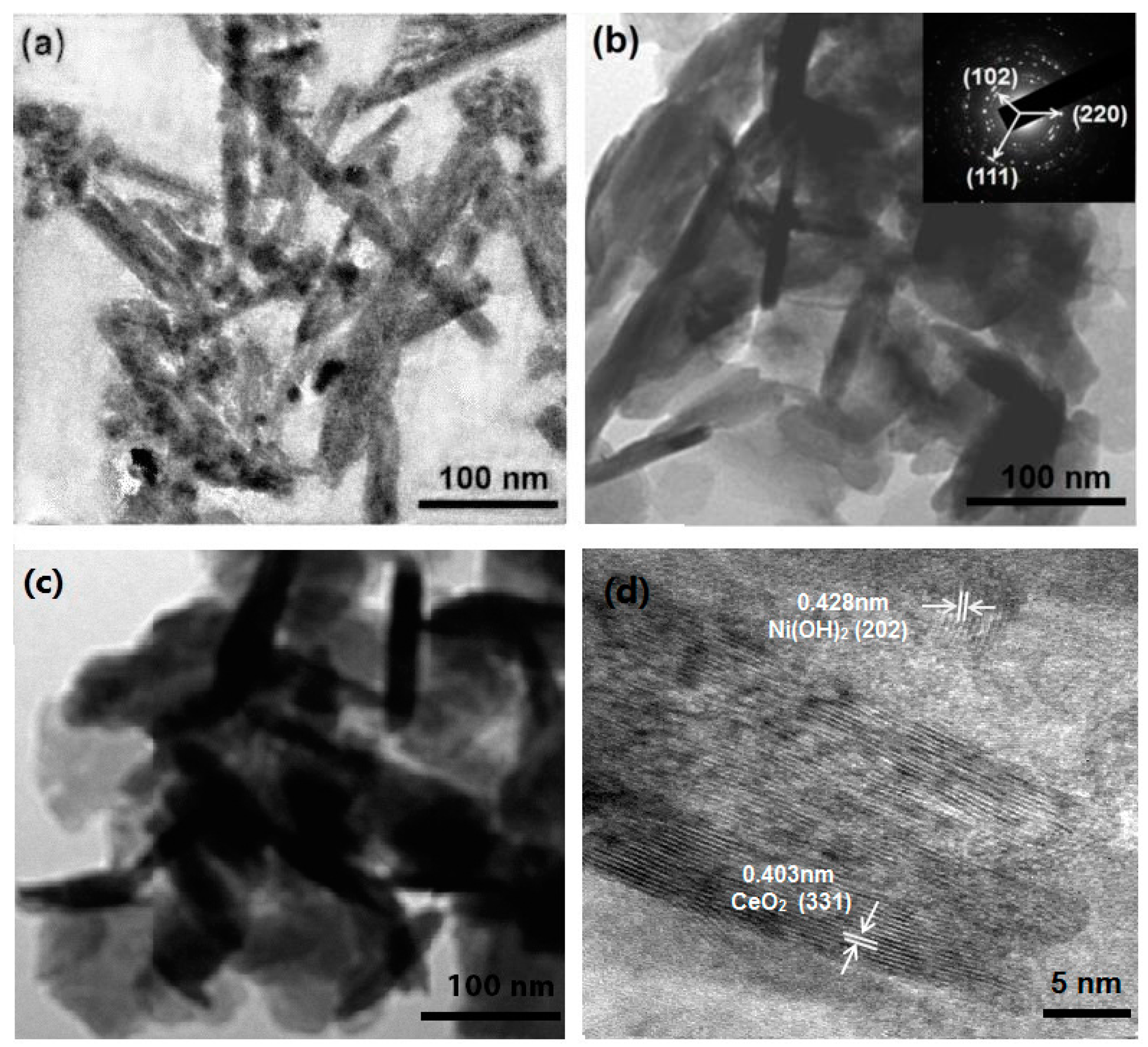
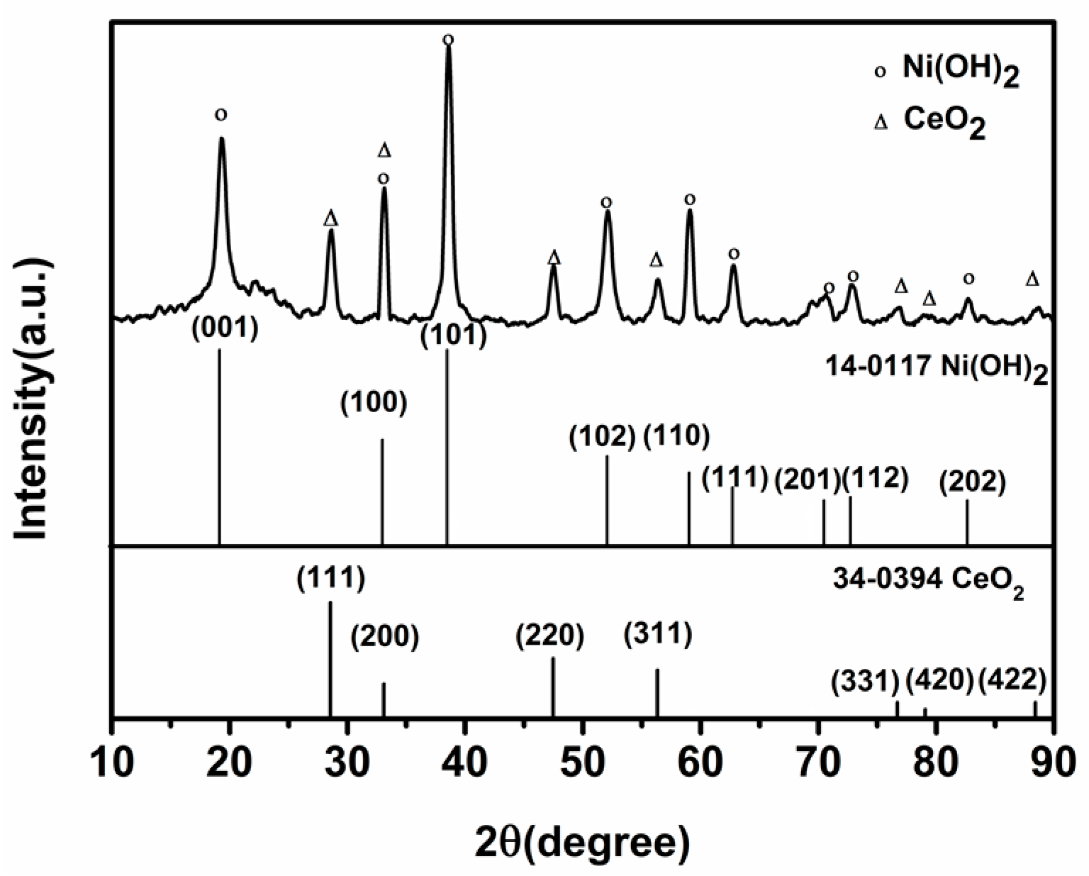
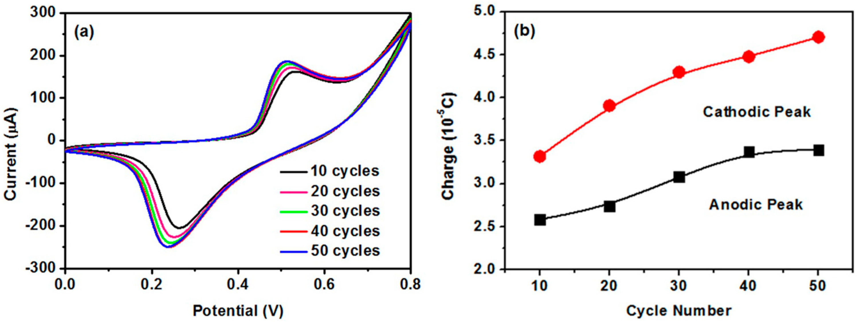
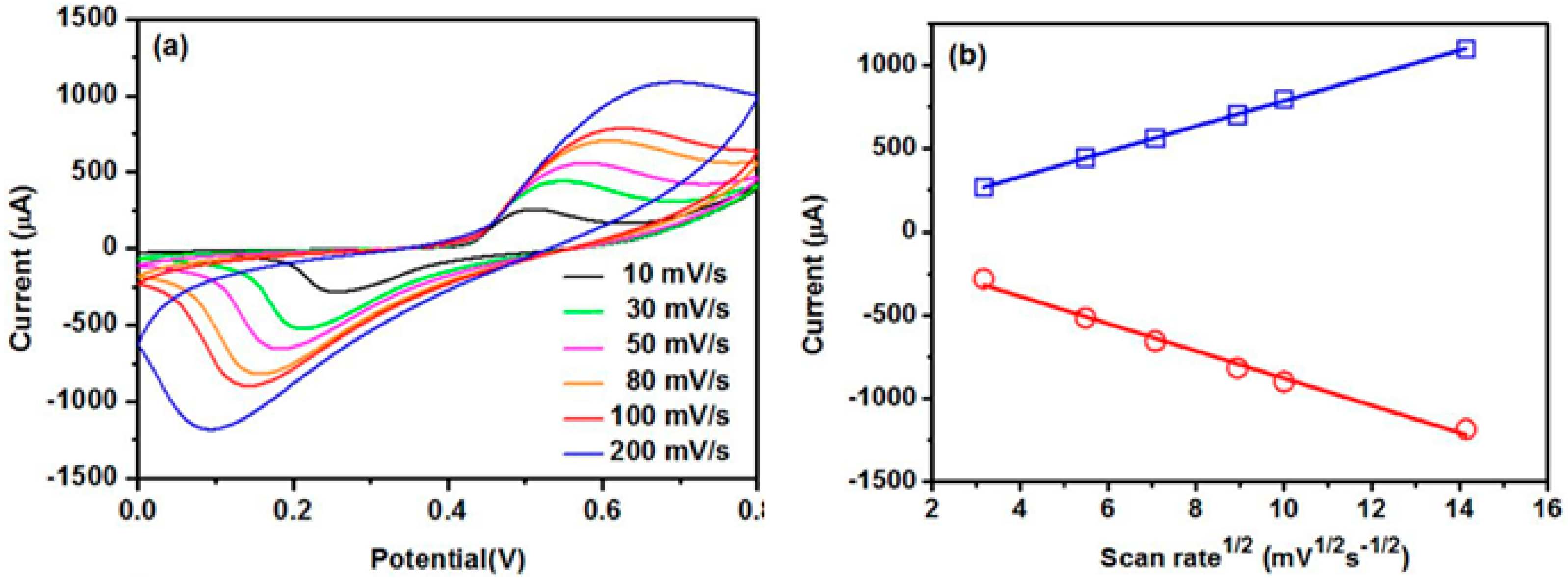
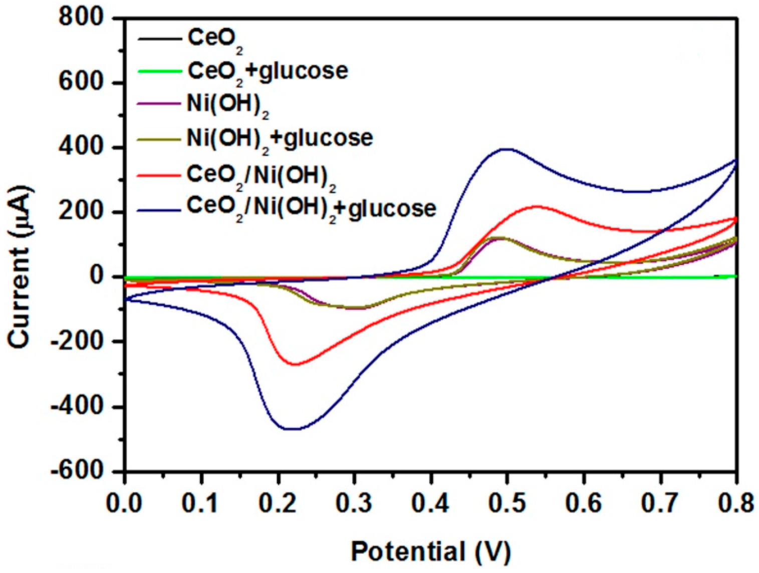
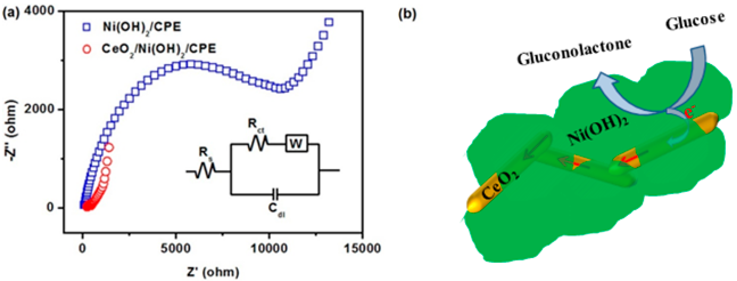
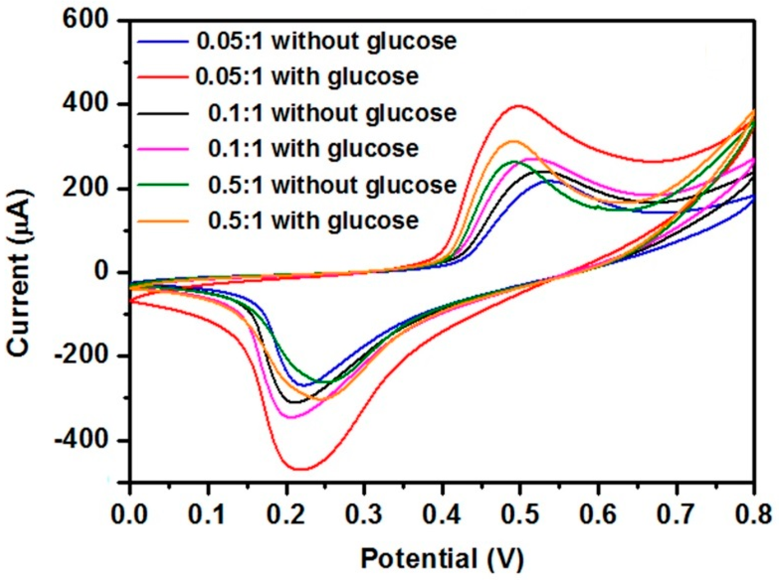
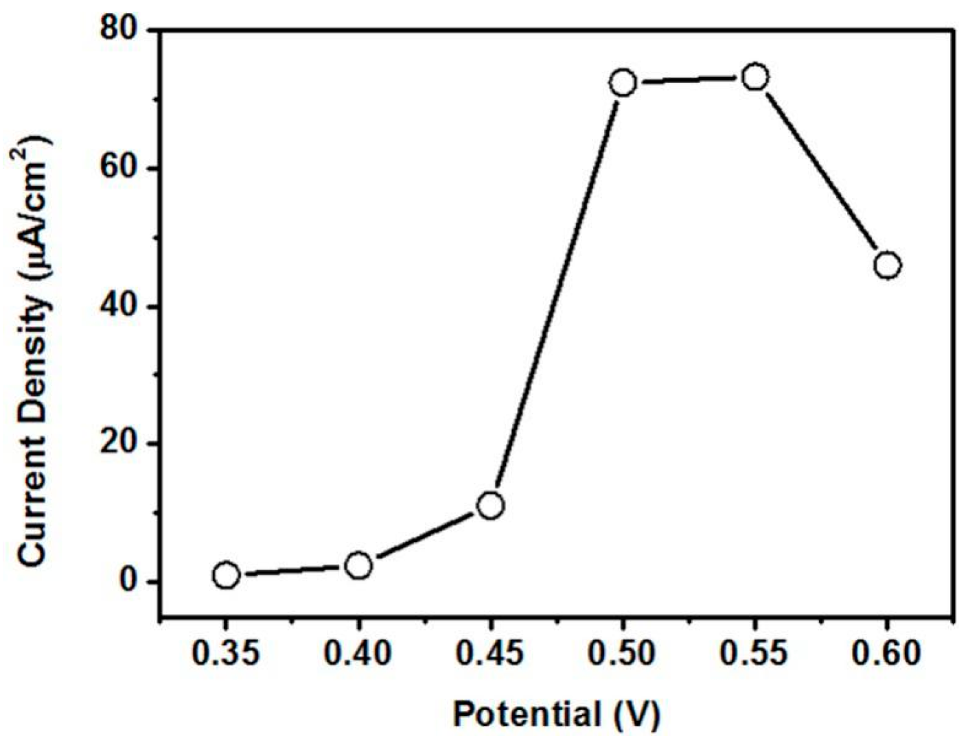
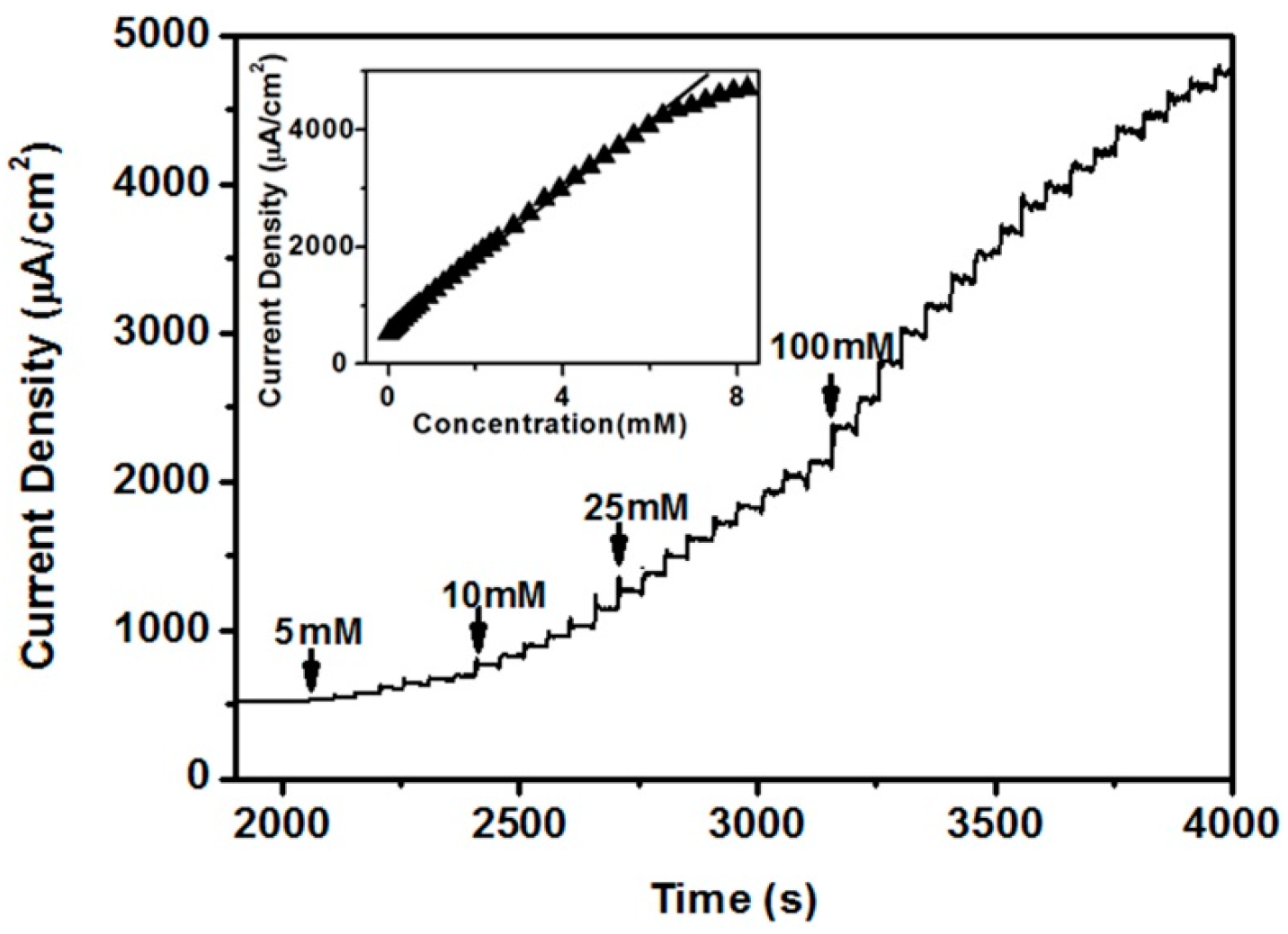
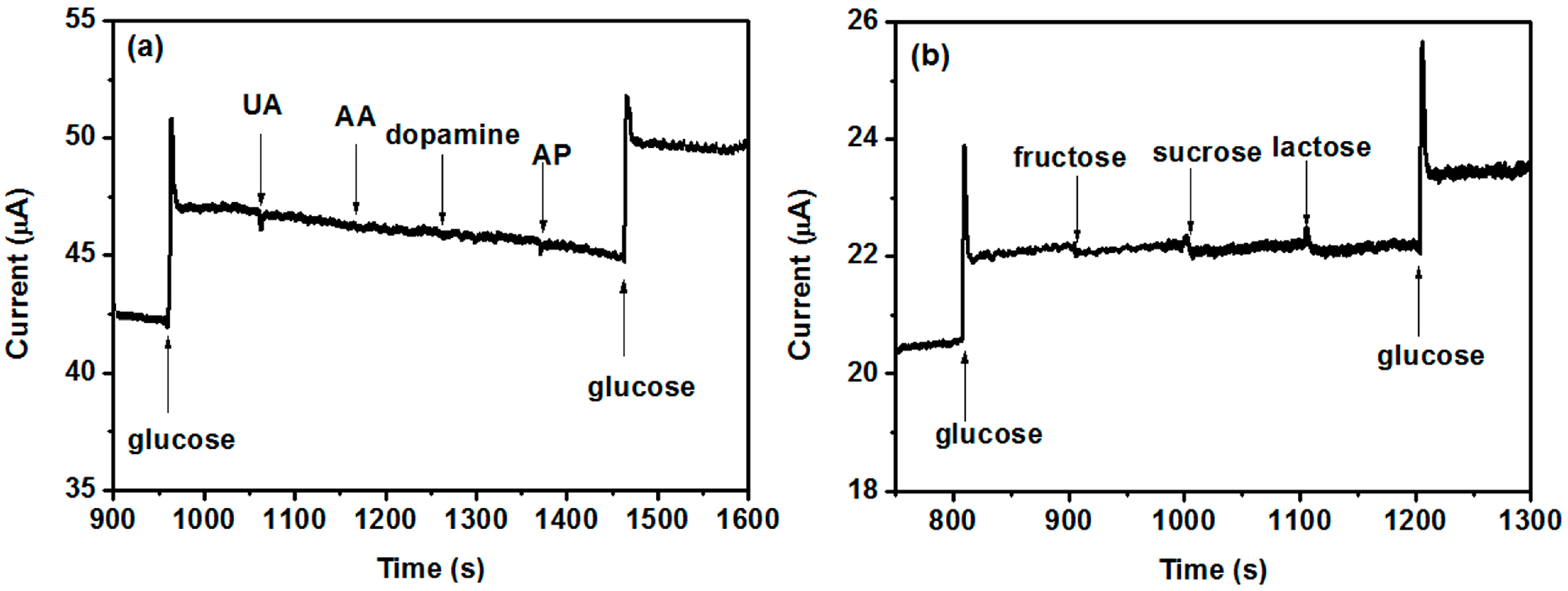
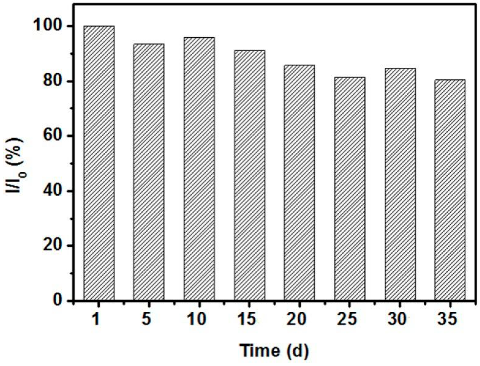
2. Results and Discussion

Transmission electron microscopy (TEM) images of CeO2 NRs and CeO2/Ni(OH)2 nanocomposites are shown in Figure 1. The length of CeO2 NRs is approximately 100 nm to 200 nm, whereas the diameters of NRs are approximately 10 nm to 20 nm (Figure 1a). Figure 1b clearly shows that CeO2 NRs are embedded in the Ni(OH)2 matrix. We also investigated the TEM image (Figure 1c) of the nanocomposite with a high Ce:Ni ratio (0.5:1), in which the sample exhibited similar structure to the nanocomposite with a small Ce:Ni ratio (0.05:1). The circles in the selected area electron diffraction (SAED) pattern are attributed to the diffraction planes of Ni(OH)2 (102), CeO2 (111), and CeO2 (220) (inset of Figure 1b), which indicates the polycrystalline structure of the nanocomposites. High-resolution TEM (HRTEM) image of CeO2/Ni(OH)2 nanocomposite is shown in Figure 1d. The lattice distances are in good agreement with the typical parameters of Ni(OH)2 (JCPDS 14-0117) and CeO2 (JCPDS 34-0394).
The X-ray diffraction pattern (XRD) of CeO2/Ni(OH)2 nanocomposite (Ce:Ni = 0.05:1) is shown in Figure 2. The diffraction peaks at 19.26°, 33.06°, 38.54°, 52.10°, 59.05°, 62.73°, 72.74°, 73.13° and 82.61° correspond to the (001), (100), (101), (102), (110), (111), (201), (112) and (202) planes of Ni(OH)2, respectively. The appearance of these peaks implies a hexagonal structure of Ni(OH)2 (JCPDC 14-0117). The peaks that appeared at 28.55°, 33.08°, 47.48°, 56.33°, 76.70°, 79.07° and 88.41° are related to the (111), (200), (220), (311), (331), (420) and (422) planes of CeO2 and indicate a cubic fluorite structure of CeO2 (JCPDS 34-0394).
The cyclic voltammogram (CV) obtained by continuously cycling the electrode potential between 0 and 0.8 V against Ag/AgCl reference electrode was used to investigate the electrocatalytic activities of the electrodes. CV scanning was repeated until a stationary state was obtained because nickel-based electrodes usually suffer from fouling under repeated cyclic sweeping in CV measurements, which can be attributed to the change in crystal structure [15]. Such a process is shown in Figure 3a, which demonstrates a pair of well-defined anodic and cathodic peaks corresponding to the redox reaction of Ni(II)/Ni(III) [1,12,27,28]. A stationary state was obtained after 40 scan cycles, which indicates that the crystal structures had stabilized. The relation between peak charges integrated from the CVs and scanning cycles is shown in Figure 3b. The charges gradually increased with an increase of cycle number, which indicates that electrocatalytic reactions mainly occurred on or near the surface of Ni(OH)2 [1,29].
Figure 4 shows the electrocatalytic activities of three different electrodes to glucose. Ni(OH)2/CPE (carbon paste electrode) exhibited a slight change in peak current after adding 1 mM glucose, in which the magnitude of such change actually increased with an increase of glucose concentration (not shown). By contrast, a clear non-enzymatic response to 1 mM glucose was observed from CeO2/Ni(OH)2/CPE. The enhanced response of CeO2/Ni(OH)2/CPE is a reflection of the synergetic effect obtained from the combination of CeO2 and Ni(OH)2. Pure CeO2 component had no electrocatalytic activity; however, it influenced the interfacial electron transfer process which was investigated by electrochemical impedance spectroscopy (Figure 5a). The Randles circuit (inset of Figure 5a), which contains electrolyte resistance (Rs), charge transfer resistance (Rct), double layer capacitance (Cdl), and Warburg element (W), was used to model these impedance plots. Based on the study of Patil et al. [24], lower Rct values were attributed to an accelerated electron transfer between the modified electrodes and electrolytes. Using ZSimpWin (v3.60) software, the Rct values that best fit the experimental impedance plots were approximately 11,078 and 178 Ω for Ni(OH)2/CPE and CeO2/Ni(OH)2/CPE, respectively, which indicates that CeO2/Ni(OH)2/CPE has a higher charge transfer ability than Ni(OH)2/CPE. Such an enhancement is related to the CeO2 NRs, which randomly contact each other in the Ni(OH)2 matrix (Figure 5b) and consequently form conductive paths for electron transfer.
The Ni(OH)2 component dominated the redox reaction of glucose by means of the Ni(II) /Ni(III) redox couple. The reaction in the blank NaOH solution can be expressed as Equation (1) [9]. After adding glucose, the Ni(III) ion could obtain an electron from the oxidation of glucose and deliver this electron to the electrode, thereby leading to an increase of peak current. The reaction is depicted in Equation (2).
Ni(OH)2 + OH → NiO(OH) + e + H2O
NiO(OH) + glucose →Ni(OH)2 + glucolactone
Figure 6 is the effect of the molar ratio of cerium to nickel on the sensing response to glucose. The results show that an optimum response can be obtained when the molar ratio is equal to 0.05:1. The influence of the scan rate on the current response is shown in Figure 7a. The magnitude of the peak current increased with the increasing scan rate. The peak current was quite linear to the square root of the scan rate (Figure 7b). Such linearity further confirmed the diffusion-controlled kinetics at the electrode surface.
The amperometric response of CeO2/Ni(OH)2/CPE was further investigated. Finding a suitably applied potential that can obtain maximum catalytic current is necessary because the applied potential intensely influences the amperometric response [4]. Hydrodynamic voltammetry measurement to 1 mM glucose was conducted in 0.1 M NaOH solution to find an optimum applied potential. As shown in Figure 8, CeO2/Ni(OH)2/CPE demonstrated the highest response current at 0.55 V. The current density drastically declined over this potential. Based on Dung et al. [15], such decrease is possibly attributed to the oxidation of water interfering with the oxidation of glucose or the products of glucose oxidation poisoning the electrode. Therefore, the amperometric response was investigated at 0.55 V based on the results of hydrodynamic voltammetry.
Figure 9 shows the amperometric response of the CeO2/Ni(OH)2/CPE with the successive addition of glucose in 0.1 M NaOH solution at 0.55 V. The current increased with the glucose addition because of the good catalytic properties of the CeO2/Ni(OH)2/CPE. The response time which means that the current reaches 95% of the steady state value was less than 5 s. A calibration curve was constructed from this amperometric response curve (inset of Figure 9). CeO2/Ni(OH)2/CPE exhibited a good linearity with glucose concentration, ranging from 2 μM to 6.62 mM with a correlation coefficient of 0.998. A high sensitivity of 594 μA mM−1 cm−2 was obtained from the slope of the calibration curve. Moreover, the limit of detection was 1.13 μM at a signal to noise ratio of 3. A comparison among several typical glucose biosensors reported in the last five years is shown in Table 1. The CeO2/Ni(OH)2/CPE exhibits superiority, especially with regard to sensitivity, linear range, and detection limit. This superiority is attributed to the synergetic effect by combining CeO2 NRs with Ni(OH)2, which dramatically increases the charge transfer and electrocatalytic activity of nanocomposite.
Selectivity is an essential feature of glucose biosensors for practical applications, especially for physiological test. Generally, the concentrations of interfering species such as ascorbic acid (AA), uric acid (UA), 4-acetamidophenol (AP), fructose, and lactose are no more than 0.1 mM in the physiological fluid, whereas the normal level of glucose is approximately 3–8 mM [1,6]. Therefore, the amperometric experiment regarding the selectivity of the sensor was conducted with 5 mM of glucose and 0.1 mM of various interfering species. Figure 10 shows that all these interfering species had negligible responses compared with glucose, which indicates that the CeO2/Ni(OH)2/CPE fabricated in this work has sufficient selectivity for practical application. The long-term stability of CeO2/Ni(OH)2/CPE was investigated by determining its current response (I) to 1 mM glucose every five days under ambient conditions for seven weeks, during which the sensor preserved 80.36% of its original value (I0), thereby reflecting a good stability of biosensor (Figure 11).
A kind of juice on sale was chosen as the test-target to investigate the practical application of CeO2/Ni(OH)2/CPE because one potential application of glucose biosensor is food inspection. Similar to the process in the reference [4,34,35], diluted juice samples with various standard additions of glucose were added into 0.1 M NaOH, and the corresponding amperometric responses were recorded at 0.55 V. Table 2 shows the obtained results. The biggest relative standard deviation is 3.0%, which illustrates the good reproducibility of the method [36]. The recoveries ranging from 95.2% to 100% indicate that the sensor is sufficient for analyzing glucose in food inspection application [34].

3. Materials and Methods

Nickel nitrate hexahydrate, cerium nitrate hexahydrate, glucose, AA, UA, AP, dopamine hydrochloride, fructose, sucrose, and lactose were purchased from Sigma–Aldrich (St. Louis, MO, USA) and used without further purification. Graphite, paraffin oil, sodium hydroxide, and ammonium hydroxide were purchased from Aladdin Chemicals (Shanghai, China).
CeO2 NRs were synthesized through a non-isothermal precipitation based on the study of Patil et al. [24]. In detail, ammonia was added (0.5 mL/min) into 100 mL 0.20 M cerium nitrate aqueous solution at 70 °C, in which a yellow precipitate was immediately formed. After 5 min, the reaction mixture was transferred into a 0 °C water bath in which the reaction continued for 24 h. Finally, the precipitate was filtered and washed with ethanol and de-ionized water and then dried at 60 °C in a vacuum. The precipitate was performed by calcination at 350 °C for 2 h under air resulting in the final CeO2 NRs.
CeO2/Ni(OH)2 nanocomposites were prepared via the wet impregnation method. Briefly, CeO2 NRs were dispersed in 50 mL nickel nitrate solution by magnetic stirring at 70 °C for 1 h. Thereafter, 5 mL ammonia was added (0.5 mL/min) to this solution, and a turquoise-colored precipitate was formed. Finally, this precipitate was filtered, washed three times with de-ionized water, and then dried at 60 °C in a vacuum. The amount of nickel nitrate was set as constant (10 mmol), whereas the molar ratio of cerium to nickel (Ce:Ni) was varied as 0.05:1, 0.1:1, and 0.5:1 for comparison purposes.
XRD analysis was performed with a Y2000 diffractometer (HAOYUAN, Dandong, China) with Cu Kα radiation (λ = 1.5418 Å). TEM was conducted with a JEM2010 instrument (JEOL, Tokyo, Japan).
The electrochemical measurements were conducted on a model CHI630D electrochemical analyzer (CHENHUA, Shanghai, China). All of the experiments were performed in 0.1 M NaOH solution at room temperature with a three-electrode electrochemical cell by using modified CPE as working electrodes, a platinum wire as a counter electrode, and an Ag/AgCl electrode as a reference electrode.
The modified CPE working electrodes were prepared according to reference [4]. In detail, the synthesized materials, graphite powder, and paraffin oil with a mass ratio of 2:5:1 were mixed by hand in a mortar until a uniformly wetted paste was obtained. Then, this paste was filled firmly into a Teflon tube with an inner diameter of 3 mm. The electrical contact was established by pushing a copper wire down the tube into the back of paste.

4. Conclusions

A non-enzymatic glucose biosensor based on CeO2/Ni(OH)2 nanocomposite modified CPE was successfully developed. The enhanced sensing performance of nanocomposite was related to the synergetic effects of the two components. Specifically, Ni(OH)2 dominated the redox reaction of glucose by using the Ni(II)/Ni(III) redox couple, whereas the entangled CeO2 NRs formed conductive paths to accelerate the electron transfer to the electrode. The nanocomposite biosensor had a high sensitivity of 594 μA mM−1 cm−2, a linear range of 0.002–6.62 mM, a fast response time of less than 5 s, and an estimated detection limit of 1.13 μM (S/N = 3). The biosensor also exhibited good selectivity and stability. In addition, the results of this work clearly demonstrate that pristine CeO2 can be a good candidate for enhancing the performance of conventional non-enzymatic sensing materials, although it has no electrocatalytic activity and is not suitable as a non-enzymatic sensing material (e.g., CuO, NiO, Ni(OH)2, Cu(OH)2). Apart from exploring other compositions (e.g., CeO2/Cu(OH)2, CeO2/Co3O4) for non-enzymatic biosensor, future work should focus on optimizing the structure of these nanocomposites (for example, CeO2/Ni(OH)2 coaxial tubular nanostructure or CeO2/Ni(OH)2 hierarchical nanostructure).

Acknowledgments

This work was supported by the National Natural Science Foundation of China (No. 61501318), and the Opened Fund of the State Key Laboratory on Integrated Optoelectronics (No. IOSKL2014KF01).

Author Contributions

Yongjian Li, Wei Li, and Xiaoling Xie conceived and designed the experiments; Yongjian Li, and Panpan Guan performed the experiments; Yongjian Li and Wei Li analyzed the data; Wei Li and Fucheng Yu contributed reagents/materials/analysis tools; Yongjian Li wrote the paper.

Conflicts of Interest

The authors declare no conflict of interest.

References

  1. Gao, A.; Zhang, X.; Peng, X.; Wu, H.; Bai, L.; Jin, W.; Wu, G.; Hang, R.; Chu, P.K. In situ synthesis of Ni(OH)2/TiO2 composite film on NiTi alloy for non-enzymatic glucose sensing. Sens. Actuators B Chem. 2016, 232, 150–157. [Google Scholar] [CrossRef]
  2. Rinaldi, A.L.; Carballo, R. Impedimetric non-enzymatic glucose sensor based on nickel hydroxide thin film onto gold electrode. Sens. Actuators B Chem. 2016, 228, 43–52. [Google Scholar] [CrossRef]
  3. Jiang, D.; Liu, Q.; Wang, K.; Qian, J.; Dong, X.; Yang, Z.; Du, X.; Qiu, B. Enhanced non-enzymatic glucose sensing based on copper nanoparticles decorated nitrogen-doped graphene. Biosens. Bioelectron. 2014, 54, 273–278. [Google Scholar] [CrossRef] [PubMed]
  4. Mu, Y.; Jia, D.; He, Y.; Miao, Y.; Wu, H.L. Nano nickel oxide modified non-enzymatic glucose sensors with enhanced sensitivity through an electrochemical process strategy at high potential. Biosens. Bioelectron. 2011, 26, 2948–2952. [Google Scholar] [CrossRef] [PubMed]
  5. Luo, L.; Li, F.; Zhu, L.; Ding, Y.; Zhang, Z.; Deng, D.; Lu, B. Nonenzymatic glucose sensor based on nickel(II)oxide/ordered mesoporous carbon modified glassy carbon electrode. Colloid Surf. B Biointerfaces 2013, 102, 307–311. [Google Scholar] [CrossRef] [PubMed]
  6. Guo, C.; Wang, Y.; Zhao, Y.; Xu, C. Non-enzymatic glucose sensor based on three dimensional nickel oxide for enhanced sensitivity. Anal. Methods 2013, 5, 1644–1647. [Google Scholar] [CrossRef]
  7. Toghill, K.E.; Xiao, L.; Phillips, M.A.; Compton, R.G. The non-enzymatic determination of glucose using an electrolytically fabricated nickel microparticle modified boron-doped diamond electrode or nickel foil electrode. Sens. Actuators B Chem. 2010, 147, 642–652. [Google Scholar] [CrossRef]
  8. Tee, S.Y.; Ye, E.; Pan, P.H.; Lee, C.J.J.; Hui, H.K.; Zhang, S.Y.; Koh, L.D.; Dong, Z.; Han, M.Y. Fabrication of bimetallic Cu/Au nanotubes and their sensitive, selective, reproducible and reusable electrochemical sensing of glucose. Nanoscale 2015, 7, 11190–11198. [Google Scholar] [CrossRef] [PubMed]
  9. Gao, Z.D.; Guo, J.; Shrestha, N.K.; Hahn, R.; Song, Y.Y.; Schmuki, P. Nickel hydroxide nanoparticle activated semi-metallic TiO2 nanotube arrays for non-enzymatic glucose sensing. Chem. Eur. J. 2013, 19, 15530–15534. [Google Scholar] [CrossRef] [PubMed]
  10. Zhuang, Z.; Su, X.; Yuan, H.; Sun, Q.; Xiao, D.; Choi, M.M.F. An improved sensitivity non-enzymatic glucose sensor based on a CuO nanowire modified Cu electrode. Analyst 2008, 133, 126–132. [Google Scholar] [CrossRef] [PubMed]
  11. Shearer, C.J.; Cherevan, A.; Eder, D. Application and future challenges of functional nanocarbon hybrids. Adv. Mater. 2014, 26, 2295–2318. [Google Scholar] [CrossRef] [PubMed]
  12. Chen, J.; Zheng, J. A highly sensitive non-enzymatic glucose sensor based on tremella-like Ni(OH)2 and Au nanohybrid films. J. Electroanal. Chem. 2015, 749, 83–88. [Google Scholar] [CrossRef]
  13. Ding, Y.; Liu, Y.; Parisi, J.; Zhang, L.; Lei, Y. A novel NiO-Au hybrid nanobelts based sensor for sensitive and selective glucose detection. Biosens. Bioelectron. 2011, 28, 393–398. [Google Scholar] [CrossRef] [PubMed]
  14. Dung, N.Q.; Patil, D.; Jung, H.; Kim, D. A high-performance nonenzymatic glucose sensor made of CuO-SWCNT nanocomposites. Biosens. Bioelectron. 2013, 42, 280–286. [Google Scholar] [CrossRef] [PubMed]
  15. Dung, N.Q.; Patil, D.; Jung, H.; Kim, J.; Kim, D. NiO-decorated single-walled carbon nanotubes for high-performance nonenzymatic glucose sensing. Sens. Actuators B Chem. 2013, 183, 381–387. [Google Scholar] [CrossRef]
  16. Yang, J.; Jiang, L.C.; Zhang, W.D.; Guansekaran, S. A highly sensitive non-enzymatic glucose sensor based on a simple two-step electrodeposition of cupric oxide (CuO) nanoparticles onto multi-walled carbon nanotube arrays. Talanta 2010, 82, 25–33. [Google Scholar] [CrossRef] [PubMed]
  17. Yang, M.; Jeong, J.M.; Lee, K.G.; Kim, D.H.; Lee, S.J.; Choi, B.G. Hierarchical porous microspheres of the Co3O4@graphene with enhanced electrocatalytic performance for electrochemical biosensors. Biosens. Bioelectron. 2017, 89, 612–619. [Google Scholar] [CrossRef] [PubMed]
  18. Yang, Y.; Wang, Y.; Bao, X.; Li, H. Electrochemical deposition of Ni nanoparticles decorated ZnO hexagonal prisms as an effective platform for non-enzymatic detection of glucose. J. Electroanal. Chem. 2016, 775, 163–170. [Google Scholar] [CrossRef]
  19. SoYoon, S.; Ramadoss, A.; Saravanakumar, B.; Kim, S.J. Novel Cu/CuO/ZnO hybrid hierarchical nanostructures for non-enzymatic glucose sensor application. J. Electroanal. Chem. 2014, 717, 90–95. [Google Scholar] [CrossRef]
  20. Cai, B.; Zhou, Y.; Zhao, M.; Cai, H.; Ye, Z.; Wang, L.; Huang, J. Synthesis of ZnO-CuO porous core-shell spheres and their application for non-enzymatic glucose sensor. Appl. Phys. A 2015, 118, 989–996. [Google Scholar] [CrossRef]
  21. Karuppiah, C.; Velmurugan, M.; Chen, S.M.; Tsai, S.H.; Lou, B.S.; Ali, M.A.; Al-Hemaid, F.M.A. A simple hydrothermal synthesis and fabrication of zinc oxide-copper oxide heterostructure for the sensitive determination of nonenzymatic glucose biosensor. Sens. Actuators B Chem. 2015, 221, 1299–1306. [Google Scholar] [CrossRef]
  22. Strano, V.; Mirabella, S. Hierarchical ZnO nanorods/Ni(OH)2 nanoflakes for room-temperature, cheap fabrication of nonenzymatic glucose sensors. RSC Adv. 2016, 6, 111374–111379. [Google Scholar] [CrossRef]
  23. Zhou, C.; Xu, L.; Song, J.; Xing, R.; Xu, S.; Liu, D.; Song, H. Ultrasensitive non-enzymatic glucose sensor based on three-dimensional network of ZnO-CuO hierarchical nanocomposites by electrospinning. Sci. Rep. 2014, 4, 7382. [Google Scholar] [CrossRef] [PubMed]
  24. Patil, D.; Dung, N.Q.; Jung, H.; Ahn, S.Y.; Jang, D.M.; Kim, D. Enzymatic glucose biosensor based on CeO2 nanorods synthesized by non-isothermal precipitation. Biosens. Bioelectron. 2012, 31, 176–181. [Google Scholar] [CrossRef] [PubMed]
  25. Rahman, M.M.; Ahammad, A.J.S.; Jin, J.H.; Ahn, S.J.; Lee, J.J. A comprehensive review of glucose biosensors based on nanostructured metal-oxides. Sensors 2010, 10, 4855–4886. [Google Scholar] [CrossRef] [PubMed]
  26. Guan, P.; Li, Y.; Zhang, J.; Li, W. Non-enzymatic glucose biosensor based on CuO-decorated CeO2 nanoparticles. Nanomaterials 2016, 6, 159. [Google Scholar] [CrossRef] [PubMed]
  27. Miao, Y.; Ouyang, L.; Zhou, S.; Xu, L.; Yang, Z.; Xiao, M.; Ouyang, R. Electrocatalysis and electroanalysis of nickel, its oxides, hydroxides and oxyhydroxides toward small molecules. Biosens. Bioelectron. 2014, 53, 428–439. [Google Scholar] [CrossRef] [PubMed]
  28. Tee, S.Y.; Teng, C.P.; Ye, E. Metal nanostructures for non-enzymatic glucose sensing. Mater. Sci. Eng. C 2017, 70, 1018–1030. [Google Scholar] [CrossRef] [PubMed]
  29. Iwu, K.O.; Lombardo, A.; Sanz, R.; Scirè, S.; Mirabella, S. Facile synthesis of Ni nanofoam for flexible and low-cost non-enzymatic glucose sensing. Sens. Actuators B Chem. 2016, 224, 764–771. [Google Scholar] [CrossRef]
  30. Qu, F.; Sun, H.; Zhang, S.; You, J.; Yang, M. Electrochemical sensing platform based on palladium modified ceria nanoparticles. Electrochim. Acta 2012, 61, 173–178. [Google Scholar] [CrossRef]
  31. Jiao, X.; Song, H.; Zhao, H.; Bai, W.; Zhang, L.; Lv, Y. Well-redispersed ceria nanoparticles: Promising peroxidase mimetics for H2O2 and glucose detection. Anal. Methods 2012, 4, 3261–3267. [Google Scholar] [CrossRef]
  32. Miao, F.; Tao, B.; Chu, P.K. Ordered-standing nickel hydroxide microchannel arrays: Synthesis and application for highly sensitive non-enzymatic glucose sensors. Microelectron. Eng. 2015, 133, 11–15. [Google Scholar] [CrossRef]
  33. Lu, P.; Liu, Q.; Xiong, Y.; Wang, Q.; Lei, Y.; Lu, S.; Lu, L.; Yao, L. Nanosheets-assembled hierarchical microstructured Ni(OH)2 hollow spheres for highly sensitive enzyme-free glucose sensors. Electrochim. Acta 2015, 168, 148–156. [Google Scholar] [CrossRef]
  34. Yang, S.; Li, G.; Wang, G.; Zhao, J.; Gao, X.; Qu, L. Synthesis of Mn3O4 nanoparticles/nitrogen-doped graphene hybrid composite for nonenzymatic glucose sensor. Sens. Actuators B Chem. 2015, 221, 172–178. [Google Scholar] [CrossRef]
  35. Tan, C.; Zhang, W.; Zheng, J.; You, X.; Lin, X.; Li, S. Fabrication of metal-organic single crystalline nanowires and reduced graphene oxide enhancement for an ultrasensitive electrochemical biosensor. J. Mater. Chem. B 2015, 3, 7117–7124. [Google Scholar] [CrossRef]
  36. Mu, J.; He, Y.; Wang, Y. Copper-incorporated SBA-15 with peroxidase-like activity and its application for colorimetric detection of glucose in human serum. Talanta 2016, 148, 22–28. [Google Scholar] [CrossRef] [PubMed]
Figure 1. Transmission electron microscopy (TEM) images of (a) CeO2; (b) CeO2/Ni(OH)2 nanocomposite (Ce:Ni = 0.05:1); and (c) CeO2/Ni(OH)2 nanocomposite (Ce:Ni = 0.5:1); (d) High-resolution TEM (HRTEM) image of CeO2/Ni(OH)2 nanocomposite (Ce:Ni = 0.05:1). Inset is the SAED pattern.
Nanomaterials 07 00205 g001
Figure 2. XRD patterns of CeO2/Ni(OH)2 nanocomposite (Ce:Ni = 0.05:1).
Nanomaterials 07 00205 g002
Figure 3. (a) CVs of CeO2/Ni(OH)2/CPE after 10, 20, 30, 40, and 50 consecutive cycles in 0.1 M NaOH solution at 50 mV/s scan rate; (b) Plots of charges integrated from anodic and cathodic peaks in CVs versus number of cycles.
Nanomaterials 07 00205 g003
Figure 4. CVs of CeO2/CPE, Ni(OH)2/CPE, and CeO2/Ni(OH)2/CPE in the absence and presence of 1 mM glucose in 0.1 M NaOH solution at 50 mV/s scan rate.
Nanomaterials 07 00205 g004
Figure 5. (a) Nyquist impedance plots for Ni(OH)2/CPE and CeO2/Ni(OH)2/CPE recorded in 0.1 M KCl solution containing 1 mM Fe[(CN)6]3−/4− (1:1); (b) Schematic diagram of sensing mechanism of CeO2/Ni(OH)2 nanocomposite.
Nanomaterials 07 00205 g005
Figure 6. CVs of CeO2/Ni(OH)2/CPE with different Ce:Ni molar ratios in the absence and presence of 1 mM glucose in 0.1 M NaOH solution at 50 mV/s scan rate.
Nanomaterials 07 00205 g006
Figure 7. (a) CVs of CeO2/Ni(OH)2/CPE in 0.1 M NaOH solution containing 1 mM glucose at different scan rate; (b) Plot of the peak current with square root of scan rate.
Nanomaterials 07 00205 g007
Figure 8. The response current of CeO2/Ni(OH)2/CPE measured in 0.1 M NaOH for 1 mM glucose at different potentials.
Nanomaterials 07 00205 g008
Figure 9. Amperometric response of CeO2/Ni(OH)2/CPE measured at 0.55 V in 0.1 M NaOH solution with successive addition of glucose. Inset is the plot of current response with glucose concentration.
Nanomaterials 07 00205 g009
Figure 10. (a) Interference test of CeO2/Ni(OH)2/CPE upon continuous adding 5 mM glucose, 0.1 mM UA, 0.1 mM AA, 0.1 mM dopamine, 0.1 mM AP, and 5 mM glucose at 0.55 V in 0.1 M NaOH solution; (b) Interference test of CeO2/Ni(OH)2/CPE upon continuous adding 5 mM glucose, 0.1 mM fructose, 0.1 mM sucrose, 0.1 mM lactose, and 5 mM glucose at 0.55 V in 0.1 M NaOH solution.
Nanomaterials 07 00205 g010
Figure 11. Stability of CeO2/Ni(OH)2/CPE electrode at ambient conditions for seven weeks using 1 mM glucose in 0.1 M NaOH solution at 0.55 V.
Nanomaterials 07 00205 g011
Table 1. Comparison of performance obtained from different glucose biosensors.
ElectrodeSensitivity (μA mM−1 cm−2)Linearity (mM)LOD c (μM)Potential (V)References
CeO2 NRs a0.1652~261000.80[24]
CeO2/Pd a-0.1~1010−0.20[30]
CeO2 NPs a-0.007~0.130.003-[31]
Ni(OH)2/SiMCP b2500~83.50.50[32]
Ni(OH)2-HS b223.390.0009~7.7810.10.45[33]
Ni(OH)2/TiO2 b1920.03~1480.50[1]
Au/Ni(OH)2 b-~2-0.50[2]
Ni(OH)2/TiOxCy b2400.02~1.75.00.70[9]
Ni(OH)2/Au b371.20.005~2.20.920.55[12]
CeO2/Ni(OH)2 b5940.002~6.61.130.55This work
a enzymatic biosensor; b non-enzymatic biosensor; c limit of detection (LOD).
Table 2. Determination of glucose in a juice on sale.
Spiked (μM)Found (μM)Recovery (%)RSD (%)
040.3--
2564.195.22.9
5090.31002.6
75112.796.51.9
100135.395.03.0

