
Monocyte (THP-1) Response to Silver Nanoparticles Synthesized with Rumex hymenosepalus Root Extract
 , ,
, ,  , , ,
, , ,
Abstract
1. Introduction
2. Materials and Methods
2.1. Obtaining Rumex hymenosepalus Extract
2.2. Rumex hymenosepalus Sample Preparation for Mass Spectroscopy
2.3. Rumex hymenosepalus Metabolites Global Profile by UPLC-MS-QTOF-SYNAPT
2.4. Silver Nanoparticle Synthesis and Their Characterization
2.5. UV-Vis Spectroscopy
2.6. FTIR
2.7. TGA
2.8. Transmission Electron Microscopy
2.9. Cells Assays
2.10. Statistical Analysis
3. Results and Discussion
3.1. Synthesis and Characterization
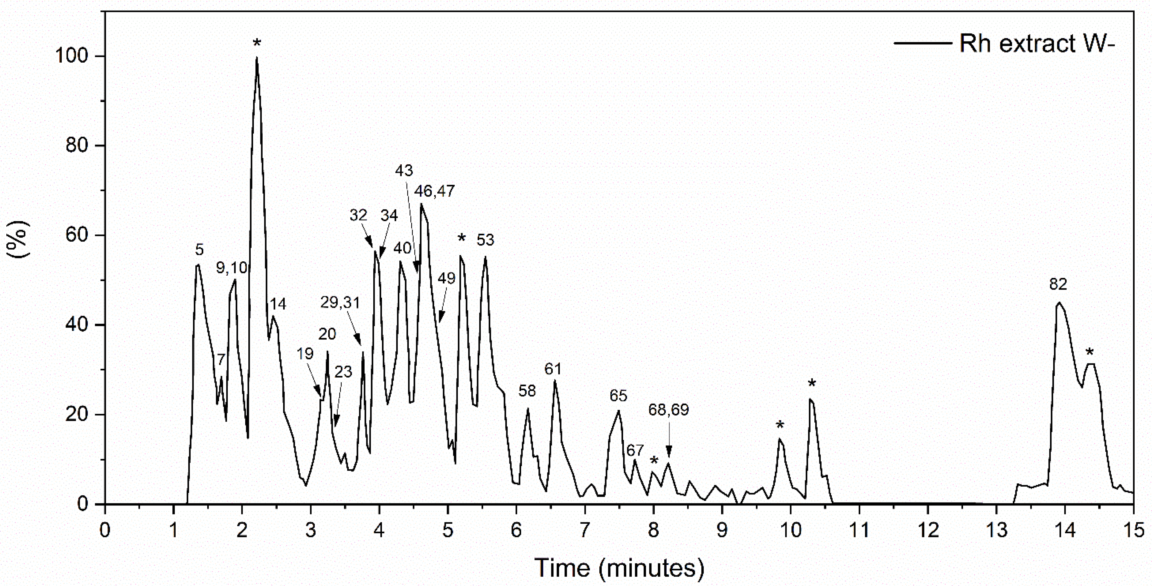
3.1.1. The Global Profile of Metabolites of Hydroethanolic Rh Extract
3.1.2. FTIR
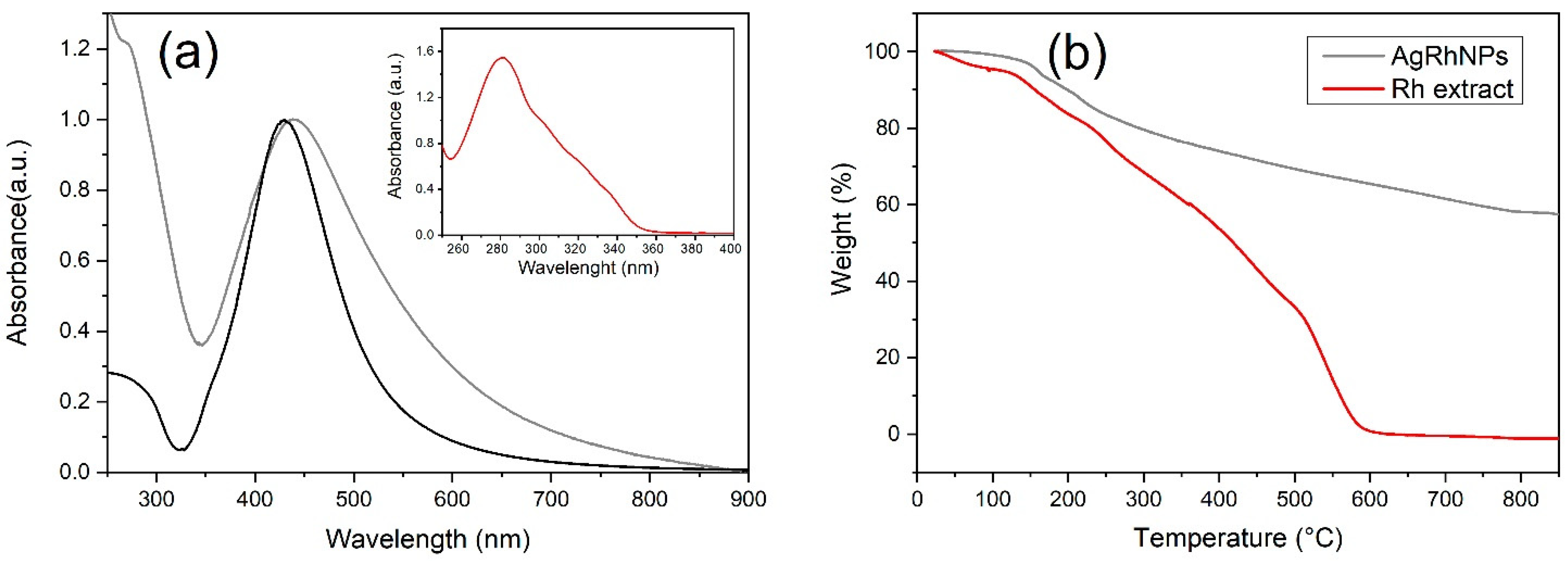
3.1.3. UV-Visible
3.1.4. UV-Vis Spectroscopy and Thermogravimetric Analysis
3.1.5. Transmission Electronic Microscopy
3.2. Cell Assays
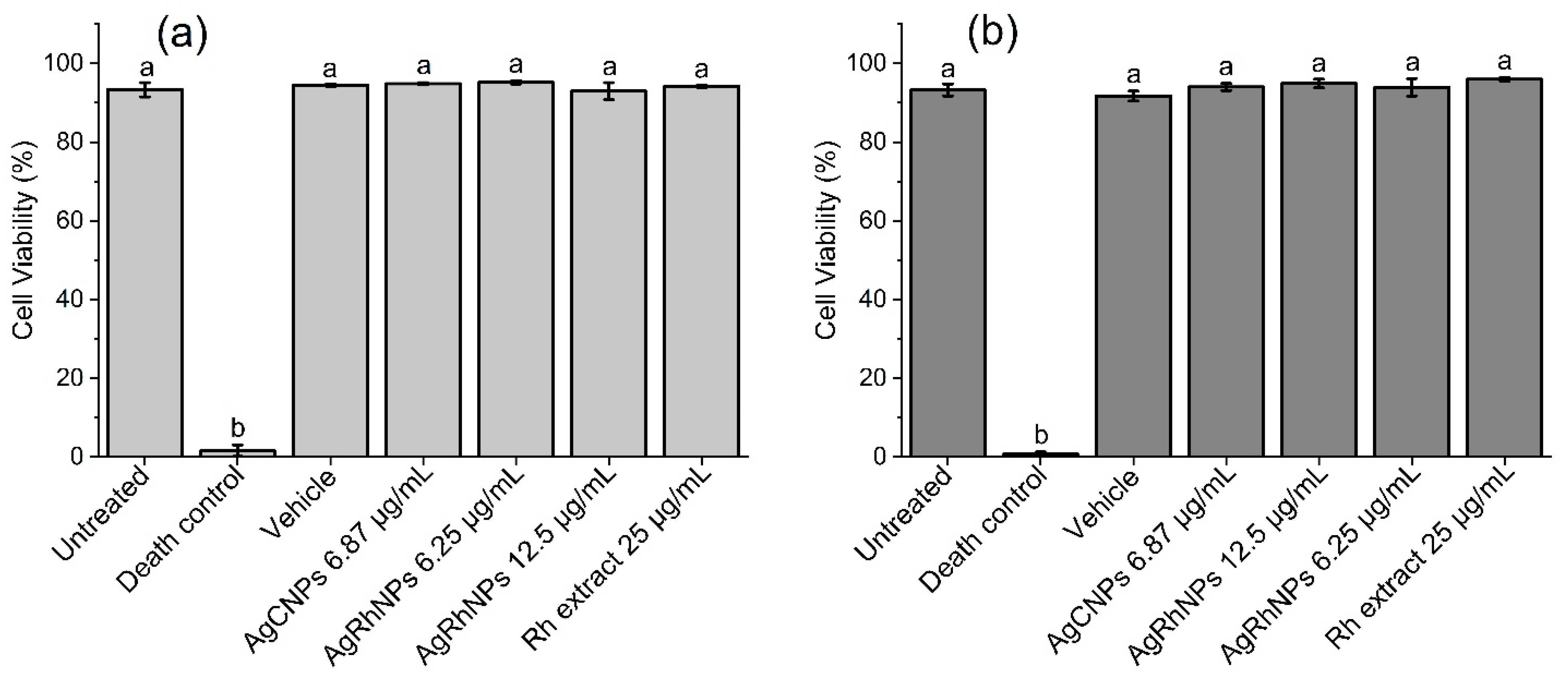
3.2.1. Viability
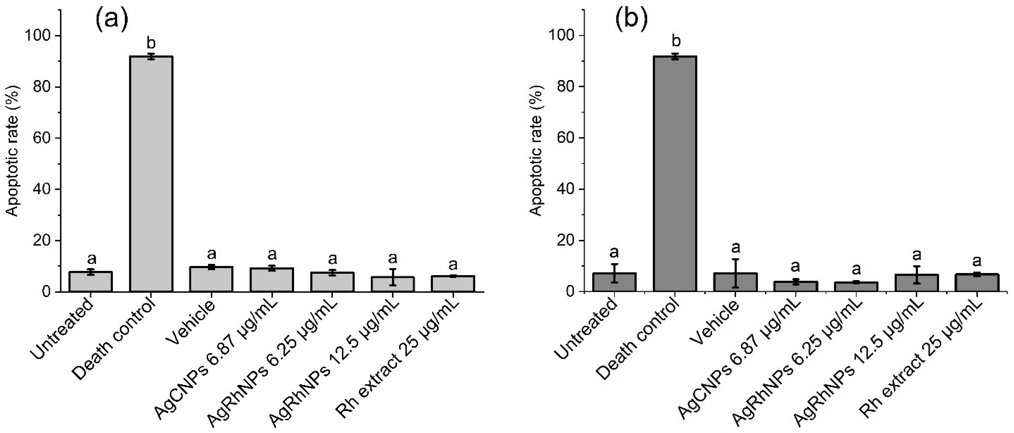
3.2.2. Annexin V-FITC Apoptosis Assay
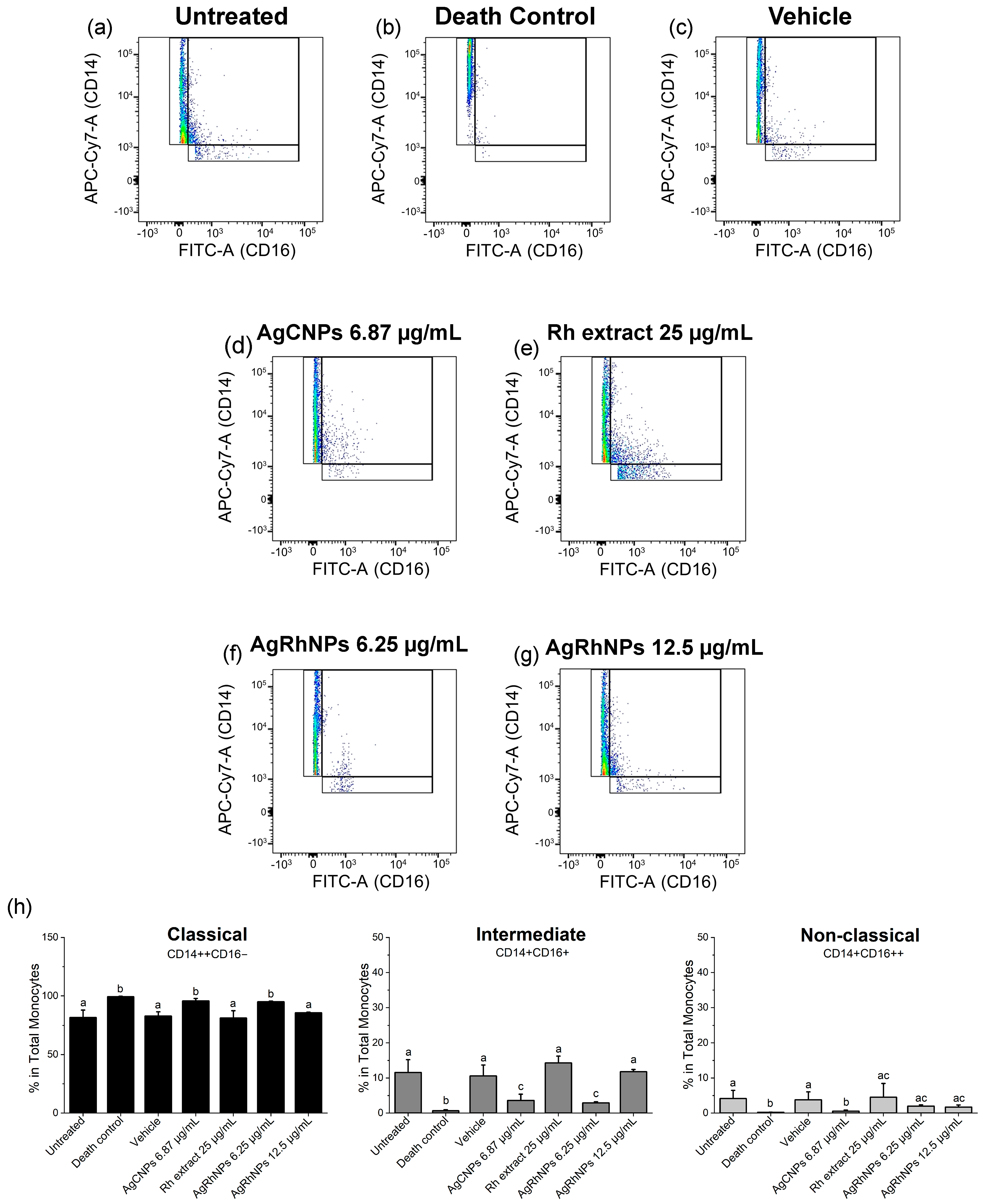
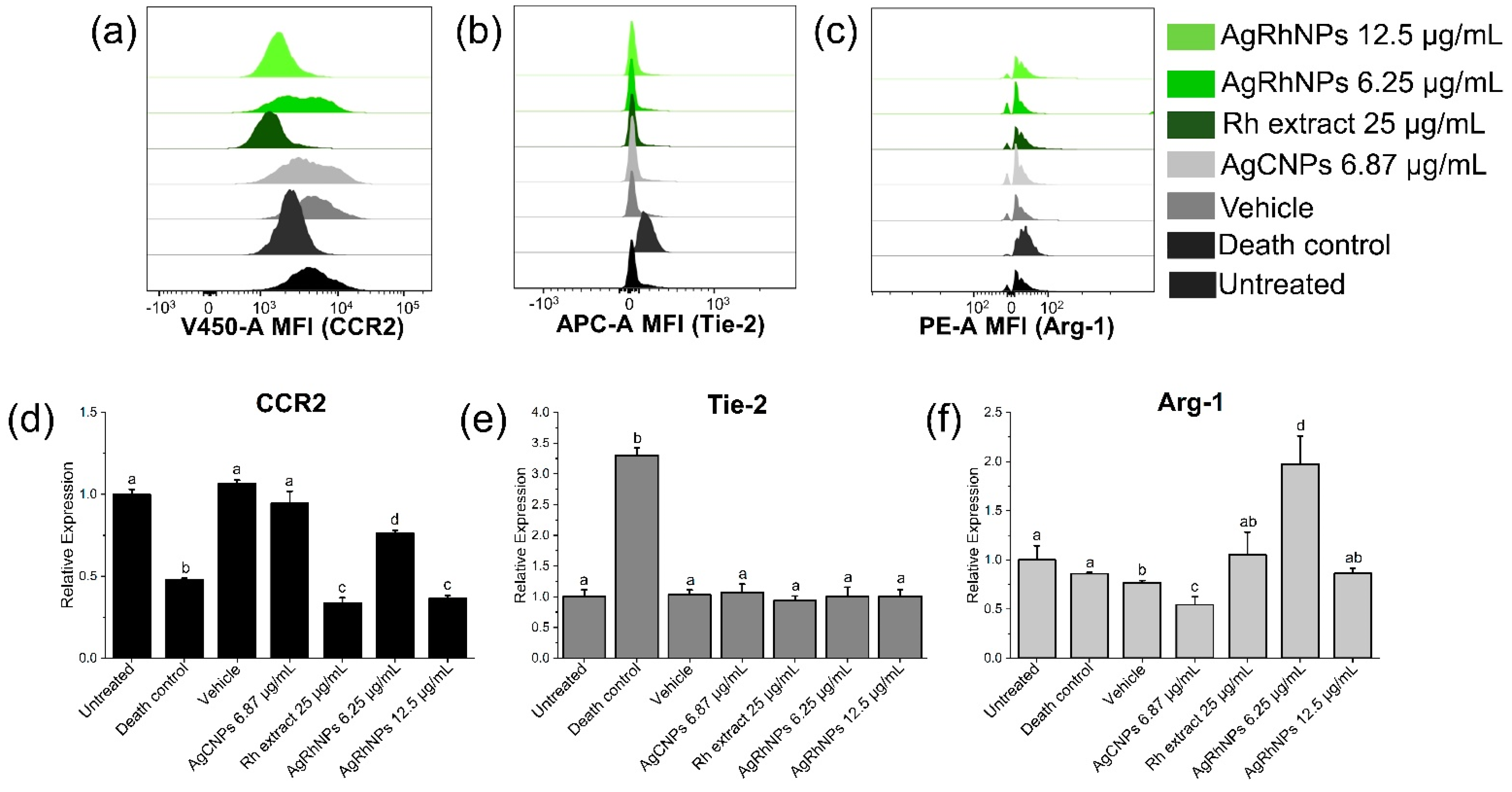
3.2.3. Monocytes Subsets
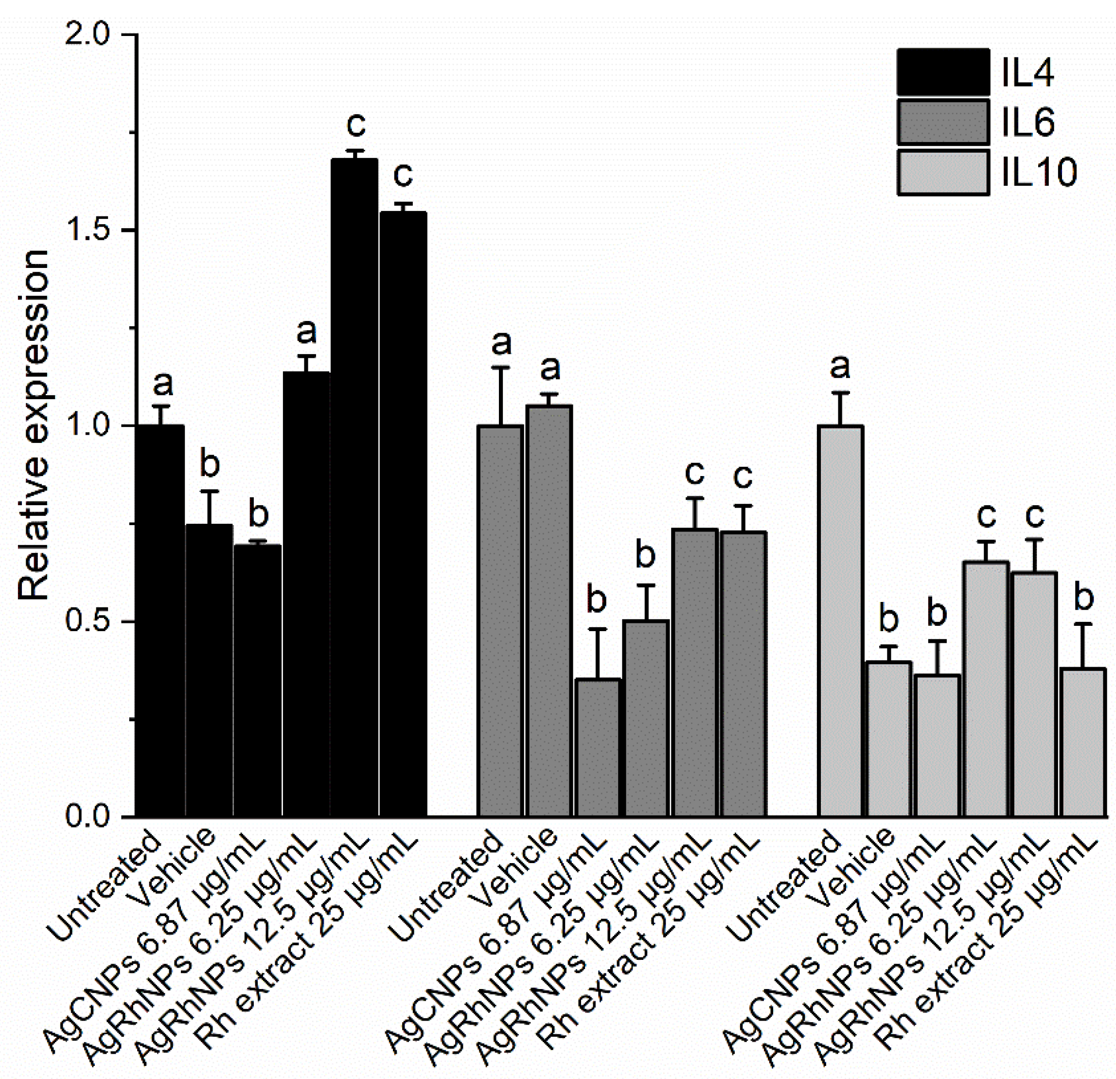
3.2.4. Interleukins Production of THP1 Cells
4. Conclusions
Supplementary Materials
Author Contributions
Funding
Data Availability Statement
Acknowledgments
Conflicts of Interest
References
- Mulvaney, P. Nanoscience vs Nanotechnology—Defining the Field. ACS Nano 2015, 9, 2215–2217. [Google Scholar] [CrossRef] [PubMed]
- Hulla, J.E.; Sahu, S.C.; Hayes, A.W. Nanotechnology: History and Future. Hum. Exp. Toxicol. 2015, 34, 1318–1321. [Google Scholar] [CrossRef] [PubMed]
- Kumar, A.; Shah, S.R.; Jayeoye, T.J.; Kumar, A.; Parihar, A.; Prajapati, B.; Singh, S.; Kapoor, D.U. Biogenic Metallic Nanoparticles: Biomedical, Analytical, Food Preservation, and Applications in Other Consumable Products. Front. Nanotechnol. 2023, 5, 1175149. [Google Scholar] [CrossRef]
- Khan, A.; Roy, A.; Bhasin, S.; Emran, T.B.; Khusro, A.; Eftekhari, A.; Moradi, O.; Rokni, H.; Karimi, F. Nanomaterials: An Alternative Source for Biodegradation of Toxic Dyes. Food Chem. Toxicol. 2022, 164, 112996. [Google Scholar] [CrossRef] [PubMed]
- Na, J.; Zheng, D.; Kim, J.; Gao, M.; Azhar, A.; Lin, J.; Yamauchi, Y. Material Nanoarchitectonics of Functional Polymers and Inorganic Nanomaterials for Smart Supercapacitors. Small 2022, 18, e2102397. [Google Scholar] [CrossRef] [PubMed]
- Babu, P.J.; Tingirikari, J.M.R. A Review on Polymeric Nanomaterials Intervention in Food Industry. Polym. Bull. 2023, 80, 137–164. [Google Scholar] [CrossRef]
- Yang, X.; Yang, M.; Pang, B.; Vara, M.; Xia, Y. Gold Nanomaterials at Work in Biomedicine. Chem. Rev. 2015, 115, 10410–10488. [Google Scholar] [CrossRef] [PubMed]
- Nguyen, N.H.A.; Falagan-Lotsch, P. Mechanistic Insights into the Biological Effects of Engineered Nanomaterials: A Focus on Gold Nanoparticles. Int. J. Mol. Sci. 2023, 24, 4109. [Google Scholar] [CrossRef]
- Metal Nanoparticles Global Market Trends, Growth Analysis, Outlook 2032. Available online: https://www.thebusinessresearchcompany.com/report/metal-nanoparticles-global-market-report (accessed on 20 November 2023).
- Arroyo, G.V.; Madrid, A.T.; Gavilanes, A.F.; Naranjo, B.; Debut, A.; Arias, M.T.; Angulo, Y. Green Synthesis of Silver Nanoparticles for Application in Cosmetics. J. Environ. Sci. Health A Tox. Hazard. Subst. Environ. Eng. 2020, 55, 1304–1320. [Google Scholar] [CrossRef]
- Vinod, T.P.; Jelinek, R. Inorganic Nanoparticles in Cosmetics. In Nanocosmetics; Springer International Publishing: Cham, Switzerland, 2019; pp. 29–46. ISBN 9783030165727. [Google Scholar]
- Szczepańska, E.; Bielicka-Giełdoń, A.; Niska, K.; Strankowska, J.; Żebrowska, J.; Inkielewicz-Stępniak, I.; Łubkowska, B.; Swebocki, T.; Skowron, P.; Grobelna, B. Synthesis of Silver Nanoparticles in Context of Their Cytotoxicity, Antibacterial Activities, Skin Penetration and Application in Skincare Products. Supramol. Chem. 2020, 32, 207–221. [Google Scholar] [CrossRef]
- Shamaila, S.; Jalil, A.; Ishfaq, M.; Sharif, R. Nano-Technological Aspects of Zinc Oxide and Silver in Cosmetics. J. Appl. Phys. 2022, 131. [Google Scholar] [CrossRef]
- Ediyilyam, S.; George, B.; Shankar, S.S.; Dennis, T.T.; Wacławek, S.; Černík, M.; Padil, V.V.T. Chitosan/Gelatin/Silver Nanoparticles Composites Films for Biodegradable Food Packaging Applications. Polymers 2021, 13, 1680. [Google Scholar] [CrossRef] [PubMed]
- de Oliveira Morais, L.; Macedo, E.V.; Granjeiro, J.M.; Delgado, I.F. Critical Evaluation of Migration Studies of Silver Nanoparticles Present in Food Packaging: A Systematic Review. Crit. Rev. Food Sci. Nutr. 2020, 60, 3083–3102. [Google Scholar] [CrossRef] [PubMed]
- Moradi, M.; Razavi, R.; Omer, A.K.; Farhangfar, A.; McClements, D.J. Interactions between Nanoparticle-Based Food Additives and Other Food Ingredients: A Review of Current Knowledge. Trends Food Sci. Technol. 2022, 120, 75–87. [Google Scholar] [CrossRef]
- Shi, C.; Pamer, E.G. Monocyte Recruitment during Infection and Inflammation. Nat. Rev. Immunol. 2011, 11, 762–774. [Google Scholar] [CrossRef]
- Libby, P. Inflammation and Cardiovascular Disease Mechanisms. Am. J. Clin. Nutr. 2006, 83, 456S–460S. [Google Scholar] [CrossRef]
- Lemus-de la Cruz, J.; Trejo-Hurtado, M.; Landa-Moreno, C.; Peña-Montes, D.; Landeros-Páramo, J.L.; Cortés-Rojo, C.; Montoya-Pérez, R.; Rosas, G.; Saavedra-Molina, A. Antioxidant Effects of Silver Nanoparticles Obtained by Green Synthesis from the Aqueous Extract of Eryngium Carlinae on the Brain Mitochondria of Streptozotocin-Induced Diabetic Rats. J. Bioenerg. Biomembr. 2023, 55, 123–135. [Google Scholar] [CrossRef]
- Alkhalaf, M.I.; Hussein, R.H.; Hamza, A. Green Synthesis of Silver Nanoparticles by Nigella Sativa Extract Alleviates Diabetic Neuropathy through Anti-Inflammatory and Antioxidant Effects. Saudi J. Biol. Sci. 2020, 27, 2410–2419. [Google Scholar] [CrossRef]
- Jini, D.; Sharmila, S. Green Synthesis of Silver Nanoparticles from Allium Cepa and Its in Vitro Antidiabetic Activity. Mater. Today 2020, 22, 432–438. [Google Scholar] [CrossRef]
- Khalaf, Y.H.; Dawood, Y.; Khashan, A.A. Green Biosynthesis of Berberine-Mediated Silver Nanorods: Their Protective and Antidiabetic Effects in Streptozotocin-Induced Diabetic Rats. Results Chem. 2023, 5, 100722. [Google Scholar] [CrossRef]
- Karuppannan, P.; Saravanan, K.; Ashokkumar, M.; Egbuna, C. Facile Green Synthesis of Silver Nanoparticles Using Ventilago Maderaspatana Leaf Extract, Physicochemical Properties and Evaluation of Antidiabetic Potential against Streptozotocin Induced Diabetic Albino Rats. Res. Sq. 2023, Preprint. [Google Scholar]
- Ullah, S.; Shah, S.W.A.; Qureshi, M.T.; Hussain, Z.; Ullah, I.; Kalsoom, U.-E.; Rahim, F.; Rahman, S.S.U.; Sultana, N.; Khan, M.K. Antidiabetic and Hypolipidemic Potential of Green AgNPs against Diabetic Mice. ACS Appl. Bio Mater. 2021, 4, 3433–3442. [Google Scholar] [CrossRef] [PubMed]
- Vijayaraghavan, K.; Ashokkumar, T. Plant-Mediated Biosynthesis of Metallic Nanoparticles: A Review of Literature, Factors Affecting Synthesis, Characterization Techniques and Applications. J. Environ. Chem. Eng. 2017, 5, 4866–4883. [Google Scholar] [CrossRef]
- Kharissova, O.V.; Dias, H.V.R.; Kharisov, B.I.; Pérez, B.O.; Pérez, V.M.J. The Greener Synthesis of Nanoparticles. Trends Biotechnol. 2013, 31, 240–248. [Google Scholar] [CrossRef] [PubMed]
- Li, J.-J.; Li, Y.-X.; Li, N.; Zhu, H.-T.; Wang, D.; Zhang, Y.-J. The Genus Rumex (Polygonaceae): An Ethnobotanical, Phytochemical and Pharmacological Review. Nat. Prod. Bioprospect. 2022, 12, 21. [Google Scholar] [CrossRef] [PubMed]
- VanderJagt, T.J.; Ghattas, R.; VanderJagt, D.J.; Crossey, M.; Glew, R.H. Comparison of the Total Antioxidant Content of 30 Widely Used Medicinal Plants of New Mexico. Life Sci. 2002, 70, 1035–1040. [Google Scholar] [CrossRef] [PubMed]
- Kanazawa, M.; Ninomiya, I.; Hatakeyama, M.; Takahashi, T.; Shimohata, T. Microglia and Monocytes/Macrophages Polarization Reveal Novel Therapeutic Mechanism against Stroke. Int. J. Mol. Sci. 2017, 18, 2135. [Google Scholar] [CrossRef] [PubMed]
- Bartnik, M.; Facey, P.C. Glycosides. In Pharmacognosy; Badal, S., Delgoda, R., Eds.; Elsevier: San Diego, CA, USA, 2017; pp. 101–161. ISBN 9780128021040. [Google Scholar]
- Li, Y.-X.; Li, N.; Li, J.-J.; Zhang, M.; Zhu, H.-T.; Wang, D.; Zhang, Y.-J. New Seco-Anthraquinone Glucoside from the Roots of Rumex Crispus. Nat. Prod. Bioprospect. 2022, 12, 29. [Google Scholar] [CrossRef]
- He, P.; Yan, S.; Wen, X.; Zhang, S.; Liu, Z.; Liu, X.; Xiao, C. Eriodictyol Alleviates Lipopolysaccharide-Triggered Oxidative Stress and Synaptic Dysfunctions in BV-2 Microglial Cells and Mouse Brain. J. Cell. Biochem. 2019, 120, 14756–14770. [Google Scholar] [CrossRef]
- Zhang, Y.; Zhang, R.; Ni, H. Eriodictyol Exerts Potent Anticancer Activity against A549 Human Lung Cancer Cell Line by Inducing Mitochondrial-Mediated Apoptosis, G2/M Cell Cycle Arrest and Inhibition of m-TOR/PI3K/Akt Signalling Pathway. Arch. Med. Sci. 2020, 16, 446–452. [Google Scholar] [CrossRef]
- Liu, Y.; Yan, X. Eriodictyol Inhibits Survival and Inflammatory Responses and Promotes Apoptosis in Rheumatoid Arthritis Fibroblast-like Synoviocytes through AKT/FOXO1 Signaling. J. Cell. Biochem. 2019, 120, 14628–14635. [Google Scholar] [CrossRef] [PubMed]
- Islam, A.; Islam, M.S.; Rahman, M.K.; Uddin, M.N.; Akanda, M.R. The Pharmacological and Biological Roles of Eriodictyol. Arch. Pharm. Res. 2020, 43, 582–592. [Google Scholar] [CrossRef] [PubMed]
- Li, M.; Shen, Y.; Ling, T.; Ho, C.-T.; Li, D.; Guo, H.; Xie, Z. Analysis of Differentiated Chemical Components between Zijuan Purple Tea and Yunkang Green Tea by UHPLC-Orbitrap-MS/MS Combined with Chemometrics. Foods 2021, 10, 1070. [Google Scholar] [CrossRef] [PubMed]
- Xiong, F.; Nie, X.; Yang, L.; Wang, L.; Li, J.; Zhou, G. Non-Target Metabolomics Revealed the Differences between Rh. Tanguticum Plants Growing under Canopy and Open Habitats. BMC Plant Biol. 2021, 21, 119. [Google Scholar] [CrossRef] [PubMed]
- Wong, C.; Ling, Y.S.; Wee, J.L.S.; Mujahid, A.; Müller, M. A Comparative UHPLC-Q/TOF-MS-Based Eco-Metabolomics Approach Reveals Temperature Adaptation of Four Nepenthes Species. Sci. Rep. 2020, 10, 21861. [Google Scholar] [CrossRef] [PubMed]
- Saeki, K.; Hayakawa, S.; Nakano, S.; Ito, S.; Oishi, Y.; Suzuki, Y.; Isemura, M. In Vitro and in Silico Studies of the Molecular Interactions of Epigallocatechin-3-O-Gallate (EGCG) with Proteins That Explain the Health Benefits of Green Tea. Molecules 2018, 23, 1295. [Google Scholar] [CrossRef] [PubMed]
- Mokra, D.; Joskova, M.; Mokry, J. Therapeutic Effects of Green Tea Polyphenol (-)-Epigallocatechin-3-Gallate (EGCG) in Relation to Molecular Pathways Controlling Inflammation, Oxidative Stress, and Apoptosis. Int. J. Mol. Sci. 2022, 24, 340. [Google Scholar] [CrossRef]
- Li, Z.; Feng, C.; Dong, H.; Jin, W.; Zhang, W.; Zhan, J.; Wang, S. Health Promoting Activities and Corresponding Mechanism of (–)-Epicatechin-3-Gallate. Food Sci. Hum. Wellness 2022, 11, 568–578. [Google Scholar] [CrossRef]
- el-Saadany, M.A.; Rawel, H.M.; Raila, J.; el-Dashloty, M.S.; Schweigert, F.J. Antioxidants Modulate the IL-6 Induced Inhibition of Negative Acute-Phase Protein Secretion in HepG2 Cells. Cell Biochem. Funct. 2008, 26, 95–101. [Google Scholar] [CrossRef]
- Prasanth, D.S.N.B.K.; Murahari, M.; Chandramohan, V.; Panda, S.P.; Atmakuri, L.R.; Guntupalli, C. In Silico Identification of Potential Inhibitors from Cinnamon against Main Protease and Spike Glycoprotein of SARS CoV-2. J. Biomol. Struct. Dyn. 2021, 39, 4618–4632. [Google Scholar] [CrossRef]
- Ksouri, A.; Klouz, A.; Bouhaouala-Zahar, B.; Moussa, F.; Bezzarga, M. Docking-Based Evidence for the Potential of ImmunoDefender: A Novel Formulated Essential Oil Blend Incorporating Synergistic Antiviral Bioactive Compounds as Promising Mpro Inhibitors against SARS-CoV-2. Molecules 2023, 28, 4296. [Google Scholar] [CrossRef] [PubMed]
- Ferraresi, A.; Esposito, A.; Girone, C.; Vallino, L.; Salwa, A.; Ghezzi, I.; Thongchot, S.; Vidoni, C.; Dhanasekaran, D.N.; Isidoro, C. Resveratrol Contrasts LPA-Induced Ovarian Cancer Cell Migration and Platinum Resistance by Rescuing Hedgehog-Mediated Autophagy. Cells 2021, 10, 3213. [Google Scholar] [CrossRef] [PubMed]
- Guo, K.; Feng, Y.; Zheng, X.; Sun, L.; Wasan, H.S.; Ruan, S.; Shen, M. Resveratrol and Its Analogs: Potent Agents to Reverse Epithelial-to-Mesenchymal Transition in Tumors. Front. Oncol. 2021, 11, 644134. [Google Scholar] [CrossRef] [PubMed]
- Mollahosseini, A.; Rahimpour, A.; Jahamshahi, M.; Peyravi, M.; Khavarpour, M. The Effect of Silver Nanoparticle Size on Performance and Antibacteriality of Polysulfone Ultrafiltration Membrane. Desalination 2012, 306, 41–50. [Google Scholar] [CrossRef]
- Wei, L.; Lu, J.; Xu, H.; Patel, A.; Chen, Z.S.; Chen, G. Silver Nanoparticles: Synthesis, Properties, and Therapeutic Applications. Drug Discov. Today 2015, 20, 595–601. [Google Scholar] [CrossRef] [PubMed]
- Sharma, V.K.; Yngard, R.A.; Lin, Y. Silver Nanoparticles: Green Synthesis and Their Antimicrobial Activities. Adv. Colloid Interface Sci. 2009, 145, 83–96. [Google Scholar] [CrossRef] [PubMed]
- Baliah, N.T.; Muthulakshmi, P.; Sheeba, P.C.; Priyatharsini, S.L. Green Synthesis and Characterization of Nanocomposites. Int. Res. J. Eng. Technol. 2018, 5, 179–186. [Google Scholar]
- Das, D.; Ghosh, R.; Mandal, P. Biogenic Synthesis of Silver Nanoparticles Using S1 Genotype of Morus Alba Leaf Extract: Characterization, Antimicrobial and Antioxidant Potential Assessment. SN Appl. Sci. 2019, 1, 498. [Google Scholar] [CrossRef]
- Zhang, Z.; Zhang, X.; Xin, Z.; Deng, M.; Wen, Y.; Song, Y. Synthesis of Monodisperse Silver Nanoparticles for Ink-Jet Printed Flexible Electronics. Nanotechnology 2011, 22, 425601. [Google Scholar] [CrossRef]
- Kang, H.; Buchman, J.T.; Rodriguez, R.S.; Ring, H.L.; He, J.; Bantz, K.C.; Haynes, C.L. Stabilization of Silver and Gold Nanoparticles: Preservation and Improvement of Plasmonic Functionalities. Chem. Rev. 2019, 119, 664–699. [Google Scholar] [CrossRef]
- Marinescu, L.; Ficai, D.; Ficai, A.; Oprea, O.; Nicoara, A.I.; Vasile, B.S.; Boanta, L.; Marin, A.; Andronescu, E.; Holban, A.-M. Comparative Antimicrobial Activity of Silver Nanoparticles Obtained by Wet Chemical Reduction and Solvothermal Methods. Int. J. Mol. Sci. 2022, 23, 5982. [Google Scholar] [CrossRef] [PubMed]
- Marinescu, L.; Ficai, D.; Oprea, O.; Marin, A.; Ficai, A.; Andronescu, E.; Holban, A.-M. Optimized Synthesis Approaches of Metal Nanoparticles with Antimicrobial Applications. J. Nanomater. 2020, 2020, 6651207. [Google Scholar] [CrossRef]
- Gherasim, O.; Puiu, R.A.; Bîrcă, A.C.; Burdușel, A.-C.; Grumezescu, A.M. An Updated Review on Silver Nanoparticles in Biomedicine. Nanomaterials 2020, 10, 2318. [Google Scholar] [CrossRef] [PubMed]
- Khan, I.; Saeed, K.; Khan, I. Nanoparticles: Properties, Applications and Toxicities. Arab. J. Chem. 2019, 12, 908–931. [Google Scholar] [CrossRef]
- Abdel-Aty, A.M.; Barakat, A.Z.; Bassuiny, R.I.; Mohamed, S.A. Statistical Optimization, Characterization, Antioxidant and Antibacterial Properties of Silver Nanoparticle Biosynthesized by Saw Palmetto Seed Phenolic Extract. Sci. Rep. 2023, 13, 15605. [Google Scholar] [CrossRef] [PubMed]
- Vazquez-Muñoz, R.; Arellano-Jimenez, M.J.; Lopez, F.D.; Lopez-Ribot, J.L. Protocol Optimization for a Fast, Simple and Economical Chemical Reduction Synthesis of Antimicrobial Silver Nanoparticles in Non-Specialized Facilities. BMC Res. Notes 2019, 12, 773. [Google Scholar] [CrossRef] [PubMed]
- Murray, P.J. Macrophage Polarization. Annu. Rev. Physiol. 2017, 79, 541–566. [Google Scholar] [CrossRef] [PubMed]
- Shapouri-Moghaddam, A.; Mohammadian, S.; Vazini, H.; Taghadosi, M.; Esmaeili, S.A.; Mardani, F.; Seifi, B.; Mohammadi, A.; Afshari, J.T.; Sahebkar, A. Macrophage Plasticity, Polarization, and Function in Health and Disease. J. Cell. Physiol. 2018, 233, 6425–6440. [Google Scholar] [CrossRef]
- Dinarello, C.A. Anti-Inflammatory Agents: Present and Future. Cell 2010, 140, 935–950. [Google Scholar] [CrossRef]
- Gaspar, N.; Zambito, G.; Löwik, C.M.W.G.; Mezzanotte, L. Active Nano-Targeting of Macrophages. Curr. Pharm. Des. 2019, 25, 1951–1961. [Google Scholar] [CrossRef]
- Poupot, R.; Goursat, C.; Fruchon, S. Multivalent Nanosystems: Targeting Monocytes/Macrophages. Int. J. Nanomed. 2018, 13, 5511–5521. [Google Scholar] [CrossRef] [PubMed]
- Ziegler-Heitbrock, L.; Ancuta, P.; Crowe, S.; Dalod, M.; Grau, V.; Hart, D.N.; Leenen, P.J.M.; Liu, Y.-J.; MacPherson, G.; Randolph, G.J.; et al. Nomenclature of Monocytes and Dendritic Cells in Blood. Blood 2010, 116, e74–e80. [Google Scholar] [CrossRef] [PubMed]
- Yang, J.; Zhang, L.; Yu, C.; Yang, X.-F.; Wang, H. Monocyte and Macrophage Differentiation: Circulation Inflammatory Monocyte as Biomarker for Inflammatory Diseases. Biomark. Res. 2014, 2, 1. [Google Scholar] [CrossRef] [PubMed]
- Merah-Mourah, F.; Cohen, S.O.; Charron, D.; Mooney, N.; Haziot, A. Identification of Novel Human Monocyte Subsets and Evidence for Phenotypic Groups Defined by Interindividual Variations of Expression of Adhesion Molecules. Sci. Rep. 2020, 10, 4397. [Google Scholar] [CrossRef] [PubMed]
- Narasimhan, P.B.; Marcovecchio, P.; Hamers, A.A.J.; Hedrick, C.C. Nonclassical Monocytes in Health and Disease. Annu. Rev. Immunol. 2019, 37, 439–456. [Google Scholar] [CrossRef] [PubMed]
- França, C.N.; Izar, M.C.; Hortêncio, M.N.; Amaral, J.B.; Ferreira, C.E.; Tuleta, I.D.; Fonseca, F.A. Monocyte Sub-Types and the CCR2 Chemokine Receptor in Cardiovascular Disease. Clin. Sci. 2017, 131, 1215–1224. [Google Scholar] [CrossRef]
- Serbina, N.V.; Pamer, E.G. Monocyte Emigration from Bone Marrow during Bacterial Infection Requires Signals Mediated by Chemokine Receptor CCR2. Nat. Immunol. 2006, 7, 311–317. [Google Scholar] [CrossRef]
- Xia, M.; Sui, Z. Recent Developments in CCR2 Antagonists. Expert Opin. Ther. Pat. 2009, 19, 295–303. [Google Scholar] [CrossRef]
- Jabir, M.S.; Saleh, Y.M.; Sulaiman, G.M.; Yaseen, N.Y.; Sahib, U.I.; Dewir, Y.H.; Alwahibi, M.S.; Soliman, D.A. Green Synthesis of Silver Nanoparticles Using Annona Muricata Extract as an Inducer of Apoptosis in Cancer Cells and Inhibitor for NLRP3 Inflam-Masome via Enhanced Autophagy. Nanomaterials 2021, 11, 384. [Google Scholar] [CrossRef]
- De Palma, M.; Murdoch, C.; Venneri, M.A.; Naldini, L.; Lewis, C.E. Tie2-Expressing Monocytes: Regulation of Tumor Angiogenesis and Therapeutic Implications. Trends Immunol. 2007, 28, 519–524. [Google Scholar] [CrossRef]
- Turrini, R.; Pabois, A.; Xenarios, I.; Coukos, G.; Delaloye, J.-F.; Doucey, M.-A. TIE-2 Expressing Monocytes in Human Cancers. Oncoimmunology 2017, 6, e1303585. [Google Scholar] [CrossRef] [PubMed]
- Bron, S.; Henry, L.; Faes-Van’t Hull, E.; Turrini, R.; Vanhecke, D.; Guex, N.; Ifticene-Treboux, A.; Marina Iancu, E.; Semilietof, A.; Rufer, N.; et al. TIE-2-Expressing Monocytes Are Lymphangiogenic and Associate Specifically with Lymphatics of Human Breast Cancer. Oncoimmunology 2016, 5, e1073882. [Google Scholar] [CrossRef] [PubMed]
- Durante, W.; Johnson, F.K.; Johnson, R.A. Arginase: A Critical Regulator of Nitric Oxide Synthesis and Vascular Function. Clin. Exp. Pharmacol. Physiol. 2007, 34, 906–911. [Google Scholar] [CrossRef] [PubMed]
- Sin, Y.Y.; Baron, G.; Schulze, A.; Funk, C.D. Arginase-1 Deficiency. J. Mol. Med. 2015, 93, 1287–1296. [Google Scholar] [CrossRef] [PubMed]
- Man, M.-Q.; Wakefield, J.S.; Mauro, T.M.; Elias, P.M. Role of Nitric Oxide in Regulating Epidermal Permeability Barrier Function. Exp. Dermatol. 2022, 31, 290–298. [Google Scholar] [CrossRef] [PubMed]
- Sin, Y.Y.; Ballantyne, L.L.; Mukherjee, K.; St. Amand, T.; Kyriakopoulou, L.; Schulze, A.; Funk, C.D. Inducible Arginase 1 Deficiency in Mice Leads to Hyperargininemia and Altered Amino Acid Metabolism. PLoS ONE 2013, 8, e80001. [Google Scholar] [CrossRef] [PubMed]
- Galbiati, V.; Cornaghi, L.; Gianazza, E.; Potenza, M.A.; Donetti, E.; Marinovich, M.; Corsini, E. In Vitro Assessment of Silver Nanoparticles Immunotoxicity. Food Chem. Toxicol. 2018, 112, 363–374. [Google Scholar] [CrossRef] [PubMed]
- Yusuf, A.; Casey, A. Surface Modification of Silver Nanoparticle (AgNP) by Liposomal Encapsulation Mitigates AgNP-Induced Inflammation. Toxicol. In Vitro 2019, 61, 104641. [Google Scholar] [CrossRef]
- Gren, S.T.; Rasmussen, T.B.; Janciauskiene, S.; Håkansson, K.; Gerwien, J.G.; Grip, O. A Single-Cell Gene-Expression Profile Reveals Inter-Cellular Heterogeneity within Human Monocyte Subsets. PLoS ONE 2015, 10, e0144351. [Google Scholar] [CrossRef]
- Parnsamut, C.; Brimson, S. Effects of Silver Nanoparticles and Gold Nanoparticles on IL-2, IL-6, and TNF-α Production via MAPK Pathway in Leukemic Cell Lines. Genet. Mol. Res. 2015, 14, 3650–3668. [Google Scholar] [CrossRef]
- Murphy, A.; Casey, A.; Byrne, G.; Chambers, G.; Howe, O. Silver Nanoparticles Induce Pro-inflammatory Gene Expression and Inflammasome Activation in Human Monocytes. J. Appl. Toxicol. 2016, 36, 1311–1320. [Google Scholar] [CrossRef]
- Ilić, K.; Kalčec, N.; Krce, L.; Aviani, I.; Turčić, P.; Pavičić, I.; Vinković Vrček, I. Toxicity of Nanomixtures to Human Macrophages: Joint Action of Silver and Polystyrene Nanoparticles. Chem. Biol. Interact. 2022, 368, 110225. [Google Scholar] [CrossRef] [PubMed]
- Alqahtani, S.; Xia, L.; Shannahan, J.H. Enhanced Silver Nanoparticle-Induced Pulmonary Inflammation in a Metabolic Syndrome Mouse Model and Resolvin D1 Treatment. Part. Fibre Toxicol. 2022, 19, 54. [Google Scholar] [CrossRef] [PubMed]
- Available online: http://www.revistagastroenterologiamexico.org/es-expresion-interleucina-il-10-con-funcionarticulo-X0375090611243237 (accessed on 16 October 2023).
- Li, K.; Gong, Q.; Lu, B.; Huang, K.; Tong, Y.; Mutsvene, T.E.; Lin, M.; Xu, Z.; Lu, F.; Li, X.; et al. Anti-Inflammatory and Antioxidative Effects of Gallic Acid on Experimental Dry Eye: In Vitro and in Vivo Studies. Eye Vis. 2023, 10, 17. [Google Scholar] [CrossRef] [PubMed]
- Lu, Q.Y.; Ma, J.Q.; Duan, Y.Y.; Sun, Y.; Yu, S.; Li, B.; Zhang, G.M. Carthamin Yellow Protects the Heart Against Ischemia/Reperfusion Injury with Reduced Reactive Oxygen Species Release and Inflammatory Response. J. Cardiovasc. Pharmacol. 2019, 74, 228–234. [Google Scholar] [CrossRef] [PubMed]
- Wu, H.; Lin, T.; Chen, Y.; Chen, F.; Zhang, S.; Pang, H.; Huang, L.; Yu, C.; Wang, G.; Wu, C. Ethanol Extract of Rosa Laevigata Michx. Fruit Inhibits Inflammatory Responses through NF-ΚB/MAPK Signaling Pathways via AMPK Activation in RAW 264.7 Macrophages. Molecules 2023, 28, 2813. [Google Scholar] [CrossRef] [PubMed]
- Thapa, R.; Afzal, O.; Alfawaz Altamimi, A.S.; Goyal, A.; Almalki, W.H.; Alzarea, S.I.; Kazmi, I.; Jakhmola, V.; Singh, S.K.; Dua, K.; et al. Galangin as an Inflammatory Response Modulator: An Updated Overview and Therapeutic Potential. Chem. Biol. Interact. 2023, 378, 110482. [Google Scholar] [CrossRef] [PubMed]
- Shi, Y.; Zhang, H.; Li, S.; Xin, D.; Li, S.; Yan, B.; Wang, S.; Liu, C. Procyanidin Improves Experimental Colitis by Regulating Macrophage Polarization. Biomed. Pharmacother. 2023, 165, 115076. [Google Scholar] [CrossRef]
- Kwon, E.-Y.; Choi, M.-S. Dietary Eriodictyol Alleviates Adiposity, Hepatic Steatosis, Insulin Resistance, and Inflammation in Diet-Induced Obese Mice. Int. J. Mol. Sci. 2019, 20, 1227. [Google Scholar] [CrossRef]
- Rasul, A.; Bao, R.; Malhi, M.; Zhao, B.; Tsuji, I.; Li, J.; Li, X. Induction of apoptosis by costunolide in bladder cancer cells is mediated through ROS generation and mitochondrial dysfunction. Molecules 2013, 18, 1418–1433. [Google Scholar] [CrossRef]
- Kummrow, A.; Frankowski, M.; Bock, N.; Werner, C.; Dziekan, T.; Neukammer, J. Quantitative assessment of cell viability based on flow cytometry and microscopy. Cytom. Part A J. Int. Soc. Anal. Cytol. 2013, 83, 197–204. [Google Scholar] [CrossRef] [PubMed]
- Romeo, S.; Sannino, A.; Scarfì, M.R.; Vernier, P.T.; Cadossi, R.; Gehl, J.; Zeni, O. ESOPE-Equivalent Pulsing Protocols for Calcium Electroporation: An In Vitro Optimization Study on 2 Cancer Cell Models. Technol. Cancer Res. Treat. 2018, 17, 1533033818788072. [Google Scholar] [CrossRef] [PubMed]
- De Leonardis, F.; Barile, S.N.; Cianci, C.; Pisano, I.; Merla, G.; Pappalettera, G.; Casavola, C.; Pappalettere, C. In Vitro Effects of Low-energy Ultrasound Treatment on Healthy CD3/CD8+ Lymphocytes, Red blood cells, Acute Myeloid leukemia cells, and Jurkat cell line. J. Cancer 2023, 14, 1088–1106. [Google Scholar] [CrossRef] [PubMed]


| No. | Accepted Description | Neutral Mass (Da) | m/z | Adducts | Retention Time (min) | Formula |
|---|---|---|---|---|---|---|
| 34 | Eriodictyol 7-(6-trans-p-coumaroylglucoside) | 596.15 | 577.13 | M-H2O-H | 3.96 | C30H28O13 |
| 61 | Epiafzelechin 3-O-gallate-(4beta->6)-epigallocatechin 3-O-gallate | 882.16 | 881.15 | M-H | 6.56 | C44H34O20 |
| 53 | (-)-Epicatechin 3-O-gallate | 442.09 | 441.08 | M-H | 5.56 | C22H18O10 |
| 23 | Pavetannin C1 | 1152.25 | 1151.23 | M-H | 3.34 | C60H48O24 |
| 88 | LysoPA(0:0/18:2(9Z,12Z)) | 434.24 | 433.24 | M-H | 18.06 | C21H39O7P |
| 58 | 6-{4-[(1E)-3-{3-[6-carboxy-5-(2,4-dihydroxyphenyl)-3-methylcyclohex-2-en-1-yl]-2,4-dihydroxyphenyl}-3-oxoprop-1-en-1-yl]-3-hydroxyphenoxy}-3,4,5-trihydroxyoxane-2-carboxylic acid | 694.18 | 729.15 | M+Cl | 6.18 | C35H34O15 |
| 82 | Galangin | 270.05 | 269.05 | M-H | 13.91 | C15H10O5 |
| 47 | Rutarin | 424.14 | 405.12 | M-H2O-H | 4.63 | C20H24O10 |
| 46 | 9,10-dihydroxy-8,8-dimethyl-2H,8H,9H,10H-pyrano[2,3-h]chromen-2-one | 262.08 | 243.07 | M-H2O-H | 4.63 | C14H14O5 |
| 14 | 3-(4-hydroxy-3-methoxyphenyl)oxirane-2-carboxylic acid | 210.05 | 255.05 | M+FA-H | 2.47 | C10H10O5 |
| 20 | 6-[4-(2-carboxyeth-1-en-1-yl)-5-hydroxy-2-methoxyphenoxy]-3,4,5-trihydroxyoxane-2-carboxylic acid | 386.09 | 407.07 | M+Na-2H | 3.26 | C16H18O11 |
| 40 | Marshrin | 290.08 | 289.07 | M-H | 4.40 | C15H14O6 |
| 32 | Procyanidin C1 | 866.21 | 865.20 | M-H | 3.88 | C45H38O18 |
| 19 | 2-{[5,7-dihydroxy-2-(4-hydroxyphenyl)-4-oxo-4H-chromen-3-yl]oxy}-4,5-dihydroxy-6-methyloxan-3-yl (2E)-3-(4-hydroxyphenyl)prop-2-enoate | 578.14 | 577.14 | M-H | 3.21 | C30H26O12 |
| 7 | 2-Galloylglucose | 332.07 | 331.07 | M-H | 1.67 | C13H16O10 |
| 49 | Aegelinol | 246.09 | 227.07 | M-H2O-H | 4.81 | C14H14O4 |
| 29 | 6-[(E)-2-(2H-1,3-benzodioxol-5-yl)ethenyl]-5-hydroxy-4-methoxy-5,6-dihydro-2H-pyran-2-one | 290.08 | 289.07 | M-H | 3.78 | C15H14O6 |
| 43 | Epicatechin 3-O-gallate-(4beta->6)-epicatechin 3-O-gallate-(4beta->8)-catechin | 1170.23 | 1169.21 | M-H | 4.50 | C59H46O26 |
| 5 | 2,5-Dioxopentanoate | 130.03 | 111.01 | M-H2O-H | 1.41 | C5H6O4 |
| 31 | Carthamin | 910.22 | 891.20 | M-H2O-H | 3.78 | C43H42O22 |
| 10 | Gallic acid | 170.02 | 169.02 | M-H | 1.98 | C7H6O5 |
| 65 | 5,6-Dihydro-5-hydroxy-6-methyl-2H-pyran-2-one (5,6-dehydrokawain) | 228.08 | 227.07 | M-H | 7.49 | C14H12O3 |
| 9 | 3-Methylglutaconic acid | 144.04 | 125.03 | M-H2O-H | 1.98 | C6H8O4 |
| 69 | Quercetin | 302.05 | 301.04 | M-H | 8.21 | C15H10O7 |
| 68 | 5-(3-methoxyphenyl)-4-(sulfooxy)pentanoic acid | 304.06 | 285.04 | M-H2O-H | 8.21 | C12H16O7S |
| 67 | Aloesol 7-glucoside | 378.14 | 377.13 | M-H | 7.72 | C19H24O9 |
Disclaimer/Publisher’s Note: The statements, opinions and data contained in all publications are solely those of the individual author(s) and contributor(s) and not of MDPI and/or the editor(s). MDPI and/or the editor(s) disclaim responsibility for any injury to people or property resulting from any ideas, methods, instructions or products referred to in the content. |
© 2024 by the authors. Licensee MDPI, Basel, Switzerland. This article is an open access article distributed under the terms and conditions of the Creative Commons Attribution (CC BY) license (https://creativecommons.org/licenses/by/4.0/).
Share and Cite
Alvarez-Cirerol, F.J.; Galván-Moroyoqui, J.M.; Rodríguez-León, E.; Candía-Plata, C.; Rodríguez-Beas, C.; López-Soto, L.F.; Rodríguez-Vázquez, B.E.; Bustos-Arriaga, J.; Soto-Guzmán, A.; Larios-Rodríguez, E.; et al. Monocyte (THP-1) Response to Silver Nanoparticles Synthesized with Rumex hymenosepalus Root Extract. Nanomaterials 2024, 14, 106. https://doi.org/10.3390/nano14010106
Alvarez-Cirerol FJ, Galván-Moroyoqui JM, Rodríguez-León E, Candía-Plata C, Rodríguez-Beas C, López-Soto LF, Rodríguez-Vázquez BE, Bustos-Arriaga J, Soto-Guzmán A, Larios-Rodríguez E, et al. Monocyte (THP-1) Response to Silver Nanoparticles Synthesized with Rumex hymenosepalus Root Extract. Nanomaterials. 2024; 14(1):106. https://doi.org/10.3390/nano14010106
Chicago/Turabian StyleAlvarez-Cirerol, Francisco Javier, José Manuel Galván-Moroyoqui, Ericka Rodríguez-León, Carmen Candía-Plata, César Rodríguez-Beas, Luis Fernando López-Soto, Blanca Esthela Rodríguez-Vázquez, José Bustos-Arriaga, Adriana Soto-Guzmán, Eduardo Larios-Rodríguez, and et al. 2024. "Monocyte (THP-1) Response to Silver Nanoparticles Synthesized with Rumex hymenosepalus Root Extract" Nanomaterials 14, no. 1: 106. https://doi.org/10.3390/nano14010106
APA StyleAlvarez-Cirerol, F. J., Galván-Moroyoqui, J. M., Rodríguez-León, E., Candía-Plata, C., Rodríguez-Beas, C., López-Soto, L. F., Rodríguez-Vázquez, B. E., Bustos-Arriaga, J., Soto-Guzmán, A., Larios-Rodríguez, E., Martínez-Soto, J. M., Martinez-Higuera, A., & Iñiguez-Palomares, R. A. (2024). Monocyte (THP-1) Response to Silver Nanoparticles Synthesized with Rumex hymenosepalus Root Extract. Nanomaterials, 14(1), 106. https://doi.org/10.3390/nano14010106