Article

Automated Approach to In Vitro Image-Guided Photothermal Therapy with Top-Down and Bottom-Up-Synthesized Graphene Quantum Dots

1
Department of Physics and Astronomy, Texas Christian University, Fort Worth, TX 76109, USA
2
Department of Physics, University of North Texas, Denton, TX 76203, USA
3
Department of Chemistry and Biochemistry, Texas Christian University, Fort Worth, TX 76109, USA
*
Author to whom correspondence should be addressed.
Nanomaterials 2023, 13(5), 805; https://doi.org/10.3390/nano13050805
Submission received: 31 January 2023 / Revised: 17 February 2023 / Accepted: 20 February 2023 / Published: 22 February 2023
(This article belongs to the Special Issue Supramolecular Nanomaterials for Biomedical Application)

Abstract

:
Graphene-based materials have been the subject of interest for photothermal therapy due to their high light-to-heat conversion efficiency. Based on recent studies, graphene quantum dots (GQDs) are expected to possess advantageous photothermal properties and facilitate fluorescence image-tracking in the visible and near-infrared (NIR), while surpassing other graphene-based materials in their biocompatibility. Several GQD structures including reduced graphene quantum dots (RGQDs) derived from reduced graphene oxide via top-down oxidation and hyaluronic acid graphene quantum dots (HGQDs) hydrothermally bottom-up synthesized from molecular hyaluronic acid were employed to test these capabilities in the present work. These GQDs possess substantial NIR absorption and fluorescence throughout the visible and NIR beneficial for in vivo imaging while being biocompatible at up to 1.7 mg/mL concentrations. In aqueous suspensions, RGQDs and HGQDs irradiated with a low power (0.9 W/cm2) 808 nm NIR laser facilitate a temperature increase up to 47.0 °C, which is sufficient for cancer tumor ablation. In vitro photothermal experiments sampling multiple conditions directly in the 96-well plate were performed using an automated simultaneous irradiation/measurement system developed on the basis of a 3D printer. In this study, HGQDs and RGQDs facilitated the heating of HeLa cancer cells up to 54.5 °C, leading to the drastic inhibition of cell viability from over 80% down to 22.9%. GQD’s fluorescence in the visible and NIR traces their successful internalization into HeLa cells maximized at 20 h suggesting both extracellular and intracellular photothermal treatment capabilities. The combination of the photothermal and imaging modalities tested in vitro makes the GQDs developed in this work prospective agents for cancer theragnostics.

Graphical Abstract

1. Introduction

Cancer remains one of the deadliest diseases, leading to almost 10 million deaths each year worldwide [1,2]. Current cancer treatment options such as surgery, chemotherapy, and radiation therapy possess side effects that affect both cancer and healthy tissue [3,4,5]. Cancer-specific treatment options that may lead to improved patient care and outcomes are now becoming a focus of active scientific inquiry. One of the promising approaches, photothermal therapy (PTT), can effectively eradicate tumors when localized specifically to the cancer site, reducing off-site side effects [3,6]. PTT utilizes highly absorbing materials termed photothermal agents, which uptake energy from an irradiation source and dissipate it in the form of heat to trigger cell death [3]. Cancer cells are less resistant to elevated temperatures compared to the healthy cells due to their compromised DNA repair mechanisms [7]. Cancer cell death is triggered by PTT at tissue temperatures above 42 °C, which leads to DNA damage and protein denaturation [8,9,10]. First, the irreversible tissue damage occurs at 42 °C, [10] while necroptosis and apoptosis are initiated at 46 °C [11] and fully achieved at 49 °C [12]. To minimize laser ablation of the adjacent healthy tissue, cancer tumors can be saturated with photothermal agents highly absorbing in the near-infrared (NIR). NIR presents two biologically safe irradiation wavelength regions at 650–950 nm (first biological window) and 1000–1350 nm (second biological window) [13]. Within these biological windows, tissue scattering and absorption are minimized, which allows NIR light to penetrate a few centimeters into the tissue [13,14,15] to be only absorbed by the photothermal agents. Nanoscale PTT agents can be preferentially targeted to tumor sites by specific receptor-mediated approaches. Nanomaterials within a size range of up to 200 nm [16] also accumulate in tumors passively due to the inherent leaky and disorganized tumor vasculature [10] via the enhanced permeability and retention (EPR) effect [17,18,19].
Current PTT research focuses on noble metal and semiconductor nanoparticles rather than conventional dyes due to the nanosized materials’ high NIR absorption and photostability [20,21,22,23,24,25]. Noble metal nanoparticles achieve PTT via the localized surface plasmon resonance when optically excited plasmon undergoes nonradiative decay via intraband and interband transitions channeling energy to heat [26,27]. In semiconductor nanoparticles, excitons generated by visible and near-infrared irradiation can experience nonradiative decay and dissipate their energy in the form of heat [26]. However, noble metal structures such as gold nanoparticles are limited in their long-term in vivo use due to their non-biodegradability [28], while the use of semiconductor nanomaterials is often restricted by their toxicity [29].
Graphene and its derivatives, such as single-walled carbon nanotubes (SWCNTs) and graphene oxide (GO), have attracted significant attention as alternative photothermal agent candidates [30] due to their advantageous properties, especially their high absorption throughout the visible and even NIR [31,32,33,34]. Their high photothermal conversion is attributed to substantial optical absorption in the NIR arising from their low dimensionality-based electronic structure and low-energy vibrational modes [20,35,36,37,38]. As an example, GO is known to exhibit successful photothermal conversion tested in vitro. Functionalized with porphyrin, GO can facilitate the ablation of glioblastoma cells (U87-MG), decreasing their viability down to 24% after 10 min of 808 nm irradiation at 2.5 W/cm2 [39]. Such laser power enables the heating of GO dispersed in cell media at 200 µg/mL from 24 °C to ~60 °C sufficient for rapid tumor ablation [39]. To enhance the effects of PTT, different therapeutics can be conjugated to GO, facilitating synergistic cancer treatment via complementary chemotherapy, [30] gene therapy, [40] or photodynamic therapy [41]. Another graphitic nanomaterial, SWCNTs, due to their intrinsic NIR fluorescence, can perform NIR image-guided PTT in vivo [42]. PEGylated SWCNTs successfully demonstrated photothermal treatment capabilities in xenograft tumor mouse models [35] and cancer cells that metastasized from the primary tumor site [42]. Conjugating SWCNTs with folic acid to target folate receptors overexpressed in cancer cells allows for their preferential accumulation and focused PTT at the cancer sites [24]. While showing high efficacy treatment and complementary diagnostic imaging capabilities, graphene-based derivatives, including SWCNTs and GO, are still limited in their translation to the clinic mainly due to the reports of potential off-target toxicity, and/or immunogenicity exacerbated by long-term tissue accumulation [43,44]. Efforts to address these issues have led to the development of biocompatible graphene derivatives such as graphene quantum dots (GQDs). These zero-dimensional nanoparticles exhibit size-dependent visible fluorescence, attributed to quantum confinement effects. Select GQD structures can also absorb and emit in the NIR, which arises from defect states and/or dopants [43,44,45]. Another benefit of the GQDs is their scalable and cost-efficient synthesis that can be performed without the use of toxic precursors [46,47]. There are two approaches to synthesizing GQDs: top-down and bottom-up. For top-down synthesis, bulk carbon materials such as coal, graphite, carbon fibers, and graphene and its derivatives undergo physical or chemical scission processes to generate nanometer-sized GQDs [48,49]. Among those, reduced graphene oxide (RGO) presents an advantageous micro- to nano-scale precursor alternative that is widely available due to its direct production from GO, which, in turn, can be synthesized from graphite [50,51]. Our recent works show the possibility of deriving GQDs from RGO via a one-step oxidation procedure employing sodium hypochlorite as a facilitator of oxidative scission [44]. As an alternative, the bottom-up synthetic approach starting with carbon-containing molecules undergoing dehydration and polymerization in hydrothermal and/or solvothermal reactions also produces nanometer-sized GQDs. The source of these carbonaceous molecules is not limited to isolated organic compounds [45,52,53,54] but can also include biomass [55,56,57,58,59]. Our previous works demonstrate the successful synthesis of bottom-up GQDs using a variety of molecular precursors including biocompatible glucosamine [60]. This process yields amphiphilic non-toxic GQDs at over 1 mg/mL concentrations that are biodegradable in cellular environments. Due to their simple synthesis and apparent biocompatibility, GQDs prove to be prospective candidates for biosensing, bioimaging, and therapeutic delivery [47,60,61]. Making GQDs also capable of photothermal treatment can enhance their capacity as standalone cancer therapeutic agents and/or facilitate irradiation-stimulated drug release from the GQD platform. The presence of substantial absorption and emission in the NIR for some GQD structures suggests the possibility of using those for both NIR imaging and PTT applications [62,63,64,65]. NIR fluorescence can provide real-time feedback on the PTT agent’s location and help focus the irradiation onto the nanomaterial’s localization area. This work aims to assess the benefits of using either top-down or bottom-up synthesized GQDs as dual PTT and bioimaging agents in vitro.
While a variety of molecular precursors are available for bottom-up synthesis, non-toxic precursors are preferable to facilitate the high biocompatibility of all synthetic products. The synthetic approach chosen in this work utilizes an acidic mucopolysaccharide, hyaluronic acid (HA), which acts as one of the main components of the extracellular matrix [54,66]. In addition to being a convenient source of carbon and oxygen, the HA backbone exhibits tumor-targeting capabilities from its specific binding to the CD-44 cell surface adhesion receptors highly expressed in many cancer cell types for their dissemination and metastasis [67,68,69]. Thus, targeting the CD-44 receptor can enhance nanomaterial’s tumor accumulation in vivo [70,71]. HA-derived carbon dots which, unlike GQDs, do not possess graphitic structure have already been previously synthesized using a bottom-up approach and retained HA targeting properties [54,72]. Thus, there is a promise that HA-synthesized GQDs (HGQDs) may also possess targeting capabilities and facilitate cancer-focused PTT. These advantages explain the choice of HA as a molecular precursor for GQD synthesis. Additionally, the availability and low cost of HA make the HGQD synthesis scalable. The top-down GQDs (RGQDs) are synthesized via the aforementioned scission approach from an RGO precursor. In addition to similar low cost and synthetic scalability, these GQD platforms are known to exhibit high-yield near-infrared fluorescence enabling both treatment and diagnostic capabilities. Therefore, as compared to other GQDs previously tested for PTT applications, the ones developed in this work concomitantly offer visible and NIR image-tracking and possess up to 10-fold higher biocompatibilities (Table S3). These nanomaterials can pave the way for the development of clinically relevant multimodal cancer theragnostic agents.
Finally, to evaluate the PTT capabilities of HGQDs and RGQDs in vitro, a novel cost-effective automated approach to larger-scale PTT studies is introduced. Automated equipment is widely used in commercial and research settings to reduce labor-intensive tasks, increase throughput, and minimize human error [73,74]. However, commercial instruments often perform only select functions and may come at a prohibitively high cost [74]. Currently, there are no instruments directly allowing for the analysis of nanomaterials’ PTT effect in vitro. Such systems, however, can be developed in-house by retrofitting a 3D printer for PTT applications. Due to their low cost, 3D printers are available to the general population, democratizing the tool for common use in personal and laboratory settings [75,76,77,78]. Both the software and hardware components of a 3D printer are retrofittable for the users’ applications. In this work, we adapted a 3D printer to design a novel in vitro PTT testing system performing 96-well plate experiments and allowing for precise individual well laser irradiation and temperature measurements to assess the effects of PTT. Moreover, a consecutive MTT assay in the same well plate can provide the evaluation of cell viability post-treatment. This automated high-throughput technique developed, for the first time, in this work can streamline PTT in vitro assessment for a variety of therapeutics allowing to select from a plethora of nanoparticle configurations and treatment conditions.

2. Experimental Section

2.1. Synthesis of Top-Down Graphene Quantum Dots

RGQDs were synthesized via a top-down approach by oxidizing high porosity reduced graphene oxide (HP-RGO-025G, Graphene Supermarket, Ronkonkoma, NY, USA). The reaction was carried out as follows: RGO was dispersed in DI water at a concentration of 0.25 mg/mL and 1 mL of sodium hypochlorite (LC246364, LabChem, Zelienople, PA, USA) was added to the aqueous RGO suspension. The vials were shaken, capped, and allowed to react for 2 days. The products were further purified using a 1 kDa dialysis bag (132104, Repligen, Waltham, MA, USA) in DI water for 24 h, where the water was changed every 30 min during the first 3 h. Finally, the purified suspension was passed through a 0.22 µm syringe filter for filtration of the unreacted RGO precursor and sterilization purposes.

2.2. Synthesis of Bottom-Up Graphene Quantum Dots

A powdered sample of 5 kDa hyaluronic acid (HA5K-5, Lifecore Biomedical, Chaska, MN, USA) was heated up to 300 °C for 5 min on a hot plate. After cooling to room temperature, the product was dispersed in water via bath ultrasonic treatment and centrifuged to isolate hydrophobic precipitates. The collected supernatant was further centrifuged in a 10 kDa centrifugation filter (10 kDa, HighSpeed™ Microcentrifuge, Southwest Science, Roebling, NJ, USA) to remove unreacted hyaluronic acid leaving larger GQD structures. Finally, the sample was passed through a 0.22 µm syringe filter for additional purification and sterilization purposes.

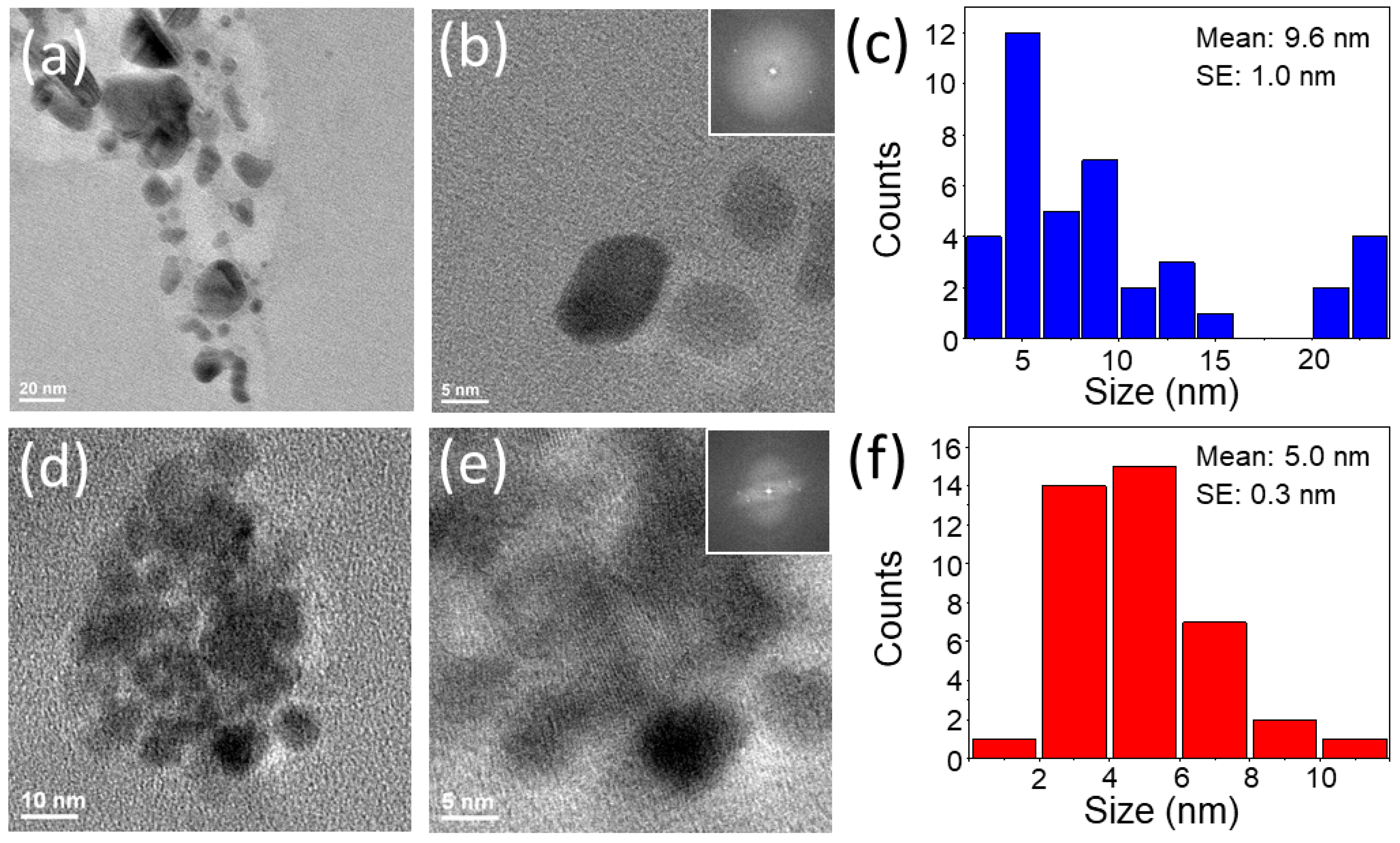
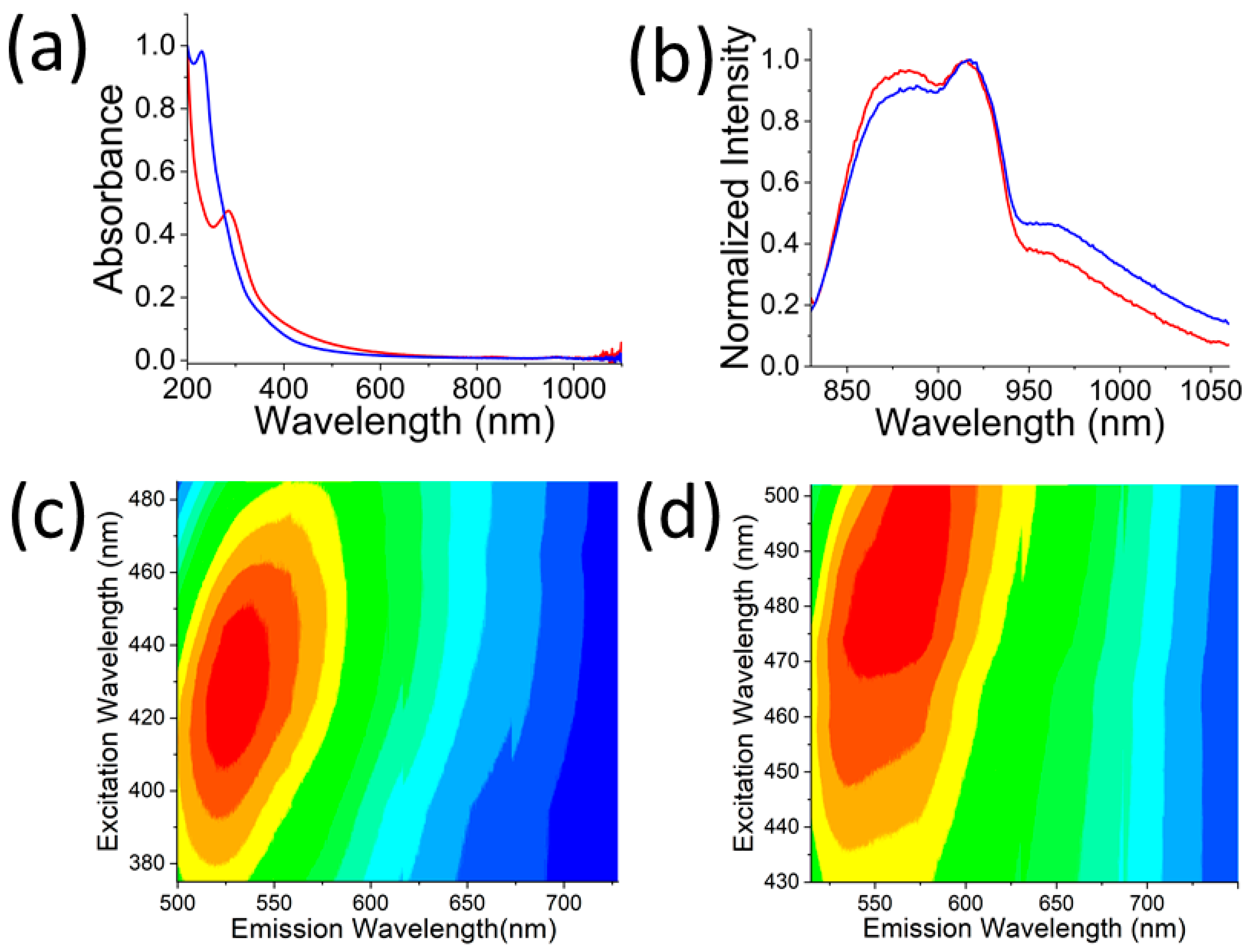
2.3. Characterization

Transmission electron microscopy (TEM, JEOL JEM-2100, Peabody, MA, USA) with energy dispersive x-ray spectroscopy (EDS, JEOL, Peabody, MA, USA) was used to assess GQD size, crystallinity, and composition. TEM sample was prepared by air-drying GQD suspensions on carbon-coated 200 mesh copper grids (Electron Microscopy Sciences, CF300-CU, Radnor, PA, USA). UV-Vis absorption spectra of all samples were acquired using Cary 60 (Agilent Technologies, Santa Clara, CA, USA) spectrophotometer in the range of 200 to 1100 nm. Visible fluorescence spectra were measured using a NanoLog (HORIBA Scientific, Piscataway, NJ, USA) spectrofluorometer, while NIR fluorescence spectra were assessed with AvaSpec-HS-TEC spectrofluorometer (Avantes, Apeldoorn, The Netherlands) with a 0.9 W/cm2 808 nm laser (808MD-12V-BL, Q-BAIHE, China) excitation.

2.4. Photothermal Effect in Solution

The photothermal effect was characterized as follows: separately, 3 mL of aqueous RGQDs at 1.5 mg/mL and 3 mL of HGQDs at 1.7 mg/mL were subjected to a 0.9 W/cm2 808 nm laser irradiation for 45 min. DI water was irradiated as a control. The temperature of each solution was continuously monitored by the submerged thermocouple. The experiments were performed in an oven held at a biological temperature of 37 °C with suspensions being magnetically stirred to maintain a constant temperature throughout the sample.

2.5. Cell Culture

HeLa cells were utilized for in vitro studies. These cells were cultured using a complete medium consisting of DMEM (D6046, Sigma-Aldrich, St. Louis, MO, USA) supplemented with 10% FBS (16140-063, Gibco, Dublin, Ireland), L-Glutamine (G7513, Sigma-Aldrich, St. Louis, MO, USA), non-essential amino acid solution (M7145, Sigma-Aldrich, St. Louis, MO, USA), and 1% penicillin/streptomycin (P4333, Sigma-Aldrich, St. Louis, MO, USA). The cell culture was held in an incubator at 37 °C with 5% CO2 (Midi CO2, Thermo Fischer Scientific, Waltham, MA, USA).

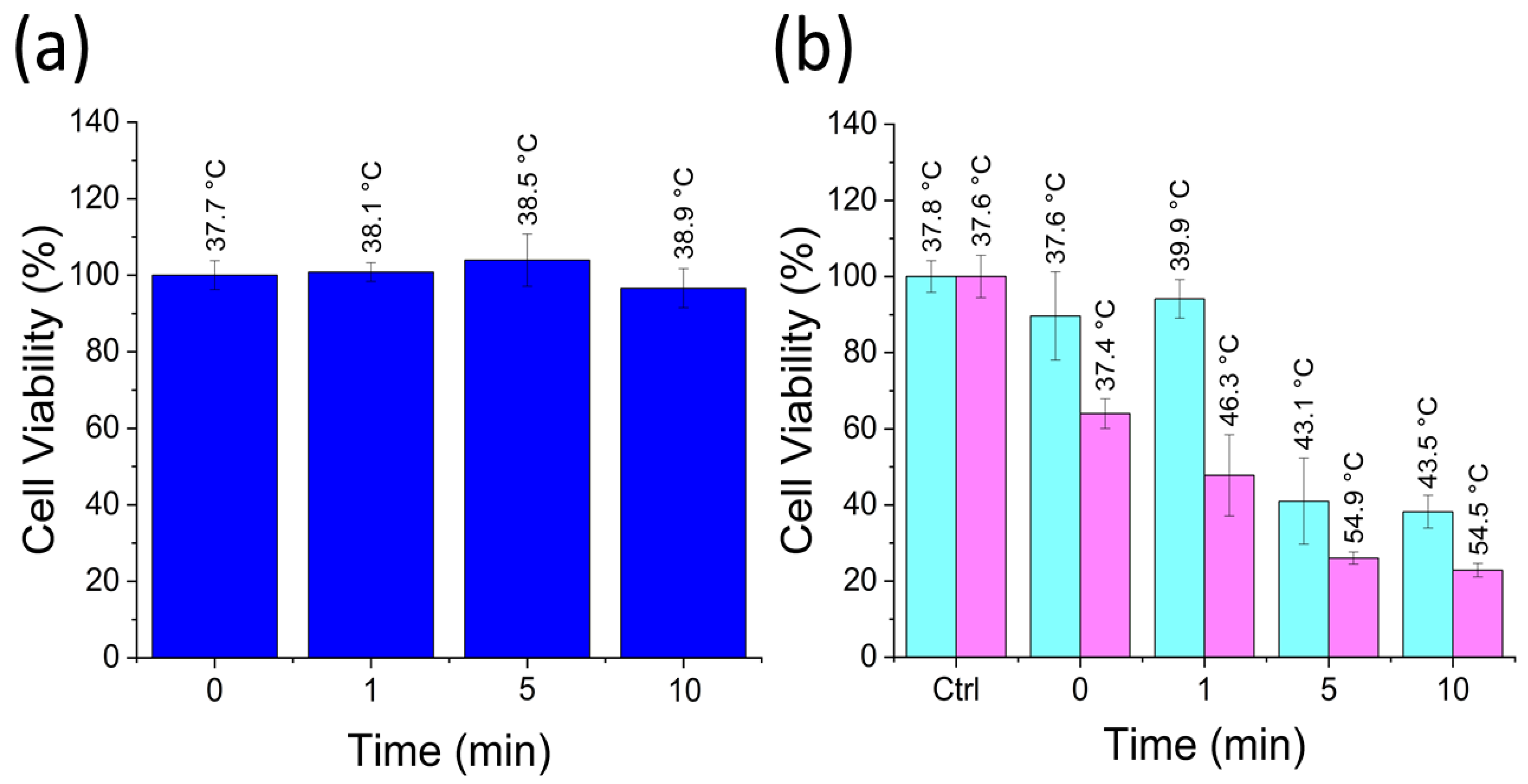
2.6. Cell Viability Assay

To assess the biocompatibility of the RGQDs and HGQDs, MTT (3-(4–dimethylthiazol-2-yl)-2,5 diphenyltetrazolium bromide) assay (Invitrogen M6494, Thermo Fischer Scientific, Waltham, MA, USA) was used. A total of 5000 cells per 200 µL were seeded into each well in a 96-well plate (701001, Nest Scientific, Wuxi, China) and kept in an incubator. RGQDs at 1.5 mg/mL and HGQDs at 1.7 mg/mL were added into different wells in two-fold serial dilutions in DMEM. After 24 h of post-treatment incubation, the complete medium in the 96-well plate was replaced with 100 µL of 1 mg/mL of MTT and stored in the incubator for 4 h. This step allowed for the metabolization of the MTT that was further replaced with 100 µL of DMSO. Finally, the 96-well plate was placed on an orbital shaker for 5 min, which was followed by the absorption measurement at 570 nm for each experimental well in an absorption plate reader (μQuant, BioTek Instruments, Winooski, VT, USA). These measurements indicated the amount of formazan generated via the reduction in the MTT by NAD(P)H-dependent cellular oxidoreductase enzymes of the viable cells, which was, therefore, proportional to the number of viable cells.

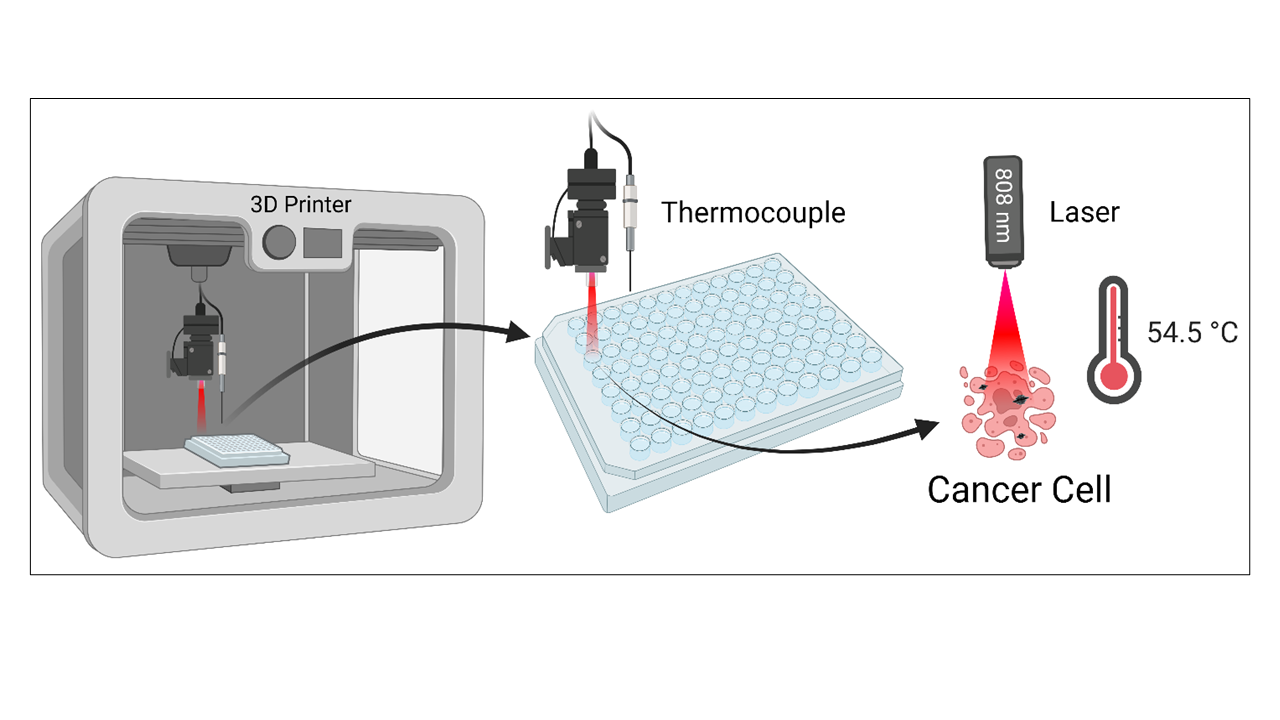
2.7. Automated Laser Irradiation and Temperature Measurement

A 3D printer (CR-10mini, Creality, Shenzhen, China) was retrofitted to be equipped with a 0.9 W/cm2 808 nm laser (808MD-12V-BL, Q-BAIHE, China) and a T-type thermocouple (TL0024, Perfect Prime, New York, NY, USA). They were attached to the extruder component of the 3D printer. The retrofitted 3D printer was placed inside a biosafety hood (Forma 1284, Thermo Fisher Scientific, Waltham, MA, USA) with a DI water reservoir to maintain the humidity of the biosafety hood environment. An iterative code was written in G-code (Table S1) to set the temperature of the heat bed of the 3D printer to biological 37 °C, position the laser and thermocouple at individual wells, and turn the laser on or off. A wireless data logger thermometer software (TC0521, Perfect Prime, New York, NY, USA) was used to collect and tabulate temperature measurements acquired by the thermocouple. The ultimate function of such retrofitted 3D printer device depicted in the schematic in Figure S1 involves irradiating the wells of the 96-well plate for 0, 1, 5, and 10 min with subsequent thermocouple well temperature measurements. The post-treatment cell viability of HeLa cells laser-treated in the 96-well plate was analyzed via the MTT assay.

2.8. Confocal Fluorescence Microscopy Imaging

Confocal fluorescence microscopy imaging was performed with a semi-motorized inverted microscope (IX73P2F, Olympus, Center Valley, PA, USA) using an IR-corrected UPLANAPO 60x/0.90na objective (1-UB831, Olympus, Center Valley, PA, USA) coupled to visible and near-infrared imaging setups. The visible fluorescence setup utilized a CMOS (Prime 95B, Photometrics, Tucson, AZ, USA) camera and a confocal disk-spinning unit (Olympus, Center Valley, PA, USA) with a 460 ± 20 nm filtered lamp excitation and 535 ± 20 nm filtered emission used in this work. The NIR setup involved an InGaAs FPA (ZephIRTM 1.7, Photon etc., Montreal, Quebec, Canada) camera and a hyperspectral imager for spectrally resolved image collection. The NIR emission was excited with an 808 nm laser and collected at the corresponding GQD emission range within 900–1200 nm.

3. Results and Discussion

In this work, GQDs were synthesized via the top-down and bottom-up approaches. RGQDs were produced from commercially available RGO in the top-down approach. The reaction was catalyzed by sodium hypochlorite (NaOCl) photodissociation, generating highly reactive oxygen free radicals to oxidize the graphitic structure [44,79]. In the bottom-up approach, HGQDs were synthesized via the dehydration reaction of the HA, during which polymeric aromatic structures were formed [45]. Size distributions of RGQDs and HGQDs assessed via TEM imaging averaged at 9.6 ± 1.0 and 5.0 ± 0.3 nm, respectively (Figure 1), indicating that nanoscale particles were created as a result of both top-down and bottom-up reactions. In vivo biodistribution studies of GQDs and carbon dots of similar sizes reported their accumulation mainly in the liver, spleen, and kidneys, where they later underwent rapid renal clearance reducing potential off-target effects, while still being capable of some EPR-based passive tumor targeting [44,65]. Some aggregates present in TEM images and reflected in GQD size distributions could have formed during the air drying of GQD suspensions on the TEM grid. In suspension, however, both GQDs showed high colloidal stability for over 6 months with substantially negative Zeta potentials of −18.7 ± 2.2 and −49.4 ± 0.4 mV for HGQDs and RGQDs, respectively (Figure S2).
EDS-based elemental analyses showed that HGQDs possessed more carbon (C: 98.1%) and less oxygen (O: 1.9%) when compared to RGQDs (C: 91.3%, O: 8.7%) (Figure S3). The presence of oxygen functional groups in the GQD structure made them amenable to chemical functionalization [44,60,61]. Furthermore, HRTEM and FFT image analyses indicated that both RGQDs and HGQDs were crystalline in structure with an interplanar distance of 0.21 nm corresponding to the lattice spacing of (100) graphene plane [80]. This finding suggests the possibility of non-covalent attachment of hydrophobic therapeutic cargo to the GQDs [47] and shows the potential for high photothermal conversion efficiency demonstrated previously for graphene and its derivatives [36,37,38]. Absorption measurements (Figure 2) indicated that both GQDs exhibited characteristic graphitic π - π * transitions along with an absorption tail extended into the longer wavelength region. While being minor when compared to the UV absorption, this tail, in part attributed to surface defect states and sub-gap transitions, still provides an avenue for photothermal conversion. Comparing both absorption spectra showed a narrower blue-shifted π - π * transition peak for RGQDs, while HGQDs possessed broader red-shifted absorption with a more extended long wavelength tail (Figure 2a).
Oxygen functional groups expected to contribute to the absorption shoulder in the 300 nm region facilitate sufficient water-solubility for RGQDs and HGQDs at the biologically relevant concentrations. The presence of oxygen functional groups was further validated by Fourier transform infrared spectroscopy (FTIR) measurements (Figure S4). Both GQDs showed stretching vibrations from O H , C H , and C = O / C = C bonds with peaks at 3380, 2909, and 1587 cm 1 for RGQDs and 3316, 2925, and 1597   cm 1 for HGQDs. Unlike RGQDs, HGQDs exhibited an additional stretching vibration for the C O bond at 1038 cm 1 . Both bottom-up and top-down approaches yielded GQDs fluorescing in the visible and NIR (Figure 2b). Their broad visible emission (Figure 2c,d) can arise from a distribution of GQD sizes in the sample. GQDs are small enough to serve as regions of electronic confinement, with each GQD size possessing a different band gap and a set of quantum confinement-dictated excitation and emission wavelengths [81]. Localized electronic defects in the GQD graphitic structure originating from oxidation and oxygen functional groups can lead to excitation-independent NIR emission. With an 808 nm laser excitation, both HGQDs and RGQDs exhibited NIR fluorescence spanning the wavelength range of 850 to 1100 nm with a peak at ~900 nm (Figure 2b). This fluorescence occurred within the biological absorption windows, which is desirable for facilitating NIR image-guided photothermal applications. The similarity of the spectral features of both GQD types can be explained by the presence of similar functional groups (Figure S4).
In order to assess maximum non-toxic concentrations for PTT applications, GQD biocompatibility was investigated in HeLa cancer cells intended as a biological test platform for the photothermal therapeutic capabilities of the GQDs. As shown by the MTT assay, RGQDs and HGQDs yielded over 80% cell viability at concentrations of up to 1.5 and 1.7 mg/mL, respectively, appearing non-toxic at high doses (Figure 3), which makes them relevant for further clinical applications. NIR imaging facilitated by RGQDs in vivo also gives promise for their clinical translation [44]. Therefore, given significant biocompatibility along with some absorption in the NIR, GQDs can serve as potential candidates to be tested for PTT applications.
Photothermal conversion efficiency of RGQDs and HGQDs was first assessed at biocompatible concentrations in an aqueous suspension. The photothermal studies reported herein utilized an 808 nm laser excitation, since the absorption coefficient of water at that wavelength is minimized [82]. Thus, unlike visible wavelengths, an 808 nm excitation in the first water window allows for deeper tissue imaging, making this excitation method potentially translatable to in vivo applications. In contrast to several previous works [24,38,65,83], the photothermal effect was assessed here at a biologically relevant baseline temperature of 37 °C in a temperature-controlled oven simulating a large heat sink provided by the human body. Upon irradiation, the HGQD and RGQD suspensions experienced respective temperature increases of up to 46 and 47 °C, while a water control sample only heated up to 37.3 °C (Figure S5). These temperatures, which are sufficient to initiate necrosis and apoptosis in cancer cells, exceed the ones reported in PTT studies with a number of nanomaterials [84,85,86]. To ensure their stability as photothermal agents, GQD samples had to undergo three cycles of laser irradiation, reaching similar temperatures averaged and reported with appropriate error ranges (Figure 4). It is worth noting that almost a maximum heating was achieved already at 10–15 min of irradiation, after which solution temperatures reached nearly an asymptotic limit. Thus, shorter time frames can be used for cancer photothermal therapy reducing potential detrimental effects on healthy tissue.
In order to test the photothermal treatment capability of the GQDs in vitro, a unique automated 3D printer-based system was developed and utilized to directly irradiate cells and measure cell culture temperatures in a 96-well plate. HeLa cells treated with RGQDs at 1.5 mg/mL and HGQDs at 1.7 mg/mL for 24 h served as a cancer cell culture model in the automated PTT treatment. This system can perform continuous in vitro experiments during which the laser is programmed via an open-source G-code software RS-274 (Table S1) to irradiate a sequence of wells with controlled time and power settings. After the irradiation of each well, the add-on thermocouple of the system measured well temperature and recorded it in the file. In order to ensure minimal cross-contamination between the wells, additional wells filled with DMEM were used to rinse and clean the thermocouple probe by dipping it into the well repeatedly. These wells also acted as cooling sinks for the temperature probe ensuring that it entered each new experimental well at the same initial temperature and, thus, did not perturb the measurement. The heat bed of the 3D printer was fitted to the 96-well plate, providing direct thermal contact to keep the base temperature at a biological 37 °C. This feature, which was rarely present in other in vitro PTT studies, simulates the massive 37 °C base temperature heat reservoir provided by the human body. Such experimental design, as well as proper calibration, enabled accurate and reproducible measurements. The temperature calibration of this instrument involved successfully measuring 37 °C in all wells that will be utilized in the experiment. In order to accomplish that, only select wells closer to the middle of the plate were used. Each experimental well was surrounded by empty wells to facilitate its thermal insulation (Figure S1). To prevent excessive evaporation of solution from the wells and maintain sterility, the experiment was performed in a biosafety hood with a large water reservoir humidifying the environment. After the irradiation and temperature measurements, the same 96-well plate was further analyzed via the MTT assay to evaluate treatment-derived effects on cell viability. All experimental conditions were tested in this high-throughput approach in a single 96-well plate. This included non-treatment control wells as well as those treated with each type of GQD and irradiated for 1, 5, and 10 min.
Temperature measurements help assess the efficacy of PTT for a particular condition. For instance, HeLa control wells irradiated for a maximum time of 10 min exhibit temperature increased only up to 38.9 ± 0.5 °C with no effect on cell viability (Figure 5a). This control test showed that the heating was negligible without the addition of the photothermal agents. In contrast, a 10 min irradiation of HeLa cells treated with RGQDs increased well temperatures to 43.5 ± 0.1 °C and dropped cell viability down to 38.2 ± 4.3%. HGQDs-treated cells upon 10 min irradiation experienced an even more drastic cell viability decrease down to 22.9 ± 1.8% due to PTT heating up to 54.5 ± 0.3 °C (Figure 5b). Again, such heating surpassed PTT temperatures achieved with multiple nanomaterial platforms [87,88,89]. The table of temperatures and cell viabilities delineates these effects per condition (Table S2), showing the reproducibility of achieved anticancer treatment. The temperatures in the range of 46 to 55 °C were reported to cause irreversible cellular damage and cell death [90], which validates the observed low cell viabilities within the treated wells. Having two assessment factors (well temperature and cell viability) allows for highly deterministic analysis of the nanomaterials’ PTT capabilities. Compared to the present work, previous studies utilizing GQDs for PTT applications (Table S3) lack to quantify these critical assessment factors. As the observed heating can originate from GQDs located both intracellularly and extracellularly, we verified their intracellular translocation and the capability for in vitro tracking via both visible and NIR fluorescence microscopies.
HeLa cells in this imaging study were treated with biocompatible concentrations of RGQDs (1.5 mg/mL) and HGQDs (1.7 mg/mL) for 20 h. This treatment time was chosen because it is close to the maximum cell internalization timeline of similar GQD structures [43,44,60]. Visible confocal fluorescence microscopy imaging suggests the internalization of both RGQDs and HGQDs showing their green 535 nm fluorescence within HeLa cells with lamp excitation at 460 ± 20 nm (Figure 6a,d). This result was further confirmed by the hyperspectral NIR imaging with an 808 nm laser excitation (Figure 6b,e). A 1080 nm bandpass setting of the hyperspectral imager was utilized to ensure that NIR fluorescence was observed only from the GQDS. While not entirely overlapping with visible, NIR emission provides a background-free assessment of GQD location and internalization. Hyperspectral microscopy-generated spectra taken from the regions of GQD fluorescence within the cells (Figure 6c,f) appeared to be similar to bulk GQD NIR fluorescence features validating the intracellular presence of GQDs (Figure 2b).
NIR emission can serve as a complementary bioimaging modality for a variety of photothermal treatment applications, including real-time image-guided ablation of tumors [83,91,92,93]. Tumor-targeted treatment can aid in locating tumors and further focus photothermal treatment to their location. Moreover, with their hydrophobic graphitic platform, an abundance of functionalizable oxygen groups, and a broad choice of potential precursor materials for top-down and bottom-up synthesis, GQDs can be nano-engineered to carry drug, gene, or targeting agents that can complement or enhance their PTT therapeutic effect [47,61,94,95,96]. Furthermore, in order to improve the therapeutic outcomes as well as enhance the platform’s biocompatibility, GQDs can also be incorporated into nanocomposite hydrogels [97,98]. Such future prospects coupled with successful PTT capabilities demonstrated in this work give HGQDs and RGQDs a substantial advantage over many other PTT nanomaterial agents in multimodal theragnostic applications.

4. Conclusions

In this work, graphene quantum dots, which were biocompatible up to 1.5–1.7 mg/mL, few nanometer-sized, water-soluble, were synthesized and tested as photothermal and bioimaging agents in vitro. Simple and scalable top-down and bottom-up approaches were utilized to produce RGQDs and HGQDs from reduced graphene oxide and hyaluronic acid precursor materials. Photothermal conversion capacity as well as PTT cancer cell treatment capabilities of these GQDs were evaluated with 0.9 W/cm2 808 nm laser irradiation in the first NIR water absorption window. Aqueous solution experiments at 37 °C ambient temperature demonstrated laser-induced heating of RGQD and HGQD suspensions up to 47 and 46 °C, respectively, after 10–15 min of irradiation with no apparent heating of water controls. Both GQD types had remarkable photothermal stability, demonstrating similar heating after three consecutive irradiation cycles. A new high-throughput irradiation/measurement system based on the retrofitted 3D printer was developed to conduct multiple in vitro PPT studies in the same 96-well plate. This device, equipped with a laser and thermocouple for in-well irradiation and temperature measurements, was programmed to test the PTT effect of different GQD samples at 1–10 min irradiation times in HeLa cells. The effects of such PTT on cancer cell viability were evaluated in the same well plate via the MTT assay. As compared to the HeLa control experiencing only minor heating and/or decrease in cell viability upon irradiation, HeLa cells treated with RGQDs and HGQDs demonstrated substantial photothermal heating to 43.5 and 54.5 °C together with the decrease in cancer cell viability down to 38.2% and 22.9%, respectively. Visible and near-infrared GQD fluorescence was successfully utilized in this work for imaging both HGQDs and RGQDs in HeLa cells to test their image tracking capabilities and verify their successful internalization. Due to its greater penetration depth, NIR fluorescence offers a broader range of potential in vivo/ex vivo applications for GQDs as dual-mode imaging/photothermal platforms. Top-down- and bottom-up-synthesized GQDs tested in this work, therefore, show a significant potential as biocompatible image-guided photothermal agents, while their prospective drug delivery capabilities can further extend GQD applications into highly desired drug/PTT combination therapeutic approaches.

Supplementary Materials

The following supporting information can be downloaded at: https://www.mdpi.com/article/10.3390/nano13050805/s1, Figure S1. In vitro PTT setup for a 96-well plate: HeLa cells control (pink), GQD-treated cells (purple), GQD-treated and laser-irradiated cells (red), and heat sink/cleaning station wells for the thermocouple (blue). Table S1. The code controlling the 3D printer for in vitro PTT experiments written using computer numerical control program language G-code. Figure S2. Zeta potential of RGQDs (blue) and HGQDs (red). Figure S3. EDX images and atomic percentages of carbon and oxygen for a region with (a) HGQDs and (b) RGQDs. Figure S4. FTIR spectra of (a) RGQDs (blue) and (b) HGQDs (red). Figure S5. Temperature of water control with 0.9 W/cm2 808 nm laser irradiation. Table S2. Tabulated results of Figure 5b. Table S3. Table of GQDs developed for PTT applications. References [99,100] are cited in the supplementary materials.

Author Contributions

Conceptualization, B.L., G.A.S., A.V. and A.V.N.; Methodology, B.L., G.A.S., A.V. and S.N.; Software, B.L.; Validation, B.L., G.A.S., A.V., S.N. and R.G.-R.; Formal analysis, B.L., G.A.S., A.V. and S.N.; Investigation, B.L., G.A.S., A.V., S.N., R.G.-R. and A.B.; Resources, J.C. and A.V.N.; Data curation, B.L., G.A.S., A.V. and S.N.; Writing—original draft, B.L. and G.A.S.; Writing—review & editing, B.L., G.A.S., S.N. and A.V.N.; Visualization, B.L., G.A.S., A.V., S.N. and R.G.-R.; Supervision, B.L. and A.V.N.; Project administration, B.L., G.A.S. and A.V.N.; Funding acquisition, A.V.N. All authors have read and agreed to the published version of the manuscript.

Funding

This research was funded by NIH NIBIB, grant # 1 R15 EB031528-01.

Institutional Review Board Statement

Not applicable.

Informed Consent Statement

Not applicable.

Data Availability Statement

The data presented in this study are available on request from the corresponding author. The data are not publicly available due to privacy.

Acknowledgments

The abstract figure and Figure S1 were created with BioRender.

Conflicts of Interest

The authors declare no conflict of interest. The funders had no role in the design of the study; in the collection, analyses, or interpretation of data; in the writing of the manuscript; or in the decision to publish the results.

References

  1. Sung, H.; Ferlay, J.; Siegel, R.L.; Laversanne, M.; Soerjomataram, I.; Jemal, A.; Bray, F. Global cancer statistics 2020: GLOBOCAN estimates of incidence and mortality worldwide for 36 cancers in 185 countries. CA Cancer J. Clin. 2021, 71, 209–249. [Google Scholar] [CrossRef]
  2. Wild, C.P.; Stewart, B.W.; Wild, C. World Cancer Report 2014; World Health Organization: Geneva, Switzerland, 2014. [Google Scholar]
  3. Liu, S.; Pan, X.; Liu, H. Two-dimensional nanomaterials for photothermal therapy. Angew. Chem. 2020, 132, 5943–5953. [Google Scholar] [CrossRef]
  4. Pérez-Herrero, E.; Fernández-Medarde, A. Advanced targeted therapies in cancer: Drug nanocarriers, the future of chemotherapy. Eur. J. Pharm. Biopharm. 2015, 93, 52–79. [Google Scholar] [CrossRef] [Green Version]
  5. Baumann, M.; Krause, M.; Hill, R. Exploring the role of cancer stem cells in radioresistance. Nat. Rev. Cancer 2008, 8, 545–554. [Google Scholar] [CrossRef]
  6. Jha, S.; Sharma, P.K.; Malviya, R. Hyperthermia: Role and risk factor for cancer treatment. Achiev. Life Sci. 2016, 10, 161–167. [Google Scholar] [CrossRef] [Green Version]
  7. Ito, A.; Shinkai, M.; Honda, H.; Yoshikawa, K.; Saga, S.; Wakabayashi, T.; Yoshida, J.; Kobayashi, T. Heat shock protein 70 expression induces antitumor immunity during intracellular hyperthermia using magnetite nanoparticles. Cancer Immunol. Immunother. 2003, 52, 80–88. [Google Scholar] [CrossRef]
  8. Han, H.S.; Choi, K.Y. Advances in nanomaterial-mediated photothermal cancer therapies: Toward clinical applications. Biomedicines 2021, 9, 305. [Google Scholar] [CrossRef]
  9. Torgovnick, A.; Schumacher, B. DNA repair mechanisms in cancer development and therapy. Front. Genet. 2015, 6, 157. [Google Scholar] [CrossRef] [Green Version]
  10. Riley, R.S.; Day, E.S. Gold nanoparticle-mediated photothermal therapy: Applications and opportunities for multimodal cancer treatment. Wiley Interdiscip. Rev. Nanomed. Nanobiotechnol. 2017, 9, e1449. [Google Scholar] [CrossRef]
  11. Zhang, Y.; Zhan, X.; Xiong, J.; Peng, S.; Huang, W.; Joshi, R.; Cai, Y.; Liu, Y.; Li, R.; Yuan, K. Temperature-dependent cell death patterns induced by functionalized gold nanoparticle photothermal therapy in melanoma cells. Sci. Rep. 2018, 8, 8720. [Google Scholar] [CrossRef] [Green Version]
  12. Knavel, E.M.; Brace, C.L. Tumor ablation: Common modalities and general practices. Tech. Vasc. Interv. Radiol. 2013, 16, 192–200. [Google Scholar] [CrossRef] [Green Version]
  13. Smith, A.M.; Mancini, M.C.; Nie, S. Second window for in vivo imaging. Nat. Nanotechnol. 2009, 4, 710–711. [Google Scholar] [CrossRef] [PubMed] [Green Version]
  14. Jaque, D.; Maestro, L.M.; del Rosal, B.; Haro-Gonzalez, P.; Benayas, A.; Plaza, J.; Rodríguez, E.M.; Solé, J.G. Nanoparticles for photothermal therapies. Nanoscale 2014, 6, 9494–9530. [Google Scholar] [CrossRef]
  15. Weissleder, R. A clearer vision for in vivo imaging. Nat. Biotechnol. 2001, 19, 316–317. [Google Scholar] [CrossRef]
  16. Rajani, C.; Borisa, P.; Karanwad, T.; Borade, Y.; Patel, V.; Rajpoot, K.; Tekade, R.K. Cancer-targeted chemotherapy: Emerging role of the folate anchored dendrimer as drug delivery nanocarrier. In Pharmaceutical Applications of Dendrimers; Elsevier: Amsterdam, The Netherlands, 2020; pp. 151–198. [Google Scholar]
  17. Park, J.; Park, J.; Ju, E.J.; Park, S.S.; Choi, J.; Lee, J.H.; Lee, K.J.; Shin, S.H.; Ko, E.J.; Park, I. Multifunctional hollow gold nanoparticles designed for triple combination therapy and CT imaging. J. Control. Release 2015, 207, 77–85. [Google Scholar] [CrossRef]
  18. Ali, E.S.; Sharker, S.M.; Islam, M.T.; Khan, I.N.; Shaw, S.; Rahman, M.A.; Uddin, S.J.; Shill, M.C.; Rehman, S.; Das, N. Seminars in cancer biology. In Targeting Cancer Cells with Nanotherapeutics and Nanodiagnostics: Current Status and Future Perspectives; Elsevier: Amsterdam, The Netherlands, 2021; pp. 52–68. [Google Scholar]
  19. Rosenblum, D.; Joshi, N.; Tao, W.; Karp, J.M.; Peer, D. Progress and challenges towards targeted delivery of cancer therapeutics. Nat. Commun. 2018, 9, 1410. [Google Scholar] [CrossRef] [Green Version]
  20. Chen, J.; Ning, C.; Zhou, Z.; Yu, P.; Zhu, Y.; Tan, G.; Mao, C. Nanomaterials as photothermal therapeutic agents. Prog. Mater. Sci. 2019, 99, 1–26. [Google Scholar] [CrossRef]
  21. Wang, Y.; Black, K.C.; Luehmann, H.; Li, W.; Zhang, Y.; Cai, X.; Wan, D.; Liu, S.-Y.; Li, M.; Kim, P. Comparison study of gold nanohexapods, nanorods, and nanocages for photothermal cancer treatment. ACS Nano 2013, 7, 2068–2077. [Google Scholar] [CrossRef]
  22. Huang, Y.; Lai, Y.; Shi, S.; Hao, S.; Wei, J.; Chen, X. Copper sulfide nanoparticles with phospholipid-PEG coating for In vivo near-infrared photothermal cancer therapy. Chem.-Asian J. 2015, 10, 370–376. [Google Scholar] [CrossRef]
  23. Li, Y.; Lu, W.; Huang, Q.; Li, C.; Chen, W. Copper sulfide nanoparticles for photothermal ablation of tumor cells. Nanomedicine 2010, 5, 1161–1171. [Google Scholar] [CrossRef] [Green Version]
  24. Zhou, F.; Da, X.; Ou, Z.; Wu, B.; Resasco, D.E.; Chen, W.R. Cancer photothermal therapy in the near-infrared region by using single-walled carbon nanotubes. J. Biomed. Opt. 2009, 14, 021009. [Google Scholar] [CrossRef]
  25. D’Acunto, M. Detection of intracellular gold nanoparticles: An overview. Materials 2018, 11, 882. [Google Scholar] [CrossRef] [Green Version]
  26. Tee, S.Y.; Win, K.Y.; Goh, S.S.; Teng, C.P.; Tang, K.Y.; Regulacio, M.D.; Li, Z.; Ye, E. Introduction to Photothermal Nanomaterials. In Photothermal Nanomaterials; Royal Society of Chemistry: London, UK, 2022. [Google Scholar]
  27. Webb, J.A.; Bardhan, R. Emerging advances in nanomedicine with engineered gold nanostructures. Nanoscale 2014, 6, 2502–2530. [Google Scholar] [CrossRef]
  28. Poon, W.; Zhang, Y.-N.; Ouyang, B.; Kingston, B.R.; Wu, J.L.; Wilhelm, S.; Chan, W.C. Elimination pathways of nanoparticles. ACS Nano 2019, 13, 5785–5798. [Google Scholar] [CrossRef]
  29. Tarantola, M.; Schneider, D.; Sunnick, E.; Adam, H.; Pierrat, S.; Rosman, C.; Breus, V.; Sonnichsen, C.; Basche, T.; Wegener, J. Cytotoxicity of metal and semiconductor nanoparticles indicated by cellular micromotility. ACS Nano 2009, 3, 213–222. [Google Scholar] [CrossRef]
  30. Zhang, W.; Guo, Z.; Huang, D.; Liu, Z.; Guo, X.; Zhong, H. Synergistic effect of chemo-photothermal therapy using PEGylated graphene oxide. Biomaterials 2011, 32, 8555–8561. [Google Scholar] [CrossRef]
  31. Shangguan, Q.; Chen, Z.; Yang, H.; Cheng, S.; Yang, W.; Yi, Z.; Wu, X.; Wang, S.; Yi, Y.; Wu, P. Design of ultra-narrow band graphene refractive index sensor. Sensors 2022, 22, 6483. [Google Scholar] [CrossRef]
  32. Cheng, Z.; Liao, J.; He, B.; Zhang, F.; Zhang, F.; Huang, X.; Zhou, L. One-step fabrication of graphene oxide enhanced magnetic composite gel for highly efficient dye adsorption and catalysis. ACS Sustain. Chem. Eng. 2015, 3, 1677–1685. [Google Scholar] [CrossRef]
  33. Shangguan, Q.; Zhao, Y.; Song, Z.; Wang, J.; Yang, H.; Chen, J.; Liu, C.; Cheng, S.; Yang, W.; Yi, Z. High sensitivity active adjustable graphene absorber for refractive index sensing applications. Diam. Relat. Mater. 2022, 128, 109273. [Google Scholar] [CrossRef]
  34. Zhang, Z.; Cai, R.; Long, F.; Wang, J. Development and application of tetrabromobisphenol A imprinted electrochemical sensor based on graphene/carbon nanotubes three-dimensional nanocomposites modified carbon electrode. Talanta 2015, 134, 435–442. [Google Scholar] [CrossRef]
  35. Moon, H.K.; Lee, S.H.; Choi, H.C. In Vivo Near-Infrared Mediated Tumor Destruction by Photothermal Effect of Carbon Nanotubes. ACS Nano 2009, 3, 3707–3713. [Google Scholar] [CrossRef]
  36. Akhavan, O.; Ghaderi, E.; Emamy, H. Nontoxic concentrations of PEGylated graphene nanoribbons for selective cancer cell imaging and photothermal therapy. J. Mater. Chem. 2012, 22, 20626–20633. [Google Scholar] [CrossRef]
  37. Leitao, M.M.; Alves, C.G.; de Melo-Diogo, D.; Lima-Sousa, R.; Moreira, A.F.; Correia, I.J. Sulfobetaine methacrylate-functionalized graphene oxide-IR780 nanohybrids aimed at improving breast cancer phototherapy. RSC Adv. 2020, 10, 38621–38630. [Google Scholar] [CrossRef]
  38. Robinson, J.T.; Tabakman, S.M.; Liang, Y.; Wang, H.; Sanchez Casalongue, H.; Vinh, D.; Dai, H. Ultrasmall reduced graphene oxide with high near-infrared absorbance for photothermal therapy. J. Am. Chem. Soc. 2011, 133, 6825–6831. [Google Scholar] [CrossRef] [PubMed]
  39. Su, S.; Wang, J.; Wei, J.; Martínez-Zaguilán, R.; Qiu, J.; Wang, S. Efficient photothermal therapy of brain cancer through porphyrin functionalized graphene oxide. New J. Chem. 2015, 39, 5743–5749. [Google Scholar] [CrossRef]
  40. Yin, F.; Hu, K.; Chen, Y.; Yu, M.; Wang, D.; Wang, Q.; Yong, K.-T.; Lu, F.; Liang, Y.; Li, Z. SiRNA delivery with PEGylated graphene oxide nanosheets for combined photothermal and genetherapy for pancreatic cancer. Theranostics 2017, 7, 1133. [Google Scholar] [CrossRef] [Green Version]
  41. Li, M.; Yang, X.; Ren, J.; Qu, K.; Qu, X. Using graphene oxide high near-infrared absorbance for photothermal treatment of Alzheimer’s disease. Adv. Mater. 2012, 24, 1722–1728. [Google Scholar] [CrossRef]
  42. Liang, C.; Diao, S.; Wang, C.; Gong, H.; Liu, T.; Hong, G.; Shi, X.; Dai, H.; Liu, Z. Tumor metastasis inhibition by imaging-guided photothermal therapy with single-walled carbon nanotubes. Adv. Mater. 2014, 26, 5646–5652. [Google Scholar] [CrossRef]
  43. Hasan, M.T.; Gonzalez-Rodriguez, R.; Lin, C.W.; Campbell, E.; Vasireddy, S.; Tsedev, U.; Belcher, A.M.; Naumov, A.V. Rare-earth metal ions doped graphene quantum dots for near-ir in vitro/in vivo/ex vivo imaging applications. Adv. Opt. Mater. 2020, 8, 2000897. [Google Scholar] [CrossRef]
  44. Hasan, M.T.; Lee, B.H.; Lin, C.-W.; McDonald-Boyer, A.; Gonzalez-Rodriguez, R.; Vasireddy, S.; Tsedev, U.; Coffer, J.; Belcher, A.M.; Naumov, A.V. Near-infrared emitting graphene quantum dots synthesized from reduced graphene oxide for in vitro/in vivo/ex vivo bioimaging applications. 2D Mater. 2021, 8, 035013. [Google Scholar] [CrossRef]
  45. Hasan, M.T.; Gonzalez-Rodriguez, R.; Ryan, C.; Pota, K.; Green, K.; Coffer, J.L.; Naumov, A.V. Nitrogen-doped graphene quantum dots: Optical properties modification and photovoltaic applications. Nano Res. 2019, 12, 1041–1047. [Google Scholar] [CrossRef]
  46. Valimukhametova, A.R.; Zub, O.S.; Lee, B.H.; Fannon, O.; Nguyen, S.; Gonzalez-Rodriguez, R.; Akkaraju, G.R.; Naumov, A.V. Dual-Mode Fluorescence/Ultrasound Imaging with Biocompatible Metal-Doped Graphene Quantum Dots. ACS Biomater. Sci. Eng. 2022, 8, 4965–4975. [Google Scholar] [CrossRef]
  47. Ajgaonkar, R.; Lee, B.; Valimukhametova, A.; Nguyen, S.; Gonzalez-Rodriguez, R.; Coffer, J.; Akkaraju, G.R.; Naumov, A.V. Detection of Pancreatic Cancer miRNA with Biocompatible Nitrogen-Doped Graphene Quantum Dots. Materials 2022, 15, 5760. [Google Scholar] [CrossRef] [PubMed]
  48. Chen, J.; Yao, B.; Li, C.; Shi, G. An improved Hummers method for eco-friendly synthesis of graphene oxide. Carbon 2013, 64, 225–229. [Google Scholar] [CrossRef]
  49. Sun, Y.-P.; Zhou, B.; Lin, Y.; Wang, W.; Fernando, K.S.; Pathak, P.; Meziani, M.J.; Harruff, B.A.; Wang, X.; Wang, H. Quantum-sized carbon dots for bright and colorful photoluminescence. J. Am. Chem. Soc. 2006, 128, 7756–7757. [Google Scholar] [CrossRef]
  50. Abdolhosseinzadeh, S.; Asgharzadeh, H.; Seop Kim, H. Fast and fully-scalable synthesis of reduced graphene oxide. Sci. Rep. 2015, 5, 10160. [Google Scholar] [CrossRef] [Green Version]
  51. Pei, S.; Cheng, H.-M. The reduction of graphene oxide. Carbon 2012, 50, 3210–3228. [Google Scholar] [CrossRef]
  52. Gul, A.R.; Le, T.N.; Kim, M.W.; Kailasa, S.K.; Oh, K.T.; Park, T.J. One-pot synthesis of carbon dots with intrinsic folic acid for synergistic imaging-guided photothermal therapy of prostate cancer cells. Biomater. Sci. 2019, 7, 5187–5196. [Google Scholar]
  53. Wu, X.; Tian, F.; Wang, W.; Chen, J.; Wu, M.; Zhao, J.X. Fabrication of highly fluorescent graphene quantum dots using L-glutamic acid for in vitro/in vivo imaging and sensing. J. Mater. Chem. C 2013, 1, 4676–4684. [Google Scholar] [CrossRef] [Green Version]
  54. Wang, H.-J.; Zhang, J.; Liu, Y.-H.; Luo, T.-Y.; He, X.; Yu, X.-Q. Hyaluronic acid-based carbon dots for efficient gene delivery and cell imaging. RSC Adv. 2017, 7, 15613–15624. [Google Scholar] [CrossRef] [Green Version]
  55. Kumawat, M.K.; Thakur, M.; Gurung, R.B.; Srivastava, R. Graphene quantum dots from mangifera indica: Application in near-infrared bioimaging and intracellular nanothermometry. ACS Sustain. Chem. Eng. 2017, 5, 1382–1391. [Google Scholar] [CrossRef]
  56. Meng, W.; Bai, X.; Wang, B.; Liu, Z.; Lu, S.; Yang, B. Biomass-derived carbon dots and their applications. Energy Environ. Mater. 2019, 2, 172–192. [Google Scholar] [CrossRef]
  57. Sahu, S.; Behera, B.; Maiti, T.K.; Mohapatra, S. Simple one-step synthesis of highly luminescent carbon dots from orange juice: Application as excellent bio-imaging agents. Chem. Commun. 2012, 48, 8835–8837. [Google Scholar] [CrossRef]
  58. Wang, L.; Zhou, H.S. Green synthesis of luminescent nitrogen-doped carbon dots from milk and its imaging application. Anal. Chem. 2014, 86, 8902–8905. [Google Scholar] [CrossRef]
  59. Gedda, G.; Lee, C.-Y.; Lin, Y.-C.; Wu, H.-f. Green synthesis of carbon dots from prawn shells for highly selective and sensitive detection of copper ions. Sens. Actuators B Chem. 2016, 224, 396–403. [Google Scholar] [CrossRef]
  60. Campbell, E.; Hasan, M.T.; Gonzalez Rodriguez, R.; Akkaraju, G.R.; Naumov, A.V. Doped graphene quantum dots for intracellular multicolor imaging and cancer detection. ACS Biomater. Sci. Eng. 2019, 5, 4671–4682. [Google Scholar] [CrossRef]
  61. Campbell, E.; Hasan, M.T.; Gonzalez-Rodriguez, R.; Truly, T.; Lee, B.H.; Green, K.N.; Akkaraju, G.; Naumov, A.V. Graphene quantum dot formulation for cancer imaging and redox-based drug delivery. Nanomed. Nanotechnol. Biol. Med. 2021, 37, 102408. [Google Scholar] [CrossRef]
  62. Bao, X.; Yuan, Y.; Chen, J.; Zhang, B.; Li, D.; Zhou, D.; Jing, P.; Xu, G.; Wang, Y.; Holá, K. In vivo theranostics with near-infrared-emitting carbon dots—Highly efficient photothermal therapy based on passive targeting after intravenous administration. Light Sci. Appl. 2018, 7, 91. [Google Scholar] [CrossRef] [Green Version]
  63. Permatasari, F.A.; Fukazawa, H.; Ogi, T.; Iskandar, F.; Okuyama, K. Design of pyrrolic-N-rich carbon dots with absorption in the first near-infrared window for photothermal therapy. ACS Appl. Nano Mater. 2018, 1, 2368–2375. [Google Scholar] [CrossRef]
  64. Lan, M.; Zhao, S.; Zhang, Z.; Yan, L.; Guo, L.; Niu, G.; Zhang, J.; Zhao, J.; Zhang, H.; Wang, P. Two-photon-excited near-infrared emissive carbon dots as multifunctional agents for fluorescence imaging and photothermal therapy. Nano Res. 2017, 10, 3113–3123. [Google Scholar] [CrossRef]
  65. Li, Y.; Bai, G.; Zeng, S.; Hao, J. Theranostic carbon dots with innovative NIR-II emission for in vivo renal-excreted optical imaging and photothermal therapy. ACS Appl. Mater. Interfaces 2019, 11, 4737–4744. [Google Scholar] [CrossRef]
  66. Tavianatou, A.G.; Caon, I.; Franchi, M.; Piperigkou, Z.; Galesso, D.; Karamanos, N.K. Hyaluronan: Molecular size-dependent signaling and biological functions in inflammation and cancer. FEBS J. 2019, 286, 2883–2908. [Google Scholar] [CrossRef] [Green Version]
  67. Senbanjo, L.; Chellaiah, M. CD44: A multifunctional cell surface adhesion receptor is a regulator of progression and metastasis of cancer cells. Front. Cell Dev. Biol. 2017, 5, 18. [Google Scholar] [CrossRef] [Green Version]
  68. Lee, J.-E.; In, I.; Lee, H.; Lee, K.D.; Jeong, J.H.; Park, S.Y. Target delivery and cell imaging using hyaluronic acid-functionalized graphene quantum dots. Mol. Pharm. 2013, 10, 3736–3744. [Google Scholar]
  69. Assmann, V.; Fieber, C.; Herrlich, P.; Hofmann, M.; Termeer, C.C.; Ahrens, T.; Simon, J.C. CD44 is the principal mediator of hyaluronic-acid-induced melanoma cell proliferation. J. Investig. Dermatol. 2001, 116, 93–101. [Google Scholar] [CrossRef]
  70. Goh, E.J.; Kim, K.S.; Kim, Y.R.; Jung, H.S.; Beack, S.; Kong, W.H.; Scarcelli, G.; Yun, S.H.; Hahn, S.K. Bioimaging of hyaluronic acid derivatives using nanosized carbon dots. Biomacromolecules 2012, 13, 2554–2561. [Google Scholar] [CrossRef]
  71. Kim, J.; Kim, K.S.; Jiang, G.; Kang, H.; Kim, S.; Kim, B.S.; Park, M.H.; Hahn, S.K. In vivo real-time bioimaging of hyaluronic acid derivatives using quantum dots. Biopolymers 2008, 89, 1144–1153. [Google Scholar] [CrossRef]
  72. Zhang, M.; Zhao, X.; Fang, Z.; Niu, Y.; Lou, J.; Wu, Y.; Zou, S.; Xia, S.; Sun, M.; Du, F. Fabrication of HA/PEI-functionalized carbon dots for tumor targeting, intracellular imaging and gene delivery. RSC Adv. 2017, 7, 3369–3375. [Google Scholar] [CrossRef] [Green Version]
  73. David, H. Why are there still so many jobs? The history and future of workplace automation. J. Econ. Perspect. 2015, 29, 3–30. [Google Scholar]
  74. Lippi, G.; Da Rin, G. Advantages and limitations of total laboratory automation: A personal overview. Clin. Chem. Lab. Med. (CCLM) 2019, 57, 802–811. [Google Scholar] [CrossRef]
  75. Spake, C.S.; Carruthers, T.N.; Crozier, J.W.; Kalliainen, L.K.; Bhatt, R.A.; Schmidt, S.T.; Woo, A.S. 3D printed N-95 masks during the COVID-19 pandemic: Lessons learned. Ann. Biomed. Eng. 2021, 49, 3666–3675. [Google Scholar] [CrossRef]
  76. Coakley, M.; Hurt, D.E. 3D printing in the laboratory: Maximize time and funds with customized and open-source labware. SLAS Technol. 2016, 21, 489–495. [Google Scholar] [CrossRef] [Green Version]
  77. Bailey, M.; Grieco, J.; Speights, A.; Weiss, R.; Orr, G. 3D printing in the classroom and laboratory. J. Comput. Sci. Coll. 2015, 31, 183–184. [Google Scholar]
  78. Petrick, I.J.; Simpson, T.W. 3D printing disrupts manufacturing: How economies of one create new rules of competition. Res.-Technol. Manag. 2013, 56, 12–16. [Google Scholar] [CrossRef]
  79. Lin, C.-W.; Bachilo, S.M.; Zheng, Y.; Tsedev, U.; Huang, S.; Weisman, R.B.; Belcher, A.M. Creating fluorescent quantum defects in carbon nanotubes using hypochlorite and light. Nat. Commun. 2019, 10, 2874. [Google Scholar] [CrossRef] [Green Version]
  80. Baskin, Y.; Meyer, L. Lattice constants of graphite at low temperatures. Phys. Rev. 1955, 100, 544. [Google Scholar] [CrossRef]
  81. Zhu, S.; Song, Y.; Wang, J.; Wan, H.; Zhang, Y.; Ning, Y.; Yang, B. Photoluminescence mechanism in graphene quantum dots: Quantum confinement effect and surface/edge state. Nano Today 2017, 13, 10–14. [Google Scholar] [CrossRef]
  82. Li, B.; Zhang, Y.; Zou, R.; Wang, Q.; Zhang, B.; An, L.; Yin, F.; Hua, Y.; Hu, J. Self-assembled WO3−x hierarchical nanostructures for photothermal therapy with a 915 nm laser rather than the common 980 nm laser. Dalton Trans. 2014, 43, 6244–6250. [Google Scholar] [CrossRef]
  83. Wang, H.; Mu, Q.; Wang, K.; Revia, R.A.; Yen, C.; Gu, X.; Tian, B.; Liu, J.; Zhang, M. Nitrogen and boron dual-doped graphene quantum dots for near-infrared second window imaging and photothermal therapy. Appl. Mater. Today 2019, 14, 108–117. [Google Scholar] [CrossRef]
  84. Li, Y.; He, L.; Dong, H.; Liu, Y.; Wang, K.; Li, A.; Ren, T.; Shi, D.; Li, Y. Fever-Inspired Immunotherapy Based on Photothermal CpG Nanotherapeutics: The Critical Role of Mild Heat in Regulating Tumor Microenvironment. Adv. Sci. 2018, 5, 1700805. [Google Scholar] [CrossRef] [Green Version]
  85. Zhang, C.; Wang, X.; Wang, J.; Qiu, Y.; Qi, Z.; Song, D.; Wang, M. TCPP-Isoliensinine Nanoparticles for Mild-Temperature Photothermal Therapy. Int. J. Nanomed. 2021, 16, 6797. [Google Scholar] [CrossRef]
  86. Jiang, A.; Liu, Y.; Ma, L.; Mao, F.; Liu, L.; Zhai, X.; Zhou, J. Biocompatible heat-shock protein inhibitor-delivered flowerlike short-wave infrared nanoprobe for mild temperature-driven highly efficient tumor ablation. ACS Appl. Mater. Interfaces 2019, 11, 6820–6828. [Google Scholar] [CrossRef]
  87. Qiu, K.; Wang, J.; Rees, T.W.; Ji, L.; Zhang, Q.; Chao, H. A mitochondria-targeting photothermogenic nanozyme for MRI-guided mild photothermal therapy. Chem. Commun. 2018, 54, 14108–14111. [Google Scholar] [CrossRef]
  88. Zhou, Z.; Yan, Y.; Hu, K.; Zou, Y.; Li, Y.; Ma, R.; Zhang, Q.; Cheng, Y. Autophagy inhibition enabled efficient photothermal therapy at a mild temperature. Biomaterials 2017, 141, 116–124. [Google Scholar] [CrossRef]
  89. Wu, J.; Bremner, D.H.; Niu, S.; Shi, M.; Wang, H.; Tang, R.; Zhu, L.-M. Chemodrug-gated biodegradable hollow mesoporous organosilica nanotheranostics for multimodal imaging-guided low-temperature photothermal therapy/chemotherapy of cancer. ACS Appl. Mater. Interfaces 2018, 10, 42115–42126. [Google Scholar] [CrossRef]
  90. Leber, B.; Mayrhauser, U.; Leopold, B.; Koestenbauer, S.; Tscheliessnigg, K.; Stadlbauer, V.; Stiegler, P. Impact of temperature on cell death in a cell-culture model of hepatocellular carcinoma. Anticancer. Res. 2012, 32, 915–921. [Google Scholar]
  91. Lee, B.H.; McKinney, R.L.; Hasan, M.T.; Naumov, A.V. Graphene quantum dots as intracellular imaging-based temperature sensors. Materials 2021, 14, 616. [Google Scholar] [CrossRef]
  92. Chen, Q.; Wang, C.; Zhan, Z.; He, W.; Cheng, Z.; Li, Y.; Liu, Z. Near-infrared dye bound albumin with separated imaging and therapy wavelength channels for imaging-guided photothermal therapy. Biomaterials 2014, 35, 8206–8214. [Google Scholar] [CrossRef]
  93. Xuan, Y.; Zhang, R.-Y.; Zhang, X.-S.; An, J.; Cheng, K.; Li, C.; Hou, X.-L.; Zhao, Y.-D. Targeting N-doped graphene quantum dot with high photothermal conversion efficiency for dual-mode imaging and therapy in vitro. Nanotechnology 2018, 29, 355101. [Google Scholar] [CrossRef]
  94. Frieler, M.; Pho, C.; Lee, B.H.; Dobrovolny, H.; Akkaraju, G.R.; Naumov, A.V. Effects of doxorubicin delivery by nitrogen-doped graphene quantum dots on cancer cell growth: Experimental study and mathematical modeling. Nanomaterials 2021, 11, 140. [Google Scholar] [CrossRef]
  95. Cheon, Y.A.; Bae, J.H.; Chung, B.G. Reduced graphene oxide nanosheet for chemo-photothermal therapy. Langmuir 2016, 32, 2731–2736. [Google Scholar] [CrossRef]
  96. Fang, J.; Liu, Y.; Chen, Y.; Ouyang, D.; Yang, G.; Yu, T. Graphene quantum dots-gated hollow mesoporous carbon nanoplatform for targeting drug delivery and synergistic chemo-photothermal therapy. Int. J. Nanomed. 2018, 13, 5991. [Google Scholar] [CrossRef] [Green Version]
  97. Zhang, Y.; Zhu, C.; Zhang, Z.; Zhao, J.; Yuan, Y.; Wang, S. Oxidation triggered formation of polydopamine-modified carboxymethyl cellulose hydrogel for anti-recurrence of tumor. Colloids Surf. B Biointerfaces 2021, 207, 112025. [Google Scholar] [CrossRef]
  98. Zhou, L.; Zhao, J.; Chen, Y.; Zheng, Y.; Li, J.; Zhao, J.; Zhang, J.; Liu, Y.; Liu, X.; Wang, S. MoS2-ALG-Fe/GOx hydrogel with Fenton catalytic activity for combined cancer photothermal, starvation, and chemodynamic therapy. Colloids Surf. B Biointerfaces 2020, 195, 111243. [Google Scholar] [CrossRef]
  99. Liu, H.; Li, C.; Qian, Y.; Hu, L.; Fang, J.; Tong, W.; Nie, R.; Chen, Q.; Wang, H. Magnetic-induced graphene quantum dots for imaging-guided photothermal therapy in the second near-infrared window. Biomaterials 2020, 232, 119700. [Google Scholar] [CrossRef]
  100. Thakur, M.; Kumawat, M.K.; Srivastava, R. Multifunctional graphene quantum dots for combined photothermal and photodynamic therapy coupled with cancer cell tracking applications. RSC Adv. 2017, 7, 5251–5261. [Google Scholar] [CrossRef] [Green Version]
Figure 1. TEM analysis of RGQDs (ac) and HGQDs (df) including representative TEM images (a,d), HRTEM scans with FFT images in the inset (b,e), and TEM-derived size distributions (c,f).
Nanomaterials 13 00805 g001
Figure 2. (a) Absorption spectra of HGQDs (red) and RGQDs (blue). (b) NIR fluorescence spectra of HGQDs (red), and RGQDs (blue) excited with 808 nm laser. Visible excitation/emission maps of (c) HGQDs and (d) RGQDs.
Nanomaterials 13 00805 g002
Figure 3. MTT assay-based viability of HeLa cells treated with different concentrations of (a) RGQDs and (b) HGQDs.
Nanomaterials 13 00805 g003
Figure 4. Temperature of (a) HGQD (1.7 mg/mL) and (b) RGQD (1.5 mg/mL) suspensions after 0.9 W/cm2 808 nm laser irradiation for up to 47 min.
Nanomaterials 13 00805 g004
Figure 5. MTT assay-based viability of HeLa cells irradiated with an 808 nm (0.9 W/cm2) laser for 0, 1, 5, and 10 min. (a) HeLa nontreatment control. (b) HeLa cells treated with RGQDs at 1.5 mg/mL (cyan) and HGQDs at 1.7 mg/mL (pink).
Nanomaterials 13 00805 g005
Figure 6. Fluorescence of RGQDs in the visible (a) and NIR (b) in HeLa cells. Fluorescence of HGQDs in the visible (d) and NIR (e) in HeLa cells. Spectra of RGQDs (c) and HGQDs (f) collected with NIR hyperspectral microscopy within HeLa cells (red) and background (green).
Nanomaterials 13 00805 g006
Disclaimer/Publisher’s Note: The statements, opinions and data contained in all publications are solely those of the individual author(s) and contributor(s) and not of MDPI and/or the editor(s). MDPI and/or the editor(s) disclaim responsibility for any injury to people or property resulting from any ideas, methods, instructions or products referred to in the content.

