Article

Cost-Effective and Facile Fabrication of a Tattoo Paper-Based SERS Substrate and Its Application in Pesticide Sensing on Fruit Surfaces

1
Department of Future Convergence Engineering, Kongju National University, Cheonan 31080, Republic of Korea
2
Department of Mechanical Engineering, Korea Advanced Institute of Science and Technology, Daejeon 34141, Republic of Korea
3
Department of Mechanical and Automotive Engineering, Kongju National University, Cheonan 31080, Republic of Korea
*
Author to whom correspondence should be addressed.
Nanomaterials 2023, 13(3), 486; https://doi.org/10.3390/nano13030486
Submission received: 9 January 2023 / Revised: 21 January 2023 / Accepted: 23 January 2023 / Published: 25 January 2023
(This article belongs to the Section Nanoelectronics, Nanosensors and Devices)

Abstract

:
Surface-enhanced Raman spectroscopy (SERS) has been transformed into a useful analytical technique with significant advantages in relation to sensitive and low-concentration chemical analyses. However, SERS substrates are expensive and the analyte sample preparation is complicated; hence, it is only used in limited areas. We have fabricated a tattoo paper-based SERS substrate by using non-complicated inkjet printing. The sensitivity of the SERS substrate was increased by removing the carbon residues via exposure to ultraviolet light without damaging the substrate. Thus, low concentrations of pesticides (up to 1 μM thiram) were measured. The SERS substrate was attached to the curved surface of an apple to demonstrate its advantages, such as the flexibility and easy attachability of tattoo paper, and its feasibility was verified by measuring 1 μM thiram on the apple’s surface. Due to its economic cost, simple usage, and rapid measurement, it will be helpful for the identification of both agricultural adulterants and food adulterants and for water-based pollutant detection. It will also possibly be helpful for medical purposes related to human body surfaces in the future.

1. Introduction

Today, as the world is growing at a fast pace in terms of developments and technological advancements, and with a great amount of success, it can be seen that various factors affect living beings on this planet, such as pollution, waste, and other hazardous elements, including pesticides and chemical adulterants, as far as the food industry is concerned. Human health depends on the consumption of various food materials, which are grown naturally and are agricultural products. These food materials are usually laced with various forms of pesticides and insecticides, which are used for crop safety and growth and which often remain as a residue. Although minute consumption of these products does not have any severe or immediate effect on living beings, long-term consumption can cause severe illnesses and harms [1,2,3]. Therefore, it is a requirement to develop a system able to detect the harmful components of food materials and, furthermore, continuously monitor them during culture and production processes.
In line with this trend, in this study, we present a facile and cost-effective way of measuring pesticides on fruit surfaces. Gas chromatography (GC), liquid chromatography (LC), gas chromatography-mass spectrometry (GC/MS), and liquid chromatography-mass spectrometry (LC-MS) are used to analyze the types and amounts of pesticide residues [4,5,6,7,8]. These methods separate an analyte into individual substances via the viscous force or inertial force and then measure the amounts of each substance. Although this method is very precise and highly reliable, it requires very large or complicated components for the separation process. Therefore, these kinds of equipment are large and expensive, and they take a long time to perform the measurements, making them unsuitable for use in the field, such as orchards. As an alternative, the enzyme-linked immunosorbent assay (ELISA) has been proposed, although it is also complicated and time-consuming [9,10,11,12]. For rapid and facile measurements, electrochemical or optical sensors using enzymes, aptamers, antibodies, cells, etc., are being widely developed [13,14,15]. However, these sensors also require pre-processes for the measurements, such as collecting samples and chopping them into extract analytes. Some methods collect analytes from the surface by rubbing or pressing the fruit surface. These methods are also cumbersome and, due to the poor repeatability, the sampling itself reduces the accuracy [16].
Therefore, a method for directly measuring the pesticides on fruit surfaces needs to be developed. Surface-enhanced Raman spectroscopy (SERS) is based on molecular vibration as the signature in the spectra and electromagnetic amplification by conductive nanostructures [17,18,19]. Raman spectroscopy provides specific information concerning the analyte as the fingerprint region of the molecule, thereby providing information on the presence of the molecule [20]. Therefore, unlike conventional methods using enzymes, aptamers, antibodies, or cells, various analytes can be measured without specifying the target in advance. However, since Raman signals are inherently weak, SERS, which can achieve high sensitivity combined with electromagnetic amplification, was developed [17,18,19,21,22]. Conductive NP clusters focus irradiated lasers and generate high plasmonic hotspots that allow SERS to detect even a single molecule [23,24,25]. Since they are based on electromagnetic amplification using conductive nanostructures, SERS devices are usually fabricated using sophisticated micro/nanostructure fabrication processes based on rigid substrates, such as silicon and quartz [20,26,27]. Although extremely high sensitivity is achieved, they are designed to be suitable for measurements in laboratories and have limitations in their applications on work sites. Due to the high sensitivity of SERS, methods for detecting pesticides based on SERS have also been developed [28,29].
Even with SERS, the sample collection processes are a fundamental drawback. Therefore, they have the same problems as chromatography and electrochemical and optical sensing [28,29]. Methods for omitting the analyte preparation processes need to be developed.
Recently, high-performance SERS substrates using randomized high-density nanostructures have been developed, and they do not require sophisticated fabrication methods [30,31,32]. Since this type of device does not need to be limited to rigid substrates, many flexible SERS substrates have been developed, thereby broadening the utilization of SERS [33,34,35]. Moreover, the production cost is also very low.
Various studies have been conducted in order to utilize these flexible SERS substrates for the detection of various components on fruit surfaces, although the majority of these methods involve pasting a substrate onto the surface of the fruit and then peeling it off, or rubbing and then peeling it off [36,37], which may result in low accuracy in terms of the detection of chemicals, as they cannot guarantee the amounts of chemicals transferred to the SERS substrate from the fruit surface. Hence, to overcome these drawbacks and ensure easier attachment, we selected tattoo paper as a substrate, which simply required attachment to the surface of the fruit followed by direct detection on the surface itself. The schematic illustration in Figure 1 shows the application of this work. A piece of tattoo paper-based SERS substrate is attached to an apple in an orchard. Several drops of water used in the attachment process lead to close contact between the silver nanoparticles (AgNPs) and pesticide molecules. The types and concentrations of the pesticides on the fruit surfaces are measured in real time and at work sites using a portable Raman machine.

2. Materials and Methods

2.1. Chemicals and Reagents

The chemicals and materials used in this work included silver ink (NBSIJ-MU01, Mitsubishi Paper Mills, Sumida, Japan), ethanol (CH3CH2OH, 99% Daejung Chemicals & Metals Co., Ltd., Siheung, Republic of Korea), benzenethiol (BT, C6H5SH, ≥99%, Sigma-Aldrich, St. Louis, MO, USA), thiram (C6H12N2S4, PESTANAL®, analytical standard, Sigma-Aldrich, St. Louis, MO, USA), and tattoo paper (Silhouette America, Inc., Lindon, UT, USA).

2.2. Fabrication of Tattoo Paper-Based AgNP SERS Substrates

The AgNP ink was printed onto the tattoo paper using an inkjet printer (Omni-100, Unijet, Seongnam, Republic of Korea). Figure 2 shows the fabrication procedure for the tattoo paper-based AgNP SERS substrates. The protective layer of the tattoo paper was detached, and the carrier layer was directed downward so that the silver nanoparticle ink was printed onto the upper adhesive layer. Patterns of AgNPs with a diameter of 2 mm at a spacing of 10 mm were printed. The AgNP printed substrates were heated at 60 °C on a hotplate to ensure the complete drying of the ink on the tattoo paper. Since AgNP ink contains organic solvents such as ethylene glycol [38], it often results in the formation of carbon residue or carbon byproduct, which tends to remain in the fabricated SERS substrates. In order to remove these carbon-based materials, the substrates are usually heated at over 300 °C, although at such a high temperature, the AgNPs may be agglomerated, and it is intrinsically impossible to apply this process to tattoo paper.
Instead of thermal decomposition, an ultraviolet (UV) light decomposition process was applied to the tattoo paper-based AgNP SERS substrates. A 254 nm wavelength UV light of 300 μW/cm2 intensity generated using a 4 W lamp (TN-4LC, Korea Ace Sci., Seoul, Republic of Korea) was irradiated to the SERS substrate for 60 h.
After the carbon decomposition process, the AgNP patterns were diced to a size of 10 mm × 10 mm for convenient use.

2.3. Analysis of Tattoo Paper-Based AgNP SERS Substrates

In order to verify the removal of the residual carbon, the SERS spectra were collected randomly at 10 points on the substrate every 5 h throughout the 60 h of the UV light decomposition process. In addition, the amounts of carbon before and after the UV light irradiation were quantitatively measured using energy-dispersive X-ray spectroscopy (EDS). Due to the charging effect of the tattoo paper, glass substrate-based samples were fabricated for the EDS.
The standard solutions of benzenethiol molar concentrations 1 μM and 1 mM were prepared using 99% ethanol as a diluent. The tattoo paper-based AgNP SERS substrates were incubated in the prepared solutions for 24 h and then air-dried. Using a Raman spectrometer (XperRAM C Series, Nanobase Inc., Seoul, Republic of Korea) with a 20× magnitude objective lens and 52.2 mW of a 633 nm laser, the identifications of the benzenethiol (1 μM and 1 mM concentrations) were carried out.

2.4. SERS Spectrum Measurement of Thiram

The 1, 10, 100, 200, and 600 μM and 1 mM thiram standard solutions were prepared using 99% ethanol as a diluent. In the same manner as with the benzenethiol, the SERS substrates were incubated in the thiram solutions of six different concentrations, and the SERS spectrum of each sample was measured.

2.5. SERS Spectrum Measurement of Thiram Using Real Fruit

Fresh Fuji apples were purchased from a grocery store. They were washed and cleaned thoroughly and repeatedly (4–5 times) using deionized water to ensure that any residual pesticides, if present, were completely removed. Once the apples were dried clean, they were subjected to high-pressured jet air cleaning in order to eliminate any possible foreign residues or dust particles, thereby creating a controlled experimental environment. The apples were then subjected to the solutions of thiram at concentrations of 1, 10, 100, 200, and 600 μM and 1 mM individually. A piece of the tattoo SERS sheet was placed on an apple and a small amount of water was supplied to wet the substrate. The carrier layer was removed, leaving only the tattoo layer on which the AgNPs were printed, and the concentrations of thiram were measured by collecting the SERS signals.

3. Results and Discussion

3.1. Characteristics of Tattoo Paper-Based AgNP SERS Substrates

The AgNP ink contains ethylene glycol to facilitate printing. The carbons in ethylene glycol have two main Raman peaks at 1320 cm−1 (the first-order D mode) and 1580 cm−1 (the first-order G mode) [39]. If a large amount of carbon residue remains, it overlaps with the main Raman peak of thiram, which is located at 1382 cm−1 [40,41], preventing accurate measurement. In order to remove these carbon components, heating at a high temperature or immersing in an organic solvent are commonly used. However, the tattoo substrate used in this work could not withstand high temperatures or harsh chemical atmospheres. In addition, not only could the substrate be damaged, but also the AgNPs could be aggregated together or separated from the substrate, which would degrade the performance of the SERS substrate. Thus, we used irradiated UV light, which is another well-known method for decomposing carbon compounds [42,43,44]. Moreover, AgNPs used as SERS particles can also act as a catalyst for carbon decomposition [45,46]. The UV lights were irradiated for a sufficiently long time (60 h) to perfectly remove the carbon residue. The SERS signals were measured at 10 random points on the SERS substrate every 5 h. Figure 3a shows the SERS spectra of the substrates before and during 60 h of UV light exposure. The carbon peaks at 1320 cm−1 and 1580 cm−1 were observed to be decreased dramatically to less than one-tenth, and the background signal was also greatly reduced. As shown in Figure 3b, a small amount of carbon remained after 40 h of irradiation. Even so, in this work, the SERS substrates were irradiated for 60 h to make reliable substrates. A 4 W UV lamp was used and the temperature increase in the substrate during the irradiation was neglectable, meaning that using a high-power UV lamp would reduce the decomposition time. The decrease in the carbon residue was indirectly proved by the intensity of the SERS signal. Thus, the EDS was measured for quantitative verification. The EDS data before and after 60 h of UV light exposure also proved the decrease in the carbon components (Figure 3c).
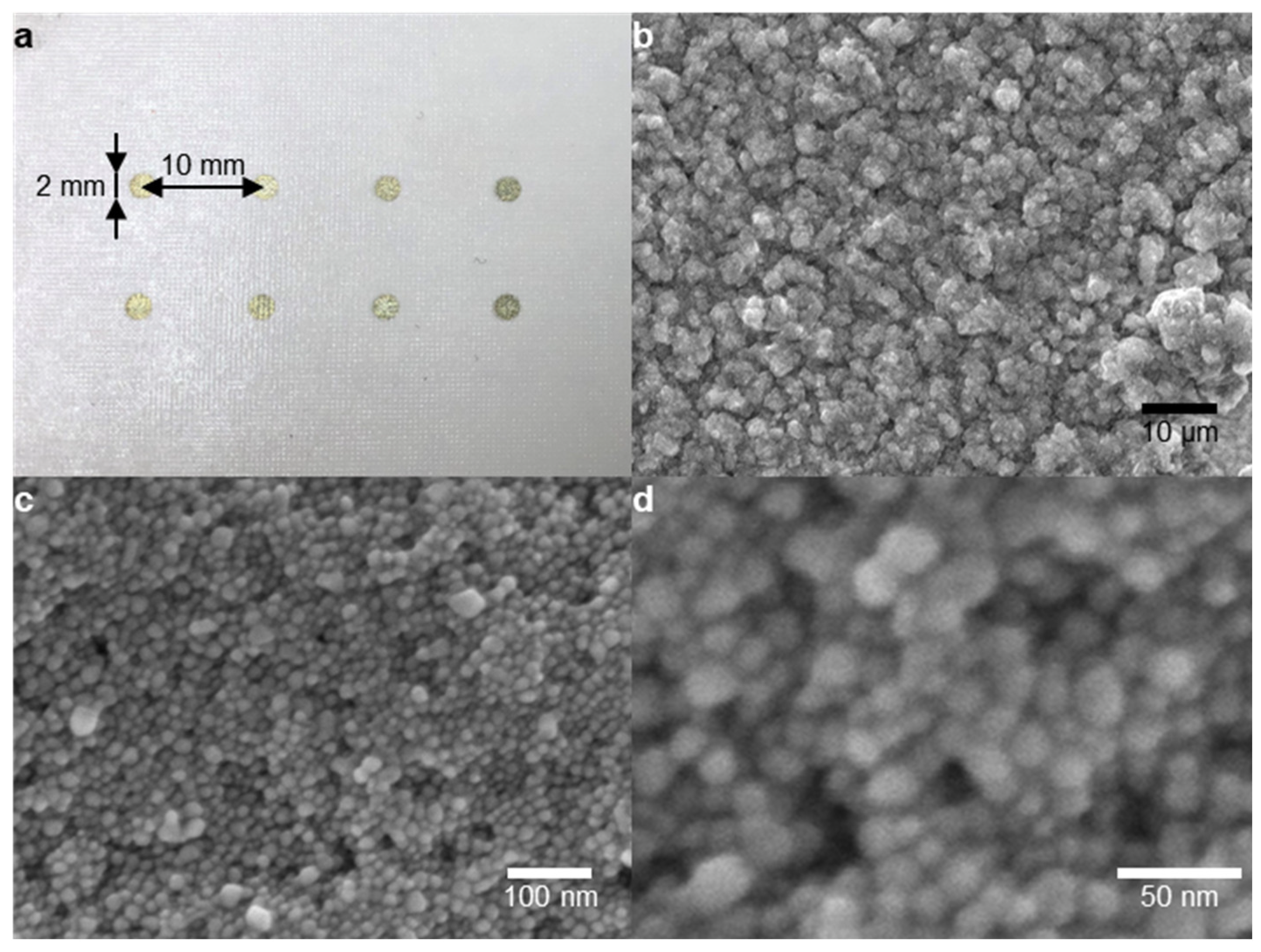
Figure 4a shows the AgNP patterns on the tattoo substrate. It was printed with a 2 mm diameter and a spacing of 10 mm, and it was diced into 10 mm × 10 mm cells for convenient use. As shown in the scanning electron microscope (SEM) images in Figure 4b–d, the AgNPs were macroscopically uniformly coated on the tattoo paper, and the microscopically rough surfaces of the AgNPs clusters were observed. The size of each particle was measured using an image analysis program (ImageJ, National Institutes of Health, USA). The average diameter was 19.5 nm and the standard deviation was 3.94 nm. The rough surfaces of the AgNP clusters concentrated the electromagnetic field and amplified the Raman signal.

3.2. Identification of Benzenethiol and Thiram

In order to evaluate the sensitivity of the tattoo paper-based SERS substrates, the diced substrates were incubated in 1 μM and 1 mM benzenethiol solutions. As shown in Figure 5a, five clear SERS peaks were observed from both the 1 μM and 1 mM benzenethiol solutions. Four peaks came from the benzenethiol and one came from the tattoo paper. These peaks were located at 999 cm−1 (in-plain ring-breathing), 1022 cm−1 (in-plain C−H bending), 1072 cm−1 (C−S stretching), 1410 cm−1 (peak from the tattoo paper), and 1574 cm−1 (C−C stretching), and they corresponded to the main peaks of benzenethiol [31,47]. These large SERS peaks prove that the tattoo paper-based substrates can be used as a SERS substrate for detecting chemicals.
The various concentrations of thiram solutions were also measured in the same way as the benzenethiol. Thiram can be identified by the SERS peaks located at 560 cm−1 (S–S stretching), 933 cm−1 (CH3N stretching), 1144 cm−1 (C–N stretching and CH3 rocking), and 1382 cm−1 (C–N stretching and symmetric CH3 deformation) [40,41,48]. The strongest peak at 1382 cm−1 was selected for the quantitative analysis. Figure 5b shows the SERS spectra of the 1, 10, 100, 200, and 600 μM and 1 mM thiram solution. Small but clear peaks were observed at 560, 933 and 1144 cm−1, and the largest and most clear peaks were located at 1382 cm−1.
According to European Food Safety Authority (EFSA) [49], the acceptable daily intake and acute reference dose of thiram are 0.01 and 0.025 mg/kg/day, respectively. Assuming that a 70 kg person eats an apple with a diameter of 8 cm per day, and that a 1 mm solution film is formed when a pesticide solution is sprayed in an orchard, the acceptable daily intake and acute reference dose of thiram are calculated to be 144.80 and 361.99 μM, respectively. Therefore, 1 μM to 1 mM thiram solutions were used in this experiment.

3.3. Identification of Thiram Using Real Fruit

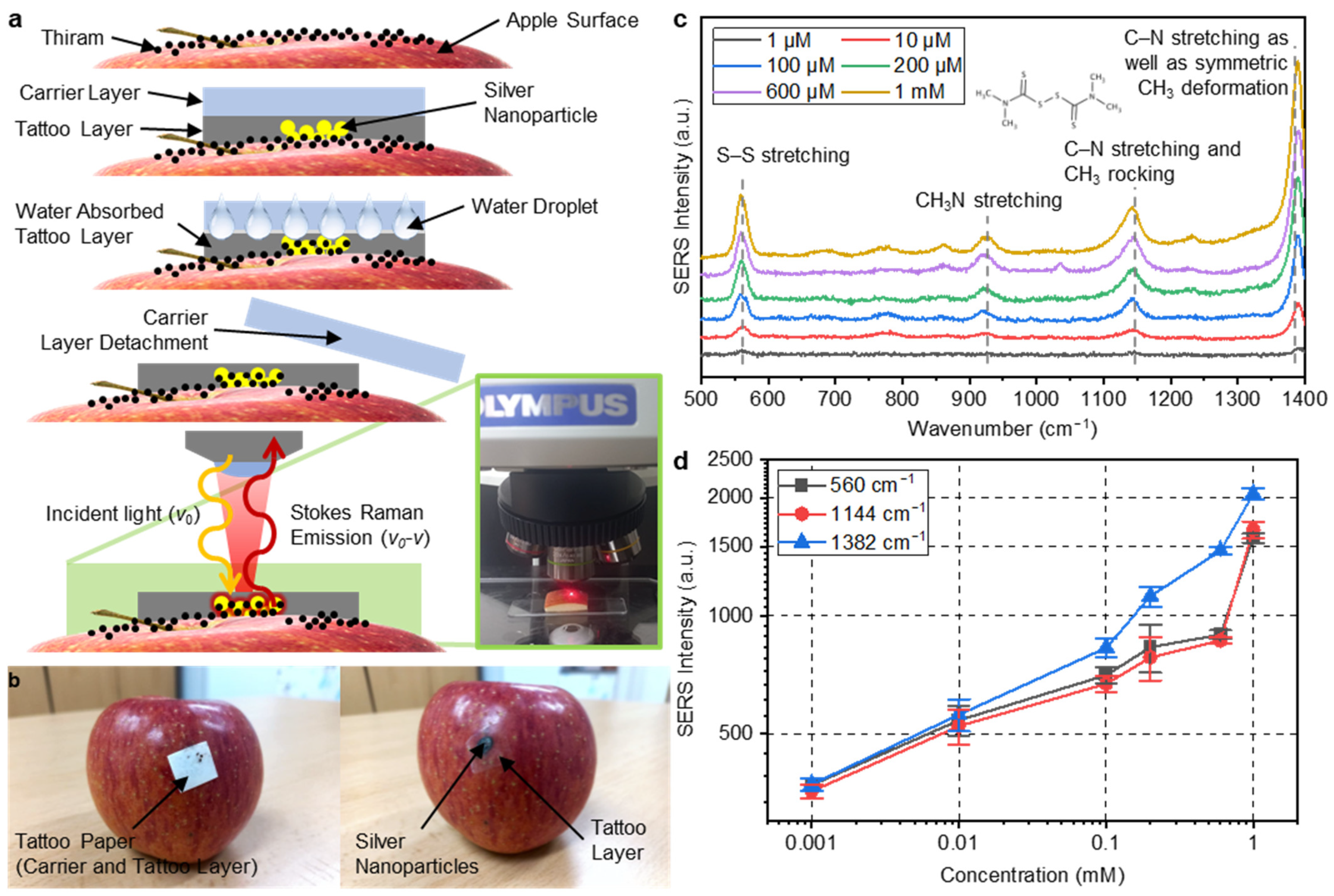
As a feasibility study, we measured the thiram on a fruit’s surface. For reliable measurement, apples purchased from a grocery store were washed clean to remove pesticides and any other impurities on the surface. Various concentrations of thiram solutions were dropped onto the surface of the apples and dried to reproduce pesticide residues. Figure 6a,b show schematic illustrations and photos of the measurement procedures. A piece of tattoo paper-based SERS sheet was placed on the surface of an apple and a few drops of water were dropped. The tattoo paper absorbed the water, and the carrier layer and the tattoo layer were gently separated. The AgNPs on the surface of the tattoo layer came into contact with the surface of the apple smeared with thiram, and the tattoo layer covered it. The water used to attach the tattoo paper dissolved the thiram molecules. This dissolved thiram solution was absorbed into the tattoo layer and increased its contact with the AgNP layer. Since the tattoo layer had very high transparency, the laser could pass through it and reach the AgNPs. In addition, since the tattoo layer covered the SERS layer, the sensing layer could be protected from the external friction that may be applied in an orchard.
Figure 6c shows the SERS spectra of the 1, 10, 100, 200, and 600 μM and 1 mM thiram solution applied to the apples. Four clear peaks at 560, 933, 1144, and 1382 cm−1 were observed. As shown in the concentration-intensity curves in Figure 6d, the SERS intensity of the three major peaks increased as the concentration of thiram increased. The tattoo paper-based SERS substrate could detect 1 μM thiram on the surface of an apple by analyzing the clearest peak at 1382 cm−1.

4. Conclusions

In this study, we have fabricated a tattoo paper-based AgNP SERS substrate. AgNPs were printed onto the surface of the tattoo paper using an inkjet printer. This method of utilizing an inkjet printer is not only helpful in ensuring speedy production, but also enables the possibility of mass production. Moreover, it is fabricated at an ambient temperature and pressure, requires a very small quantity of ink, and inexpensive tattoo paper is utilized. Thus, it is a cost-effective technique. We ensured the sensing performance of the tattoo paper-based SERS substrate by measuring as low as 1 μM benzenethiol and thiram standard solutions. To demonstrate its practicality, this substrate was applied to fruit. Being flexible and adhesive, the tattoo paper was effortlessly attached to an apple’s surface. It succeeded in detecting as low as 1 μM thiram on the apple’s surface. Further studies are needed to develop more sensitive SERS substrates for lower detection limits, to diversify the detectable chemicals, and to elaborate the quantification processes.
As this study presents a novel method of attaching SERS substrates onto curved surfaces, it can be used to analyze materials on the surface of various fruits and vegetables. In addition, since tattoo paper-based SERS substrates are considerably environment-friendly and cost-effective, very easy to handle, and involve simpler sample collection methodology, they can be widely used for onsite pollutant detection in water and other chemical-based food adulterants and additives in future works and applications. Furthermore, as tattoo paper can be easily attached to human and animal skin, it is expected to expand its use for medical purposes in the near future.

Author Contributions

D.Y., P.P.M., M.K., I.P., and B.K. conceived and designed the experiments; P.P.M. and M.K. performed the experiments; D.Y. and P.P.M. analyzed the data; D.Y. and P.P.M. evaluated the validation; D.Y. and P.P.M. wrote the article; D.Y., P.P.M., I.P., and B.K. reviewed the article. All authors have read and agreed to the published version of the manuscript.

Funding

This work was supported by a research grant from Kongju National University in 2019.

Institutional Review Board Statement

Not applicable.

Informed Consent Statement

Not applicable.

Data Availability Statement

Data are available from the corresponding author upon request.

Conflicts of Interest

The authors declare no conflict of interest.

References

  1. Sivaperumal, P.; Thasale, R.; Kumar, D.; Mehta, T.G.; Limbachiya, R. Human health risk assessment of pesticide residues in vegetable and fruit samples in Gujarat State, India. Heliyon 2022, 8, e10876. [Google Scholar]
  2. Lerro, C.C.; Beane Freeman, L.E.; DellaValle, C.T.; Andreotti, G.; Hofmann, J.N.; Koutros, S.; Parks, C.G.; Shrestha, S.; Alavanja, M.C.R.; Blair, A.; et al. Pesticide exposure and incident thyroid cancer among male pesticide applicators in agricultural health study. Environ. Int. 2021, 146, 106187. [Google Scholar] [CrossRef]
  3. Islam, M.A.; Amin, S.M.N.; Rahman, M.A.; Juraimi, A.S.; Uddin, M.K.; Brown, C.L.; Arshad, A. Chronic effects of organic pesticides on the aquatic environment and human health: A review. Environ. Nanotechnol. Monit. Manag. 2022, 18, 100740. [Google Scholar] [CrossRef]
  4. Alder, L.; Greulich, K.; Kempe, G.; Vieth, B. Residue analysis of 500 high priority pesticides: Better by GC-MS or LC-MS/MS? Mass. Spectrom. Rev. 2006, 25, 838–865. [Google Scholar] [CrossRef]
  5. Khetagoudar, M.C.; Chetti, M.B.; Bilehal, D.C. Gas Chromatographic-Mass Spectrometric Detection of Pesticide Residues in Grapes. In Gas Chromatography; Kusch, P., Ed.; IntechOpen: Rijeka, Croatia, 2019; Chapter 5; pp. 1–11. [Google Scholar]
  6. Hogendoorn, E.A. High-Performance Liquid Chromatography Methods in Pesticide Residue Analysis. In Encyclopedia of Analytical Chemistry, 1st ed.; John Wiley & Sons, LTD.: Hoboken, NJ, USA, 2006; pp. 1–36. [Google Scholar]
  7. Albero, B.; Sánchez-Brunete, C.; Tadeo, J.L. Determination of Thiabendazole in Orange Juice and Rind by Liquid Chromatography with Fluorescence Detection and Confirmation by Gas Chromatography/Mass Spectrometry After Extraction by Matrix Solid-Phase Dispersion. J. AOAC Int. 2019, 87, 664–670. [Google Scholar] [CrossRef] [Green Version]
  8. Ping, H.; Wang, B.; Li, C.; Li, Y.; Ha, X.; Jia, W.; Li, B.; Ma, Z. Potential health risk of pesticide residues in greenhouse vegetables under modern urban agriculture: A case study in Beijing, China. J. Food Compos. Anal. 2022, 105, 104222. [Google Scholar] [CrossRef]
  9. Nunes, G.S.; Toscano, I.A.; Barceló, D. Analysis of pesticides in food and environmental samples by enzyme-linked immunosorbent assays. TrAC Trends Anal. Chem. 1998, 17, 79–87. [Google Scholar] [CrossRef]
  10. Wu, S.; Yan, C.; Fan, X.; Wang, H.; Wang, Y.; Peng, D. Development of enzyme-linked immunosorbent assay and colloidal gold-based immunochromatographic assay for the rapid detection of gentamicin in chicken muscle and milk. Chin. J. Anal. Chem. 2022, 50, 100142. [Google Scholar] [CrossRef]
  11. Huang, R.; Huang, Y.; Liu, H.; Guan, K.; Chen, A.; Zhao, X.; Wang, S.; Zhang, L. A bifunctional AuNP probe-based enzyme-linked immunosorbent assay for facile and ultrasensitive detection of trace zearalenone in coix seed. Microchem. J. 2023, 184, 108152. [Google Scholar] [CrossRef]
  12. Feng, R.; Wang, M.; Qian, J.; He, Q.; Zhang, M.; Zhang, J.; Zhao, H.; Wang, B. Monoclonal antibody-based enzyme-linked immunosorbent assay and lateral flow immunoassay for the rapid screening of paraquat in adulterated herbicides. Microchem. J. 2022, 180, 107644. [Google Scholar] [CrossRef]
  13. Pang, S.; Labuza, T.P.; He, L. Development of a single aptamer-based surface enhanced Raman scattering method for rapid detection of multiple pesticides. Analyst 2014, 139, 1895–1901. [Google Scholar] [CrossRef]
  14. Khadem, M.; Faridbod, F.; Norouzi, P.; Foroushani, A.R.; Ganjali, M.R.; Shahtaheri, S.J. Biomimetic electrochemical sensor based on molecularly imprinted polymer for dicloran pesticide determination in biological and environmental samples. J. Iran. Chem. Soc. 2016, 13, 2077–2084. [Google Scholar] [CrossRef]
  15. Moriwaki, H.; Yamada, K.; Nakanishi, H. Evaluation of the Interaction between Pesticides and a Cell Membrane Model by Surface Plasmon Resonance Spectroscopy Analysis. J. Agric. Food Chem. 2017, 65, 5390–5396. [Google Scholar] [CrossRef]
  16. Kumar, S.; Goel, P.; Singh, J.P. Flexible and robust SERS active substrates for conformal rapid detection of pesticide residues from fruits. Sens. Actuators B Chem. 2017, 241, 577–583. [Google Scholar] [CrossRef]
  17. Kahraman, M.; Mullen, E.R.; Korkmaz, A.; Wachsmann-Hogiu, S. Fundamentals and applications of SERS-based bioanalytical sensing. Nanophotonics 2017, 6, 831–852. [Google Scholar] [CrossRef] [Green Version]
  18. Pilot, R.; Signorini, R.; Durante, C.; Orian, L.; Bhamidipati, M.; Fabris, L. A Review on Surface-Enhanced Raman Scattering. Biosensors 2019, 9, 57. [Google Scholar] [CrossRef] [Green Version]
  19. Langer, J.; Jimenez de Aberasturi, D.; Aizpurua, J.; Alvarez-Puebla, R.; Auguié, B.; Baumberg, J.J.; Bazan, G.C.; Bell, S.E.J.; Boisen, A.; Brolo, A.G.; et al. Present and Future of Surface-Enhanced Raman Scattering. ACS Nano 2020, 14, 28–117. [Google Scholar] [CrossRef] [Green Version]
  20. He, S.; Xie, W.; Fang, S.; Huang, X.; Zhou, D.; Zhang, Z.; Du, J.; Du, C.; Wang, D. Silver films coated inverted cone-shaped nanopore array anodic aluminum oxide membranes for SERS analysis of trace molecular orientation. Appl. Surf. Sci. 2019, 488, 707–713. [Google Scholar] [CrossRef]
  21. Liu, Y.; Wu, S.; Du, X.; Sun, J. Plasmonic Ag nanocube enhanced SERS biosensor for sensitive detection of oral cancer DNA based on nicking endonuclease signal amplification and heated electrode. Sens. Actuators B Chem. 2021, 338, 129854. [Google Scholar] [CrossRef]
  22. Mandavkar, R.; Lin, S.; Pandit, S.; Kulkarni, R.; Burse, S.; Habib, M.A.; Kunwar, S.; Lee, J. Hybrid SERS platform by adapting both chemical mechanism and electromagnetic mechanism enhancements: SERS of 4-ATP and CV by the mixture with GQDs on hybrid PdAg NPs. Surf. Interfaces 2022, 33, 102175. [Google Scholar] [CrossRef]
  23. Moronshing, M.; Bhaskar, S.; Mondal, S.; Ramamurthy, S.S.; Subramaniam, C. Surface-enhanced Raman scattering platform operating over wide pH range with minimal chemical enhancement effects: Test case of tyrosine. J. Raman Spectrosc. 2019, 50, 826–836. [Google Scholar] [CrossRef]
  24. Bhaskar, S.; Thacharakkal, D.; Ramamurthy, S.S.; Subramaniam, C. Metal–Dielectric Interfacial Engineering with Mesoporous Nano-Carbon Florets for 1000-Fold Fluorescence Enhancements: Smartphone-Enabled Visual Detection of Perindopril Erbumine at a Single-molecular Level. ACS Sustain. Chem. Eng. 2023, 11, 78–91. [Google Scholar] [CrossRef]
  25. Parmigiani, M.; Albini, B.; Pellegrini, G.; Genovesi, M.; De Vita, L.; Pallavicini, P.; Dacarro, G.; Galinetto, P.; Taglietti, A. Surface-Enhanced Raman Spectroscopy Chips Based on Silver Coated Gold Nanostars. Nanomaterials 2022, 12, 3609. [Google Scholar] [CrossRef]
  26. Gao, T.; Xu, Z.; Fang, F.; Gao, W.; Zhang, Q.; Xu, X. High performance surface-enhanced Raman scattering substrates of Si-based Au film developed by focused ion beam nanofabrication. Nanoscale Res. Lett. 2012, 7, 399. [Google Scholar] [CrossRef] [Green Version]
  27. Melekhov, E.; Penn, T.; Weidauer, T.; Abb, V.; Kammler, M.; Lechner, A. Tunable nanopillar array on a quartz-fiber tip for surface enhanced Raman scattering (SERS) detection. Tech. Mess. 2022, 89, 70–81. [Google Scholar] [CrossRef]
  28. Chen, X.; Lin, M.; Sun, L.; Xu, T.; Lai, K.; Huang, M.; Lin, H. Detection and quantification of carbendazim in Oolong tea by surface-enhanced Raman spectroscopy and gold nanoparticle substrates. Food Chem. 2019, 293, 271–277. [Google Scholar] [CrossRef]
  29. Sitjar, J.; Liao, J.; Lee, H.; Liu, B.H.; Fu, W. SERS-Active Substrate with Collective Amplification Design for Trace Analysis of Pesticides. Nanomaterials 2019, 9, 664. [Google Scholar] [CrossRef] [Green Version]
  30. Calis, B.; Yilmaz, M. Fabrication of gold nanostructure decorated polystyrene hybrid nanosystems via poly(L-DOPA) and their applications in surface-enhanced Raman Spectroscopy (SERS), and catalytic activity. Colloids Surf. A Physicochem. Eng. Asp. 2021, 622, 126654. [Google Scholar] [CrossRef]
  31. Yang, D.; Cho, H.; Koo, S.; Vaidyanathan, S.R.; Woo, K.; Yoon, Y.; Choo, H. Simple, Large-Scale Fabrication of Uniform Raman-Enhancing Substrate with Enhancement Saturation. ACS Appl. Mater. Interfaces 2017, 9, 19092–19101. [Google Scholar] [CrossRef] [Green Version]
  32. Izquierdo-Lorenzo, I.; Jradi, S.; Adam, P. Direct laser writing of random Au nanoparticle three-dimensional structures for highly reproducible micro-SERS measurements. RSC Adv. 2014, 4, 4128–4133. [Google Scholar] [CrossRef]
  33. Liu, S.; Tian, X.; Guo, J.; Kong, X.; Xu, L.; Yu, Q.; Wang, A.X. Multi-functional plasmonic fabrics: A flexible SERS substrate and anti-counterfeiting security labels with tunable encoding information. Appl. Surf. Sci. 2021, 567, 150861. [Google Scholar] [CrossRef]
  34. Chen, Y.; Ge, F.; Guang, S.; Cai, Z. Low-cost and large-scale flexible SERS-cotton fabric as a wipe substrate for surface trace analysis. Appl. Surf. Sci. 2018, 436, 111–116. [Google Scholar] [CrossRef]
  35. Lin, S.; Lin, X.; Han, S.; Liu, Y.; Hasi, W.; Wang, L. Flexible fabrication of a paper-fluidic SERS sensor coated with a monolayer of core–shell nanospheres for reliable quantitative SERS measurements. Anal. Chim. Acta 2020, 1108, 167–176. [Google Scholar] [CrossRef]
  36. Luo, J.; Wang, Z.; Li, Y.; Wang, C.; Sun, J.; Ye, W.; Wang, X.; Shao, B. Durable and flexible Ag-nanowire-embedded PDMS films for the recyclable swabbing detection of malachite green residue in fruits and fingerprints. Sens. Actuators B Chem. 2021, 347, 130602. [Google Scholar] [CrossRef]
  37. Zhang, S.; Xu, J.; Liu, Z.; Huang, Y.; Fu, R.; Jiang, S. Facile and scalable preparation of solution-processed succulent-like silver nanoflowers for 3D flexible nanocellulose-based SERS sensors. Surf. Interfaces 2022, 34, 102391. [Google Scholar] [CrossRef]
  38. Cai, Y.; Yao, X.; Piao, X.; Zhang, Z.; Nie, E.; Sun, Z. Inkjet printing of particle-free silver conductive ink with low sintering temperature on flexible substrates. Chem. Phys. Lett. 2019, 737, 136857. [Google Scholar] [CrossRef]
  39. Botti, S.; Rufoloni, A.; Rindzevicius, T.; Schmidt, M.S. Surface-Enhanced Raman Spectroscopy Characterization of Pristine and Functionalized Carbon Nanotubes and Graphene. In Raman Spectroscopy; Morari do Nascimento, G., Ed.; IntechOpen: Rijeka, Croatia, 2018; Chapter 10; pp. 203–219. [Google Scholar]
  40. Pu, H.; Huang, Z.; Xu, F.; Sun, D. Two-dimensional self-assembled Au-Ag core-shell nanorods nanoarray for sensitive detection of thiram in apple using surface-enhanced Raman spectroscopy. Food Chem. 2021, 343, 128548. [Google Scholar] [CrossRef]
  41. Zheng, X.; Chen, Y.; Chen, Y.; Bi, N.; Qi, H.; Qin, M.; Song, D.; Zhang, H.; Tian, Y. High performance Au/Ag core/shell bipyramids for determination of thiram based on surface-enhanced Raman scattering. J. Raman Spectrosc. 2012, 43, 1374–1380. [Google Scholar] [CrossRef]
  42. Varadachari, C.; Mitra, S.; Ghosh, K. Photochemical oxidation of soil organic matter by sunlight. Proc. Indian Natl. Sci. Acad. 2017, 83, 223–229. [Google Scholar]
  43. Böhnke, P.R.; Kruppke, I.; Hoffmann, D.; Richter, M.; Häntzsche, E.; Gereke, T.; Kruppke, B.; Cherif, C. Matrix Decomposition of Carbon-Fiber-Reinforced Plastics via the Activation of Semiconductors. Materials 2020, 13, 3267. [Google Scholar] [CrossRef]
  44. Austin, A.T.; Méndez, M.S.; Ballaré, C.L. Photodegradation alleviates the lignin bottleneck for carbon turnover in terrestrial ecosystems. Proc. Natl. Acad. Sci. USA 2016, 113, 4392–4397. [Google Scholar] [CrossRef] [Green Version]
  45. Ravikumar, S.; Mani, D.; Rizwan Khan, M.; Ahmad, N.; Gajalakshmi, P.; Surya, C.; Durairaj, S.; Pandiyan, V.; Ahn, Y. Effect of silver incorporation on the photocatalytic degradation of Reactive Red 120 using ZnS nanoparticles under UV and solar light irradiation. Environ. Res. 2022, 209, 112819. [Google Scholar] [CrossRef]
  46. Gola, D.; Kriti, A.; Bhatt, N.; Bajpai, M.; Singh, A.; Arya, A.; Chauhan, N.; Srivastava, S.K.; Tyagi, P.K.; Agrawal, Y. Silver nanoparticles for enhanced dye degradation. Curr. Res. Green Sustain. Chem. 2021, 4, 100132. [Google Scholar] [CrossRef]
  47. Joo, T.H.; Kim, M.S.; Kim, K. Surface-enhanced Raman scattering of benzenethiol in silver sol. J. Raman. Spectrosc. 1987, 18, 57–60. [Google Scholar] [CrossRef]
  48. Chen, J.; Huang, Y.; Kannan, P.; Zhang, L.; Lin, Z.; Zhang, J.; Chen, T.; Guo, L. Flexible and Adhesive Surface Enhance Raman Scattering Active Tape for Rapid Detection of Pesticide Residues in Fruits and Vegetables. Anal. Chem. 2016, 88, 2149–2155. [Google Scholar] [CrossRef]
  49. European Food Safety Authority. Peer review of the pesticide risk assessment of the active substance thiram. EFSA J. 2017, 15, e04700. [Google Scholar]
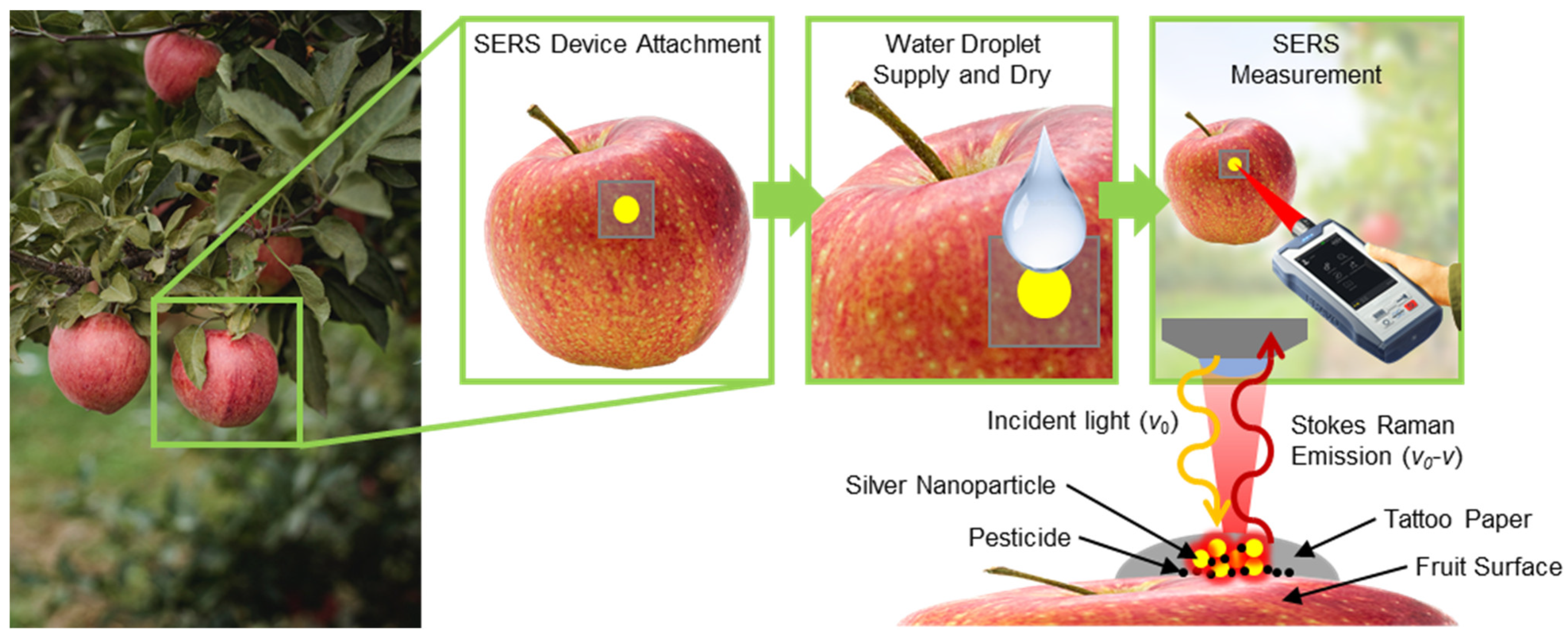
Figure 1. Application of the tattoo paper-based AgNP SERS substrates on a real fruit (apple) sample and identification of the pesticide using a Raman spectrometer.
Nanomaterials 13 00486 g001
Figure 2. Fabrication steps for the tattoo paper-based AgNP SERS substrates.
Nanomaterials 13 00486 g002
Figure 3. (a) SERS intensity spectra of substrates showing gradual carbon decomposition by UV irradiation within the time period from 0 to 60 h; (b) reduction in carbon intensity during UV irradiation; and (c) EDS data before and after the substrates were subjected to UV irradiation.
Nanomaterials 13 00486 g003
Figure 4. (a) Photo of the printed AgNP dot array on tattoo paper; and (bd) SEM images of the AgNP clusters printed on the tattoo paper substrate.
Nanomaterials 13 00486 g004
Figure 5. (a) SERS intensities at 999, 1022, 1072, 1410, and 1574 cm−1, which were measured during the identification of the 1 μM and 1 mM concentrations of benzenethiol solutions; and (b) SERS intensity spectrum without thiram and with thiram at concentrations of 1, 10, 100, 200, and 600 μM and 1 mM, exhibiting peak identification at 560, 933, 1144, and 1382 cm−1 for all the concentrations.
Nanomaterials 13 00486 g005
Figure 6. (a) Working mechanism of fabricated tattoo paper-based AgNP SERS substrates using a thiram contaminated fruit (apple) sample; (b) real apple specimen (thiram contaminated) used for the analysis; (c) SERS intensity spectra of the apple specimens showing peak intensities at 560, 933, 1144, and 1382 cm−1 for the different concentration of 1, 10, 100, 200, and 600 μM and 1 mM of thiram solutions; and (d) intensity curve showing increasing absorption at the 560, 1144, and 1382 cm−1 peaks with the higher concentration level of thiram solutions.
Nanomaterials 13 00486 g006
Disclaimer/Publisher’s Note: The statements, opinions and data contained in all publications are solely those of the individual author(s) and contributor(s) and not of MDPI and/or the editor(s). MDPI and/or the editor(s) disclaim responsibility for any injury to people or property resulting from any ideas, methods, instructions or products referred to in the content.