Article

Interfacial Charge Transfer Effects of MoS2/α-Fe2O3 Nano-Heterojunction and Efficient Photocatalytic Hydrogen Evolution under Visible-Light Irradiation

Coastal Water and Environment Center, College of Hydrosphere Science, National Kaohsiung University of Science and Technology, Kaohsiung 81157, Taiwan
*
Authors to whom correspondence should be addressed.
Nanomaterials 2023, 13(20), 2763; https://doi.org/10.3390/nano13202763
Submission received: 6 September 2023 / Revised: 7 October 2023 / Accepted: 8 October 2023 / Published: 15 October 2023

Abstract

:
Researchers have made efforts to develop high-productivity photocatalysts for photocatalytic hydrogen production to reduce the problem of a lack of energy. Bulk semiconductor photocatalysts mainly endure particular limitations, such as low visible light application, a quick recombination rate of electron–hole pairs, and poor photocatalytic efficiency. The major challenge is to improve solar-light-driven heterostructure photocatalysts that are highly active and stable under the photocatalytic system. In this study, the proposed nano-heterojunction exhibits a great capacity for hydrogen production (871.2 μmol g−1 h−1), which is over 8.1-fold and 12.3-fold higher than that of the bare MoS2 and bare α-Fe2O3 samples, respectively. It is demonstrated that the MoS2/α-Fe2O3 heterojunction gives rise to an enhanced visible light response and accelerated photoinduced charge carrier separation. This work provides an improved visible light absorption efficiency and a narrowed energy band gap, and presents a “highway” for electron–hole pairs to promote transfer and inhibit the combination of photoinduced charge carriers for the utilization of nano-heterojunction photocatalysts in the field of hydrogen production.

1. Introduction

Solar-driven photocatalytic hydrogen evolution (PHE) via water splitting has garnered increasing attention because of the remarkably favorable and useful strategies used to transform renewable solar light into neat chemical energy [1,2]. Because it is essential that supportable growth diminishes the movement of carbon dioxide (CO2) and organic compounds to their surroundings, practical nanoparticles for novel clear energy utilization processes like photocatalysis and electrocatalysis routes have been effectively probed for decades [3]. Visible-light-driven nanomaterials are used to split water and transform solar light energy into purified energy [4,5]. Within all observed semiconductor photocatalysts, all efforts to produce hydrogen (H2) through heterojunction-based photocatalysis to date have unfavorably fallen short of the capability rates needed for efficient utilization [6]. To figure out this issue and obtain sustainable growth, renewable energy sources that could be employed and offered as the main renewable power source, like H2, need to be evaluated [7]. Nearly all scientists have taken advantage of its facilely managed catalyst configuration [8,9] and handily managed the band gap energy to regularly realize it as one of the most favorable semiconductors. Photocatalytic H2 evolution from water with visible light activity has attracted much attention owing to its achievable utilization in the transition and capacity of abundant solar sources. Cocatalysts commonly act as reactive locations for the production of photoinduced carriers and decrease the response energy barriers for photocatalytic H2 production. Among these semiconductor photocatalysts, hematite (α-Fe2O3) has great photocatalytic efficiency for hydrogen generation [10]. The photo-Fenton photocatalyst is broad because of its proper energy band gap (~2.1 eV), inexpensiveness, high stability, and environmental kindness [11,12]. The photo-Fenton reaction is a procedure in which Fe2+ and H2O2 join to generate OH. On the other hand, the photocatalytic efficiency of α-Fe2O3 is constrained by its short diffusion length, high electron–hole recombination rate, and the poor electrical conductivity of charge carriers [13]. The suggested MoS2/α-Fe2O3 (MF) nano-heterojunction exhibits a greater H2 generation rate of 4356 μmol h−1 g−1 (20 mg sample), which corresponds to that of pristine MoS2 and α-Fe2O3. In addition, the MF nano-heterojunction displays superior stability and recyclability in the 5th cycle test of photocatalytic hydrogen generation.
So far, with regard to traditional ZnO and TiO2 photocatalysts, particular photocatalysts like MoS2, Co3O4, Cu2O, WS2, and Fe2O3 have been more broadly investigated in the area of photocatalysis [14,15]. MoS2, as the favorable substitute, has garnered great attention owing to its abundance and the suitable free energy of the H2 production rate [16]. Nowadays, MoS2 is known to be a good semiconductor for photocatalytic H2 generation with visible light activity [17]. The photocatalyst has a broad range of solar light absorption abilities, has a high capacity for photoinduced charge carrier separation, and a high transfer ability, meaning that it can meet the terms of high-performance interfacial photocatalysis [18]. The great photoinduced charge carrier migration capacity, high H2 generation rate and excellent photocatalytic stability of MoS2-based semiconductors make them favorable for photocatalytic H2 generation. In prior studies, phase junctions on the same material have displayed good photocatalytic efficiency because of their particular benefits, including involving elements, having good electron affinity, and vigorous coupling [19,20].The confined active locations and the unfortunate inherent conductibility of MoS2 prohibit the whole process of PHE. Therefore, integrating the advantages of great conductivity and abundant active sites is still a strenuous mission. Various fabrication and treatment methods have been presented for altering the band gap energy in order to collect solar light and enhance the electron–hole pair separation efficiency in photocatalysis [21,22]. Between these materials, the utilization of a co-catalyst has attracted immense interest due to the heterojunction structure, which could be useful regarding the removal rate of photoexcited electrons and when aiming to avoid the surface electron–hole pair recombination of bulk material [23]. As one of the significant photocatalysts, α-Fe2O3 has excellent properties, a good capacity for visible photoelectric response and a large specific surface area [24,25]. Meanwhile, α-Fe2O3 has great electrical and optical properties, and superior utilization in the fields of photocatalytic and environmental science.
To overcome the above problems, several procedures have been used to improve the photocatalytic efficiency by enhancing the charge carrier separation rate; these include the addition of chemicals to alter the energy band gap of heterojunctions with cocatalysts and the building of Z-scheme routes. Semiconductor heterojunctions have been suggested to improve interfacial electron–hole pair transfer and the separation of charge carriers, driven by an internal electrostatic field in the heterojunction structure. Consequently, it is beneficial to create photocatalyst nano-heterojunctions that not only improve the practical electron and hole separation rate, but also promote their transfer efficiency. Up to now, Fe2O3/MoS2 heterostructure photocatalysts have been developed and studied for the degradation of various organic compounds, such as a Fe2O3/MoS2 heterojunction for the photo-Fenton reaction [26,27], and a Fe2O3/MoS2 nanocomposite and QDs for the degradation of dye [28,29]. Moreover, nanocomposites can be quickly formed to form intimate contact heterostructures with different kinds of semiconductors. When two materials come into contact, the charge carriers around the interface of the two materials diffuse through the interface, owing to the density of the charge carrier elevation. Furthermore, nano-scale photocatalysts are more easily transferred from the inner part to the surface because of their small dimensions, which make it much less probable that charge carrier recombination will occur [30,31]. In addition, Chen [32] proposed a novel Z-scheme Fe2O3/MoS2 nanohybrid, in which potential photoinduced electron–hole pairs are saved in the conduction band of the nanohybrid, while inhibition photoexcited electrons and holes are recombined under visible light irradiation. Therefore, the formation of nano-heterojunctions via the merging of narrow-energy-band-gap MoS2 nanoparticles with other nanomaterials with more positive acceleration leads to the separation and transfer of photoexcited carriers for photocatalytic hydrogen evolution.
In this case, the fabricated MF nano-heterojunction will promote the transfer of electrons and inhibit the electron–hole pair recombination rate on the surface of the nanocomposite, as well as contribute great reactive sites for photocatalytic H2 generation. Herein, these heterojunctions not only provide abundant active sites to improve the capacity for photocatalysis, but also inhibit the recombination efficiency of photo-response charge carriers on the surface of the photocatalyst [33]. Furthermore, an appropriate photocatalysis mechanism was presented depending on the active sites catching test, ESR analysis and photoelectrochemical experiments. This study provides guidance for the design and formation of Z-scheme heterojunction photocatalysts toward photocatalytic H2 production.

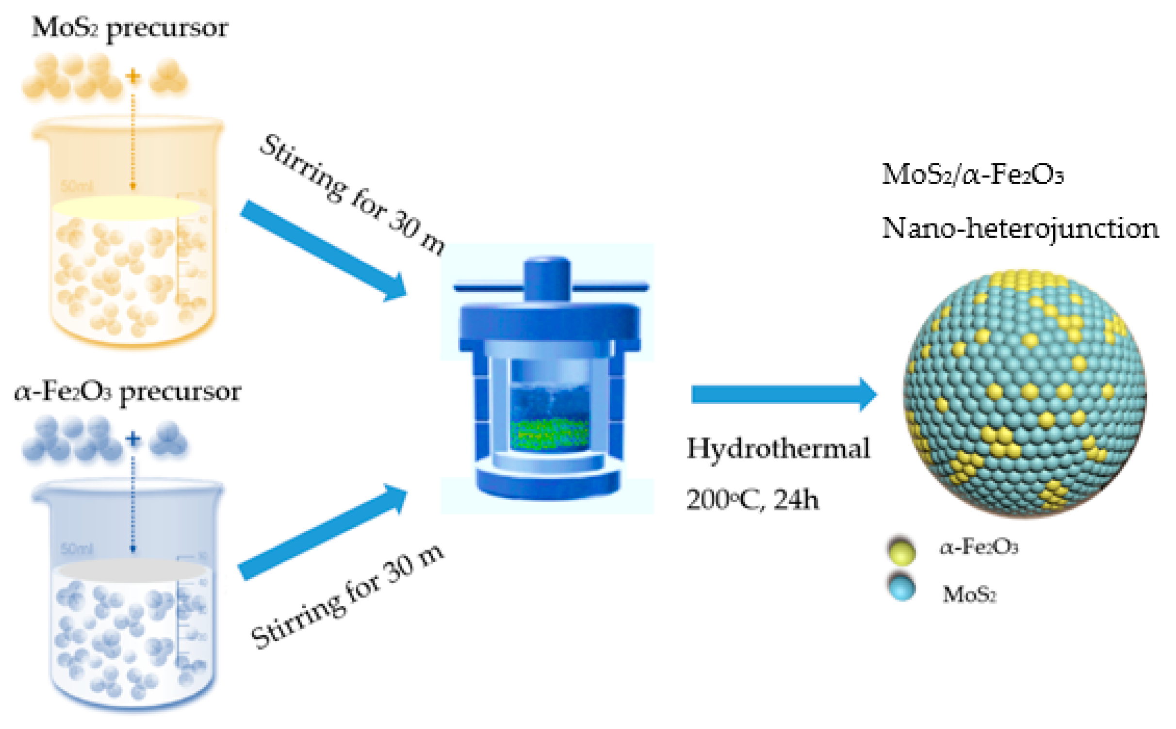
2. Materials and Methods
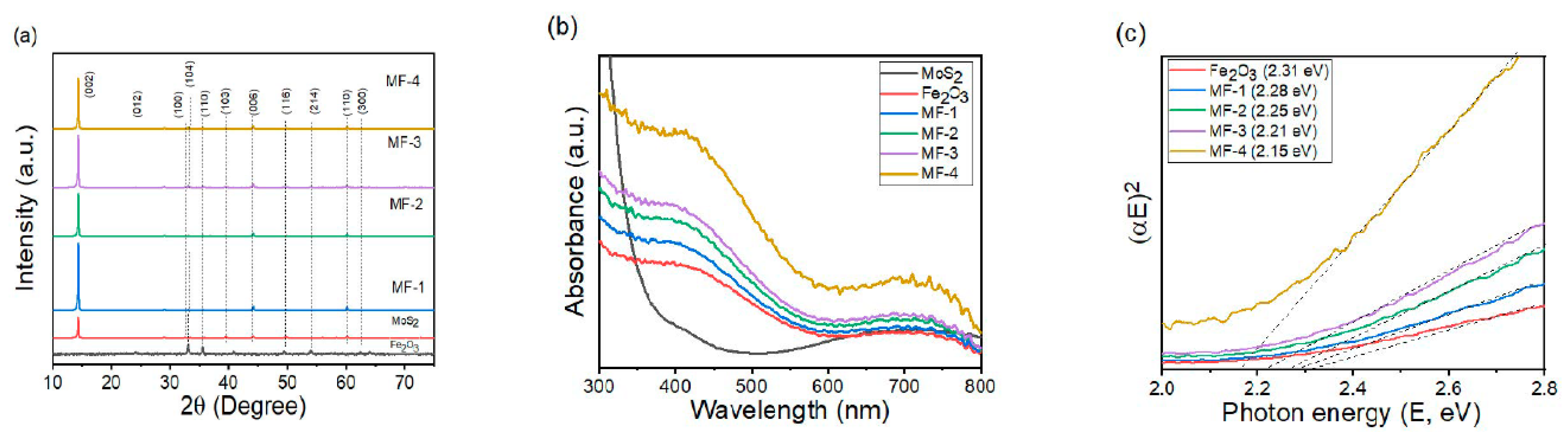
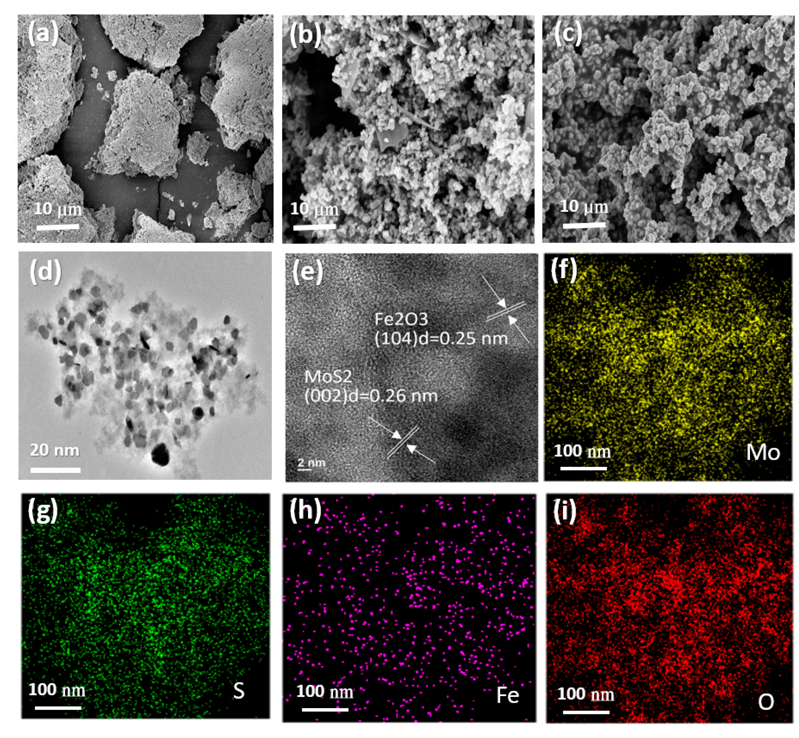
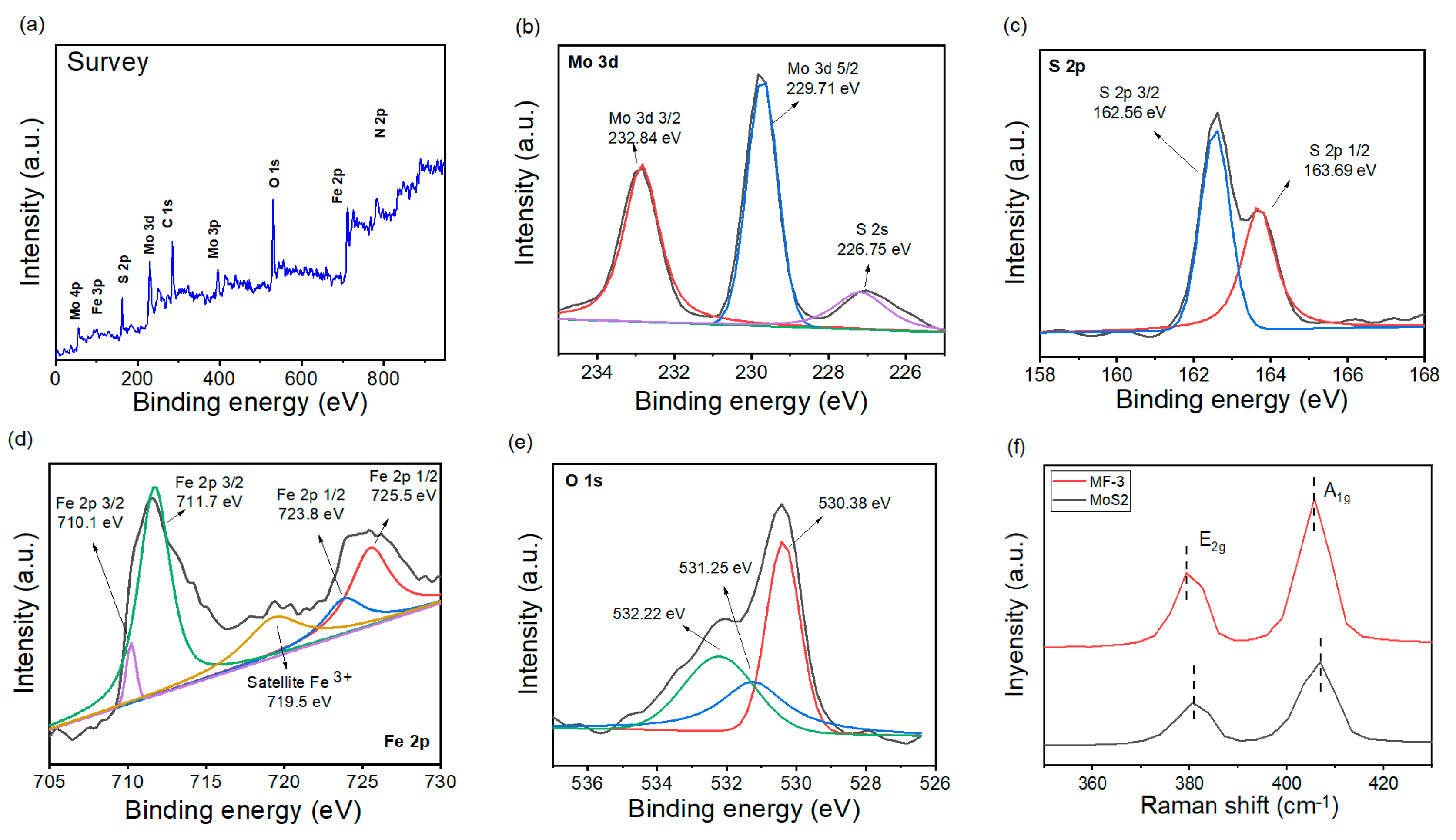
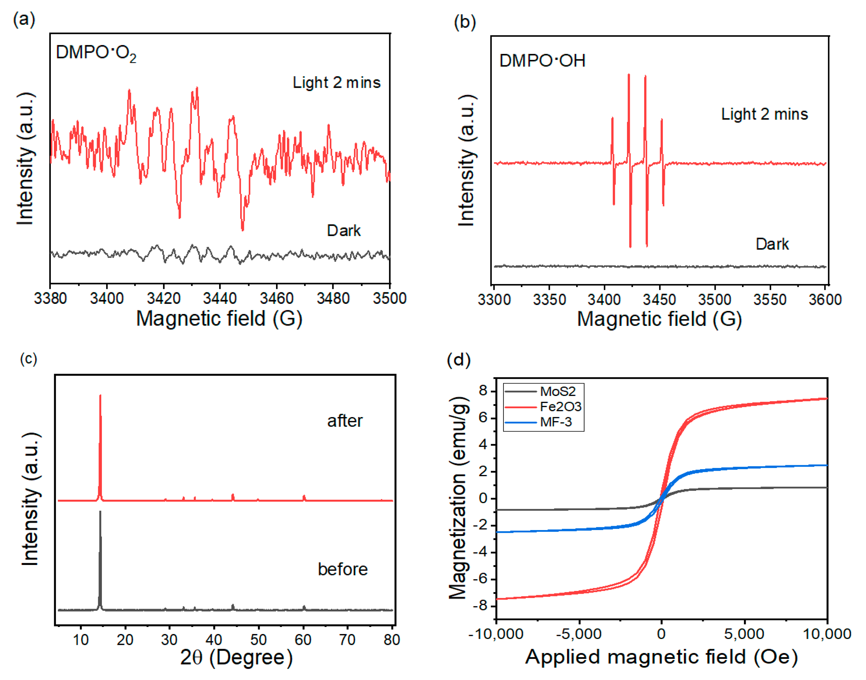
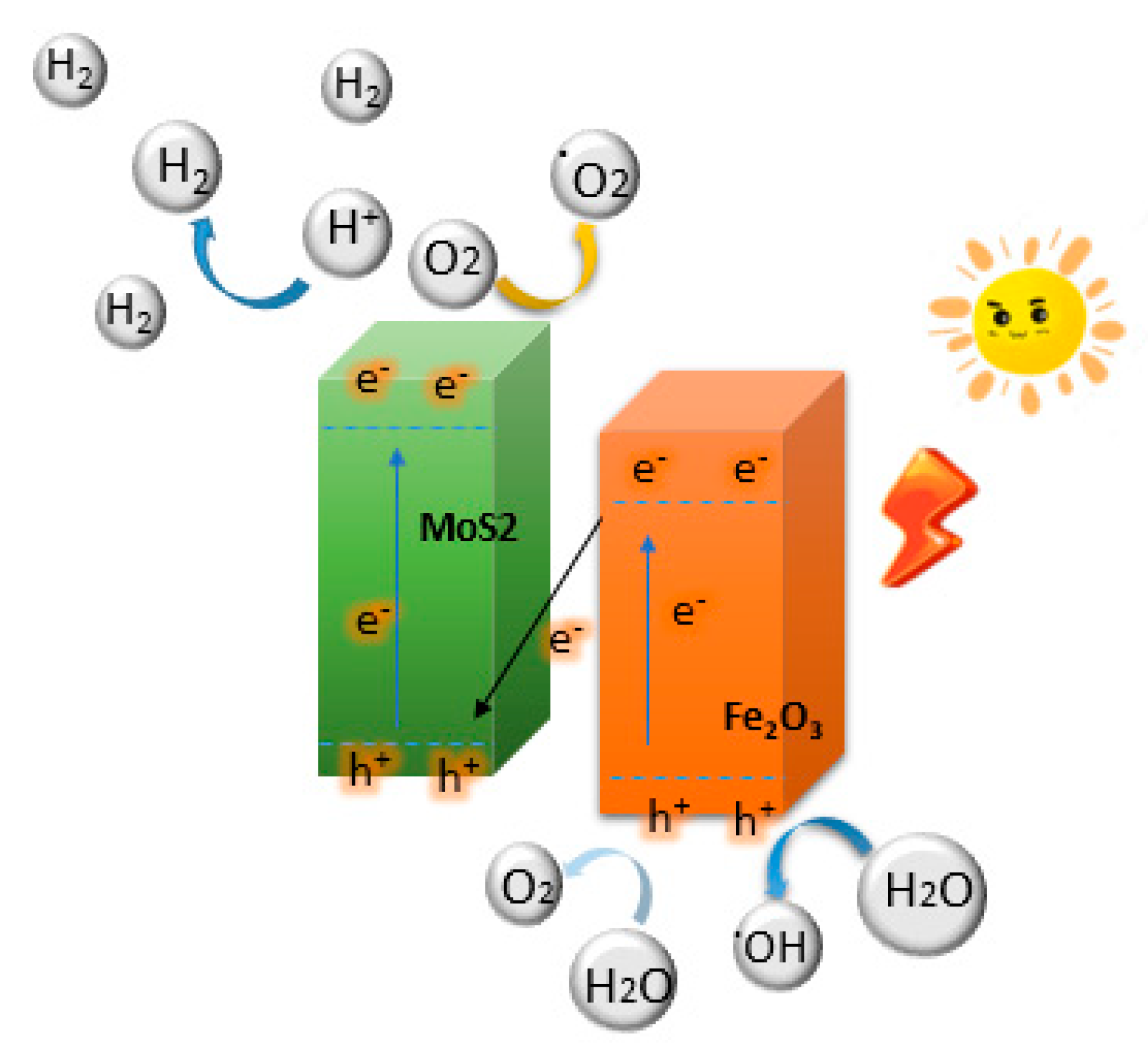
Sodium molybdate dihydrate (Na2MoO4·2H2O), thiourea (CH4N2S), iron(III) nitrate nonahydrate (Fe(NO3)3·9H2O), and hydrochloric acid (HCl, 36.5%) were adopted as precursors for the fabrication of MoS2 and α-Fe2O3, respectively, and purchased from Sigma Aldrich. All the chemical reagents used in this study were provided without further purification.
As shown in Scheme 1, the MF nano-heterojunction used in this work was synthesized with a hydrothermal process [34], in which CH4N2S (2.5 mmol) and Na2MoO4·2H2O (1.5 mmol) were mixed and sonicated evenly in 40 mL of water and kept at 30 °C for 30 min (solution 1). Fe(NO3)3·9H2O was dissolved in 40 mL of distilled water and kept at 30 °C for 30 min (solution 2). The pH value of the mixture solution was set to 7. The mixture solution was distributed constantly using an ultrasonic process, stirred extensively for 30 min, then delivered into a 100 mL Teflon-lined stainless steel autoclave and kept at 200 °C for 24 h. After the hydrothermal process, the sample was cooled down naturally and washed with diluted HCl, H2O, EtOH and NMP three times. Then, the products were dried at 70 °C under vacuum overnight. The adding content of α-Fe2O3 was easily altered by the addition of Fe(NO3)3·9H2O. The nominal amounts of Fe(NO3)3·9H2O added to the mixture solution were 0.5, 1, 1.5, and 2 mmol, and the ratios of α-Fe2O3 for the received samples were MF-1, MF-2, MF-3, and MF-4, respectively.
The photocatalytic tests were performed in a 50 mL glass reactor equipped with a 350 W Xe lamp and a UV cut-off filter (λ > 420 nm). Firstly, the 20 mg photocatalytic product was distributed in a 50 mL mixture solution of 0.25 M/0.35 M Na2SO3/Na2S and lactic acid of 10% sacrifice reagent into a quartz bottle reactor. Before illumination, the suspension of the photocatalyst was distributed using an ultrasonic conductor for 20 min, and thus degassed via the bubbling of N2 for 30 min. The extent of photocatalysis H2 production was evaluated using gas chromatography (GC 7890, TCD detector).
The crystal structure of the photocatalytic materials was studied using X-ray diffraction (XRD) on a Bruker D8 Advanced with Cu Kα radiation. The morphologies of the product were displayed using field emission scanning electron microscopy (FE-SEM, ZEISS AURIGA, Tokyo, Japan). Transition electron microscopy (TEM) observations were characterized with a TEM, JEOL JEM-2100F (Tokyo, Japan) electron microscope. The elemental mapping and composition were collected using an energy-dispersive X-ray spectrometer and evaluated using TEM analysis. The UV–vis spectra (UV–vis) were determined on a spectrophotometer (Hitachi UV-4100, Tokyo, Japan) in the range of 300–800 nm to record the optical response of the prepared samples. The photoluminescence (PL) emission spectrum of the products was recorded using a spectrophotometer (F-7000 FL, Tokyo, Japan), with a xenon lamp acting as the visible light source in the PL spectrophotometer. The elemental compositions and chemical states were recorded using X-ray photoelectron spectroscopy (XPS, Thermo ESCALAB, UK). Electron spin resonance (ESR) analysis was performed using a spectrometer (JEOL JES-FA200, Tokyo, Japan) under visible light activity.

3. Results

XRD analysis was performed to check the crystal and phase structure of the as-fabricated samples and the received results are exhibited in Figure 1. The diffraction peaks located at 14.5°, 32.3°, 39.2°, 44.3° and 60.2° are assigned to the (002), (100), (103), (006) and (110) planes, which are properly matched with the crystal phase of MoS2 and correspond to the JCPDS card (JCPDS No. 73-1503) [35]. The XRD analysis suggests that the semi-crystalline behavior of the fabricated MoS2/α–Fe2O3 nano-heterojunction and events are beneficially matched with the results received. The feature peaks are comparable with the anatase phase of α-Fe2O3. Several additional feature peaks located at 2θ of 24.4°, 33.6°, 35.6°, 49.3°, 54.2°, and 64.5° correspond to the (012), (104), (110), (116), (214), and (300) planes (JCPDS No. 79-0007) [36] of the rhombohedra phase of α-Fe2O3, respectively, in the heterojunction. Owing to the covering of the peaks of MoS2 and α-Fe2O3, some slight altering and extending of the feature peaks were noted. Divided peaks comparable to MoS2 in the XRD analysis of the MoS2/α-Fe2O3 nano-heterojunction were not found, which could be owing to the presence of a low amount. The findings regarding MoS2 do not change the structural and crystallinity characteristics of the heterostructures. The gradual decrease in the peak intensity as the content of α-Fe2O3 increased apparently implies that α-Fe2O3 was successfully loaded on the binary MF nano-heterojunction. When α-Fe2O3 is combined with the MF nano-heterojunction, the diffraction feature peak of α-Fe2O3 does not emerge clearly. This is possibly due to the low amount of α-Fe2O3 and the well-featured peak of the MF nano-heterojunction that covers the diffraction feature peak of α-Fe2O3. In addition, the XRD analysis results demonstrate that the feature peak of the MF nano-heterojunction gradually diminishes with the increase in the α-Fe2O3 amount. This could be due to the addition of α-Fe2O3 having a respective influence on the crystal structure of the MF nano-heterojunction, proving that the nano-heterojunction was successfully fabricated.
To confirm the optical properties of the fabricated photocatalysts, the products were measured using a UV–vis spectrometer in the 300–800 nm wavelength range. Figure 1b exhibits the UV–vis measurement of MoS2, α-Fe2O3 and the MF nano-heterojunction. It was evident that the heterojunction had a stronger capacity to absorb light in the visible light range than MoS2 and α-Fe2O3, which could be attributed to the α-Fe2O3 experiencing more significant absorbance in the range (λ > 450 nm) than the MoS2 samples. The change in absorbance from the UV to visible range extends with a rise in the amount of α-Fe2O3 added. A decrease in the energy band gap of the MF nano-heterojunction was observed with the addition of α-Fe2O3 over the MF nano-heterojunction. The low energy band gap of the heterostructures illustrated the efficient excitation under visible light activity and the improved photocatalytic performance of the MF nano-heterojunction. The improved absorption of the photocatalyst means that many photons are absorbed, inducing more charge carriers in the photocatalyst and promotes the efficiency of H2 evolution. As can be observed in Figure 1c, the forbidden band value of the bare α-Fe2O3 was calculated as 2.31 eV; in addition, the band gap values of the MF-1, MF-2, MF-3, and MF-4 were 2.28, 2.25, 2.21 and 2.15 eV, respectively. Then, the heterostructure developed via the addition of α-Fe2O3 alters the band gap value of the MF nano-heterostructure photocatalyst. The analysis results demonstrate that the energy band gap of the MF nano-heterojunction decreased as the amount of α-Fe2O3 sample increased, suggesting that the introduction of the Fe component into the MoS2 lattice altered the band gap levels.
The morphologies and dimensions of the MoS2, α-Fe2O3, and MF nano-heterojunction are presented in SEM (Figure 2a–c) images. MoS2 displayed a granular stacked structure (Figure 2a), and α-Fe2O3 displayed a sphere structure (Figure 2b). The heterojunction MF photocatalyst obtained by joining MoS2 with α-Fe2O3 is illustrated in Figure 2c. Several clusters of particles for the heterojunction were further obtained. Less clustered nanomaterials influence the morphology and dimension of the nanostructure [37]. The TEM test of the characteristic MF-3 nano-heterojunction that displayed the greatest photocatalytic efficiency is exhibited in Figure 2d. The TEM analysis showed that the nanomaterials of the fabricated photocatalysts were consistently distributed and sphere-shaped in nature, with dimensions of 6–18 nm. Furthermore, it can be noted from the HRTEM analysis (Figure 2e) that the MoS2 and α-Fe2O3 material form a great heterojunction interface mode, which promotes the charge transport between photocatalysts and thus might improve the capabilities of the heterojunction photocatalysts. The interplanar lattice spacing of the heterojunction, which results in lattice fringe spacings of 0.26 nm and 0.25 nm in the MF-3 nano-heterojunction, belongs to the (002) planes of MoS2 and the (104) planes of α-Fe2O3, respectively. Figure 2f–i shows the EDS mapping of the MF nano-heterojunction. The EDS mapping spectrum of the MF-3 nano-heterojunction establishes the existence of Mo, S, Fe, and O elements, and corresponds to the results of the XPS spectrum. The mapping measure confirmed the construction of the MF photocatalyst with the constant distribution of the elements in the nano-heterojunction.
XPS measurements were recorded to check the elemental components and the chemical states of the elements in the heterojunction product displaying the greatest photocatalysis performance. Therefore, the XPS survey spectrum of the MF nano-heterojunction is exhibited in Figure 3a, which confirms the existence of four elements, including Mo, S, Fe, and O in the sample. The peak position was observed to investigate the chemical state and elements of the product. The fine spectra of Mo 3d are shown in Figure 3b. The Mo in the MF nano-heterojunction could be split into two feature peaks at 232.84 and 229.71 eV, which could be ascribed to Mo 3d3/2 and Mo 3d5/2, respectively [38]. Displayed in Figure 3c are the fine spectra of S 2p, split into two feature peaks at 162.56 and 163.69 eV, which are ascribed to S 2p3/2 and S 2p1/2, respectively [35]. Figure 2d shows that the two significant peaks of Fe 2p1/2 and Fe 2p3/2 were balanced and ascribed to binding energies of 724.63 eV and 711.51 eV, respectively, which verified the good recombination rate of MoS2 and α-Fe2O3. In addition, there was one weak stray peak that was obtained at 719.23 eV, which could have been acquired from trace Fe3+ components [31]. As displayed in Figure 3e, the distinguished electron spectra peaks of O1s at 530.38 eV, 531.25 eV and 532.22 eV, respectively, are ascribed to the adsorption of the chemical oxygen, hydroxyl group, and lattice oxygen of α-Fe2O3 [33]. In Figure 3f, the peaks of the Raman spectra at 406.9 cm−1 and 380.8 cm−1 are shown to be ascribed to the out-of-plane A1g and in-plane E12g vibrational situations of MoS2, which were in agreement with earlier studies [39]. Comparable to those for MoS2, both the A1g and E12g forms of MF-3 switched to lower positions (379.3 cm−1 and 405.7 cm−1, respectively). This switch was attributed to the interaction between the MoS2 and Fe3O4 samples that formed the Fe–S bonds noted in the XPS analysis [40]. This altered the strain stress in the lattice of MoS2, which was influenced by its heterojunction structure, and ultimately caused an alteration in the lattice vibrational frequency of the photocatalyst [41].
The PHE efficiency of the sole photocatalysts MoS2 and α-Fe2O3 is rather low, but the MF heterojunction displays a high PHE efficiency, which is due to the heterojunction having a better capacity for electron/hole transfer. The PHE activity of MF-3 in the 0.25/0.35 M Na2SO3/Na2S solution reaches 4356 μmol g−1 for 5 h, which is 8.1-fold greater than that of MoS2 and 12.3-fold greater than that of α-Fe2O3, respectively. As depicted in Figure 4b, the PHE efficiency of six photocatalysts with various ratios was collected in the 0.35/0.25 M Na2S/Na2SO3 solution, and the analysis results suggested that MF-3 had a higher PHE efficiency. To promote the study of the influence of the α-Fe2O3 co-catalyst, we checked the PHE performance of various amounts of the MF nano-heterojunction. As shown in Figure 4c, 5 mg, 10 mg, 20 mg, 40 mg and 50 mg of the MF-3 nano-heterojunction all maintained enhanced PHE efficiencies (384.5, 682.1, 871.2, 872.6, and 873.7 μmol g−1 h−1) owing to the α-Fe2O3, which suggests that electron transfer occurred. With the increased content of the added α-Fe2O3 sample, the better the electron/hole transfer performance, and the greater the PHE efficiency. When the amount of addition approaches a definite level, it will influence the application efficiency of MF-3, so MF-3 is the optimal ratio of heterojunction. The charge carrier on the surface of the photocatalyst studies the pH value of the solution, which qualitatively alters and responds directly to the charge mobility of the surface of the photocatalyst. The presence of various contents of OH or H+ has a comparable influence on the redox reaction of the H2 production system. Hence, the H2 production tests were performed using the MF-3 nano-heterojunction at a H2 production rate of 871.2 μmol g−1 h−1 and various pH values to explore the influence of pH value on the PHE efficiency. As can be observed in Figure 4d, the photocatalytic nano-heterojunction performed well with various pH values of 7, 8, 9, 10, 11 and 12, respectively; the PHE activity exhibited a trend of rising with the pH when it was altered from low to high. It can be seen that a higher amount of NaOH correlates with a poor capacity for PHE, which can be attributed to the lower number of protons in the alkaline solution. The nano-heterojunction achieved optimum PHE efficiency with pH values of 7. Based on the studies in the literature [42,43,44], it is obvious that the pH value of the solution for improved PHE activity may adapt with respect to the qualities of the heterostructure and other reaction parameters. It could be concluded that adding α-Fe2O3 to a heterojunction successfully enhances PHE efficiencies. The best PHE efficiency is found to be 871.2 μmol g−1 h−1 for the MF-3 nano-heterojunction; this is 12.3-fold greater than that of pristine α-Fe2O3 and 8.1-fold greater than that of MoS2. The cycle stability of the MF-3 nano-heterojunction was recorded and is shown in Figure 4e. The photocatalytic performance of the heterojunction decreased from 99.2% to 98.6, 97.4, 97.1 and 96.8% for the 2nd, 3rd, 4th and 5th cyclic tests, respectively. In five consecutive tests, the H2 production efficiency of production after recycling did not decrease. This slight decay in the performance of the heterojunction could be attributed to a decrease in certain mass during the reformation and prevention of some active species following its repeated use. The analysis results show that the MF nano-heterojunction is more stable under photodegradation systems, which is great for the possibility of utilization [45]. Varied sacrificial agents have a significant effect on the optimal analysis results regarding the efficiency of PHE with photocatalysts. Figure 4f shows the effect of other sacrificial reagents (Na2S/Na2SO3 of 0.35/0.25 M, methanol of 10%, TEOA of 15%, ethanol of 10%) on the PHE efficiency of the photocatalyst. The 5 h PHE efficiency test results show that the MF-3 photocatalyst exhibits a better PHE efficiency in Na2S/Na2SO3 of 0.35/0.25 M, compared to a rather better PHE efficiency in the 0.35/0.25 M Na2S/Na2SO3 solution. The PHE performance is found to be caused by a reduced residual sacrifice agent. The enhanced stability of the MF nano-heterojunction may be attributed to heterostructures adding in α-Fe2O3 with the stabilized configuration of MoS2 with visible light illumination [46]. The HER efficiency of heterojunction photocatalysts is also compared with the results and reports exhibited in Table 1. For comparison, the results demonstrated that the present study reported a higher photocatalytic HER efficiency.
On the other hand, electron spin resonance (ESR) was used to also check the production of O2 and OH radicals with regard to the PHE efficiency. As shown in Figure 5a,b, no comparable signal characteristic peak was obtained under the dark condition. With visible light activity, the feature signal peak of DMPO-O2 and DMPO-OH could apparently be received, which also suggests that O2 and OH radicals are produced in the photocatalytic step. Following the above results, the major active sites for PHE activity were found to be O2 radicals, while the dominant species was OH radicals. The photocatalytic activity over the MF-3 nano-heterojunction occurred via an oxygen-excited route. The X-ray diffraction analysis of the MF-3 nano-heterojunction before and after the H2 generation cycling test is exhibited in Figure 5c. The diffraction feature peak shape is not altered, suggesting that the photocatalyst has great stability. In addition, the MF-3 catalyst has a relatively stable PHE efficiency in the 0.35/0.25 M Na2S/Na2SO3 solution. After 5 cycle tests of the 25 h PHE efficiency experiment without an alternative sacrifice reagent, the rate of PHE efficiency was not substantially decayed, which implied that the MF-3 heterojunction photocatalyst has higher PHE efficiency stability in the 0.35/0.25 M Na2S/Na2SO3 solution. The magnetic test of MF-3 was performed in an applied magnetic field of −10–10 kOe. Figure 5d shows that the MoS2 was found to have nearly no magnetism; periodically, the magnetic saturation values of the MF-3 heterojunction and α-Fe2O3 were 2.1 emu/g and 7.5 emu/g, respectively. This shows that the saturation magnetization of the MF-3 nano-heterostructure was improved and that it was typical of soft magnetic semiconductors, which verified that the amount of paramagnetic α-Fe2O3 sample used was conducive to the magnetic recycling and separation of the MF-3 heterojunction.
In addition, the transfer and separation efficiencies of the electron–hole pairs of the as-fabricated products were studied and the photocurrent–time curve for estimating their H2 production capacity was constructed. Under the electrochemical analysis, the solution of the electrolyte adopted 0.25 M Na2SO4 (pH = 7). The greater the charge carrier separation rate, the greater the transient photocurrent response of the sample. In the work, a 350 W xenon lamp was used as the visible light source. The samples displayed altering grades of photocurrent intensity (Figure 6a). The MF-3 nano-heterojunction displayed the best intensity of photocurrent response, showing that the two bare semiconductors were developed among the heterojunction interfaces that conduct electron–hole pairs and construct valid charge migration routes, thus suppressing the recombination efficiency of charge carriers. In addition, the rate of electron transfer was also checked using electrochemical impedance spectrum (EIS) analysis. In addition, the smaller radius of the curvature of the arc suggests that more rapid charge transport at the interface of the photocatalysts occurred and that the photoinduced electron–hole pairs performed better regarding separation [47,48]. In Figure 6b, it can be observed that MF-3 has lower resistivity as a photocatalyst. The resistance of the heterojunction reduces after a small amount of α-Fe2O3 is added; this shows that the joining of α-Fe2O3 decreases the nano-heterojunction resistance, permitting the photoinduced charge carriers to further operatively transfer to the active species to contribute to the H2 evolution activity. The results of the photocurrent stability test of MF-3 were confirmed and are exhibited in Figure 6c. It can be observed that the photocurrent produced by MF-3 is the best of all the tested photocatalysts, which is in good agreement with that of the PHE system. Then, the recombination rate of the charge carriers and the electrons/holes transfer was further studied using photoluminescence (PL) analysis. In Figure 6d, it can be observed that, with the addition of α-Fe2O3 to heterojunctions, the PL intensity diminishes compared to pristine MoS2 and α-Fe2O3. Among the photocatalysts, the MF-3 nano-heterojunction displays the minimum intensity of PL, suggesting the lowest recombination in the photoinduced charge carriers; this was helpful when enhancing the performance of the photocatalyst for H2 production. When further adding the α-Fe2O3 co-catalyst, the weaker peak intensity of PL was observed on the MF-3 nano-heterojunction, implying that the electron and hole recombination rates were successfully inhibited.
The above-mentioned results establish that, in the photocatalytic system, the nano-heterojunction-structured Z-scheme MF was able to powerfully separate the photoinduced charge carrier, and possessed strong redox abilities in its electron–hole pairs. These phenomena provided the marvelous photocatalytic efficiency of the fabricated Z-scheme MF nano-heterojunction in this study. The photocatalytic H2 evolution mechanism of the MF nano-heterojunction is shown in Figure 7. Generally, due to the reduced dimensions of its material, the enlarged surface area of the heterojunction contributes more response active species, which facilitates the migration of photoinduced charge carriers to the surface reaction of the photocatalyst [49]. Simultaneously, the addition of α-Fe2O3 leads to alterations in the conduction band (CB) and valence band (VB), enhancing the capacity for oxidation and improving the PHE efficiency [50]. α-Fe2O3 serves as an electron and hole catching center to successfully restrain the charge carrier recombination rate and enhance the stability of the MF nano-heterojunction. Furthermore, the reaction mechanism involves the migration of the photoinduced carriers of MoS2 to α-Fe2O3, effectively owing to the construction of the heterojunction, and also their migration to the surface of the photocatalyst [51,52]. As a result, the MF nano-heterojunction approaches high PHE efficiency and stability with visible light illumination.

4. Conclusions

In summary, we designed a new and efficient approach to making MF nano-heterojunctions and studied their utilization in promoting H2 production through photocatalysis. In addition, the α-Fe2O3 co-catalyst formed on the surface of the MF nano-heterojunction maintains a better H2 production performance (871.2 μmol g−1 h−1), which is nearly 12.3-fold better compared to pristine α-Fe2O3. The construction of the MF nano-heterojunction exhibited a smaller particle size and a greater specific surface area, and the construction of the α-Fe2O3 altered the energy band gap structure of the fabricated heterostructure and enhanced the capacity for migration and separation rate of the photoinduced electron–hole pairs. As a result, the final MF system displays outstanding photocatalytic efficiency during H2 production with visible light irradiation. In particular, this work exhibits a new approach to the formation of heterostructures on the surface of photocatalysts during the process of photocatalysis.

Author Contributions

Writing—original draft, methodology, T.-M.T.; writing—review and editing, E.L.C.; data curation, formal analysis, T.-M.T.; funding acquisition, T.-M.T.; writing— review and editing, T.-M.T.; validation, E.L.C.; resources, T.-M.T.; formal analysis, data collection, E.L.C.; conceptualization, writing—review and editing, E.L.C.; supervision, investigation, E.L.C.; supervision, project administration, T.-M.T. All authors have read and agreed to the published version of the manuscript.

Funding

This research received no external funding.

Data Availability Statement

The data presented in this study are available upon request from the corresponding author.

Acknowledgments

The MOST and the NKUST are gratefully acknowledged for their general support. The authors gratefully acknowledge the use of HRTEM equipment belonging to the Instrument Center of the National Cheng Kung University.

Conflicts of Interest

The authors declare no conflict of interest.

References

  1. Yang, X.; Roy, A.; Alhabradi, M.; Alruwaili, M.; Chang, H.; Tahir, A.A. Fabrication and Characterization of Tantalum–Iron Composites for Photocatalytic Hydrogen Evolution. Nanomaterials 2023, 13, 2464. [Google Scholar] [CrossRef] [PubMed]
  2. Chen, L.J.; Lee, C.R.; Chuang, Y.J.; Wu, Z.H.; Chen, C. Synthesis and Optical Properties of Lead-Free Cesium Tin Halide Perovskite Quantum Rods with High-Performance Solar Cell Application. J. Phys. Chem. Lett. 2016, 7, 5028–5035. [Google Scholar] [CrossRef]
  3. Meda, U.S.; Rajyaguru, Y.V.; Pandey, A. Generation of green hydrogen using self-sustained regenerative fuel cells: Opportunities and challenges. Int. J. Hydrogen Energy 2023, 48, 28289–28314. [Google Scholar] [CrossRef]
  4. Hunge, Y.M.; Yadav, A.A.; Kang, S.W.; Lim, S.J.; Kim, H. Visible light activated MoS2/ZnO composites for photocatalytic degradation of ciprofloxacin antibiotic and hydrogen production. J. Photochem. Photobiol. A Chem. 2023, 434, 114250. [Google Scholar] [CrossRef]
  5. Chen, L.J. Tunable photoluminescence emission from Cadmium Tellurium nanorods with ethylenediamine template-assistance at a low temperature. Mater. Lett. 2013, 101, 83–85. [Google Scholar] [CrossRef]
  6. Shrestha, N.K.; Lee, K.; Kirchgeorg, R.; Hahn, R.; Schmuki, P. Self-organization and zinc doping of Ga2O3 nanoporous architecture: A potential nano-photogenerator for hydrogen. Electrochem. Commun. 2013, 35, 112–115. [Google Scholar] [CrossRef]
  7. Balan, B.; Xavier, M.M.; Mathew, S. MoS2-Based Nanocomposites for Photocatalytic Hydrogen Evolution and Carbon Dioxide Reduction. ACS Omega 2023, 8, 25649–25673. [Google Scholar] [CrossRef]
  8. Ren, R.; Zhao, H.; Sui, X.; Guo, X.; Huang, X.; Wang, Y.; Dong, Q.; Chen, J. Exfoliated Molybdenum Disulfide Encapsulated in a Metal Organic Framework for Enhanced Photocatalytic Hydrogen Evolution. Catalysts 2019, 9, 89. [Google Scholar] [CrossRef]
  9. Chen, L.J.; Chuang, Y.J.; Chen, C. Surface passivation assisted lasing emission in the quantum dots doped cholesteric liquid crystal resonating cavity with polymer template. RSC Adv. 2014, 4, 18600. [Google Scholar] [CrossRef]
  10. Lee, C.R.; Chen, L.J. Polyvinylbutyral assisted synthesis and characterization of kesterite quaternary semiconductor Cu2ZnSnSe4 nanofibers by electrospinning route. Sol. Energy Mater. Sol. Cells 2016, 151, 24–29. [Google Scholar] [CrossRef]
  11. Geng, Y.; Chen, D.; Li, N.; Xu, Q.; Li, H.; He, J.; Lu, J. Z-Scheme 2D/2D α-Fe2O3/g-C3N4 heterojunction for photocatalytic oxidation of nitric oxide. Appl. Catal. B Environ. 2021, 280, 119409. [Google Scholar] [CrossRef]
  12. Alp, E.; İmamoǧlu, R.; Savac, U.; Turan, S.; Kazmanl, M.K.; Genc, A. Plasmon-enhanced photocatalytic and antibacterial activity of gold nanoparticles-decorated hematite nanostructures. J. Alloys Compd. 2021, 852, 157021. [Google Scholar] [CrossRef]
  13. Arzaee, N.A.; Noh, M.F.M.; Halim, A.A.; Rahim, M.A.F.A.; Ita, N.S.H.M.; Mohamed, N.A.; Nasir, S.N.F.M.; Ismail, A.F.; Teridi, M.A.M. Cyclic voltammetry-A promising approach towards improving photoelectrochemical activity of hematite. J. Alloys Compd. 2021, 852, 156757. [Google Scholar] [CrossRef]
  14. Hu, C.-C.; Nian, J.-N.; Teng, H. Electrodeposited p-type Cu2O as photocatalyst for H2 evolution from water reduction in the presence of WO3. Sol. Energy Mater. Sol. Cells 2008, 92, 1071–1076. [Google Scholar] [CrossRef]
  15. Chen, L.J.; Chuang, Y.J. Directly electrospinning growth of single crystal Cu2ZnSnS4 nanowires film for high performance thin film solar cell. J. Power Sources 2013, 241, 259–265. [Google Scholar] [CrossRef]
  16. Vamvasakis, I.; Andreou, E.K.; Armatas, G.S. Mesoporous Dual-Semiconductor ZnS/CdS Nanocomposites as Efficient Visible Light Photocatalysts for Hydrogen Generation. Nanomaterials 2023, 13, 2426. [Google Scholar] [CrossRef] [PubMed]
  17. Mohsin, M.; Ishaq, T.; Bhatti, I.A.; Maryam, M.; Jilani, A.; Melaibari, A.A.; Abu-Hamdeh, N.H. Semiconductor Nanomaterial Photocatalysts for Water-Splitting Hydrogen Production: The Holy Grail of Converting Solar Energy to Fuel. Nanomaterials 2023, 13, 546. [Google Scholar] [CrossRef]
  18. Tien, T.M.; Chuang, Y.; Chen, E.L. Z-scheme driven of MoS2/Co3O4 nano-heterojunction for efficient photocatalysis hydrogen evolution and Rhodamine B degradation. J. Photochem. Photobiol. A Chem. 2023, 444, 114986. [Google Scholar] [CrossRef]
  19. Guo, C.; Chu, L.; Zhang, Q.; Li, Z.; Yang, G.; Peng, F. The Zinc Vacancy Induced CdS/ZnS Z-Scheme Structure as a Highly Stable Photocatalyst for Hydrogen Production. J. Alloys Compd. 2021, 888, 161620. [Google Scholar] [CrossRef]
  20. Chen, L.J.; Chuang, Y.J. Quaternary Semiconductor Derived and formation mechanism by non-vacuum Route from Solvothermal Nanostructures for High-Performance application. Mater. Lett. 2013, 91, 372–375. [Google Scholar] [CrossRef]
  21. Abd-Elkader, O.H.; Abdelsalam, H.; Sakr, M.A.S.; Shaltout, A.A.; Zhang, Q. First-Principles Study of MoS2, WS2, and NbS2 Quantum Dots: Electronic Properties and Hydrogen Evolution Reaction. Crystals 2023, 13, 994. [Google Scholar] [CrossRef]
  22. Chuang, Y.J.; Liao, J.D.; Chen, L.J. Polyvinylbutyral-assisted synthesis and characterization of mesoporous silica nanofibers by electrospinning route. J. Compos. Mater. 2012, 46, 227–236. [Google Scholar] [CrossRef]
  23. Tien, T.M.; Chen, E.L. A Novel MoS2/TiO2/Graphene Nanohybrid for Enhanced Photocatalytic Hydrogen Evolution under Visible Light Irradiation. Catalysts 2023, 13, 1152. [Google Scholar] [CrossRef]
  24. Lassoued, A.; Dkhil, B.; Gadri, A.; Ammar, S. Control of the shape and size of iron oxide (α-Fe2O3) nanoparticles synthesized through the chemical precipitation method. Results Phys. 2017, 7, 3007–3015. [Google Scholar] [CrossRef]
  25. Chen, L.J.; Chuang, Y.J. Hydrothermal Synthesis and Characterization of Hexagonal Zinc Oxide Nanorods with a Hexamethylenetetramine (HMTA) Template-assisted at a Low Temperature. Mater. Lett. 2012, 68, 460–462. [Google Scholar] [CrossRef]
  26. Wang, S.; Tang, B.; Yang, W.; Wu, F.; Zhang, G.; Zhao, B.; He, X.; Yang, Y.; Jiang, J. The flower-like heterostructured Fe2O3/MoS2 coated by amorphous Si-Oxyhydroxides: An effective surface modification method for sulfide photocatalysts in photo-Fenton reaction. J. Alloys Compd. 2019, 784, 1099–1105. [Google Scholar] [CrossRef]
  27. Jia, L.; Wang, C.; Liu, H.; Chen, R.; Wu, K. Preparation of defect-enriched (SiO32−, Cu2+)–α-Fe2O3/MoS2 Z-scheme composites with enhanced photocatalytic-Fenton performance. J. Alloys Compd. 2022, 923, 166293. [Google Scholar] [CrossRef]
  28. Bagheri, F.; Chaibakhsh, N. Efficient visible-light photocatalytic ozonation for dye degradation using Fe2O3/MoS2 nanocomposite. Sep. Sci. Technol. 2021, 56, 3022–3032. [Google Scholar] [CrossRef]
  29. Khabiri, G.; Aboraia, A.M.; Soliman, M.; Guda, A.A.; Butova, V.V.; Yahia, I.S.; Soldatov, A.V. A novel α-Fe2O3@MoS2 QDs heterostructure for enhanced visible-light photocatalytic performance using ultrasonication approach. Ceram. Int. 2020, 46, 19600–19608. [Google Scholar] [CrossRef]
  30. Chen, L.J.; Chuang, Y.; Yang, W.D.; Tsai, K.C.; Chen, C.W.; Dong, C.D. All-inorganic perovskite CsPbX3 electrospun nanofibers with color-tunable photoluminescence and high performance optoelectronic applications. J. Alloys Compd. 2021, 856, 157426. [Google Scholar] [CrossRef]
  31. Chen, L.J.; Chen, C.W.; Dong, C.D. Direct Z-Scheme Heterostructures Based on MoSSe Quantum Dots for Visible Light-Driven Photocatalytic Tetracycline Degradation. ACS Appl. Nano Mater. 2021, 4, 1038–1047. [Google Scholar] [CrossRef]
  32. Chen, L.J.; Arshad, M.; Chuang, Y.; Nguyen, T.B.; Wu, C.H.; Chen, C.W.; Dong, C.D. A novel nano-heterojunction MoS2/α-Fe2O3 photocatalysts with high photocatalytic and photoelectrochemical performance under visible light irradiation. J. Alloys Compd. 2023, 947, 169577. [Google Scholar] [CrossRef]
  33. Cho, Y.; Huh, Y. Preparation of Hyperbranched Structures of α-Fe2O3. Bull. Korean Chem. Soc. 2009, 30, 1413–1415. [Google Scholar]
  34. Chen, L.J. Synthesis and optical properties of lead-free cesium germanium halide perovskite quantum rods. RSC Adv. 2018, 18, 18396–18399. [Google Scholar] [CrossRef] [PubMed]
  35. Hinnemann, B.; Moses, P.G.; Bonde, J.; Jørgensen, K.P.; Nielsen, J.H.; Horch, S.; Chorkendorff, I.; Nørskov, J.K. Biomimetic hydrogen evolution: MoS2 nanoparticles as catalyst for hydrogen evolution. J. Am. Chem. Soc. 2005, 127, 5308–5309. [Google Scholar] [CrossRef]
  36. Khasawneh, O.; Palaniandy, P. Removal of organic pollutants from water by Fe2O3/TiO2 based photocatalytic degradation: A review. Environ. Technol. Innov. 2021, 21, 101230. [Google Scholar] [CrossRef]
  37. Chen, L.J.; Chuang, Y.J. Diethylenetriamine assisted synthesis and characterization of stannite quaternary semiconductor Cu2ZnSnSe4 nanorods by self-assembly. J. Cryst. Growth 2013, 376, 11–16. [Google Scholar] [CrossRef]
  38. Sun, Z.; He, J.; Yuan, M.; Lin, L.; Zhang, Z.; Kang, Z.; Liao, Q.; Li, H.; Sun, G.; Yang, X. Li+-clipping for edge S-vacancy MoS2 quantum dots as an efficient bifunctional electrocatalyst enabling discharge growth of amorphous Li2O2 film. Nano Energy 2019, 65, 103996. [Google Scholar] [CrossRef]
  39. Ramakrishna Matte, H.S.S.; Gomathi, A.; Manna, A.K.; Late, D.J.; Datta, R.; Pati, S.K.; Rao, C.N.R. MoS2 and WS2 Analogues of Graphene. Angew. Chem. Int. Ed. 2010, 49, 4059–4062. [Google Scholar] [CrossRef]
  40. Zhang, Z.; Shi1, R.; Wang, F.; Wang, S.; Fu, G.; Zou, X.; Li, L.; Yu, L.; Tian, Y.; Luo, F. Separable magnetic MoS2@Fe3O4 nanocomposites with multi-exposed active edge facets toward enhanced adsorption and catalytic activities. J. Mater. Sci. 2021, 56, 5015–5030. [Google Scholar] [CrossRef]
  41. Yu, J.; Yin, W.; Zheng, X.; Tian, G.; Zhang, X.; Bao, T.; Dong, X.; Wang, Z.; Gu, Z.; Ma, X.; et al. Smart MoS2/Fe3O4 Nanotheranostic for Magnetically Targeted Photothermal Therapy Guided by Magnetic Resonance/Photoacoustic Imaging. Theranostics 2015, 5, 931. [Google Scholar] [CrossRef]
  42. Shi, F.; Xing, C.; Wang, X. Preparation of TiO2/MoSe2 heterostructure composites by a solvothermal method and their photocatalytic hydrogen production performance. Int. J. Hydrogen Energy 2021, 46, 38636–38644. [Google Scholar] [CrossRef]
  43. Zhang, W.; Zhou, X.; Huang, J.; Zhang, S.; Xu, X. Noble metal-free core-shell CdS/iron phthalocyanine Z-scheme photocatalyst for enhancing photocatalytic hydrogen evolution. J. Mater. Sci. Technol. 2022, 115, 199–207. [Google Scholar] [CrossRef]
  44. Yang, C.; Zhu, Y.; Liu, Y.; Wang, H.; Yang, D. Ternary red phosphorus/CoP2/SiO2 microsphere boosts visible-light-driven photocatalytic hydrogen evolution from pure water splitting. J. Mater. Sci. Technol. 2022, 125, 59–66. [Google Scholar] [CrossRef]
  45. Yang, W.; Ma, G.; Fu, Y.; Peng, K.; Yang, H.; Zhan, X.; Yang, W.; Wang, L.; Hou, H. Rationally designed Ti3C2 MXene@TiO2/CuInS2 Schottky/S-scheme integrated heterojunction for enhanced photocatalytic hydrogen evolution. Chem. Eng. J. 2022, 429, 132381. [Google Scholar] [CrossRef]
  46. Ge, Y.C.; Chu, H.; Chen, J.Y.; Zhuang, P.Y.; Feng, Q.Y.; Smith, W.R.; Dong, P.; Ye, M.X.; Shen, J.F. Ultrathin MoS2 Nanosheets Decorated Hollow CoP Heterostructures for Enhanced Hydrogen Evolution Reaction. ACS Sustain. Chem. Eng. 2019, 7, 10105–10111. [Google Scholar] [CrossRef]
  47. Mansour, H.; Omri, K.; Bargougui, R.; Ammar, S. Novel α-Fe2O3/TiO2 nanocomposites with enhanced photocatalytic activity. Appl. Phys. A 2020, 126, 151. [Google Scholar] [CrossRef]
  48. Chen, L.J.; Liao, J.D.; Chuang, Y.J.; Fu, Y.S. Synthesis and Characterization of Cu(InxB1-x)Se2 Nanocrystals for Low-Cost Thin Film Photovoltaics. J. Am. Chem. Soc. 2011, 133, 3704–3707. [Google Scholar] [CrossRef]
  49. Zhai, Z.; Li, C.; Zhang, L.; Wu, H.-C.; Zhang, L.; Tang, N.; Wang, W.; Gong, J. Dimensional construction and morphological tuning of heterogeneous MoS2/NiS electrocatalysts for efficient overall water splitting. J. Mater. Chem. A 2018, 6, 9833–9838. [Google Scholar] [CrossRef]
  50. Huang, R.; Liang, R.; Fan, H.; Ying, S.; Wu, L.; Wang, X. Enhanced Photocatalytic Fuel Denitrification over TiO2/α-Fe2O3 Nanocomposites under Visible Light irradiation. Sci. Rep. 2017, 7, 7858. [Google Scholar] [CrossRef]
  51. Poudel, M.B.; Awasthi, G.P.; Kim, H.J. Novel insight into the adsorption of Cr (VI) and Pb (II) ions by MOF derived Co-Al layered double hydroxide @ hematite nanorods on 3D porous carbon nanofiber network. Chem. Eng. J. 2021, 417, 129312. [Google Scholar] [CrossRef]
  52. Poudel, M.B.; Kim, H.J. Confinement of Zn-Mg-Al-layered double hydroxide and α-Fe2O3 nanorods on hollow porous carbon nanofibers: A free-standing electrode for solid-state symmetric supercapacitors. Chem. Eng. J. 2022, 429, 132345. [Google Scholar] [CrossRef]
Scheme 1. Process of preparation of heterojunction.
Nanomaterials 13 02763 sch001
Figure 1. (a) X-ray diffraction graphs; (b) UV–Vis DRS spectra; (c) Mott–Schottky plot of MoS2, α-Fe2O3, and MF heterojunction with different additions of α-Fe2O3.
Nanomaterials 13 02763 g001
Figure 2. The (ac) SEM, (d) TEM, (e) HRTEM, and (fi) EDS mapping of the synthetic MF heterojunction photocatalysts.
Nanomaterials 13 02763 g002
Figure 3. (a) XPS spectra of MF nano-heterojunction survey; (b) Mo 3d, (c) S 2p, (d) Fe 2p, (e) and O1s; Raman spectra of MoS2 and MF-3 photocatalysts (f).
Nanomaterials 13 02763 g003
Figure 4. Photocatalytic hydrogen evolution rate of (a,b) MF-based photocatalysts; (c) different amounts and (d) pH values of MF-3 nano-heterojunction; (e) photocatalytic stability of MF-3 nano-heterojunction; (f) PHE efficiency of MF-3 in various sacrificial reagents for 5 h.
Nanomaterials 13 02763 g004
Figure 5. (a,b) ESR/EPR signals of MF-3 under visible light irradiation; (c) the XRD analysis of MF-3 before and after the photocatalytic process; (d) magnetization vs. magnetic field loops of MF-3 heterojunction for photocatalysis.
Nanomaterials 13 02763 g005
Figure 6. (a) Transient photocurrent curves; (b) EIS Nyquist plots; (c) photocurrent response; (d) PL spectrum of the photocatalysts.
Nanomaterials 13 02763 g006
Figure 7. Photocatalytic hydrogen evolution mechanism of the MF heterojunction.
Nanomaterials 13 02763 g007
Table 1. The photocatalytic HER efficiency of different heterojunction-based photocatalysts.
PhotocatalystsMaximum Rates
(μmol h−1 g−1)
Light SourceReferences
TiO2/MoSe2401300 W Xe lamp[42]
CdS/FePc73.01300 W Xe lamp[43]
phosphorus/CoP2/SiO211.79300 W Xe lamp[44]
Ti3C2 MXene@TiO2/CuInS2356.27300 W Xe lamp[45]
MoS2/α-Fe2O3871.2350 W Xe lampThis work
Disclaimer/Publisher’s Note: The statements, opinions and data contained in all publications are solely those of the individual author(s) and contributor(s) and not of MDPI and/or the editor(s). MDPI and/or the editor(s) disclaim responsibility for any injury to people or property resulting from any ideas, methods, instructions or products referred to in the content.

