1. Introduction
Phospholipids are bipolar substances consisting of hydrophilic heads and hydrophobic tails [
1,
2,
3]. When a large number of phospholipids are placed in water, liposomes are formed with a structure in which the outside is surrounded by hydrophilic heads and the inside is connected in the form of a phospholipid bilayer sphere with hydrophobic tails [
4,
5]. This spherical shape has the advantage of capturing all bipolar materials, such as hydrophilic and hydrophobic materials [
6]. Owing to these advantages, liposomes can be used in various applications such as cosmetics, medicine, and drug delivery [
7,
8,
9,
10].
When liposomes are used as media for cosmetics or drug delivery, the encapsulation efficiency must be optimized to efficiently deliver functional substances. Because encapsulation efficiency is closely related to the size of liposomes, size control is a critical factor in liposome preparation [
11]. Liposomes are classified as small unilamellar vesicles (SUV) and large unilamellar vesicles (LUV) based on their size [
12,
13,
14]. If the size of a liposome is too large, the stability of the liposome is reduced, and it cannot properly penetrate the skin epidermis and dermis, thereby reducing the skin absorption ratio. However, if the size is too small, the space to encapsulate the material decreases, reducing the encapsulation efficiency of the functional materials [
11,
15]. Therefore, it is important to make nano-sized liposomes of an appropriately small size. In addition, the particle size distribution, which indicates the uniformity of the liposome size, is also important. The delivery efficiency of the functional substances increases when the particle size of the liposomes is uniform [
16]. Finally, stability is also an important factor that determines whether the liposomes can be preserved for a long period of time [
17].
The existing methods for producing liposomes include the method involving the use of a mixer [
18] and the high-pressure emulsification method [
19,
20,
21,
22]. In addition, sonication using ultrasonic technology is mainly used to produce nano-sized liposomes [
23,
24,
25,
26]. Although bath and horn types of sonication apparatuses are commonly used, they present several disadvantages, such as low energy, non-uniform dispersion, and non-adjustable frequency or power of the ultrasonic waves [
27]. In this study, we aim to overcome these disadvantages and use a focused ultrasound device that can control frequency and power, thereby producing uniform dispersion.
Focused ultrasound technology effectively utilizes ultrasound energy by focusing it at the center of a cylindrical piezoelectric ceramic [
28]. Therefore, the energy distribution is more uniform than that of other ultrasonic equipment, such as bath or horn sonicators. The principle of focused ultrasound technology is based on the effect of cavitation. When ultrasonic waves pass through a liquid medium, microbubbles are formed. The microbubbles grow and shrink repeatedly according to the sound pressure generated by the ultrasonic waves. The bubbles burst when they reach a certain size, releasing a large amount of heat, energy, and pressure [
29,
30,
31]. Through this emitted energy, the particles are physically split to reduce their size [
32] or dispersed to prevent aggregation [
33,
34], thereby increasing stability. In addition, focused ultrasound can control the frequency and power of ultrasonic waves, compensating for the disadvantages of existing ultrasound equipment.
In this study, liposome solutions were prepared using conventional methods such as using a mixer and microfluidizer, sonication methods such as bath and horn types, and focused ultrasound equipment. Subsequently, the size and polydispersity index (PDI) were measured using dynamic light scattering (DLS), the stability was measured using a stability analyzer, and, finally, the shape of the liposomes was observed under a microscope using cryogenic electron microscopy (Cryo-EM) [
35,
36,
37]. Liposomes with smaller and more uniform sizes were expected to be produced through this process.
2. Materials and Methods
2.1. Materials
A total of 18.2 MΩ distilled (DI) water was used as the aqueous phase, and 95% ethanol (EtOH, Duksan, Ansan-si, Republic of Korea) was used as the oil phase. Among several types of lecithin [
38,
39], hydrogenated soy lecithin (GL-SPC 75 H, Goshen Biotech, Namyangju-si, Republic of Korea) was chosen to produce phospholipids. Cholesterol (Nippon Fine Chemical, Osaka, Japan) was used as an auxiliary material for strengthening the liposome structure [
3,
40,
41].
Table 1 summarizes the materials used for liposome preparation.
2.2. Methods
2.2.1. Homo Mixer
The aqueous phase, DI water, was heated to approximately 70 °C, and the oil phase, EtOH, in which lecithin and cholesterol were dissolved, was heated to approximately 78 °C. The two solutions were then mixed in a homo mixer (PRIMIX, Awaji, Japan) at 5000 rpm for 20 min.
2.2.2. High-Pressure Emulsification
The aqueous and oil phases were heated in the same way as described in
Section 2.2.1. After heating, the aqueous phase was placed into the microfluidics (MF) equipment (Ilsin Autoclave, Daejeon, Republic of Korea), and the oil phase was slowly added using a pipette. After operating the MF equipment to prepare the liposome solution in one pass, the one-pass solution was placed into the MF equipment again. The liposome solution was processed in a total of two passes.
2.2.3. Bath Sonication
The heating process was the same as that described in
Section 2.2.1. After heating, the two solutions were mixed and sonicated for 30 min using a bath sonicator (MUJIGAE, Seoul, Republic of Korea). The ultrasonic frequency was 40 kHz, and the power was approximately 100 W.
2.2.4. Horn Sonication
The heating process was the same as that described in
Section 2.2.1. The two solutions were mixed after heating. Because heat was generated when using the horn sonication equipment (Branson Ultrasonics, Brookfield, CT, USA), the mixed solution was immediately placed in an ice bath. The mixed solution was sonicated for 30 min. The ultrasonic frequency was 20 kHz, and the power was approximately 110 W.
2.2.5. Focused Ultrasonication
A high-intensity focused ultrasound device (FS-R01K1, FUST Lab, Daejeon, Republic of Korea) was used for the focused ultrasonic method. The focused ultrasonic equipment consists of a cylindrical piezoelectric ceramic that concentrates ultrasonic waves onto a sample positioned at its center [
28]. Accordingly, the sample uniformly absorbs strong mechanical energy, resulting in uniform dispersion forces compared to other ultrasonic equipment. Additionally, the focused ultrasound equipment grants direct control of power and frequency. Hence, in this experiment, liposome production could be optimized by adjusting the power and frequency settings of the focused ultrasonic equipment. Two lead zirconate titanates (PZT) were used to maximize the sonication effect. Both PZT frequencies were 380 kHz [
42]; the first PZT had a power of 100 W, and the second PZT had a power of 150 W (
Figure S1). The amount and temperature of the aqueous and oil phases were the same as those in the previous experiments. The oil phase was injected at a rate of 1.0 mL/min, and the aqueous phase was injected at a rate of 17.79 mL/min. It took approximately 1 h and 40 min to inject all the oil phase, and the solution was circulated for another 2 h.
2.3. Characterization
2.3.1. Visual Confirmation
After liposome solutions were prepared, each solution was placed in a glass bottle. Subsequently, the presence of bubbles and transparency of the liposome solution were observed with the naked eye against a dark background.
2.3.2. Size Distribution
Zetasizer Nano ZSP (Malvern Panalytical, Malvern, UK) is an analytical device capable of measuring the particle size and PDI in a solution. The particle size was analyzed by measuring the scattering intensity over time in a solution under Brownian motion [
43,
44]. The measurement was performed after diluting the liposome solution 100 times in DI water. The liposome solution was measured daily for 4 d from the day of preparation, and changes in the size and PDI of the liposomes were evaluated.
2.3.3. Stability
Turbiscan AGS (Formulation, Toulouse, France) is a device that can analyze the stability of a solution through changes in aggregation or phase separation by measuring the degree of transmission of the solution at regular time intervals [
45]. Stability was analyzed by measuring the liposome solution every 6 h for 1 week.
2.3.4. Cryo-EM
Cryo-EM (Talos L 120C, FEI company, OR, USA) is used to observe frozen biological samples through a transmission electron microscope [
46,
47]. Unlike other electron microscopes, Cryo-EM has the advantage of easily preventing the samples from deteriorating [
48]. The accelerating voltage was 120 kV, and the ice growth rate was less than 0.7 nm/h.
3. Results
3.1. Size Distribution
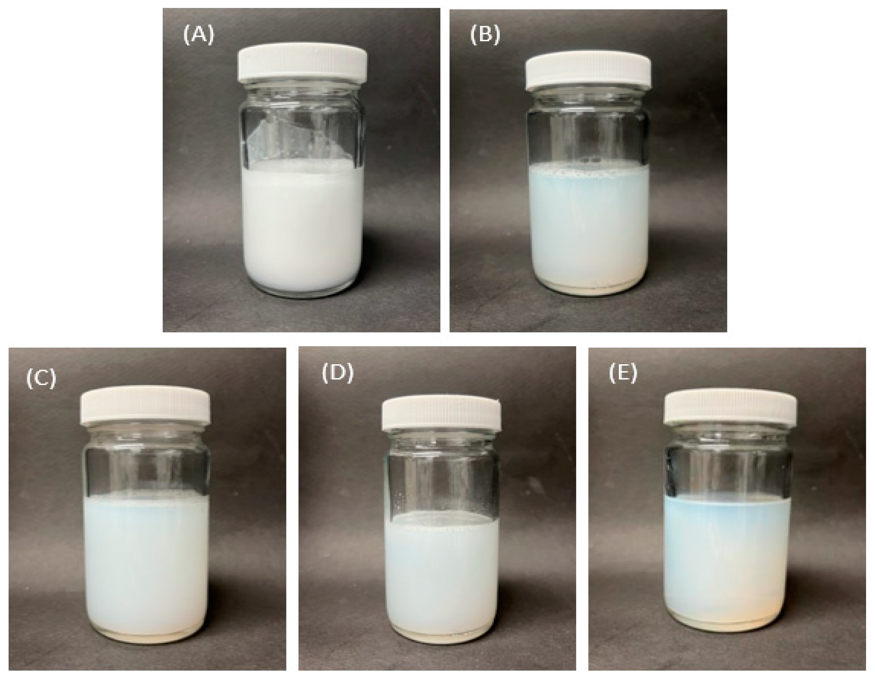
Figure 1 shows photographs of the prepared liposome solutions. The liposome solution prepared using the homo mixer was opaque with numerous bubbles. In contrast, the liposome solutions prepared using MF, bath sonication, horn sonication, and focused ultrasonication had no bubbles and were more transparent. In the case of the liposome solution prepared using focused ultrasonication, there were no bubbles in the upper layer of the solution, and the color of the solution was the most transparent. Transparency is a correlation between particle size and light scattering, with larger particles scattering more light and smaller particles scattering less light [
49].
Figure 2 shows the size and size distribution results measured using DLS, and
Table 2 lists the size and PDI. As expected, the liposome solution prepared using the homo mixer had a much larger size and PDI value than those of other liposome solutions. In addition, in terms of the stability measurement taken 3 d after the experiment, the size change was large, making it highly unstable. The liposome solution prepared using the MF method consisted of relatively small liposomes (127.8 nm on the day of the experiment); however, it was difficult to treat it as a stable solution because the size increased to 199.6 nm after 2 d. The solution prepared using bath sonication was judged to be relatively stable, considering that the liposome size was measured to be near 150 nm for 3 d. The size of the liposomes prepared using horn sonication increased from 159.2 nm to 185.8 nm on Day 2 and then decreased to 153.1 nm, showing an unstable appearance. In addition, its PDI value was 0.138 on the day of the experiment, and it increased over time. Finally, in the case of the liposome solution prepared using the focused ultrasound method, the liposome size measured on the day of the experiment was the smallest (113.6 nm), and the solution also seemed to be considerably stable as a result of the measurement taken after a total of 4 d. In terms of the PDI, the size distribution seemed to be highly uniform, as the PDI value was measured to be approximately 0.1.
3.2. Stability
Figure 3 shows the result of the Turbiscan measurement of the degree of delta transmission every 6 h for 1 week, which is an important result indicating the stability of the liposome solution. The x-axis represents the height of the bottle containing the sample, and the y-axis represents the delta transmission change (%). First, in the case of the solution prepared using a homo mixer, the delta transmission percentage was approximately 50% or higher, indicating that many liposome particles aggregated in the solution. Next, the liposome solution prepared using MF was less unstable than that prepared using the homo mixer, but it seems that some aggregation and phase separation occurred. In the case of the solution prepared using bath sonication, a graph in the form of phase separation was observed at the bottom and top of the solution, and the particles aggregated. However, its delta transmission percentage was approximately 10%, indicating that it was more stable than the MF liposome solution. Next, in the case of the liposome solution prepared using horn-type sonication, it appears that the delta transmission percentage was high across the whole solution, indicating aggregation. Finally, the solution prepared using the focused ultrasound device had the lowest degree of data transmission, indicating that it was considerably stable and did not show much aggregation or phase separation compared to other liposome solutions.
3.3. Cryo-EM
Figure 4 shows Cryo-EM images of the liposomes. The circular shape shown in the images is a grid appearing on the Cryo-EM measuring plate. The concentration of liposomes in the solution prepared using the homo mixer was remarkably low. This is because the liposome solution was phase-separated, and many bubbles were generated. In the case of the liposome solution prepared using the MF equipment, the size of liposomes was non-uniform, although most liposomes had a large size of approximately 200 nm. The liposome solution prepared using bath sonication also exhibited non-uniform particle sizes, and a significant number of double-layer or multi-layer liposomes were observed. Next, the liposome solution prepared using horn sonication produced many large liposomes with non-uniform particle sizes. Finally, the liposome solution prepared using focused ultrasound showed liposomes with a size smaller than 200 nm; however, the size distribution was not very uniform. This result demonstrated that the focused ultrasound method was capable of producing smaller liposomes than those produced using other methods.
Figure 5 shows the Cryo-EM images of the liposome solution prepared using the focused ultrasound method. Compared to the liposomes prepared using other methods, which had a size of approximately 200 nm, the liposomes prepared using focused ultrasound had a size of approximately 100 nm. In addition, it was confirmed that single-layer liposomes with uniform shape and size distribution were produced.
4. Discussion
In this study, liposomes were prepared using five different methods. First, the mixer manufacturing approach, being a simple mixing method, generated numerous bubbles, leading to larger size and PDI values in DLS measurements. Next, the high-pressure emulsification method, widely employed in liposome preparation, was found to be relatively simple and time-consuming but lacked stability. Among the ultrasonic methods used to reduce the size of liposomes, bath sonication showed minimal change in size during the 4-day measurement period. However, the size itself did not reach the desired small size, which was considered a disappointing result. Similarly, horn sonication yielded a size similar to bath sonication, albeit slightly less stable. This could be attributed to the uneven concentration of ultrasonic energy within the liposome solution. Finally, the focused ultrasonic method proved to be the most promising. It not only resulted in the smallest size and uniform particle distribution, but also demonstrated superior stability compared to other methods.
As noted in the Introduction, liposomes become more stable as their size decreases. Achieving a uniform and small size enhances the efficiency of collecting functional materials. Upon analyzing the above results, it is evident that the focused ultrasonication method yielded liposomes with the smallest and most uniform size, compared to simple techniques like homogenization and high-pressure emulsification, as well as existing sonication methods such as bath-type and horn-type methods.
This favorable outcome is attributed to the focused ultrasound method’s ability to apply appropriate frequency and energy to the liposome solution while also directing concentrated energy to the circulating solution.
Consequently, employing the focused ultrasound method is expected to lead to the creation of nano-sized liposomes with uniform structures, enhanced stability, and significantly improved encapsulation efficiency. These liposomes are likely to find applications in diverse fields such as drug delivery and cosmetics.
5. Conclusions
In this study, liposomes with a small particle size and uniform size distribution were prepared using focused ultrasound technology and compared with the liposomes prepared using the existing manufacturing methods, such as homo mixer, microfluidizer, and bath and horn types of sonication. The liposomes prepared using focused ultrasound equipment had an average size of 113.6 nm and a PDI of 0.124, demonstrating a relatively small particle size and uniform size distribution. In addition, the stability measurement up to 3 d after the experiment showed little size change, and the size was 123.8 nm, 113.7 nm, and 116.5 nm, on Days 1, 2, and 3, respectively. This level of stability surpassed that of solutions prepared using other methods. Therefore, it was confirmed that this focused ultrasound method was more effective than the existing liposome preparation methods. This focused ultrasound technology can be applied to produce stable liposome solution with small and uniform sizes for applications in various fields, such as cosmetics, food, medicine, and drug delivery.
Author Contributions
Conceptualization, S.-A.H. and Y.-G.J.; methodology, J.-S.Y.; validation, J.-S.Y.; investigation, S.-A.H. and Y.-G.J.; resources, S.-A.H. and Y.-G.J.; data curation, J.-S.Y.; writing—original draft preparation, J.-S.Y.; writing—review and editing, S.-A.H. and Y.-G.J.; visualization, J.-S.Y.; supervision, S.-A.H. and Y.-G.J.; project administration, S.-A.H.; funding acquisition, S.-A.H. All authors have read and agreed to the published version of the manuscript.
Funding
This work was funded by the Technology Innovation Program (Development of reference standard for tubular piezoelectric ceramics for supply and diffusion of ultrasonic dispersion/emulsification technology (grant number 20011794)) funded by the Ministry of Trade, Industry, and Energy (MOTIE, Republic of Korea) and by ‘Technical start-up corporation fostering project’ through the Commercialization Promotion Agency for R&D Outcomes(COMPA) grant funded by the Korea government(MSIT) (No. 2023 Incubating 1_08) and by the Technology Innovation Program (Development of smart ultrasound equipment and application technology for the manufacture of monodisperse nanoemulsions for nanopharmaceuticlas (grant number 20023461)) funded by the Ministry of Trade, Industry, and Energy (MOTIE, Republic of Korea).
Data Availability Statement
Not applicable.
Conflicts of Interest
The authors declare no conflict of interest. The funders had no role in the design of the study; in the collection, analyses, or interpretation of data; in the writing of the manuscript, or in the decision to publish the results.
References
- Nsairat, H.; Khater, D.; Sayed, U.; Odeh, F.; Al Bawab, A.; Alshaer, W. Liposomes: Structure, composition, types, and clinical applications. Heliyon 2022, 8, e09394. [Google Scholar] [CrossRef]
- Rovira-Bru, M.; Thompson, D.H.; Szleifer, I. Size and structure of spontaneously forming liposomes in lipid/PEG-lipid mixtures. Biophys. J. 2002, 83, 2421–2433. [Google Scholar] [CrossRef]
- Nakhaei, P.; Margiana, R.; Dmitry, O.; Bokov, D.O.; Abdelbasset, W.K.; Kouhbanani, M.A.J.; Rajender, S.; Varma, R.S.; Marofi, F.; Jarahian, M.; et al. Liposomes: Structure, biomedical applications, and stability parameters with emphasis on cholesterol. Front. Bioeng. Biotechnol. 2021, 9, 705856. [Google Scholar]
- Lasic, D.D.; Joannic, R.; Keller, B.C.; Frederik, P.M. Spontaneous vesiculation. Adv. Colloid. Interface Sci. 2001, 89–90, 337–349. [Google Scholar] [CrossRef]
- Hauser, H. Mechanism of spontaneous vesiculation. Proc. Natl. Acad. Sci. USA 1989, 86, 5351–5355. [Google Scholar] [CrossRef]
- Ge, L.; Tan, X.; Sheng, R.; Xiao, J. Layer-by-layer self-assembly of giant polyelectrolyte microcapsules templated by microbubbles as potential hydrophilic or hydrophobic drug delivery system. Colloid. Interface Sci. Commun. 2022, 47, 100603. [Google Scholar] [CrossRef]
- Pradhan, B.; Kumar, N.; Saha, S.; Roy, A. Liposome: Method of preparation, advantages, evaluation and its application. Int. J. Appl. Biol. Pharm. 2015, 3, 1–8. [Google Scholar]
- Allison, A.C.; Gregoriadis, G. Liposomes as immunological adjuvants. Nature 1974, 252, 252. [Google Scholar] [CrossRef]
- Allena, T.M.; Cullis, P.R. Liposomal drug delivery systems: From concept to clinical applications. Adv. Drug Deliv. Rev. 2013, 65, 36–48. [Google Scholar] [CrossRef]
- Torchilin, V.P. Recent advances with liposomes as pharmaceutical carriers. Nat. Rev. Drug Discov. 2005, 4, 145–160. [Google Scholar] [CrossRef]
- Ong, S.G.M.; Ming, L.C.; Lee, K.S.; Yuen, K.H. Influence of the encapsulation efficiency and size of liposome on the oral bioavailability of griseofulvin-loaded liposomes. Pharmaceutics 2016, 8, 25. [Google Scholar] [CrossRef]
- Jones, M.N. The surface properties of phospholipid liposome systems and their characterization. Adv. Colloid. Interface Sci. 1995, 54, 93–128. [Google Scholar] [CrossRef]
- Duong, T.T.; Isomäki, A.; Paaver, U.; Laidmäe, I.; Tõnisoo, A.; Yen, T.T.H.; Kogermann, K.; Raal, A.; Heinämäki, J.; Pham, T. Nano formulation and evaluation of oral berberine-loaded liposomes. Molecules 2021, 26, 2591. [Google Scholar] [CrossRef]
- Szoka, F., Jr.; Papahadjopoulos, D. Comparative properties and methods of preparation of lipid vesicles (Liposomes). Annu. Rev. Biophys. Bioeng. 1980, 9, 467–508. [Google Scholar] [CrossRef]
- Sharma, A.; Sharma, U.S. Liposomes in drug delivery: Progress and limitations. Int. J. Pharm. 1997, 154, 123–140. [Google Scholar] [CrossRef]
- Ong, S.G.M.; Ming, L.C.; Lee, K.S.; Yuen, K.H. Impact of particle size and polydispersity index on the clinical applications of lipidic nanocarrier systems. Pharmaceutics 2018, 10, 57. [Google Scholar]
- Yu, J.Y.; Chuesiang, P.; Shin, G.H.; Park, H.J. Post-processing techniques for the improvement of liposome stability. Pharmaceutics 2021, 13, 1023. [Google Scholar] [CrossRef]
- Jung, S.H.; Cho, Y.S.; Jun, S.S.; Koo, J.S.; Cheon, H.G.; Shin, B.C. Topical application of liposomal cobalamin hydrogel for atopic dermatitis therapy. Pharmazie 2011, 66, 430–435. [Google Scholar]
- Vemuri, S.; Yu, C.; Wangatorntanakun, V.; Roosdorp, N. Large-scale production of liposomes by a microfluidizer. Drug Dev. Ind. Pharm. 1990, 16, 2243–2256. [Google Scholar] [CrossRef]
- Thompson, A.K.; Singh, H. Preparation of liposomes from milk fat globule membrane phospholipids using a microfluidizer. J. Dairy. Sci. 2006, 89, 410–419. [Google Scholar] [CrossRef]
- Taisma, H.; Ozer, A.Y.; van Bloois, L.; Crommelin, D.J.A. The size reduction of liposomes with a high pressure homogenizer (Microfluidizer™). Characterization of prepared dispersions and comparison with conventional methods. Drug Dev. Ind. Pharm. 1989, 15, 197–207. [Google Scholar]
- Lajunen, T.; Hisazumi, K.; Kanazawa, T.; Okada, H.; Seta, Y.; Yliperttula, M.; Urtti, A.; Takashima, Y. Topical drug delivery to retinal pigment epithelium with microfluidizer produced small liposomes. Eur. J. Pharm. Sci. 2014, 62, 23–32. [Google Scholar] [CrossRef] [PubMed]
- Woodbury, D.J.; Richardson, D.S.; Gigg, A.W.; Welling, R.D.; Knudson, B.H. Reducing liposome size with ultrasound: Bimodal size distributions. J. Liposome Res. 2006, 16, 57–80. [Google Scholar] [CrossRef]
- Yamaguchi, T.; Nomura, M.; Matsuoka, T.; Koda, S. Effects of frequency and power of ultrasound on the size reduction of liposome. Chem. Phys. Lipids 2009, 160, 58–62. [Google Scholar] [CrossRef]
- Lapinski, M.M.; Castro-Forero, A.; Greiner, A.J.; Ofoli, R.Y.; Blanchard, G.J. Comparison of liposomes formed by sonication and extrusion: Rotational and translational diffusion of an embedded chromophore. Langmuir 2007, 23, 11677–11683. [Google Scholar] [CrossRef] [PubMed]
- Taladrid, D.; Marín, D.; Alemán, A.; Álvarez-Acero, I.; Montero, P.; Gómez-Guillén, M.C. Effect of chemical composition and sonication procedure on properties of food-grade soy lecithin liposomes with added glycerol. Food Res. Int. 2017, 100, 541–550. [Google Scholar] [CrossRef]
- Hwangbo, S.A.; Kwak, K.; Kim, J.; Lee, T.G. Novel surfactant-free water dispersion technique of TiO2 NPs using focused ultrasound system. Nanomaterials 2021, 11, 427. [Google Scholar] [CrossRef] [PubMed]
- Choi, Y.M.; Lee, Y.L.; Lim, E.S.; Trimzi, M.A.; Hwangbo, S.A.; Ham, Y.B. Performance improvement of ring-type PZT ceramics for ultrasonic dispersion system. Micromachines 2020, 11, 144. [Google Scholar] [CrossRef]
- Hwangbo, S.A.; Choi, Y.M.; Lee, T.G. Influence of piezoelectric properties on the ultrasonic dispersion of TiO2 nanoparticles in aqueous suspension. Micromachines 2021, 12, 52. [Google Scholar] [CrossRef]
- Suslick, K.S.; Didenko, Y.; Fang, M.M.; Hyeon, T.; Kolbeck, K.J.; McNamara, W.B.; Mdleleni, M.M.; Wong, M. Acoustic cavitation and its chemical consequences. Philos. Trans. R. Soc. London. Ser. A Math. Phys. Eng. Sci. 1999, 357, 335–353. [Google Scholar] [CrossRef]
- Lauterborn, W.; Ohl, C.D. Cavitation bubble dynamics. Ultrason. Sonochem. 1997, 4, 65–75. [Google Scholar] [CrossRef]
- Sivakumara, M.; Tangb, S.Y.; Tanc, K.W. Cavitation technology–A greener processing technique for the generation of pharmaceutical nano emulsions. Ultrason. Sonochem. 2014, 21, 2069–2083. [Google Scholar] [CrossRef]
- Bai, L.; Chen, X.; Zhu, G.; Xu, W.; Lin, W.; Wu, P.; Li, C.; Xu, D.; Yan, J. Surface tension and quasi-emulsion of cavitation bubble cloud. Ultrason. Sonochem. 2017, 35, 405–414. [Google Scholar] [CrossRef]
- Lad, V.N.; Murthy, Z.V.P. Enhancing the stability of oil-in-water emulsions emulsified by coconut milk protein with the application of acoustic cavitation. Ind. Eng. Chem. Res. 2012, 51, 4222–4229. [Google Scholar] [CrossRef]
- Barone, A.; Cristiano, M.C.; Cilurzo, F.; Locatelli, M.; Iannotta, D.; DiMarzio, L.; Celia, C.; Paolino, D. Ammonium glycyrrhizate skin delivery from ultradeformable liposomes: A novel use as an anti-inflammatory agent in topical drug delivery. Colloids Surf. B Biointerfaces 2020, 193, 111152. [Google Scholar] [CrossRef] [PubMed]
- Molinaro, R.; Evangelopoulos, M.; Hoffman, J.R.; Corbo, C.; Taraballi, F.; Martinez, J.O.; Hartman, K.A.; Cosco, D.; Costa, G.; Romeo, I.; et al. Design and development of biomimetic nanovesicles using a microfluidic approach. Adv. Mater. 2018, 30, e1702749. [Google Scholar] [CrossRef] [PubMed]
- Zhang, Y.; He, W.; Du, Y.; Du, Y.; Zhao, C.; Zhang, Y.; Zhang, H.; Yin, L.; Li, X. Dimeric artesunate phospholipid-conjugated liposomes as promising anti-inflammatory therapy for rheumatoid arthritis. Int. J. Pharm. 2020, 579, 119178. [Google Scholar] [CrossRef] [PubMed]
- Le, N.T.T.; Cao, V.D.; Nguyen, T.N.Q.; Le, T.T.H.; Tran, T.T.; Thi, T.T.H. Soy lecithin-derived liposomal delivery systems: Surface modification and current applications. Int. J. Mol. Sci. 2019, 20, 4706. [Google Scholar] [CrossRef] [PubMed]
- Hermansson, U.; Johannisson, A.; Axnér, E. Cryopreservation of dog semen in a Tris extender with two different 1% soybean preparations compared with a Tris egg yolk extender. Vet. Med. Sci. 2021, 7, 812–819. [Google Scholar] [CrossRef]
- Briuglia, M.L.; Rotella, C.; McFarlane, A.; Lamprou, D.A. Influence of cholesterol on liposome stability and on in vitro drug release. Drug Deliv. Transl. Res. 2015, 5, 231–242. [Google Scholar] [CrossRef]
- Sułkowski, W.W.; Pentak, D.; Nowak, K.; Sułkowska, A. The influence of temperature, cholesterol content and pH on liposome stability. J. Mol. Struct. 2005, 744–747, 737–747. [Google Scholar] [CrossRef]
- Hwangbo, S.A.; Lee, S.Y.; Kim, B.A.; Chang Kwon Moon, C.K. Preparation of surfactant-free nano oil particles in water using ultrasonic system and the mechanism of emulsion stability. Nanomaterials 2022, 12, 1547. [Google Scholar] [CrossRef] [PubMed]
- Hassan, P.A.; Rana, S.; Verma, G. Making sense of Brownian motion: Colloid characterization by dynamic light scattering. Langmuir 2015, 31, 3–12. [Google Scholar] [CrossRef]
- Weitz, D.A.; Pine, D.J.; Pusey, P.N.; Tough, R.J.A. Nondiffusive Brownian motion studied by diffusing-wave spectroscopy. Phys. Rev. Lett. 1989, 63, 1747–1750. [Google Scholar] [CrossRef] [PubMed]
- Kaombe, D.D.; Lenes, M.; Toven, K.; Glomm, W.R. Turbiscan as a tool for studying the phase separation tendency of pyrolysis oil. Energy Fuels 2013, 27, 1446–1452. [Google Scholar] [CrossRef]
- Danev, R.; Yanagisawa, H.; Kikkawa, M. Cryo-electron microscopy methodology: Current aspects and future directions. Trends Biochem. Sci. 2019, 44, 837–848. [Google Scholar] [CrossRef]
- Nogales, E.; Scheres, S.H.W. Cryo-EM: A unique tool for the visualization of macromolecular complexity. Mol. Cell 2015, 58, 677–689. [Google Scholar] [CrossRef] [PubMed]
- Uluata, S.; McClements, D.J.; Decker, E.A. Riboflavin-induced oxidation in fish oil-in-water emulsions: Impact of particle size and optical transparency. Food Chem. 2016, 213, 457–461. [Google Scholar] [CrossRef]
- Sinclair, D.; La Mer, V.K. Light scattering as a measure of particle size in aerosols. The production of monodisperse aerosols. Chem. Rev. 1949, 44, 245–267. [Google Scholar] [CrossRef]
| Disclaimer/Publisher’s Note: The statements, opinions and data contained in all publications are solely those of the individual author(s) and contributor(s) and not of MDPI and/or the editor(s). MDPI and/or the editor(s) disclaim responsibility for any injury to people or property resulting from any ideas, methods, instructions or products referred to in the content. |
© 2023 by the authors. Licensee MDPI, Basel, Switzerland. This article is an open access article distributed under the terms and conditions of the Creative Commons Attribution (CC BY) license (https://creativecommons.org/licenses/by/4.0/).


