
Synthesis of Sandwiched Composite Nanomagnets by Epitaxial Growth of Fe3O4 Layers on SrFe10Cr2O19 Nanoplates in High-Boiling Organic Solvent

 ,
,  , , and
Abstract
1. Introduction
2. Materials and Methods
2.1. Materials
2.2. Hexaferrite Nanoparticles Synthesis
2.3. Hexaferrite/Magnetite Composites Synthesis
2.4. Characterization of Samples
3. Results and Discussion
4. Conclusions
Author Contributions
Funding
Data Availability Statement
Conflicts of Interest
Appendix A


References
- McCallum, R.W.; Lewis, L.H.; Skomski, R.; Kramer, M.J.; Anderson, I.E. Practical Aspects of Modern and Future Permanent Magnets. Annu. Rev. Mater. Res. 2014, 44, 451–477. [Google Scholar] [CrossRef]
- Gorbachev, E.A.; Kozlyakova, E.S.; Trusov, L.A.; Sleptsova, A.E.; Zykin, M.A.; Kazin, P.E. Design of Modern Magnetic Materials with Giant Coercivity. Russ. Chem. Rev. 2021, 90, 1287–1329. [Google Scholar] [CrossRef]
- Odenbach, S. Colloidal Magnetic Fluids: Basics, Development and Application of Ferrofluids; Odenbach, S., Ed.; Springer: Berlin/Heidelberg, Germany, 2009. [Google Scholar]
- Ohkoshi, S.; Namai, A.; Imoto, K.; Yoshikiyo, M.; Tarora, W.; Nakagawa, K.; Komine, M.; Miyamoto, Y.; Nasu, T.; Oka, S.; et al. Nanometer-Size Hard Magnetic Ferrite Exhibiting High Optical-Transparency and Nonlinear Optical-Magnetoelectric Effect. Sci. Rep. 2015, 5, 14414. [Google Scholar] [CrossRef] [PubMed]
- Gorbachev, E.; Soshnikov, M.; Wu, M.; Alyabyeva, L.; Myakishev, D.; Kozlyakova, E.; Lebedev, V.; Anokhin, E.; Gorshunov, B.; Brylev, O.; et al. Tuning the Particle Size, Natural Ferromagnetic Resonance Frequency and Magnetic Properties of ε-Fe2O3 Nanoparticles Prepared by a Rapid Sol–Gel Method. J. Mater. Chem. C 2021, 9, 6173–6179. [Google Scholar] [CrossRef]
- Gich, M.; Frontera, C.; Roig, A.; Taboada, E.; Molins, E.; Rechenberg, H.R.; Ardisson, J.D.; Macedo, W.A.A.; Ritter, C.; Hardy, V.; et al. High- and Low-Temperature Crystal and Magnetic Structures of ε-Fe2O3 and Their Correlation to Its Magnetic Properties. Chem. Mater. 2006, 18, 3889–3897. [Google Scholar] [CrossRef]
- Kubániová, D.; Brázda, P.; Závěta, K.; Kmječ, T.; Klementová, M.; Kohout, J. Identification of Ferric Oxide Polymorphs in Nanoparticles Prepared by Sol-Gel Method and Maximization of ε-Fe2O3 Content. J. Magn. Magn. Mater. 2019, 472, 96–103. [Google Scholar] [CrossRef]
- Pullar, R.C. Hexagonal Ferrites: A Review of the Synthesis, Properties and Applications of Hexaferrite Ceramics. Prog. Mater. Sci. 2012, 57, 1191–1334. [Google Scholar] [CrossRef]
- de Julian Fernandez, C.; Sangregorio, C.; de la Figuera, J.; Belec, B.; Makovec, D.; Quesada, A. Topical Review: Progress and Prospects of Hard Hexaferrites for Permanent Magnet Applications. J. Phys. D Appl. Phys. 2020, 54, 153001. [Google Scholar] [CrossRef]
- Lisjak, D.; Mertelj, A. Anisotropic Magnetic Nanoparticles: A Review of Their Properties, Syntheses and Potential Applications. Prog. Mater. Sci. 2018, 95, 286–328. [Google Scholar] [CrossRef]
- Eliseev, A.A.; Eliseev, A.A.; Trusov, L.A.; Chumakov, A.P.; Boesecke, P.; Anokhin, E.O.; Vasiliev, A.V.; Sleptsova, A.E.; Gorbachev, E.A.; Korolev, V.V.; et al. Rotational Dynamics of Colloidal Hexaferrite Nanoplates. Appl. Phys. Lett. 2018, 113, 113106. [Google Scholar] [CrossRef]
- Eliseev, A.A.; Trusov, L.A.; Anokhin, E.O.; Chumakov, A.P.; Korolev, V.V.; Sleptsova, A.E.; Boesecke, P.; Pryakhina, V.I.; Shur, V.Y.; Kazin, P.E.; et al. Tunable Order in Colloids of Hard Magnetic Hexaferrite Nanoplatelets. Nano Res. 2022, 15, 898–906. [Google Scholar] [CrossRef]
- Lukatskaya, M.R.; Trusov, L.A.; Eliseev, A.A.; Lukashin, A.V.; Jansen, M.; Kazin, P.E.; Napolskii, K.S. Controlled Way to Prepare Quasi-1D Nanostructures with Complex Chemical Composition in Porous Anodic Alumina. Chem. Commun. 2011, 47, 2396–2398. [Google Scholar] [CrossRef] [PubMed]
- Cao, W.; Yin, S.; Plank, M.; Chumakov, A.; Opel, M.; Chen, W.; Kreuzer, L.P.; Heger, J.E.; Gallei, M.; Brett, C.J.; et al. Spray-Deposited Anisotropic Ferromagnetic Hybrid Polymer Films of PS-b-PMMA and Strontium Hexaferrite Magnetic Nanoplatelets. ACS Appl. Mater. Interfaces 2021, 13, 1592–1602. [Google Scholar] [CrossRef] [PubMed]
- Kushnir, S.E.; Kazin, P.E.; Trusov, L.A.; Tretyakov, Y.D. Self-Organization of Micro- and Nanoparticles in Ferrofluids. Russ. Chem. Rev. 2012, 81, 560. [Google Scholar] [CrossRef]
- Shirk, B.; Buessem, W. Theoretical and Experimental Aspects of Coercivity versus Particle Size for Barium Ferrite. IEEE Trans. Magn. 1971, 7, 659–663. [Google Scholar] [CrossRef]
- Kazin, P.E.; Trusov, L.A.; Kushnir, S.E.; Yaroshinskaya, N.V.; Petrov, N.A.; Jansen, M. Hexaferrite Submicron and Nanoparticles with Variable Size and Shape via Glass-Ceramic Route. J. Phys. Conf. Ser. 2010, 200, 72048. [Google Scholar] [CrossRef]
- Trusov, L.A.; Babarkina, O.V.; Anokhin, E.O.; Sleptsova, A.E.; Gorbachev, E.A.; Eliseev, A.A.; Filippova, T.V.; Vasiliev, A.V.; Kazin, P.E. Crystallization of Magnetic Particles in nNa2O-9SrO-6Fe2O3-8B2O3 (n = 1 and 4) Glasses. J. Magn. Magn. Mater. 2019, 476, 311–316. [Google Scholar] [CrossRef]
- Kazin, P.E.; Trusov, L.A.; Zaitsev, D.D.; Tretyakov, Y.D.; Jansen, M. Formation of Submicron-Sized SrFe12−xAlxO19 with Very High Coercivity. J. Magn. Magn. Mater. 2008, 320, 1068–1072. [Google Scholar] [CrossRef]
- Trusov, L.A.; Sleptsova, A.E.; Duan, J.; Gorbachev, E.A.; Kozlyakova, E.S.; Anokhin, E.O.; Eliseev, A.A.; Karpov, M.A.; Vasiliev, A.V.; Brylev, O.A.; et al. Glass-Ceramic Synthesis of Cr-Substituted Strontium Hexaferrite Nanoparticles with Enhanced Coercivity. Nanomaterials 2021, 11, 924. [Google Scholar] [CrossRef]
- Anokhin, E.O.; Trusov, L.A.; Kozlov, D.A.; Chumakov, R.G.; Sleptsova, A.E.; Uvarov, O.V.; Kozlov, M.I.; Petukhov, D.I.; Eliseev, A.A.; Kazin, P.E. Silica Coated Hard-Magnetic Strontium Hexaferrite Nanoparticles. Adv. Powder Technol. 2019, 30, 1976–1984. [Google Scholar] [CrossRef]
- Primc, D.; Makovec, D. Composite Nanoplatelets Combining Soft-Magnetic Iron Oxide with Hard-Magnetic Barium Hexaferrite. Nanoscale 2015, 7, 2688–2697. [Google Scholar] [CrossRef] [PubMed]
- Primc, D.; Belec, B.; Makovec, D. Synthesis of Composite Nanoparticles Using Co-Precipitation of a Magnetic Iron-Oxide Shell onto Core Nanoparticles. J. Nanopart. Res. 2016, 18, 64. [Google Scholar] [CrossRef]
- Belec, B.; Dražić, G.; Gyergyek, S.; Podmiljšak, B.; Goršak, T.; Komelj, M.; Nogués, J.; Makovec, D. Novel Ba-Hexaferrite Structural Variations Stabilized on the Nanoscale as Building Blocks for Epitaxial Bi-Magnetic Hard/Soft Sandwiched Maghemite/Hexaferrite/Maghemite Nanoplatelets with out-of-Plane Easy Axis and Enhanced Magnetization. Nanoscale 2017, 9, 17551–17560. [Google Scholar] [CrossRef] [PubMed]
- Haynes, W.M.; Lide, D.R.; Bruno, T.J. CRC Handbook of Chemistry and Physics, 97th ed.; Haynes, W.M., Ed.; CRC Press: Boca Raton, FL, USA, 2016. [Google Scholar]
- Lutterotti, L. Total Pattern Fitting for the Combined Size–Strain–Stress–Texture Determination in Thin Film Diffraction. Nucl. Instrum. Methods Phys. Res. Sect. B Beam Interact. Mater. At. 2010, 268, 334–340. [Google Scholar] [CrossRef]
- Klinger, M.; Jäger, A. Crystallographic Tool Box (CrysTBox): Automated Tools for Transmission Electron Microscopists and Crystallographers. J. Appl. Crystallogr. 2015, 48, 2012–2018. [Google Scholar] [CrossRef]
- Klinger, M. More Features, More Tools, More CrysTBox. J. Appl. Crystallogr. 2017, 50, 1226–1234. [Google Scholar] [CrossRef]
- Obradors, X.; Solans, X.; Collomb, A.; Samaras, D.; Rodriguez, J.; Pernet, M.; Font-Altaba, M. Crystal Structure of Strontium Hexaferrite SrFe12O19. J. Solid State Chem. 1988, 72, 218–224. [Google Scholar] [CrossRef]
- Shannon, R.D. Revised Effective Ionic Radii and Systematic Studies of Interatomic Distances in Halides and Chalcogenides. Acta Crystallogr. Sect. A 1976, 32, 751–767. [Google Scholar] [CrossRef]
- Makovec, D.; Belec, B.; Goršak, T.; Lisjak, D.; Komelj, M.; Dražić, G.; Gyergyek, S. Discrete Evolution of the Crystal Structure during the Growth of Ba-Hexaferrite Nanoplatelets. Nanoscale 2018, 10, 14480–14491. [Google Scholar] [CrossRef]
- Makovec, D.; Dražić, G.; Gyergyek, S.; Lisjak, D. A New Polymorph of Strontium Hexaferrite Stabilized at the Nanoscale. CrystEngComm 2020, 22, 7113–7122. [Google Scholar] [CrossRef]
- Markelova, M.; Nygaard, R.; Tsymbarenko, D.; Shurkina, A.; Abramov, A.; Amelichev, V.; Makarevich, A.; Vasiliev, A.; Kaul, A. Multiferroic H-LuFeO3 Thin Films on (111) and (100) Surfaces of YSZ Substrates: An Experimental and Theoretical Study. ACS Appl. Electron. Mater. 2021, 3, 1015–1022. [Google Scholar] [CrossRef]
- Momma, K.; Izumi, F. VESTA3 for Three-Dimensional Visualization of Crystal, Volumetric and Morphology Data. J. Appl. Crystallogr. 2011, 44, 1272–1276. [Google Scholar] [CrossRef]
- Gorbachev, E.A.; Trusov, L.A.; Kovalenko, A.D.; Morozov, A.V.; Kazin, P.E. Sandwiched CoFe2O4/SrFe11.5Al0.5O19/CoFe2O4 Nanoparticles with Exchange-Coupling Effect. Nanoscale 2021, 13, 18340–18348. [Google Scholar] [CrossRef] [PubMed]
- Trusov, L.A.; Vasiliev, A.V.; Lukatskaya, M.R.; Zaytsev, D.D.; Jansen, M.; Kazin, P.E. Stable Colloidal Solutions of Strontium Hexaferrite Hard Magnetic Nanoparticles. Chem. Commun. 2014, 50, 14581–14584. [Google Scholar] [CrossRef] [PubMed]
- Zeng, H.; Li, J.; Liu, J.P.; Wang, Z.L.; Sun, S. Exchange-Coupled Nanocomposite Magnets by Nanoparticle Self-Assembly. Nature 2002, 420, 395–398. [Google Scholar] [CrossRef]
- Kojima, H. Chapter 5 Fundamental Properties of Hexagonal Ferrites with Magnetoplumbite Structure. In Handbook of Ferromagnetic Materials; Elsevier: Amsterdam, The Netherlands, 1982; Volume 3, pp. 305–391. [Google Scholar]
- Cao, L.; Xie, D.; Guo, M.; Park, H.S.; Fujita, T. Size and Shape Effects on Curie Temperature of Ferromagnetic Nanoparticles. Trans. Nonferrous Met. Soc. China 2007, 17, 1451–1455. [Google Scholar] [CrossRef]
- Guimarães, A.P. Principles of Nanomagnetism, 1st ed.; Springer: Berlin/Heidelberg, Germany, 2009; ISBN 978-3-642-01482-6. [Google Scholar]
- Nguyen, T.N.A.; Knut, R.; Fallahi, V.; Chung, S.; Le, Q.T.; Mohseni, S.M.; Karis, O.; Peredkov, S.; Dumas, R.K.; Miller, C.W.; et al. Depth-Dependent Magnetization Profiles of Hybrid Exchange Springs. Phys. Rev. Appl. 2014, 2, 44014. [Google Scholar] [CrossRef]


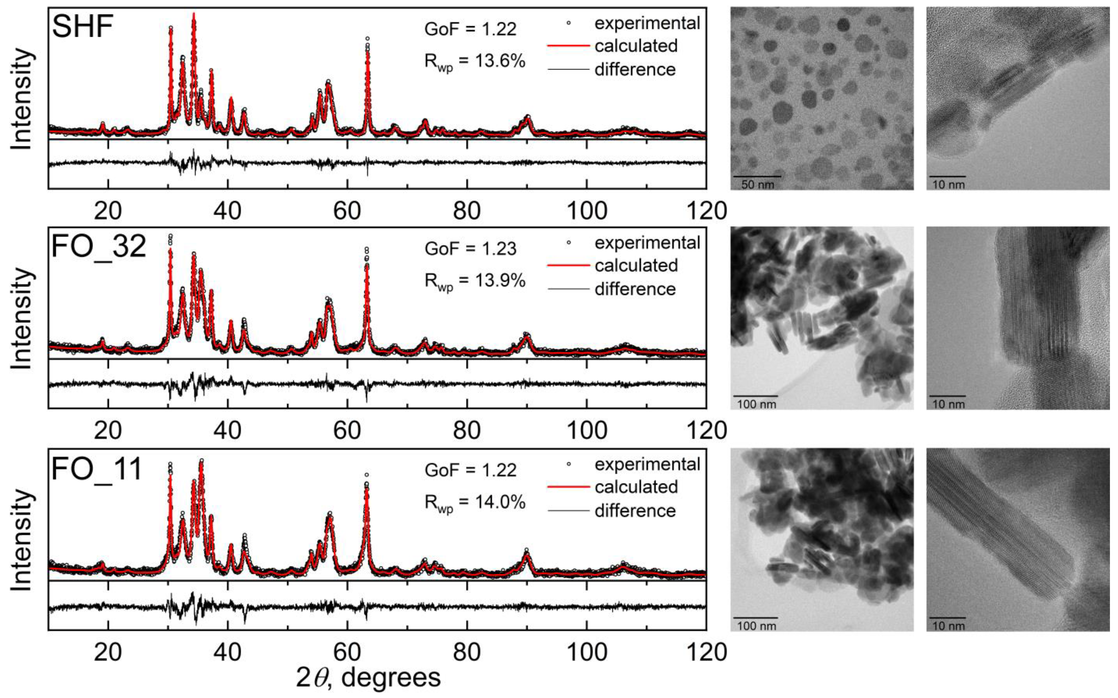
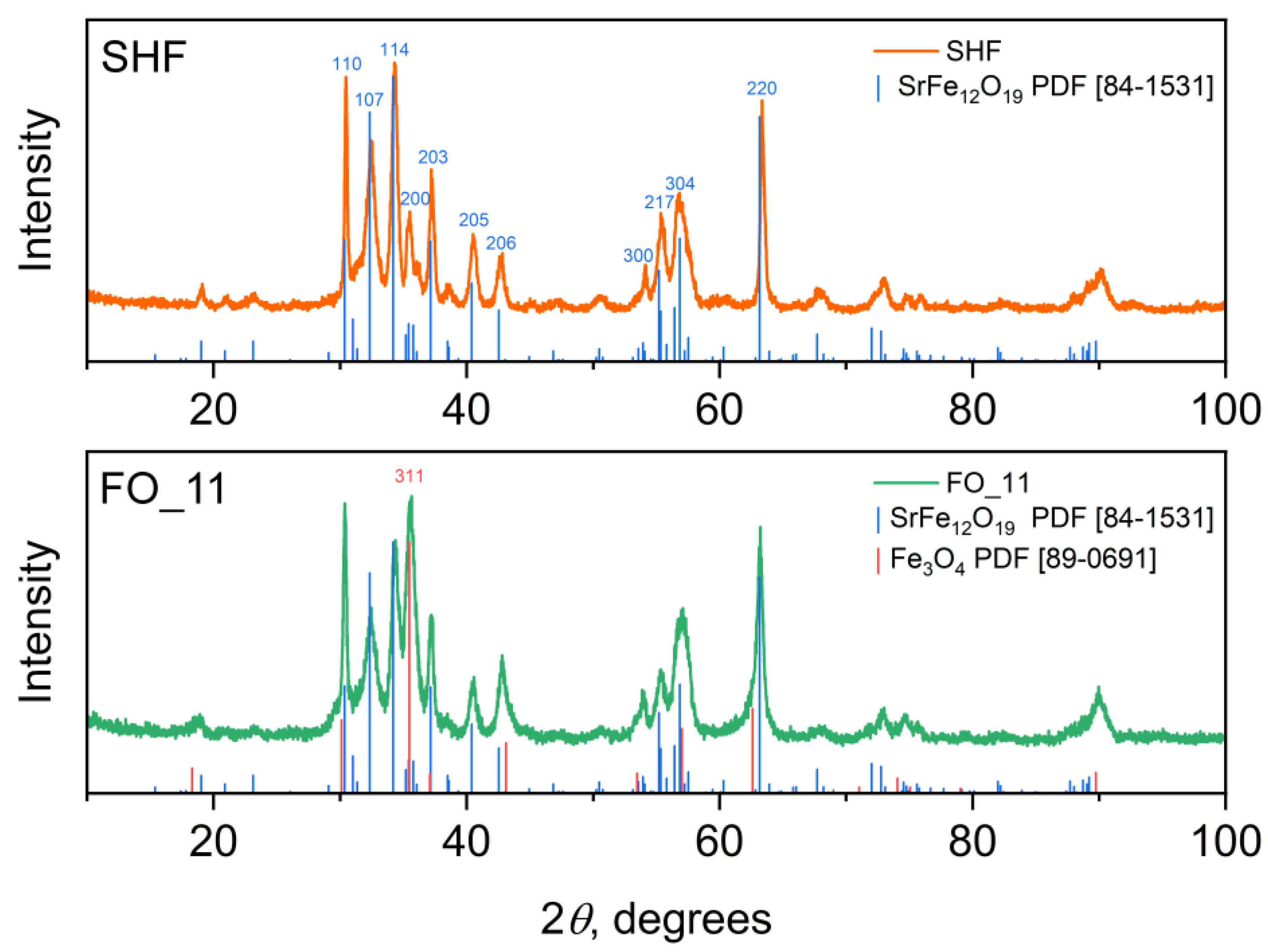
| Sample | Fe:Cr Ratio (ICP-MS) | ω (Fe3O4), wt % (ICP-MS) | ω (Fe3O4), wt % (XRD) | ω (Fe3O4), wt % (Nominal) |
|---|---|---|---|---|
| SHF | 10.0:2.0 | 0 | 0 | 0 |
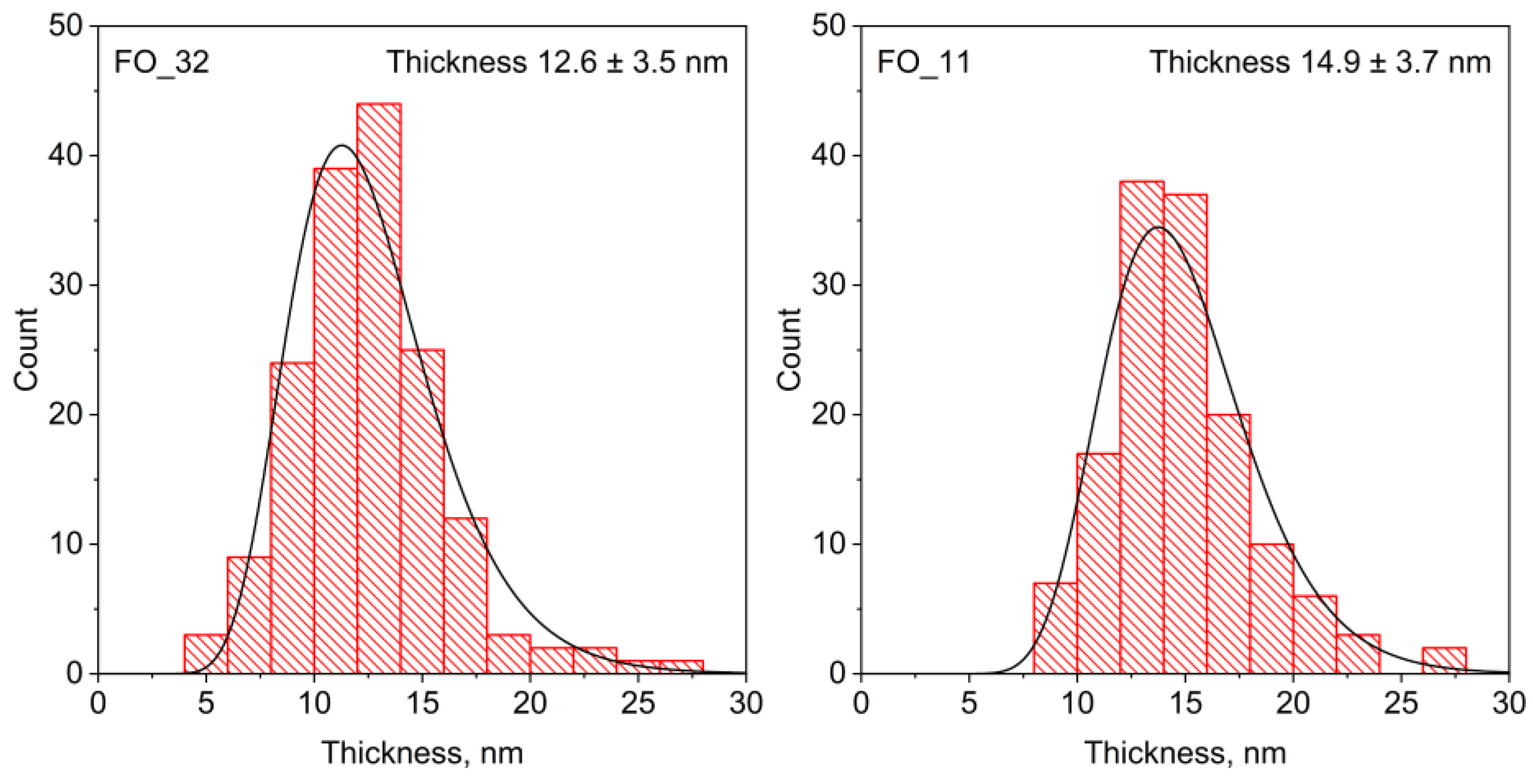
| FO_32 | 14.6:2.0 | 27.1 | 27.5 | 40 |
| FO_11 | 18.2:2.0 | 39.8 | 37.5 | 50 |
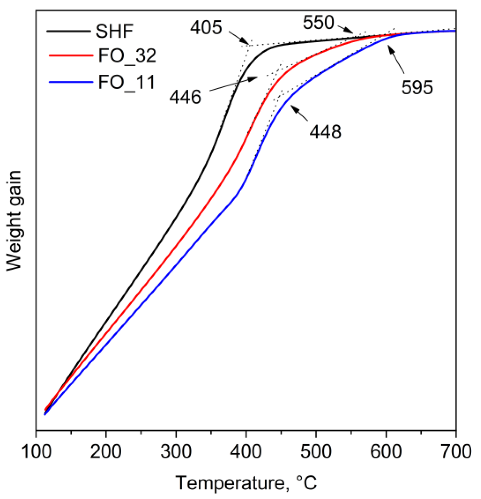
| Sample | Curie Temperature TC, °C (K) | |
|---|---|---|
| SHF | 405 (678) | — |
| FO_32 | 446 (719) | 550 (823) |
| FO_11 | 448 (721) | 595 (868) |
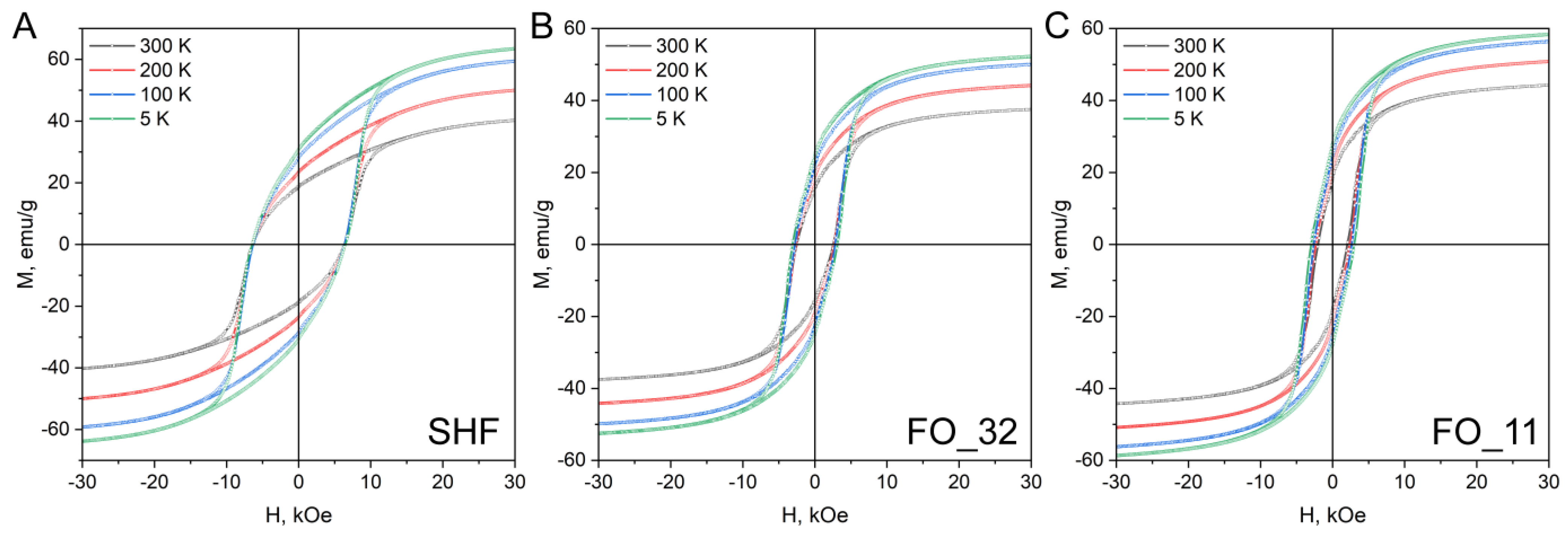
| Sample | Temperature, K | 300 | 200 | 100 | 5 |
|---|---|---|---|---|---|
| SHF | HC, Oe | 6350 | 6430 | 6320 | 6450 |
| MS, emu/g | 40.2 | 50.0 | 59.3 | 63.6 | |
| MR, emu/g | 18.7 | 23.5 | 28.3 | 30.8 | |
| MR/MS | 0.47 | 0.47 | 0.48 | 0.48 | |
| FO_32 | HC, Oe | 2520 | 2630 | 2780 | 3120 |
| MS, emu/g | 37.6 | 44.2 | 49.9 | 52.4 | |
| MR, emu/g | 15.7 | 19.0 | 22.2 | 24.7 | |
| MR/MS | 0.42 | 0.43 | 0.44 | 0.47 | |
| FO_11 | HC, Oe | 2020 | 2330 | 2640 | 2990 |
| MS, emu/g | 44.3 | 50.8 | 56.3 | 58.5 | |
| MR, emu/g | 18.2 | 21.2 | 24.7 | 27.9 | |
| MR/MS | 0.41 | 0.42 | 0.44 | 0.48 |
Disclaimer/Publisher’s Note: The statements, opinions and data contained in all publications are solely those of the individual author(s) and contributor(s) and not of MDPI and/or the editor(s). MDPI and/or the editor(s) disclaim responsibility for any injury to people or property resulting from any ideas, methods, instructions or products referred to in the content. |
© 2022 by the authors. Licensee MDPI, Basel, Switzerland. This article is an open access article distributed under the terms and conditions of the Creative Commons Attribution (CC BY) license (https://creativecommons.org/licenses/by/4.0/).
Share and Cite
Anokhin, E.O.; Deyankov, D.A.; Xia, Z.; Kozlyakova, E.S.; Lebedev, V.A.; Morozov, A.V.; Kozlov, D.A.; Nygaard, R.R.; Petukhov, D.I.; Trusov, L.A. Synthesis of Sandwiched Composite Nanomagnets by Epitaxial Growth of Fe3O4 Layers on SrFe10Cr2O19 Nanoplates in High-Boiling Organic Solvent. Nanomaterials 2023, 13, 167. https://doi.org/10.3390/nano13010167
Anokhin EO, Deyankov DA, Xia Z, Kozlyakova ES, Lebedev VA, Morozov AV, Kozlov DA, Nygaard RR, Petukhov DI, Trusov LA. Synthesis of Sandwiched Composite Nanomagnets by Epitaxial Growth of Fe3O4 Layers on SrFe10Cr2O19 Nanoplates in High-Boiling Organic Solvent. Nanomaterials. 2023; 13(1):167. https://doi.org/10.3390/nano13010167
Chicago/Turabian StyleAnokhin, Evgeny O., Danila A. Deyankov, Zitian Xia, Ekaterina S. Kozlyakova, Vasily A. Lebedev, Anatolii V. Morozov, Daniil A. Kozlov, Roy R. Nygaard, Dmitry I. Petukhov, and Lev A. Trusov. 2023. "Synthesis of Sandwiched Composite Nanomagnets by Epitaxial Growth of Fe3O4 Layers on SrFe10Cr2O19 Nanoplates in High-Boiling Organic Solvent" Nanomaterials 13, no. 1: 167. https://doi.org/10.3390/nano13010167
APA StyleAnokhin, E. O., Deyankov, D. A., Xia, Z., Kozlyakova, E. S., Lebedev, V. A., Morozov, A. V., Kozlov, D. A., Nygaard, R. R., Petukhov, D. I., & Trusov, L. A. (2023). Synthesis of Sandwiched Composite Nanomagnets by Epitaxial Growth of Fe3O4 Layers on SrFe10Cr2O19 Nanoplates in High-Boiling Organic Solvent. Nanomaterials, 13(1), 167. https://doi.org/10.3390/nano13010167