Article

Transparent Heat Shielding Properties of Core-Shell Structured Nanocrystalline CsxWO3@TiO2

College of Science, Inner Mongolia University of Science and Technology, Baotou 014010, China
*
Authors to whom correspondence should be addressed.
Nanomaterials 2022, 12(16), 2806; https://doi.org/10.3390/nano12162806
Submission received: 13 July 2022 / Revised: 7 August 2022 / Accepted: 9 August 2022 / Published: 16 August 2022
(This article belongs to the Special Issue 2D Nanostructures for Optoelectronic and Green Energy Devices)

Abstract

:
Nanocrystalline tungsten bronze is an excellent near-infrared absorbing material, which has a good potential application in the field of transparent heat shielding materials on windows of automobiles or buildings, but it exhibits serious instability in the actual environment, which hinders its further application. In this paper, we coated the CsxWO3 nanoparticles with TiO2 to prepare core-shell structured CsxWO3@TiO2, and its crystal structure and optical properties were studied. The results show that the surface of CsxWO3 nanoparticles is coated with a layer of TiO2 particles with the size of several nanometers, and the shell thickness can be adjusted by the amount of Ti source. The measurement of optical properties illustrates that TiO2-coated CsxWO3 exhibits good stability in actual environment, and its transparent heat shielding performance will decrease with the increase in TiO2 shell thickness. This work provides a new route to promote the applications of tungsten bronze as heat shielding materials.

1. Introduction

Nearly half of the solar radiation energy comes from near infrared light (NIR) in the range of about 760–2500 nm. Therefore, if the glass of buildings or automobiles can block NIR light while maintaining high transmittance of visible light, it can effectively reduce the room temperature, thus reducing the utilization of air conditioning and achieving the purpose of energy saving and emission reduction. At present, the most common commercial transparent heat shielding glass is Low-E glass or ITO glass [1,2], but the popularity of these materials is not very high because of its complex preparation process and high cost. Therefore, the current research has focused on new transparent heat shielding materials such as rare-earth hexaborides, VO2 or tungsten bronzes [3,4,5,6,7,8,9,10,11]. All of these materials have good application prospects in the field of transparent heat shielding materials, but there are also some problems. It is difficult to prepare nanocrystalline rare-earth hexaborides and the cost is high; high phase transition temperature of 68 °C and low visible transmittance hinders the further development of VO2; although tungsten bronze has excellent properties, its stability in the practical environment is insufficient.
The chemical formula of tungsten bronzes can be written as MxWO3, where the M represents alkaline earth metal, alkali metal, ammonium or rare earth metal ion. When the M cations are inserted into the whole gap of MxWO3, then x = 1. In the actual environment, MxWO3 is easily oxidized, and M+ escapes from the particle and forms WO3 on the surface, which leads to serious instability of NIR absorption of MxWO3 [12]. A good way to solve this problem is coating MxWO3 nanoparticles with suitable materials. Jin et al. prepared core-shell structured CsxWO3@SiO2 and CsxWO3@ZnO nanoparticles and achieved high stability of NIR absorption [13,14]. In the previous theoretical calculation, we found that TiO2-coated CsxWO3 (CWO) also has good transparent heat shielding properties [15]. Considering the better stability of TiO2, we synthesized core-shell structured nanocrystalline CWO@TiO2 in this work, and the stability and optical behavior of nanoparticles was investigated in detail.

2. Materials and methods

2.1. Preparation of CWO Nanoparticles

A total of 0.7554 g of cesium hydroxide monohydrate (CsOH·H2O) was added to 300 mL benzyl alcohol (C7H8O) solution and stirred for half an hour. Then, an appropriate amount of tungsten hexachloride (WCl6) was added and the WCl6 concentration of precursor solution was kept at 0.015 M. After that, the orange precursor solution was heated in an autoclave at 200 °C for 4 h. Finally, the obtained blue powder was washed with alcohol and deionized water several times, and dried in a vacuum at 60 °C for 2 h.

2.2. Preparation of CWO@TiO2 Nanoparticles

An amount of 3 g CWO powder was added to 400 mL ethanol and dispersed by sonication for 30 min. Then, a different amount (0.2, 0.4 and 0.6 mL) of titanium isopropoxide (TTIP) was added to the solution for forming TiO2 shell on the surface of CWO particles. After that, deionized water was added to the solution with strong stirring. Then, the mixture was heated in the autoclave at 200 °C for 18 h. Finally, the product was washed several times with deionized water, and dried at 50 °C in vacuum overnight. According to the different amount of TTIP used in the reaction process, the obtained three samples are expressed as [email protected]2, [email protected]2 and [email protected]2, respectively.

2.3. Fabrication Process for CWO@TiO2 Coated Glass

The heat-shielding glass was prepared by the spin coating method. First, 0.2 g CWO@TiO2 was dispersed in 20 mL ethanol solution by ultrasonic for 30 min, then 5 g polyvinyl butyral (PVB) resin was added under strong stirring for 20 min to obtain coating slurry. After spin-coating with a centrifugal speed of 2000 rad/min for 40 s, the coated glass was kept at 40 °C for 1 h to remove residual liquid.

2.4. Discrete Dipole Approximation Method

Discrete dipole approximation method (DDA) is an effective way to calculate the far-field and near-field optical responses of nanoparticles with complex refractive indexes and arbitrary geometries [16,17,18,19]. In this work, an open-source Fortran-90 code DDSCAT 7.3 applying the DDA method [20] was used to simulate the extinction efficiencies of CWO@TiO2. The program has given ellipsoid, regular tetrahedron, cuboid, cylinder, hexagonal prism, regular tetrahedron and other structural models. These models can be combined with the dielectric function of the corresponding material to calculate the extinction, absorption and scattering efficiency. Specific parameters such as the effective radius of particles, the number of dipoles and the wavelength range should be set in the program during calculation. In our calculation, a complex dielectric constant of CWO measured by Sato et al. was used to simulate the optical response [21] and the dielectric constants of TiO2 were obtained from the diel files of DDSCAT 7.3. The calculated wavelength range was 300 nm–2500 nm with 100 steps; The effective radius of the CWO@TiO2 with different TiO2 shell thicknesses are set as 55 nm, 60 nm, 65 nm, 70 nm and 80 nm; The dipole ratios are 11:10, 6:5, 13:10, 7:5, 8:5; The refractive index is set to 1.

3. Results and Discussions

The phase composition and crystallographic structure of the samples were confirmed by XRD measurement and results are given in Figure 1. The pure CWO presented hexagonal structure of Cs0.32WO3 (JCPDS 83-1334), and no impurity peaks were observed in the pattern. For TiO2-coated CWO nanoparticles, the extra peaks appeared in the pattern which belongs to TiO2 (JCPDS 21-1272). With the increase in the amount of Ti source, the peak intensity at 25° is obviously increasing, indicating that the content of TiO2 is increasing. These XRD results indicate that there are both CWO and TiO2 crystals in the sample. The two structures exist independently and the formation of TiO2 did not affect the structure of CWO.
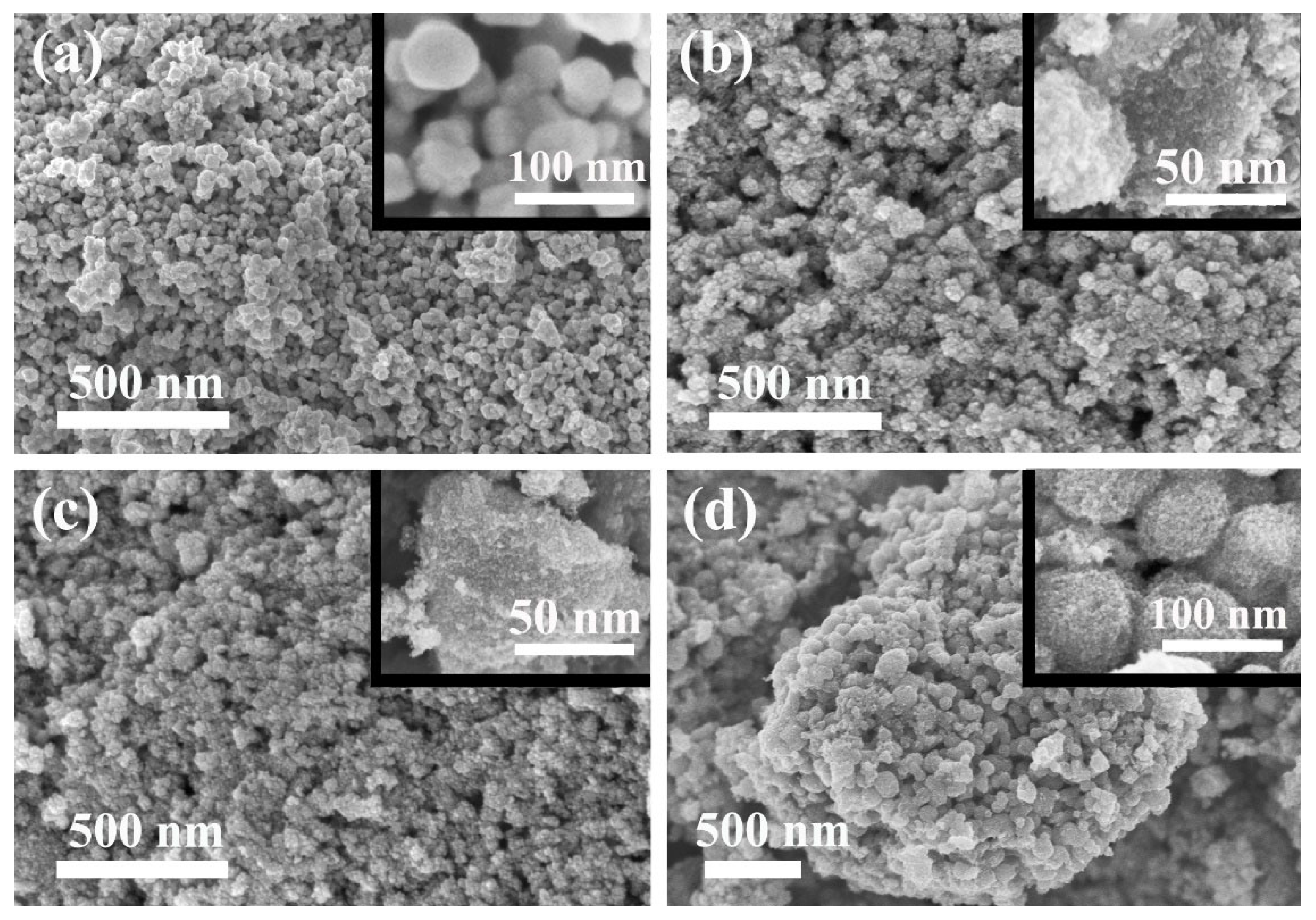
Figure 2 shows the SEM images of uncoated CWO and TiO2-coated CWO nanoparticles. The element mapping and corresponding element spectrum for [email protected]2 are also given in Figure 2. The uncoated CWO is composed of irregular particles with good dispersion, and the size is about tens of nanometers. Unlike pure CWO samples, it is clearly seen that a layer of several nanometer-sized small particles appeared on the surface of the CWO particles for the TiO2 coated samples. With the increase in TiO2 content, [email protected]2 exhibits obvious spherical shape with largest size. Combined with the XRD results, it can be concluded that CWO is coated with a layer of TiO2. The element mapping of all samples and corresponding element spectrum for [email protected]2 are also given in Figure 3, which illustrate that Cs, W, O and Ti elements are uniformly distributed in the selected area on coated samples, and no other elements are found.
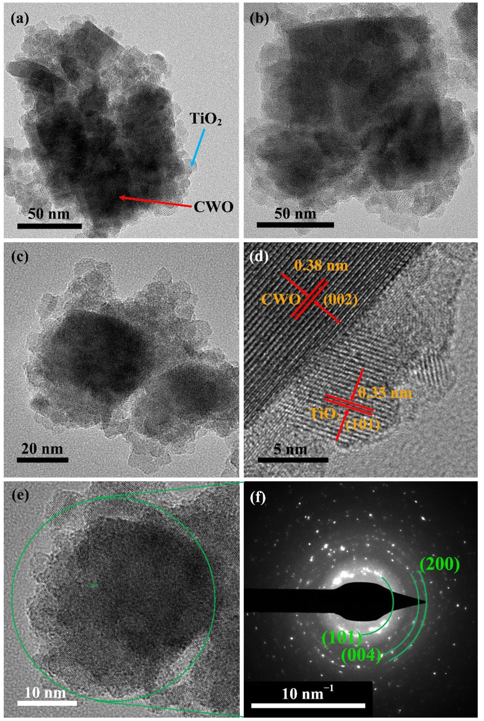
Figure 4 shows the TEM images of coated CWO samples. It can be clearly seen in Figure 4a–c that tens of nanometers of particles are coated by several nanometers of small particles, and the small particles in the outer layer are increasing with the increase in Ti source. Figure 4d clearly exhibits the single-crystalline nature of the two kinds of particles. The lattice fringes of d = 0.38 nm have good agreement with the (002) crystal planes of CWO structure shown in Figure 1, while the lattice fringe of d = 0.35 nm corresponds with the (101) crystal plane of tetragonal TiO2 phase. In Figure 4e,f, the diffraction rings of TiO2 such as (101), (004) and (220) are obtained, which is consistent with the XRD analysis (JCPDS 21-1272). The TEM results confirm that the surface of CWO is coated with a layer of crystalline TiO2.
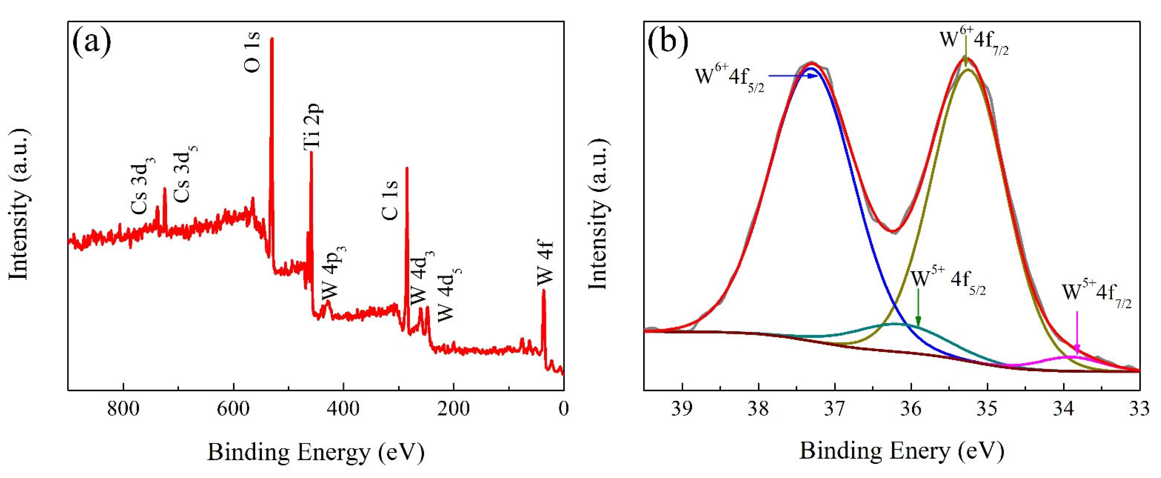
The chemical states of the coated samples was carefully determined by XPS. The full range XPS spectra and W4f core-level spectra of [email protected]2 are given in Figure 5. Except the existence of Cs, W, O, and Ti elements, no other impurity elements were found in the full range XPS spectra, which is consistent with the element spectrum results in Figure 3e. The W4f core-level XPS spectra of [email protected]2 can be fitted as two spin-orbit doublets. The peaks at 37.3 and 35.2 eV were attributed to W6+, and the peaks at 36.1 and 33.8 eV were attributed to W5+, respectively. The NIR shielding properties of CWO are determined by the plasmon resonance of free electrons. The addition of Cs element into WO3 structure can reduce part of W6+ to W5+, so as to enhance the carrier concentration and small polaron mechanism [22], which is the reason why CWO material has high NIR shielding performance.
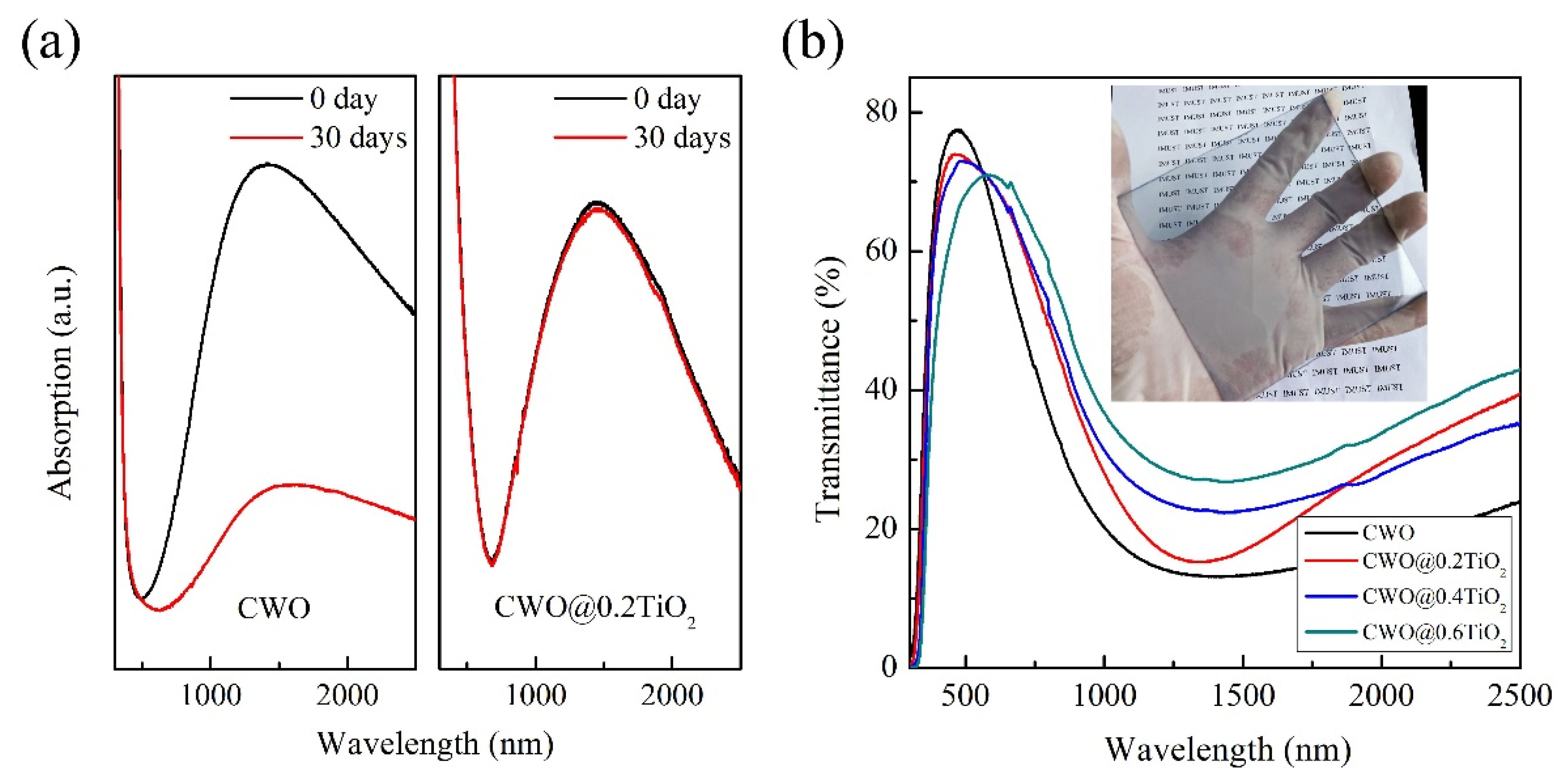
In order to observe the stability of coated CWO in the actual environment, the obtained powders were dispersed in ethanol and made into thin films on glass slides to test their absorption behavior. Figure 6a shows the absorption curve of CWO and [email protected]2 nanoparticles after different durations. For the uncoated CWO nanoparticles, the NIR absorption ability degraded seriously after 30 days. While the NIR absorption of [email protected]2 nanoparticles showed only slight degradation, indicating that TiO2 showed a good protective effect that TiO2 prevents Cs+ escape from the particle surface and form WO3. To determine the effect of different Ti sources content on the transmittance behavior of CWO particles, the three CWO@TiO2 samples were dispersed with PVB resin and coated on a glass slide (size of 10 cm × 10 cm) using a spin coating method. By observing the [email protected]2 coated glass photo in Figure 6b, no opaque or haze-like property was found. The transmittance curve of three CWO@TiO2-coated glass are revealed in Figure 6b, and the transmittance curve of CWO-coated glass is also given for comparison purpose. It can be clearly seen that the four samples show good transparent NIR shielding properties. The transmittance in the visible light decreases with the increase in Ti source and increases obviously in the NIR region. This shows that the thicker the TiO2 shell, the worse the transparent heat shielding performance. This can be attributed to two reasons, one is the influence of the intrinsic optical properties of TiO2, and the other is the increasing particle size with the increase in TiO2 shell thickness. Our previous research shows that the larger the particle size of transparent heat shielding material, the worse the NIR shielding performance [15].
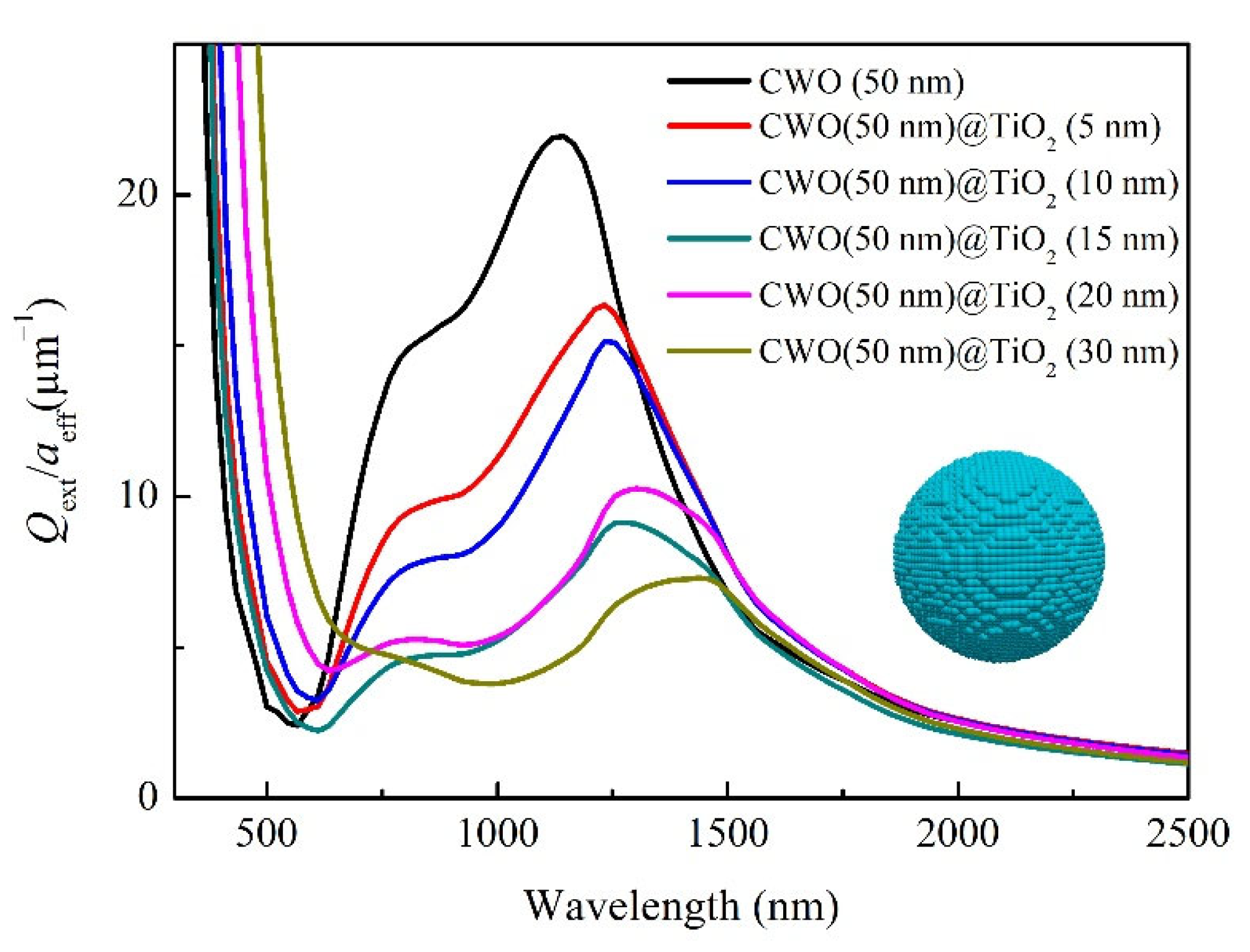
However, it is difficult to accurately control the particle size, shell thickness and other factors in the experiment. In order to systematically study the effect of shell thickness on the optical properties of CWO, we calculated the extinction characteristics of CWO with different TiO2 thickness by using the discrete dipole approximation (DDA) method. Figure 7 presents the extinction efficiencies of CWO spherical particles with different TiO2 shell thicknesses. It is discernible that uncoated CWO shows high extinction in the NIR region and low extinction in the visible region, indicating the transparent heat-shielding properties of CWO materials. With the increase in TiO2 thickness, the absorption edge in the visible region is red-shifted, and the extinction in the NIR region is weakened. We infer that the red shift of the absorption edge in the visible region is related to the intrinsic optical properties of TiO2, while the weakening of the extinction in the NIR region is related to the increase in particle size. Such a trend of optical response with TiO2 shell thickness obtained by DDA calculation is in good agreement with the experimental results in Figure 6b, indicating that the shell thickness should not be too thick when coating CWO with TiO2.
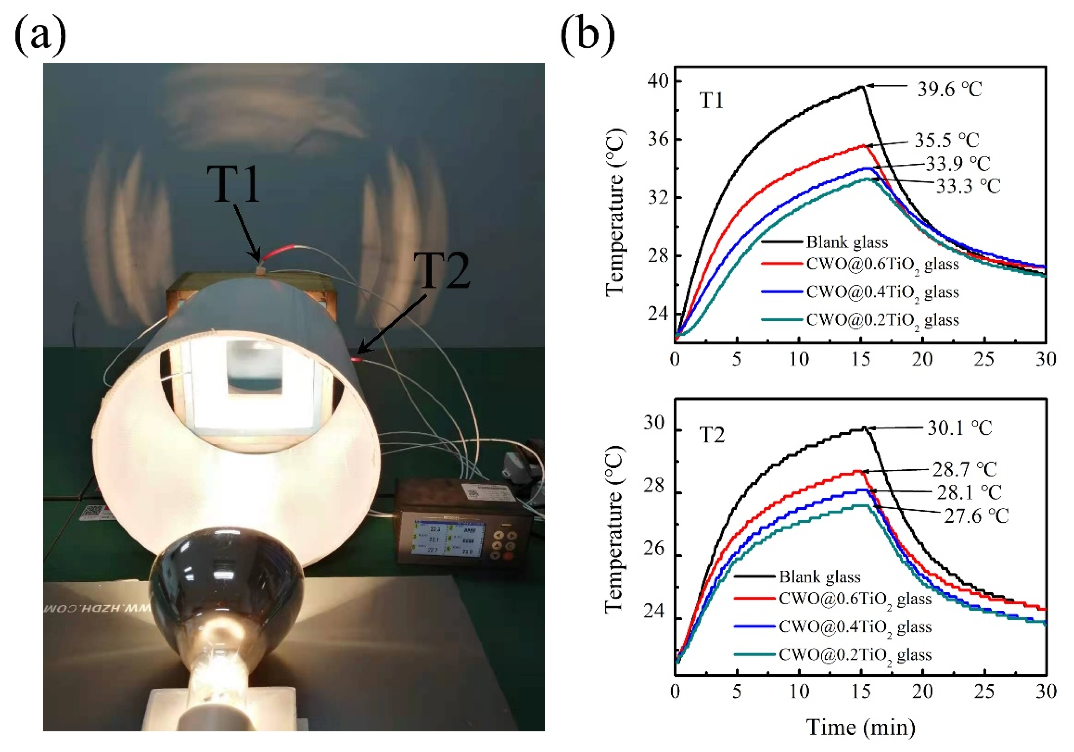
Finally, to verify the temperature control effect of CWO@TiO2-coated glass, a model house has been designed to test the temperature change as shown in the Figure 8a. The [email protected]2-coated glass is placed on the center of a cement wall and directly irradiated by the light from a NIR lamp (PHILIPS, R125) 50 cm away. Two thermocouples of T1 and T2 are placed behind the glass where they are directly illuminated by light and behind the cement wall where not directly illuminated by light, respectively. In addition, a blank glass was used in the control group test. Figure 8b shows the temperature changes with time measured by T1 and T2 in the model house. The results show that the [email protected]2 coated glass reduces the T1 and T2 temperature by 6.3 °C and 2.5 °C, respectively, indicating that TiO2-coated CWO still has good heat shielding effect. However, although the other two samples [email protected]2 and [email protected]2 also have the cooling effect, the effect is not as good as [email protected]2, because the thicker the coating, the greater the near-infrared transmittance, which is consistent with the results in Figure 6b.

4. Conclusions

In this article, nanocrystalline CWO particles were prepared by solvothermal method and coated with small TiO2 crystals. The XRD, SEM and TEM results show that the surface of CWO is coated with a layer of crystalline TiO2, and the thickness of TiO2 shell increases with the increase in TTIP amount in the reaction process. The absorption spectrum illustrates that the NIR absorption stability of [email protected]2 is much better than that of CWO after 30 days, indicating that TiO2 coating significantly improves the stability of tungsten bronze. The transmittance of CWO@TiO2-coated glass in the visible region decreases with the increase in Ti source and increases obviously in the NIR region, indicating that the thicker the TiO2 shell, the worse the transparent heat shielding performance. The DDA simulation results also confirm this trend. The measurement of temperature control effect in the model house gives that the [email protected]2-coated glass reduces the indoor temperature by 6.3 °C and 2.5 °C at different places in the room, respectively, which demonstrated the good heat shielding performance of TiO2-coated CWO.

Author Contributions

L.C.: Resources, Original draft, Supervision, Funding acquisition. C.S.: Investigation, Data curation, Formal analysis. J.L. (Jiaxin Li): Data curation. M.S.: Formal analysis. J.L. (Jia Liu): Supervision. Y.M.: Project administration. All authors have read and agreed to the published version of the manuscript.

Funding

This work was supported by the Natural Science Foundation of Inner Mongolia (No. 2019MS05015), Innovation Fund of Inner Mongolia University of Science and Technology (No. 2019QDL-B35).

Institutional Review Board Statement

Not applicable.

Informed Consent Statement

Not applicable.

Data Availability Statement

No data reported other than that presented.

Conflicts of Interest

The authors declare no conflict of interest. The funders had no role in the design of the study; in the collection, analyses, or interpretation of data; in the writing of the manuscript, or in the decision to publish the results.

References

  1. Karlsson, J.; Roos, A. Annual energy window performance vs. glazing thermal emittance—The relevance of very low emittance values. Thin. Solid. Film. 2001, 92, 345–348. [Google Scholar] [CrossRef]
  2. Kim, C.; Park, J.W.; Kim, J.; Hong, S.J.; Lee, M.J. A highly efficient indium tin oxide nanoparticles (ITO-NPs) transparent heater based on solution-process optimized with oxygen vacancy control. J. Alloys Compd. 2017, 726, 712–719. [Google Scholar] [CrossRef]
  3. Chao, L.; Bao, L.; Wei, W.; Tegus, O. A review of recent advances in synthesis, characterization and NIR shielding property of nanocrystalline rare-earth hexaborides and tungsten bronzes. Sol. Energy 2019, 190, 10–27. [Google Scholar] [CrossRef]
  4. Schelm, S.; Smith, G.B. Dilute LaB6 Nanoparticles in Polymer as Optimized Clear Solar Control Glazing. Appl. Phys. Lett. 2003, 82, 4346. [Google Scholar] [CrossRef]
  5. Takeda, H.; Kuno, H.; Adachi, K. Solar Control Dispersions and Coatings with Rare-Earth Hexaboride Nanoparticles. J. Am. Ceram. Soc. 2018, 91, 2897. [Google Scholar] [CrossRef]
  6. Long, L.; Ye, H.; Gao, Y.; Zou, R. Performance demonstration and evaluation of the synergetic application of vanadium dioxide glazing and phase change material in passive buildings. Appl. Energy 2014, 136, 89–97. [Google Scholar] [CrossRef]
  7. Shen, N.; Dong, B.; Cao, C.; Chen, Z.; Liu, J.; Luo, H.; Guo, Y. Lowered phase transition temperature and excellent solar heat shielding properties of well-crystallized VO2 by W doping. Phys. Chem. Chem. Phys. 2016, 18, 28010–28017. [Google Scholar] [CrossRef] [PubMed]
  8. Iken, O.; Fertahi, S.; Dlimi, M.; Agounoun, R.; Kadiri, I.; Sbai, K. Thermal and energy performance investigation of a smart double skin facade integrating vanadium dioxide through CFD simulations. Energy Convers. Manag. 2019, 195, 650–671. [Google Scholar] [CrossRef]
  9. Liang, X.; Guo, C.; Chen, M.; Guo, S.; Zhang, L.; Li, F.; Guo, S.; Yang, H. A roll-to-roll process for multi-responsive soft-matter composite films containing CsxWO3 nanorods for energy-efficient smart window applications. Nanoscale Horiz. 2017, 2, 319–325. [Google Scholar] [CrossRef] [PubMed]
  10. Ran, S.; Liu, J.; Shi, F.; Fan, C.; Chen, B.; Zhang, H.; Yu, L.; Liu, S.H. Greatly improved heat-shielding performance of KxWO3 by trace Pt doping for energy-saving window glass applications. Sol. Energy. Mater. Sol. Cells 2018, 174, 342–350. [Google Scholar] [CrossRef]
  11. Liu, J.X.; Ando, Y.; Dong, X.L.; Shi, F.; Yin, S.; Adachi, K.; Chonan, T.; Tanaka, A.; Sato, T. Microstructure and electrical–optical properties of cesium tungsten oxides synthesized by solvothermal reaction followed by ammonia annealing. J. Solid. State. Chem. 2010, 183, 2456–2460. [Google Scholar] [CrossRef]
  12. Adachi, K.; Ota, Y.; Tanaka, H.; Okada, M.; Oshimura, N.; Tofuku, A. Chromatic instabilities in cesium-doped tungsten bronze nanoparticles. J. Appl. Phys. 2013, 114, 194304. [Google Scholar] [CrossRef]
  13. Chen, Y.; Zeng, X.; Zhou, Y.; Li, R.; Yao, H.L.; Cao, X.; Jin, P. Core-shell structured CsxWO3@ZnO with excellent stability and high performance on near-infrared shielding. Ceram. Int. 2018, 44, 2738–2744. [Google Scholar] [CrossRef]
  14. Zeng, X.; Zhou, Y.; Jin, S.; Luo, H.; Yao, H.; Huang, X.; Jin, P. The preparation of a high performance nearinfrared shielding CsxWO3/SiO2 composite resin coating and research on its optical stability under ultraviolet illumination. J. Mater. Chem. C 2015, 3, 8050–8060. [Google Scholar] [CrossRef]
  15. Sun, C.; Liu, J.; Chao, L. Theoretical analysis on optical properties of Cs0.33WO3 nanoparticles with different sizes, shapes and structures. Materials. Lett. 2020, 272, 127847. [Google Scholar] [CrossRef]
  16. Draine, B.T. The discrete-dipole approximation and its application to interstellar graphite grains. Astrophysical. J. 1988, 333, 848–872. [Google Scholar] [CrossRef]
  17. Draine, B.T.; Flatau, P.J. Discrete-dipole approximation for scattering calculations. J. Opt. Soc. Am. 1994, 11, 1491–1499. [Google Scholar] [CrossRef]
  18. Purcell, E.M.; Pennypacker, C.R. Scattering and absorption of light by nonspherical dielectric grains. Astrophysical. J. 1973, 186, 705–714. [Google Scholar] [CrossRef]
  19. Yurkin, M.A.; Hoekstra, A.G. The discrete-dipole-approximation code ADDA: Capabilities and known limitations. J. Quant. Spectrosc. Radiat. Transf. 2011, 112, 2234–2247. [Google Scholar] [CrossRef]
  20. Draine, B.T.; Flatau, P.J. User Guide for the Discrete Dipole Approximation Code DDSCAT 7.3. arXiv 2013. [Google Scholar] [CrossRef]
  21. Sato, Y.; Terauchi, M.; Adachi, K. High energy-resolution electron energy-loss spectroscopy study on the near-infrared scattering mechanism of Cs0.33WO3 crystals and nanoparticles. J. Appl. Phys. 2012, 112, 074308. [Google Scholar] [CrossRef]
  22. Li, G.; Guo, C.; Yan, M.; Liu, S. CsxWO3, nanorods: Realization of full-spectrum-responsive photocatalytic activities from UV, visible to near-infrared region. Appl. Catal. B Environ. 2016, 183, 142–148. [Google Scholar] [CrossRef]
Figure 1. XRD patterns of the uncoated CsxWO3 and TiO2 coated CsxWO3 nanoparticles.
Nanomaterials 12 02806 g001
Figure 2. SEM image of the (a) CWO, (b) [email protected]2, (c) [email protected]2, (d) [email protected]2 (inset shows a magnification of one segment).
Nanomaterials 12 02806 g002
Figure 3. Element mapping of (a) CWO, (b) [email protected]2, (c) [email protected]2, (d) [email protected]2; (e) Element spectrum of [email protected]2.
Nanomaterials 12 02806 g003
Figure 4. TEM images of the (a) [email protected]2, (b) [email protected]2, (c) [email protected]2; HRTEM image of the (d) [email protected]2; SAED of the [email protected]2 (e) selected area, (f) diffraction rings.
Nanomaterials 12 02806 g004
Figure 5. (a) Full range XPS spectra and (b) W4f core-level XPS spectra of [email protected]2.
Nanomaterials 12 02806 g005
Figure 6. (a) The absorption curve of CWO and [email protected]2 nanoparticles after different durations, (b) Transmittance curve of coated glass (inset shows the [email protected]2-coated glass used in the transmittance test).
Nanomaterials 12 02806 g006
Figure 7. Extinction efficiencies of CWO spherical particles with different TiO2 shell thicknesses.
Nanomaterials 12 02806 g007
Figure 8. (a) Photographs of devices used in temperature control test, (b) The temperature changes with time measured by T1 and T2 in the model house.
Nanomaterials 12 02806 g008
Publisher’s Note: MDPI stays neutral with regard to jurisdictional claims in published maps and institutional affiliations.