The Effect of Mesoporous Bioactive Glass Nanoparticles/Graphene Oxide Composites on the Differentiation and Mineralization of Human Dental Pulp Stem Cells

 , ,
, ,
Abstract
1. Introduction
2. Materials and Methods
2.1. Synthesis of MBN
2.2. Preparation and Characterization of the MBN/GO Composite
2.3. MBN/GO Composite Coating
2.4. Cell Culture
2.5. Cell Viability Assay
2.6. Alkaline Phosphatase (ALP) Activity Assay
2.7. Quantitative Real-Time Polymerase Chain Reaction (qRT-PCR)
2.8. Western Blot
2.9. Alizarin Red S Staining and Quantitative Analysis
2.10. Analysis of Wnt/β-Catenin Signaling Pathway-Related GENE expression in hDPSCs Cultured on MBN/GO Composites
2.11. Statistical Analysis
3. Results
3.1. Characterization of the MBN/GO Composite
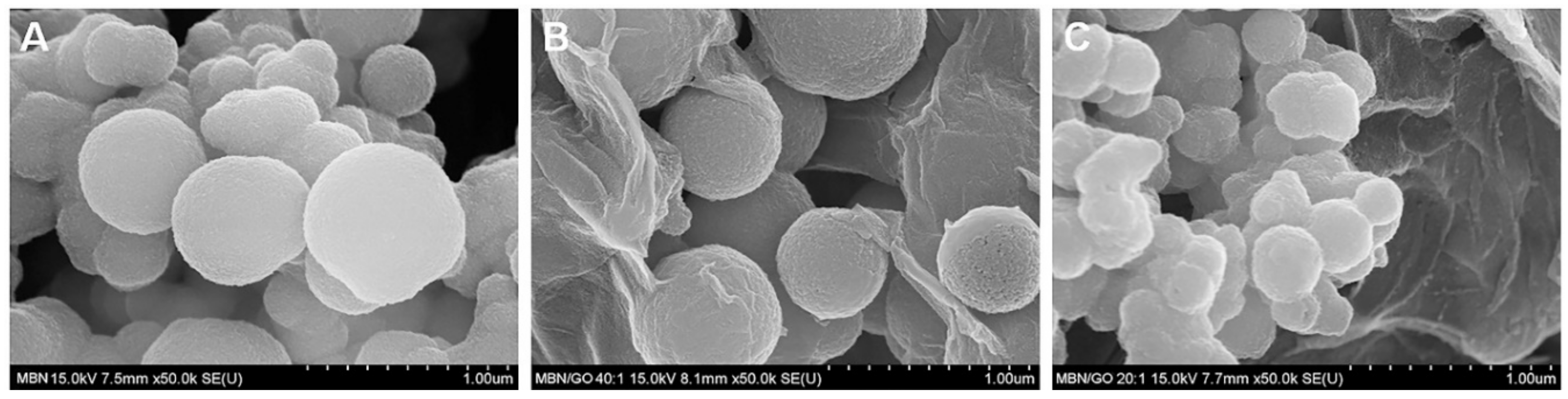
3.1.1. FESEM
3.1.2. XRD
3.1.3. FTIR
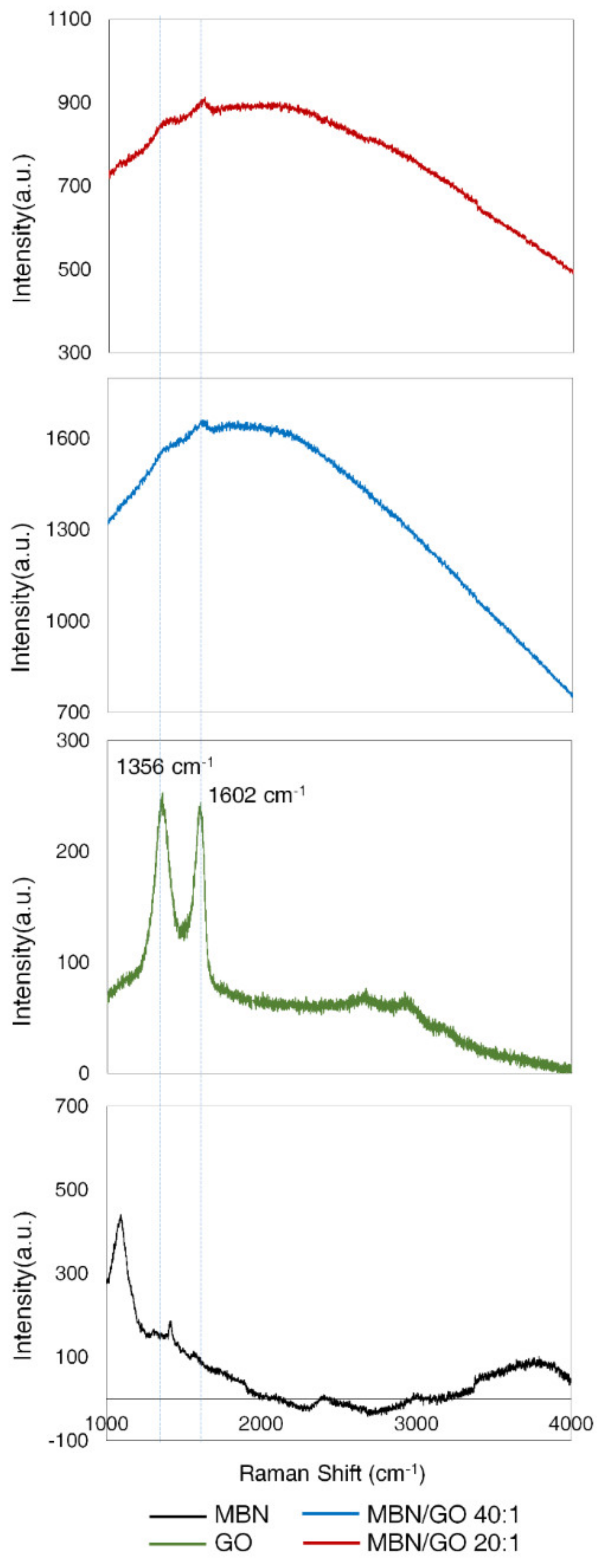
3.1.4. Raman Spectroscopy
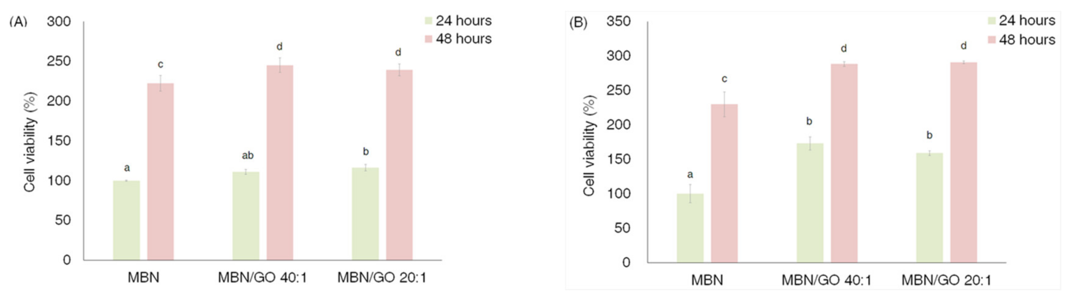
3.2. Viability of hDPSCs
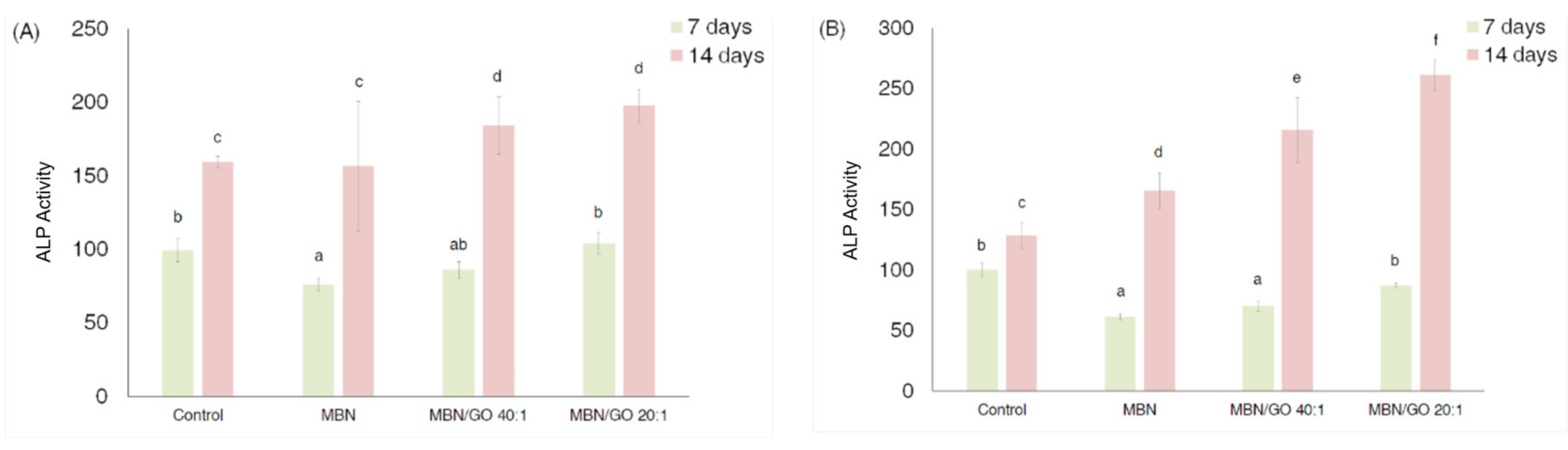
3.3. ALP Activity in hDPSCs
3.4. qRT-PCR
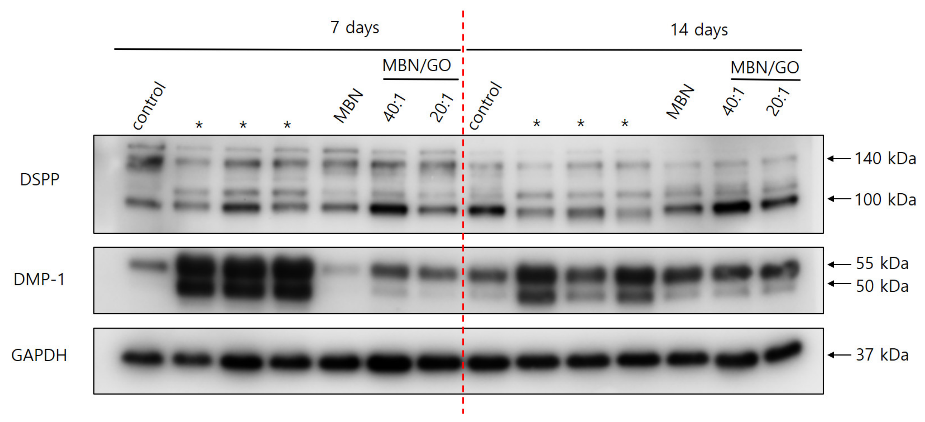
3.5. Western Blot
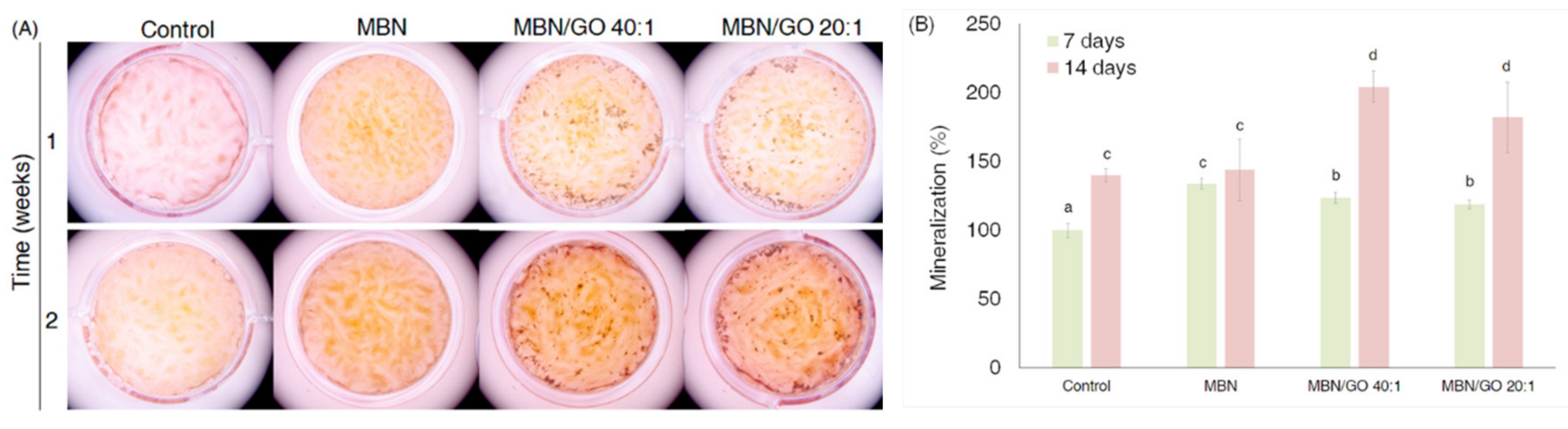
3.6. Alizarin red S Staining and Analysis
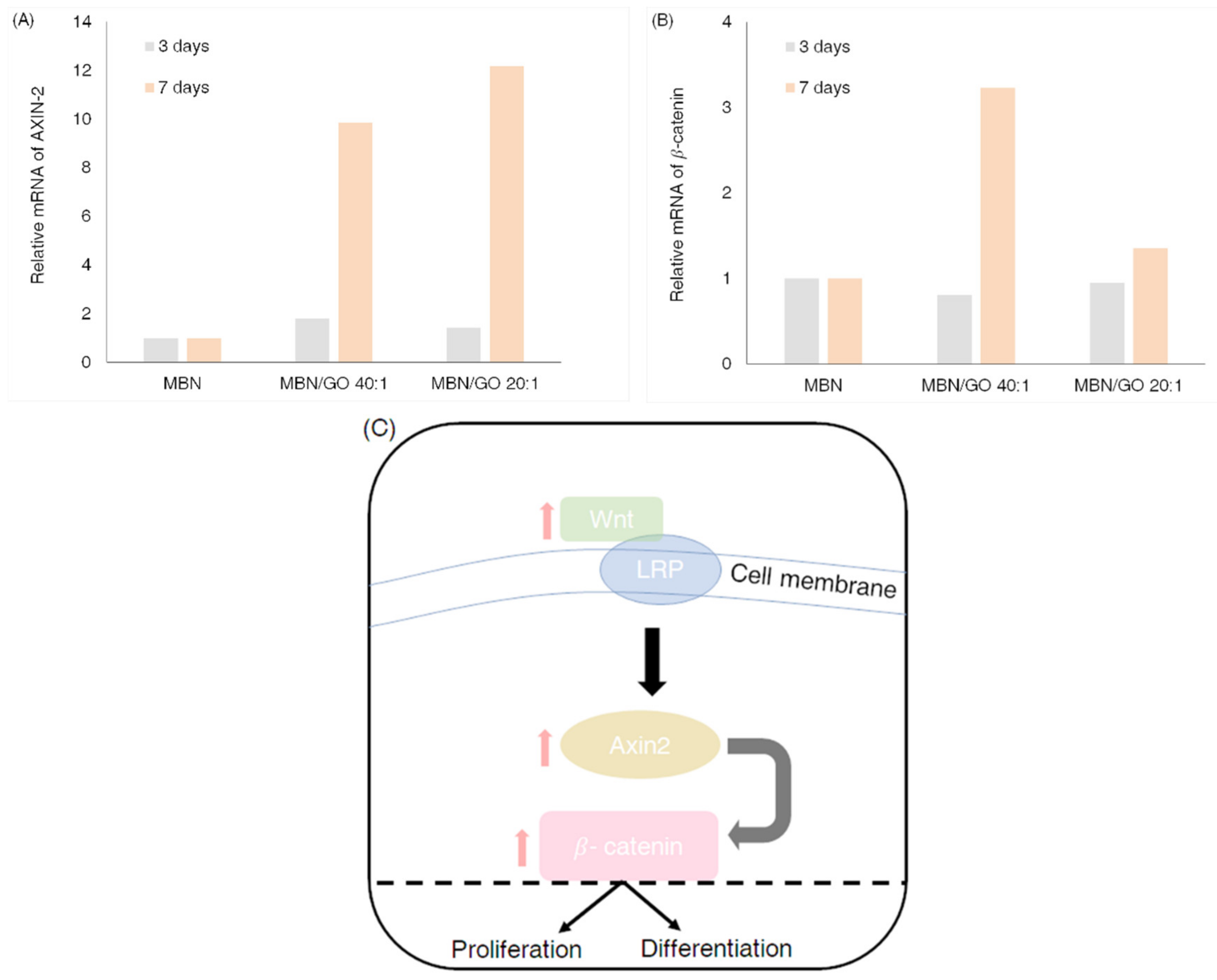
3.7. Wnt/β-Catenin Signaling Pathway-Related Gene Expression in hDPSCs Cultured on MBN/GO Composites
4. Discussion
5. Conclusions
Author Contributions
Funding
Conflicts of Interest
References
- Sloan, A.J.; Smith, A.J. Stem cells and the dental pulp: Potential roles in dentine regeneration and repair. Oral Dis. 2007, 13, 151–157. [Google Scholar] [CrossRef] [PubMed]
- Couble, M.L.; Farges, J.C.; Bleicher, F.; Perrat-Mabillon, B.; Boudeulle, M.; Magloire, H. Odontoblast differentiation of human dental pulp cells in explant cultures. Calcif. Tissue Int. 2000, 66, 129–138. [Google Scholar] [CrossRef] [PubMed]
- Gronthos, S.; Mankani, M.; Brahim, J.; Robey, P.G.; Shi, S. Postnatal human dental pulp stem cells (DPSCs) In Vitro and In Vivo. Proc. Natl. Acad. Sci. USA 2000, 97, 13625–13630. [Google Scholar] [CrossRef] [PubMed]
- Hoppe, A.; Guldal, N.S.; Boccaccini, A.R. A review of the biological response to ionic dissolution products from bioactive glasses and glass-ceramics. Biomaterials 2011, 32, 2757–2774. [Google Scholar] [CrossRef] [PubMed]
- Fernando, D.; Attik, N.; Pradelle-Plasse, N.; Jackson, P.; Grosgogeat, B.; Colon, P. Bioactive glass for dentin remineralization: A systematic review. Mater. Sci. Eng. C Mater. Biol. Appl. 2017, 76, 1369–1377. [Google Scholar] [CrossRef]
- Rahaman, M.N.; Day, D.E.; Bal, B.S.; Fu, Q.; Jung, S.B.; Bonewald, L.F.; Tomsia, A.P. Bioactive glass in tissue engineering. Acta Biomater. 2011, 7, 2355–2373. [Google Scholar] [CrossRef] [PubMed]
- Ma, J.; Chen, C.Z.; Wang, D.G.; Meng, X.G.; Shi, J.Z. Influence of the sintering temperature on the structural feature and bioactivity of sol–gel derived SiO2–CaO–P2O5 bioglass. Ceram. Int. 2010, 36, 1911–1916. [Google Scholar] [CrossRef]
- Boccaccini, A.R.; Erol, M.; Stark, W.J.; Mohn, D.; Hong, Z.; Mano, J.F. Polymer/bioactive glass nanocomposites for biomedical applications: A review. Compos. Sci. Technol. 2010, 70, 1764–1776. [Google Scholar] [CrossRef]
- Kim, T.H.; Lee, T.; El-Said, W.A.; Choi, J.W. Graphene-Based Materials for Stem Cell Applications. Materials 2015, 8, 8674–8690. [Google Scholar] [CrossRef]
- Chen, G.Y.; Pang, D.W.; Hwang, S.M.; Tuan, H.Y.; Hu, Y.C. A graphene-based platform for induced pluripotent stem cells culture and differentiation. Biomaterials 2012, 33, 418–427. [Google Scholar] [CrossRef]
- Lee, J.H.; Shin, Y.C.; Jin, O.S.; Kang, S.H.; Hwang, Y.S.; Park, J.C.; Hong, S.W.; Han, D.W. Reduced graphene oxide-coated hydroxyapatite composites stimulate spontaneous osteogenic differentiation of human mesenchymal stem cells. Nanoscale 2015, 7, 11642–11651. [Google Scholar] [CrossRef] [PubMed]
- Ashok raja, C.; Balakumar, S.; Bargavi, P.; Rajashree, P.; Anandkumar, B.; George, R.P.; Kamachi Mudali, U. Decoration of 1-D nano bioactive glass on reduced graphene oxide sheets: Strategies and In Vitro bioactivity studies. Mater. Sci. Eng. C Mater. Biol. Appl. 2018, 90, 85–94. [Google Scholar]
- Intiso, A.; Martinez-Triguero, J.; Cucciniello, R.; Proto, A.; Palomares, A.E.; Rossi, F. A Novel Synthetic Route to Prepare High Surface Area Mayenite Catalyst for TCE Oxidation. Catalysts 2019, 9, 27. [Google Scholar] [CrossRef]
- Ogomi, D.; Serizawa, T.; Akashi, M. Bioinspired organic-inorganic composite materials prepared by an alternate soaking process as a tissue reconstitution matrix. J. Biomed. Mater. Res. Part A 2003, 67, 1360–1366. [Google Scholar] [CrossRef] [PubMed]
- Maeda, H.; Kasuga, T. Preparation of poly(lactic acid) composite hollow spheres containing calcium carbonates. Acta Biomater. 2006, 2, 403–408. [Google Scholar] [CrossRef] [PubMed]
- Kumar, G.S.; Girija, E.K.; Thamizhavel, A.; Yokogawa, Y.; Kalkura, S.N. Synthesis and characterization of bioactive hydroxyapatite-calcite nanocomposite for biomedical applications. J. Colloid Interface Sci. 2010, 349, 56–62. [Google Scholar] [CrossRef]
- Radev, L.; Vladov, D.; Michailova, I.; Cholakova, E.; Fernandes, M.F.; Salvado, I.M. In Vitro bioactivity of polycaprolactone/bioglass composites. Int. J. Mater. Chem. 2013, 3, 91–98. [Google Scholar]
- Zhang, W.L.; Choi, H.J. Silica-graphene oxide hybrid composite particles and their electroresponsive characteristics. Langmuir 2012, 28, 7055–7062. [Google Scholar] [CrossRef]
- Fan, Z.; Wang, J.; Liu, F.; Nie, Y.; Ren, L.; Liu, B. A new composite scaffold of bioactive glass nanoparticles/graphene: Synchronous improvements of cytocompatibility and mechanical property. Colloids Surf. B Biointerfaces 2016, 145, 438–446. [Google Scholar] [CrossRef]
- Chen, W.; Yan, L.; Bangal, P.R. Preparation of graphene by the rapid and mild thermal reduction of graphene oxide induced by microwaves. Carbon 2010, 48, 1146–1152. [Google Scholar] [CrossRef]
- Zheng, K.; Boccaccini, A.R. Sol-gel processing of bioactive glass nanoparticles: A review. Adv. Colloid Interface Sci. 2017, 249, 363–373. [Google Scholar] [CrossRef]
- Yan, X.; Yu, C.; Zhou, X.; Tang, J.; Zhao, D. Highly ordered mesoporous bioactive glasses with superior In Vitro bone-forming bioactivities. Angew. Chem. Int. Ed. Engl. 2004, 43, 5980–5984. [Google Scholar] [CrossRef]
- El-Fiqi, A.; Lee, J.H.; Lee, E.J.; Kim, H.W. Collagen hydrogels incorporated with surface-aminated mesoporous nanobioactive glass: Improvement of physicochemical stability and mechanical properties is effective for hard tissue engineering. Acta Biomater. 2013, 9, 9508–9521. [Google Scholar] [CrossRef] [PubMed]
- Fu, Q.; Saiz, E.; Rahaman, M.N.; Tomsia, A.P. Bioactive glass scaffolds for bone tissue engineering: State of the art and future perspectives. Mater. Sci. Eng. C Mater. Biol. Appl. 2011, 31, 1245–1256. [Google Scholar] [CrossRef] [PubMed]
- Ilyas, K.; Zahid, S.; Batool, M.; Chaudhry, A.A.; Jamal, A.; Iqbal, F.; Nawaz, M.H.; Goerke, O.; Gurlo, A.; Shah, A.T.; et al. In-Vitro investigation of graphene oxide reinforced bioactive glass ceramics composites. J. Non Cryst. Solids 2019, 505, 122–130. [Google Scholar] [CrossRef]
- Porwal, H.; Grasso, S.; Cordero-Arias, L.; Li, C.; Boccaccini, A.R.; Reece, M.J. Processing and bioactivity of 45S5 Bioglass((R))-graphene nanoplatelets composites. J. Mater. Sci. Mater. Med. 2014, 25, 1403–1413. [Google Scholar] [CrossRef]
- Kim, J.; Choi, K.S.; Kim, Y.; Lim, K.T.; Seonwoo, H.; Park, Y.; Kim, D.H.; Choung, P.H.; Cho, C.S.; Kim, S.Y.; et al. Bioactive effects of graphene oxide cell culture substratum on structure and function of human adipose-derived stem cells. J. Biomed. Mater. Res. Part A 2013, 101, 3520–3530. [Google Scholar] [CrossRef]
- Meng, D.; Rath, S.N.; Mordan, N.; Salih, V.; Kneser, U.; Boccaccini, A.R. In Vitro evaluation of 45S5 Bioglass(R)-derived glass-ceramic scaffolds coated with carbon nanotubes. J. Biomed. Mater. Res. Part A 2011, 99, 435–444. [Google Scholar] [CrossRef]
- Jung, J.H.; Park, S.B.; Yoo, K.H.; Yoon, S.Y.; Bae, M.K.; Lee, D.J.; Ko, C.C.; Kwon, Y.H.; Kim, Y.I. Effect of different sizes of bioactive glass-coated mesoporous silica nanoparticles on dentinal tubule occlusion and mineralization. Clin. Oral Investig. 2019, 23, 2129–2141. [Google Scholar] [CrossRef]
- Zeng, Y.; Pei, X.; Yang, S.; Qin, H.; Cai, H.; Hu, S.; Sui, L.; Wan, Q.; Wang, J. Graphene oxide/hydroxyapatite composite coatings fabricated by electrochemical deposition. Surf. Coat. Technol. 2016, 286, 72–79. [Google Scholar] [CrossRef]
- Baradaran, S.; Moghaddam, E.; Basirun, W.J.; Mehrali, M.; Sookhakian, M.; Hamdi, M.; Nakhaei Moghaddam, M.R.; Alias, Y. Mechanical properties and biomedical applications of a nanotube hydroxyapatite-reduced graphene oxide composite. Carbon 2014, 69, 32–45. [Google Scholar] [CrossRef]
- Dimiev, A.M.; Tour, J.M. Mechanism of graphene oxide formation. ACS Nano 2014, 8, 3060–3068. [Google Scholar] [CrossRef] [PubMed]
- Chang, Y.; Yang, S.T.; Liu, J.H.; Dong, E.; Wang, Y.; Cao, A.; Liu, Y.; Wang, H. In Vitro toxicity evaluation of graphene oxide on A549 cells. Toxicol. Lett. 2011, 200, 201–210. [Google Scholar] [CrossRef] [PubMed]
- Wang, K.; Ruan, J.; Song, H.; Zhang, J.; Wo, Y.; Guo, S.; Cui, D. Biocompatibility of Graphene Oxide. Nanoscale Res. Lett. 2011, 6, 8. [Google Scholar] [CrossRef]
- Nam, S.; Won, J.E.; Kim, C.H.; Kim, H.W. Odontogenic differentiation of human dental pulp stem cells stimulated by the calcium phosphate porous granules. J. Tissue Eng. 2011, 2011, 812547. [Google Scholar] [CrossRef]
- Hong, S.J.; Yu, H.S.; Noh, K.T.; Oh, S.A.; Kim, H.W. Novel scaffolds of collagen with bioactive nanofiller for the osteogenic stimulation of bone marrow stromal cells. J. Biomater. Appl. 2010, 24, 733–750. [Google Scholar] [CrossRef]
- Liu, X.; Shen, H.; Song, S.; Chen, W.; Zhang, Z. Accelerated biomineralization of graphene oxide-incorporated cellulose acetate nanofibrous scaffolds for mesenchymal stem cell osteogenesis. Colloids Surf. B Biointerfaces 2017, 159, 251–258. [Google Scholar] [CrossRef]
- Kim, J.W.; Yamakoshi, Y.; Iwata, T.; Hu, Y.Y.; Zhang, H.; Hu, J.C.; Zhang, H.; Hu, J.; Simmer, J.P. Porcine dentin matrix protein 1: Gene structure, cDNA sequence, and expression in teeth. Eur. J. Oral Sci. 2006, 114, 33–41. [Google Scholar] [CrossRef][Green Version]
- Tartaix, P.H.; Doulaverakis, M.; George, A.; Fisher, L.W.; Butler, W.T.; Qin, C.; Salih, E.; Tan, M.; Fujimoto, Y.; Spevak, L.; et al. In Vitro effects of dentin matrix protein-1 on hydroxyapatite formation provide insights into In Vivo functions. J. Biol. Chem. 2004, 279, 18115–18120. [Google Scholar] [CrossRef]
- Qin, C.; Baba, O.; Butler, W.T. Post translational modifications of sibling proteins and their roles in osteogenesis and dentinogenesis. Crit. Rev. Oral Biol. Med. 2004, 15, 126–136. [Google Scholar] [CrossRef]
- Pan, K.; Sun, Q.; Zhang, J.; Ge, S.; Li, S.; Zhao, Y.; Yang, P. Multilineage differentiation of dental follicle cells and the roles of Runx2 over-expression in enhancing osteoblast/cementoblast-related gene expression in dental follicle cells. Cell Prolif. 2010, 43, 219–228. [Google Scholar] [CrossRef] [PubMed]
- Suzuki, S.; Haruyama, N.; Nishimura, F.; Kulkarni, A.B. Dentin sialophosphoprotein and dentin matrix protein-1: Two highly phosphorylated proteins in mineralized tissues. Arch. Oral Biol. 2012, 57, 1165–1175. [Google Scholar] [CrossRef] [PubMed]
- von Marschall, Z.; Fisher, L.W. Dentin sialophosphoprotein (DSPP) is cleaved into its two natural dentin matrix products by three isoforms of bone morphogenetic protein-1 (BMP1). Matrix Biol. 2010, 29, 295–303. [Google Scholar] [CrossRef] [PubMed]
- Zhang, X.; Zhao, J.; Li, C.; Gao, S.; Qiu, C.; Liu, P.; Wu, G.; Qiang, B.; Lo, W.H.; Shen, Y. DSPP mutation in dentinogenesis imperfecta Shields type II. Nat. Genet. 2001, 27, 151–152. [Google Scholar] [CrossRef] [PubMed]
- Sun, Y.; Lu, Y.; Chen, S.; Prasad, M.; Wang, X.; Zhu, Q.; Zhang, J.; Ball, H.; Feng, J.; Butler, W.T.; et al. Key proteolytic cleavage site and full-length form of DSPP. J. Dent. Res. 2010, 89, 498–503. [Google Scholar] [CrossRef] [PubMed]
- Boskey, A.; Spevak, L.; Tan, M.; Doty, S.B.; Butler, W.T. Dentin sialoprotein (DSP) has limited effects on In Vitro apatite formation and growth. Calcif. Tissue Int. 2000, 67, 472–478. [Google Scholar] [CrossRef] [PubMed]
- He, G.; Ramachandran, A.; Dahl, T.; George, S.; Schultz, D.; Cookson, D.; Veis, A.; George, A. Phosphorylation of phosphophoryn is crucial for its function as a mediator of biomineralization. J. Biol. Chem. 2005, 280, 33109–33114. [Google Scholar] [CrossRef]
- Aplin, H.M.; Hirst, K.L.; Crosby, A.H.; Dixon, M.J. Mapping of the human dentin matrix acidic phosphoprotein gene (DMP1) to the dentinogenesis imperfecta type II critical region at chromosome 4q21. Genomics 1995, 30, 347–349. [Google Scholar] [CrossRef]
- Qin, C.; Brunn, J.C.; Cook, R.G.; Orkiszewski, R.S.; Malone, J.P.; Veis, A.; Butler, W.T. Evidence for the proteolytic processing of dentin matrix protein 1. Identification and characterization of processed fragments and cleavage sites. J. Biol. Chem. 2003, 278, 34700–34708. [Google Scholar] [CrossRef]
- Sun, Y.; Chen, L.; Ma, S.; Zhou, J.; Zhang, H.; Feng, J.Q.; Qin, C. Roles of DMP1 processing in osteogenesis, dentinogenesis and chondrogenesis. Cells Tissues Organs 2011, 194, 199–204. [Google Scholar] [CrossRef]
- Tecles, O.; Laurent, P.; Zygouritsas, S.; Burger, A.S.; Camps, J.; Dejou, J.; About, I. Activation of human dental pulp progenitor/stem cells in response to odontoblast injury. Arch. Oral Biol. 2005, 50, 103–108. [Google Scholar] [CrossRef] [PubMed]
- Boskey, A.L. Biomineralization: Conflicts, challenges, and opportunities. J. Cell. Biochem. 1998, 72, 83–91. [Google Scholar] [CrossRef]
- Hoikkala, N.J.; Wang, X.; Hupa, L.; Smatt, J.H.; Peltonen, J.; Vallittu, P.K. Dissolution and mineralization characterization of bioactive glass ceramic containing endodontic sealer Guttaflow Bioseal. Dent. Mater. J. 2018, 37, 988–994. [Google Scholar] [CrossRef] [PubMed]
- Ahmadzadeh, A.; Norozi, F.; Shahrabi, S.; Shahjahani, M.; Saki, N. Wnt/beta-catenin signaling in bone marrow niche. Cell Tissue Res. 2016, 363, 321–335. [Google Scholar] [CrossRef] [PubMed]
- Yu, H.M.; Jerchow, B.; Sheu, T.J.; Liu, B.; Costantini, F.; Puzas, J.E.; Birchmeier, W.; Hsu, W. The role of Axin2 in calvarial morphogenesis and craniosynostosis. Development 2005, 132, 1995–2005. [Google Scholar] [CrossRef]
- Duan, P.; Bonewald, L.F. The role of the wnt/beta-catenin signaling pathway in formation and maintenance of bone and teeth. Int. J. Biochem. Cell Biol. 2016, 77, 23–29. [Google Scholar] [CrossRef]
- Wu, C.; Xia, L.; Han, P.; Xu, M.; Fang, B.; Wang, J.; Chang, J.; Xiao, Y. Graphene-oxide-modified β-tricalcium phosphate bioceramics stimulate In Vitro and In Vivo osteogenesis. Carbon 2015, 93, 116–129. [Google Scholar] [CrossRef]


| Sample Powder’s Concentration % (w/v) | Samples | |
|---|---|---|
| 0.05% | 0.01% | |
| Surface coating concentration at culture plates (mg·cm–2) | 0.15625 mg·cm–2 | 0.03125 mg·cm–2 |
| Gene | Primer | Sequence (5’-3’) | Size (bp) |
|---|---|---|---|
| β-actin | Forward | GCACTCTTCCAGCCTTCCTT | 150 |
| Reverse | AATGCCAGGGTACATGGTGG | ||
| DMP-1 | Forward | GGAGAGACAGCAAGGGTGAC | 87 |
| Reverse | CACTGCTGGGACCATCTACG | ||
| DSPP | Forward | GCTGGCCTGGATAATTCCGA | 135 |
| Reverse | CTCCTGGCCCTTGCTGTTAT | ||
| ALP | Forward | AATGTGGACACAGTGGCTGGA | 78 |
| Reverse | TCTCCTGCTCAGTCATCTGCT | ||
| BMP-2 | Forward | AAGCCAAACACAAACAGCGG | 104 |
| Reverse | GGGAGCCACAATCCAGTCAT | ||
| RUNX-2 | Forward | TCTGGCCTTCCACTCTCAGTA | 134 |
| Reverse | TGGATAGTGCATTCGTGGGT | ||
| MEPE | Forward | GCAGCTATCCACACCAGAAAG | 113 |
| Reverse | GTTGAAATGTTGGTGCTGCC | ||
| AXIN-2 | Forward | CCCTGCTGACTTGAGAGAGAC | 82 |
| Reverse | CCCACTGAGTCTGGAATCTC | ||
| β-catenin | Forward | CAGCGTGGACAATGGCTACT | 101 |
| Reverse | AGATTCCTGCTGGTGGCTTG |
© 2020 by the authors. Licensee MDPI, Basel, Switzerland. This article is an open access article distributed under the terms and conditions of the Creative Commons Attribution (CC BY) license (http://creativecommons.org/licenses/by/4.0/).
Share and Cite
Ahn, J.H.; Kim, I.-R.; Kim, Y.; Kim, D.-H.; Park, S.-B.; Park, B.-S.; Bae, M.-K.; Kim, Y.-I. The Effect of Mesoporous Bioactive Glass Nanoparticles/Graphene Oxide Composites on the Differentiation and Mineralization of Human Dental Pulp Stem Cells. Nanomaterials 2020, 10, 620. https://doi.org/10.3390/nano10040620
Ahn JH, Kim I-R, Kim Y, Kim D-H, Park S-B, Park B-S, Bae M-K, Kim Y-I. The Effect of Mesoporous Bioactive Glass Nanoparticles/Graphene Oxide Composites on the Differentiation and Mineralization of Human Dental Pulp Stem Cells. Nanomaterials. 2020; 10(4):620. https://doi.org/10.3390/nano10040620
Chicago/Turabian StyleAhn, Jae Hwa, In-Ryoung Kim, Yeon Kim, Dong-Hyun Kim, Soo-Byung Park, Bong-Soo Park, Moon-Kyoung Bae, and Yong-Il Kim. 2020. "The Effect of Mesoporous Bioactive Glass Nanoparticles/Graphene Oxide Composites on the Differentiation and Mineralization of Human Dental Pulp Stem Cells" Nanomaterials 10, no. 4: 620. https://doi.org/10.3390/nano10040620
APA StyleAhn, J. H., Kim, I.-R., Kim, Y., Kim, D.-H., Park, S.-B., Park, B.-S., Bae, M.-K., & Kim, Y.-I. (2020). The Effect of Mesoporous Bioactive Glass Nanoparticles/Graphene Oxide Composites on the Differentiation and Mineralization of Human Dental Pulp Stem Cells. Nanomaterials, 10(4), 620. https://doi.org/10.3390/nano10040620