Article

Design of Trabecular Bone Mimicking Voronoi Lattice-Based Scaffolds and CFD Modelling of Non-Newtonian Power Law Blood Flow Behaviour

by
Haja-Sherief N. Musthafa
1,* and
Jason Walker
2
1
Department of Computer Science, Electrical Engineering and Mathematical Sciences, Western Norway University of Applied Sciences, 5063 Bergen, Norway
2
Center for Design and Manufacturing Excellence, The Ohio State University, Columbus, OH 43210, USA
*
Author to whom correspondence should be addressed.
Computation 2024, 12(12), 241; https://doi.org/10.3390/computation12120241
Submission received: 10 October 2024 / Revised: 25 November 2024 / Accepted: 26 November 2024 / Published: 5 December 2024
(This article belongs to the Section Computational Engineering)

Abstract

:
Designing scaffolds similar to the structure of trabecular bone requires specialised algorithms. Existing scaffold designs for bone tissue engineering have repeated patterns that do not replicate the random stochastic porous structure of the internal architecture of bones. In this research, the Voronoi tessellation method is applied to create random porous biomimetic structures. A volume mesh created from the shape of a Zygoma fracture acts as a boundary for the generation of random seed points by point spacing to create Voronoi cells and Voronoi diagrams. The Voronoi lattices were obtained by adding strut thickness to the Voronoi diagrams. Gradient Voronoi scaffolds of pore sizes (19.8 µm to 923 µm) similar to the structure of the trabecular bone were designed. A Finite Element Method-based computational fluid dynamics (CFD) simulation was performed on all designed Voronoi scaffolds to predict the pressure drops and permeability of non-Newtonian blood flow behaviour using the power law material model. The predicted permeability (0.33 × 10⁻⁹ m2 to 2.17 × 10⁻⁹ m2) values of the Voronoi scaffolds from the CFD simulation are comparable with the permeability of scaffolds and bone specimens from other research works.

1. Introduction

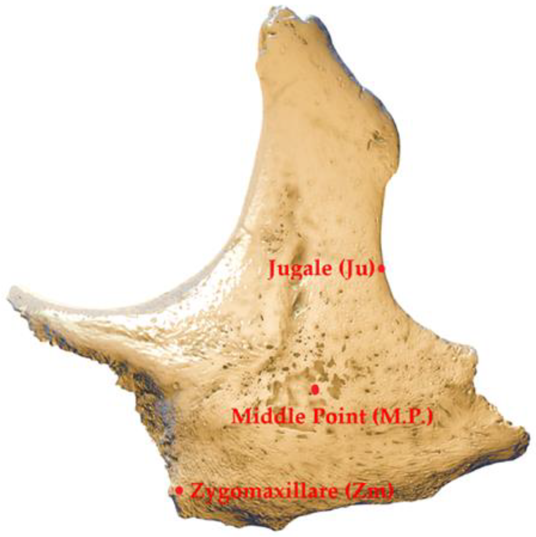
A human bone is a stiff organ with outer concentrated tissues (cortical bone) and inner spongy tissues (trabecular bone/cancellous bone). Trabecular bone patterns such as the femur (thigh bone) and the zygoma (cheekbone) (Figure 1 and Figure 2) possess distinct random and interconnected irregular porous arrangements of different thicknesses (50 to 300 µm) and pore sizes [1]. Bones, which protect internal organs such as the brain, lungs, and heart, act as a storehouse of minerals and a production site of red blood cells. Bone defects occur due to infections, genetic disorders, trauma, diseases, and tumours. Although bone grafts are the gold standard in the repair of bone defects, they have demerits of limited bone tissue availability, donor site morbidity, immune rejection of transplanted cells, long recovery times, and transmission of infections [2]. Three-dimensional (3D) scaffolds, which are porous temporary structures that are utilised in bone tissue engineering (BTE), are alternatives to bone grafts for replacement, repair, and regeneration of bone tissues in bone defects, trauma and injuries [3,4,5]. BTE scaffolds have been applied in various applications, such as large segmental defects in long bones [6], oral and maxillofacial defects [7], articular osteochondral defects (OCD) [8] and ipsilateral femoral bone defects [9]. These scaffolds provide support to withstand the applied mechanical loading, mass transport of nutrients, biofactors, cells, and the removal of wastes to achieve the goal of bone tissue regeneration. Mechanical loading and fluid flow shear stress act as mechanical stimuli transduced into various biophysical factors for osteogenesis to proliferate and differentiate implanted stem cells into functional bone tissues and de novo blood vessels (angiogenesis) [10,11,12].
With modern additive manufacturing (3D printing) machines, manufacturing complex geometries of tissue engineering scaffolds is practically feasible [17,18]. The design of scaffolds for BTE is highly demanding in creating a structure that mimics the architecture of natural trabecular bone [19,20]. The scaffold architecture must be able to replicate the in vivo nature of bones by providing adequate pore distribution with the required structural integrity and maintaining permeability between scaffolds and native bone tissues [21,22]. It is also vital for cell attachment, proliferation (multiplication of cells), and cell differentiation into functional bone tissues. It is feasible to distinguish BTE scaffold designs into two rudimentary groups: one is nonparametric design, such as cubic [23], diamond, or honeycomb [24,25], and the other is parametric design, such as triply periodic minimal surfaces (TPMS) [26,27,28,29]. Unfortunately, the nonparametric and TPMS-based scaffolds are not able to fully replicate the non-periodic and irregular porous microarchitecture of the natural bones. There is a need for a design of scaffolds to mimic the architecture of trabecular bone, which has gradient porous structures that enhance bone regeneration activities such as cell attachment on the scaffold surface, cell growth, and osteogenesis. Such irregular stochastic designs require complex algorithms, which are complicated, and the scaffold design based on the Voronoi tessellation method is one of the possibilities to achieve the biomimetic structure of bones, providing realistic physiological habitat for human cells [30].
The Voronoi tessellation (tiling) method is based on a Voronoi diagram, where several points (seeds) are distributed in a given plane. A circle is drawn around every seed, and it extends until two circles touch each other to form an edge or boundary, further extending to create polygons. The graph formed from the seeds given is known as the Voronoi diagram. The given plane is, therefore, divided into several regions known as Voronoi cells or Thiessen polygons [31]. Each polygon surrounds a point, and the boundary between two or more points is equal to their distance (Figure 3). This concept of Voronoi diagrams can be extended to a 3D space for a given random point cloud to create a stochastic random polyhedral lattice structure that can resemble the internal architecture of a trabecular bone for BTE applications (Figure 4) [32,33,34,35]. Nguyen et al. generated 3D random lattice geometrical models based on Voronoi diagrams for additive manufacturing and presented a workflow to create a final assembled product from random points [36]. Rezapourian et al. performed a numerical study for orthopaedic applications using finite element analysis (FEA) to examine the variation of mechanical properties of Hydroxyapatite based irregular Voronoi lattice bone scaffolds by altering their morphological parameters, such as the thickness of the trabeculae and the point spacing distance [37]. Liu et al. combined the topological optimisation method with Voronoi-based random porous structures generated from Voronoi diagrams using the surface mesh superposition concept for medical applications [38]. Xiao et al. studied the effect of the variation of porosity of Voronoi tessellation-based random pore structured geometry models on their permeabilities using the Lattice Boltzmann method (LBM) [39].
Permeability is a measure for scaffolds to allow the mass transport movement of biological cells, oxygen, nutrients for activities of the cells, and waste materials [41,42]. It depends on the microstructure, the shape of the scaffolds and well-interconnected pores, which allow substantial fluid transport to enrich bone tissue regeneration [43,44]. Large pores and higher porosity of scaffolds lead to higher permeability due to a high quantity of mass transfer but with a reduction in mechanical strength. Jiao et al. concluded that the trabecular porous scaffolds of higher porosities have superior bone integration, bone formation and bone ingrowth than the scaffolds with lower porosities in defects of femoral bones [45]. However, higher permeabilities can cause increased cell washout by the fluid and non-adhesion to the scaffolds, while lower permeabilities can lead to malnutrition, which hinders bone tissue growth.
Computational fluid dynamics (CFD) is a numerical modelling scheme for the prediction of flow properties such as permeability, tortuosity, wall shear stress (WSS), and fluid pressure within scaffolds [46,47]. As the governing equations of fluid flow related to partial difference Equations (PDEs), numerical methods involving finite difference methods (FDM), finite volume methods (FVM), and finite element methods (FEM) have been applied to solve these equations. The problem with FDM methods is that they apply structured grids for discretising the given fluid domain, and they have difficulty approximating complex geometries. This problem is solved by FVM and FEM, which use unstructured meshes with tetrahedral, hexahedral, and polyhedral elements [48]. Even though tetrahedral meshes are convenient for discretising complex geometry, their discretised cells are overstretched in highly curved structures. Such meshes have a large aspect ratio and are not aligned with the direction of flow, causing false diffusion while dealing with advection terms and making the solution diverge in the case of FVM [49]. FEM applies several streamline upwinding advection dominated PDE methods such as monotone streamline upwind method [50,51], streamline upwind Petrov–Galerkin (SUPG) stabilisation method [52,53], min-mod Petrov–Galerkin method, flux-corrected transport (FCT) stabilisation method [54,55], and the discontinuous Petrov–Galerkin (DPG) method [56] to achieve numerical stability to make the solution converge while dealing with unstructured tetrahedral meshes [57]. Many previous CFD simulation research works used a Newtonian fluid material such as water to predict the flow properties of permeable BTE scaffolds [58,59,60]. However, having a non-Newtonian fluid of varying viscosity, such as blood—due to constituents of plasma, platelets, white blood cells (WBCs), and red blood cells (RBCs)—gives realistic predictions of flow properties [61,62,63]. Different blood viscosity models such as Casson, Cross, Powell–Erying, Carreau, Carreau–Yasuda [64,65], Generalised power law, and Power law have been applied in CFD simulation research applications. The power law model is the simplest model, which requires only two parameters: consistency index and power law index, and other non-Newtonian models require four to five parameters, such as zero shear rate viscosity, infinite shear rate viscosity, yield stress and relaxation time [66]. The power law model is apt for non-Newtonian fluids with lower ranges of shear rates, as the blood exhibits non-Newtonian behaviour, such as shear thinning at shear rates less than 100 s−1 [67]. Blood plays a significant role in transporting stem cells, growth factors, and oxygen to the scaffolds for cell adhesion, proliferation and cell differentiation, as well as the removal of toxins and waste metabolites [68]. Ali et al. applied power law-based non-Newtonian models to rectangular pore lattice and gyroid scaffolds to predict their permeability and WSS [69]. Mahammod et al. also applied a power law model to predict the permeability of irregularly shaped scaffolds designed from computed tomography (CT) images of composite scaffolds manufactured from solvent-leaching techniques [70]. Singh et al. utilised power law-based CFD models to simulate the flow behaviour of non-Newtonian blood on open cell neovius TPMS structures for BTE applications [71]. CFD was applied on Voronoi lattice scaffolds using Newtonian fluid models to study the properties of fluid flow affected by the morphological parameters of scaffolds in applications of osteogenesis [72] and using discrete phase model to study the ability of adhesion of stem cells on the scaffolds [73]. The prediction of fluid flow properties using CFD modelling for non-Newtonian viscosity models of Voronoi scaffolds for BTE applications has not been widely reported in the literature.
The goal of this research is to develop a Voronoi lattice-based scaffold design that mimics the functionally graded trabecular porous structure of a natural bone and to apply CFD modelling of non-Newtonian blood flow through a variety of scaffold geometries. It is accomplished by modulating the scaffold porosity and using the power law model and monotone streamline upwind FEM method to predict their fluid properties.

2. Materials and Methods

2.1. Design of Voronoi Lattice-Based Scaffolds

A 3D scanned (EinScan-SE V2 3D scanner, SHINNING 3D Tech, Hangzhou, China) zygomatic bone model (Figure 5) was obtained and was licensed under CC BY 4.0 from Edgars Edelmers, Institute of Anatomy and Anthropology, Riga Stradins University, Latvia (https://skfb.ly/owx6M, accessed on 12 February 2024). nTopology software (version 4.22.2) was used to design Voronoi scaffolds of porosities 90%, 85%, 80%, 75% and 70% based on a virtual fracture performed on the zygoma.

2.1.1. Extraction of Shape (Macrostructure) for Scaffolds

The zygoma model obtained from a 3D scanner was imported as a wavefront (.OBJ) file, which has the surface geometry of the given bone model in 3D format. A virtual fracture was performed using a box (3 × 4 × 3 mm3) in the Ju region of the zygoma to create the bone defect (Appendix A Figure A1). The fracture shape was obtained by a Boolean difference operation between the normal zygoma and the fractured zygoma (Figure 6). The extracted fracture shape acts as a shape or macrostructure for scaffolds.

2.1.2. Design of Voronoi Lattice (Microstructure) for Scaffolds

For generation of a Voronoi lattice, three things are required: (a) seed points, (b) boundary volume, and (c) strut (trabeculae) thickness.
(i.)
Creation of random seed points inside the implicit body of scaffold shape using point spacing.
The vital task in the design of the Voronoi lattice is generating random seed points within the implicit body. The shape of the scaffold, which was obtained from the defect region, is represented as an implicit body (Figure A1c). These seed points are cores for forming polyhedral Voronoi cells, and the combination of these cells with their edges and vertices creates a Voronoi diagram. The distance between the seed points is known as point spacing (PS), which influences the porosity (Figure 7) (Figure A2a). A random seed value of 10 was used to obtain the randomness of seed points. The larger point spacing gives large porosity and vice versa (Section 3.1).
(ii.)
Creation of a boundary volume from the shape of the scaffold
The bone defect region was obtained as a shape (macrostructure) of the scaffold. The shape obtained was converted to a surface mesh with a tolerance of 0.01 mm, and then it was converted to a volume mesh (tetrahedral element, edge length 2 mm and growth rate of 1.2). The created volume mesh became a boundary layer or a design volume, where the Voronoi cells were generated (Figure 8) (Figure A2b).
(iii.)
Creation of Voronoi lattice (microstructure) by adding strut thickness
By applying strut thickness (ST) to the Voronoi cells (Figure A3c), the Voronoi lattice-based scaffolds can be formed (Figure A2d). Therefore, PS and ST are essential specifications for altering the microarchitecture of scaffolds. In this research project, five Voronoi scaffolds of porosities 90% (V90), 85% (V85), 80% (V80), 75% (V75) and 70% (V70) were designed with constant strut thickness (ST) of 0.11 mm [16] and different PS values of 0.851 mm, 0.668 mm, 0.570 mm, 0.502 and 0.449 mm, respectively (Figure 9). The porosity of a scaffold is calculated by dividing its porous volume by the total volume.

2.1.3. Conversion of the Implicit Body (Voronoi Lattice) to CAD Body

The Voronoi lattice (implicit body) was converted to a Parasolid binary CAD file (.x_b) and exported to Autodesk Inventor Professional 2024 (version 28.0.153). In Inventor, the Parasolid file was converted to a lightweight Jupiter Tessellation (.JT) CAD file [74] (Figure 10). The JT file is smaller in size than the Parasolid binary file (as shown in Supplementary Table S1). The created JT file was then exported to Autodesk CFD 2024 (version 24.1) for CFD simulation.

2.2. CFD Modelling of Scaffolds

The CFD modelling in this research involves four steps: (a) geometry preparation (Import of CAD file and creation of fluid domain); (b) setting known values for CFD simulation (material properties, boundary conditions, and generation of volume mesh); (c) setting FEM-based solver; and (d) visualisation of results and analysis. All these steps were performed in Autodesk CFD 2024 (version 24.1) to inspect the flow properties of Voronoi scaffolds of distinct porosities. The CFD simulations were performed on a computer with 128 GB RAM and an i7-9700 CPU of 8 cores.

2.2.1. Geometry Preparation and Fluid Domain Generation for CFD Simulation

The CAD (.JT) file of the Voronoi lattice was imported into Autodesk CFD. A model assessment toolkit was used to assess the imported CAD model for any issues such as surface slivers, model slivers, edge lengths, part gaps, model gaps and interferences for assembly and part problems to improve the CAD geometry and ensure defectless mesh quality. A surface wrapping tool was used to create an external flow volume (fluid domain) of 3.5 × 4 × 3.5 mm3 surrounding the Voronoi lattice (Figure 11).

2.2.2. Setting Known Values for CFD Simulation

(i.)
Material Properties
In Newtonian fluids, which obey Newton’s viscosity law, the shear stress produced is directly proportional to the shear rate or the velocity gradient. The viscosity is constant for the Newtonian fluids for a given constant temperature.
τ = µ d u d y = µ γ
where τ is the shear stress produced, µ is the dynamic viscosity of a given fluid, which is the ratio of viscosity of the given fluid and density of that fluid, and γ is the velocity gradient (or) the shear strain rate. However, biofluids such as blood do not obey Newton’s law due to the presence of hematocrit and other blood components, which lead to variable viscosity.
In this CFD simulation study, blood was considered as an incompressible non-Newtonian power law fluid material of variable dynamic viscosity for CFD simulation with the given parameters (Figure 12a): Density (ρ) = 1050 kg/m3, consistency index (K) = 0.017 Pa.s, and power law index (n) = 0.708 [70,71]. The formula for the non-Newtonian power law fluid model with shear rate (γ) dependence, which was applied to determine the dynamic viscosity (µ) of blood, is given here:
τ = K d u d y n = K γ n
µ = K γ n 1 ,   where   µ min < µ < µ max
The scaffold material (solid domain) was assigned as PEEK (polyether ether ketone). It was considered a rigid material to ignore the effects of fluid–structure interaction (fsi).
(ii.)
Boundary Conditions
The boundary conditions designate the input conditions to the CFD models required for the simulation. The three steady state boundary conditions are an inlet fluid velocity, an outlet pressure and slip wall conditions. The applied inlet velocity was assigned 0.7 mm/s to analyse the fluid flow scenarios for different scaffolds [75]. The outlet pressure was 0 Pa, and a no-slip condition was applied to the walls of the scaffolds (Figure 12b).
(iii.)
Volume Mesh Generation
The given geometry domain must be divided into smaller elements to transform the given geometry model into a simulation model, and the process is known as meshing. In the case of CFD simulations, having a finer mesh may lead to better results. However, increasing the number of elements in a mesh leads to more computational time. So, a tradeoff is often required between the number of elements and the computational cost depending on the hardware of a given computer. A mesh independence test was performed to choose the optimal element size for getting a converged solution irrespective of mesh element size (Supplementary Figure S1). A volume mesh (Figure 13) with quadratic order tetrahedral elements (four-noded elements with every node having five degrees of freedom for laminar flow) was generated for CFD simulation with the parameters given in Supplementary Table S2.

2.2.3. Setting FEM-Based Solver for CFD Simulation

The applied governing equation to solve the CFD of the incompressible laminar flow of blood passing through scaffolds using FEM is the steady-state Navier–Stokes equation of mass conservation and momentum [76]. As the governing equations are represented in PDEs, FEM was used to reduce these PDEs to algebraic equations, which are achieved by meshing or discretisation of the given domain for solving them numerically with the required boundary conditions. A monotone streamline upwind-based FEM scheme was used to discretise the advection terms of the governing equations in this CFD simulation to accomplish numerical stability to avoid errors of numerical diffusion [50]. Advection is the transportation of a quantity, such as velocity, in a fluid domain. It is handled by upwind methods such as the monotone streamline method. In this method, the advection terms are modified to streamline coordinates. This method, whose order is between 1 and 2, is a more stable numerical method compared to other streamline methods such as Petrov–Galerkin and min-mod and is suitable for complex porous structures [77]. An absolute criteria value of 10−5 was set to check the convergence of continuity, x/y/z velocity components residuals and pressure residual (Supplementary Figure S2).

2.2.4. Visualisation of Results and Analysis

The main goal of the research work is to predict the permeability of the given Voronoi lattice-based scaffolds. Darcy law for calculating the permeability (k) of Newtonian fluid through porous scaffolds:
k = u µ L p
where k is the permeability through the given scaffold, µ is the dynamic viscosity, L is the length of the given scaffold, and ∆p is the pressure drop predicted from the CFD simulation and is the difference of pressures at inlet and outlet of the given fluid domain.
As the given fluid (blood) is considered a non-Newtonian power law material, an altered Darcy’s equation to estimate the permeability through porous scaffolds is given below:
k = u K L p 1 / n
where k is the permeability through the given scaffold, L is the length of the scaffold (3.4217 mm), n is the power law exponent, and ∆p is the predicted pressure drop from CFD simulation.
The CFD simulation results were used in Autodesk CFD to determine the values of velocities and pressure variations inside the fluid domain. The velocity and pressure contours were plotted from the simulation results. The pressure drop values were obtained and applied to find the permeability of the fluid passing through the Voronoi scaffolds.

3. Results and Discussion

3.1. Design of Voronoi Lattice-Based Scaffolds

The architecture (pore dimensions and porosity) of the Voronoi scaffold can be varied by varying the strut thickness (ST) of the Voronoi volume lattice (or) point spacing (PS) of seed points inside the volume of the lattice. Different porous architectures in this research were obtained by varying the PS in the design of the Voronoi lattice with a constant strut thickness of 0.11 mm (Appendix B). By modifying the point spacing, the number of seed points varied, leading to a change in the number of Voronoi cells to have variations in the morphological parameters of scaffolds (Table 1). The obtained porous structures are functionally graded, which implies that the scaffolds have different pore sizes.

3.1.1. Gradient Pore Size Distribution

Having a gradient porous structure is beneficial such that the larger pores help in the transfer of nutrients and cell infiltration for beneficial bone ingrowth, and smaller pores lead to cell attachment to the extracellular matrix [78]. It was reported that a mean pore size of 596 µm was beneficial for uniform Voronoi-based trabecular biomimetic scaffolds [3], while a pore size range of 458 µm to 989 µm was found to be optimal [72] for regenerating bone tissues (BT) in graded Voronoi scaffolds. In another study, it was found in an animal model that a pore size of 300 µm was required for BT regeneration in bone defects of zygoma [79]. The pore sizes of BT scaffolds between 20 µm and 1500 µm were reported in different research works about their effects on cell proliferation, cell attachment, blood vessel growth, osteogenic differentiation, increase of alkaline phosphate and calcium deposition [80,81,82].
In this research work, the given gradient Voronoi scaffolds have an extensive range of pore sizes from 19.8 µm to 923 µm, which can satisfy the requirements of bone regeneration (Table 2). Figure 14 shows the scalar point maps, which are the outputs of lattice pore size calculations of the diameter of the largest sphere fitting in each Voronoi cell. Every point map denotes the centre point and diameter of every sphere. The V90 scaffold has 216 pores, and the V70 scaffold has the largest number of pores, 1362.

3.1.2. Consequences of Surface Area and Surface Area to Volume Ratio

Figure 15a illustrates that as scaffold porosity increases for a given strut diameter, surface area decreases. Higher porosity (such as 90% in the V90 scaffold) enhances the mass transport of cells, nutrients, and oxygen. This increases scaffold permeability, promoting bone tissue growth by supporting more significant cell proliferation [83]. On the other hand, lower porosity results in a larger surface area, providing more sites for cell attachment but reducing permeability [84]. The V70 scaffold, with the largest surface area of 185 mm2, supports better cell adhesion, facilitating bone matrix accumulation, osteogenic gene expression, and effective osseointegration [85]. The surface area is dependent on the size of the pores and the porosities of scaffolds. Scaffolds with heterogeneous pore sizes mimicking natural bone structures are essential in bone remodelling [86].
Apart from the surface area, the parameter surface area to volume ratio (SA: V), also known as specific surface area, is vital for bone growth. Figure 15b shows that the highest SA: V was observed in the scaffold (V70) of 70% porosity. It means that there is ample room for the transport of nourishment and oxygen for cell consumption due to increased collision with scaffold walls. An increase in SA: V leads more cells to adhere to the surface of scaffolds. Lower SA: V means that there is less growth of cells due to lower chances of oxygen collision with scaffold walls and less consumption of oxygen by the cells living on the surface [87].

3.2. CFD Simulation

A pressure drop is a decline in pressure when the fluid passes through scaffolds, and it is a decisive part of CFD for designing scaffolds, as it is involved in calculating permeability so that the performance of scaffolds is evaluated. As the fluid passes through the scaffolds, the scaffold walls restrict the flow, leading to energy loss, and ultimately, the pressure drop occurs. The main factors that contribute to pressure drop are viscosity, fluid velocity or flow rate, density, types of flow (laminar flow in this study), and flow obstructions. Pressure drops are calculated from the difference of pressures at inlet and outlet regions near scaffolds.

3.2.1. Reliability of CFD Models

Even though the inlet velocity of 0.7 mm/s was applied for this CFD simulation research (Section 2.2.2: (ii.)), the reliability of all CFD models and the accomplishment of given boundary conditions were tested using five inlet velocities 0.5, 0.6, 0.7, 0.75 and 0.8 mm/s [71]. Figure 16 depicts the variation of pressure drop for all Voronoi scaffolds based on the given inlet velocities. It is a noticeable fact that when the inlet velocity rises, the pressure drop also increases. The highest pressure drop is found in the V70 scaffold due to the high number of Voronoi cells found in the scaffold, which restrict the fluid flow. The lowest pressure drop was found in the V90 scaffold due to a smaller number of Voronoi cells.

3.2.2. Velocity and Pressure Distributions Within Voronoi Scaffolds

The cross-sectional velocity contours and the velocity streamlines of the given Voronoi scaffolds with different porosities are shown in Figure 17 and Figure 18. It was noticed that all Voronoi scaffolds exhibited higher velocities inside the scaffolds compared to the inlet velocity of 0.7 mm/s. The reason was the change in the internal architecture of scaffolds between the inlet and outlet zones, and the fluid flow was hindered, leading to the acceleration of the flow. Therefore, the fluid velocity inside the scaffolds is influenced by the inhomogeneous microarchitecture of the scaffolds. The 3D velocity streamlines show the travel of the fluid particles through the scaffolds. V90 scaffold showed a lower velocity change compared to other scaffolds. This phenomenon is helpful in determining how smoothly the cells and nutrients pass through the scaffold pores to achieve the goal of bone tissue regeneration. Even though the higher velocities are beneficial for the transport of nutrients, they do not help the attached cells on the surface of scaffolds absorb the nutrients. A similar trend of higher velocities in the central region of scaffold surfaces due to complex architectures was found in permeability simulation by Ali et al. [69].
Figure 19 depicts the pressure contours of Voronoi scaffolds at the inlet velocity of the fluid. It displays the difference in pressures at the inlet and outlet sections of both fluid domains and scaffolds. There was a gradual drop in pressure from the maximum value at the inlet to the minimal value of pressure at the outlet. This indistinguishable trend of pressure distribution was observed in all Voronoi scaffolds. The pressure drops of scaffolds decreased with an increase in porosity (Figure 20).

3.2.3. Permeability Evaluation

Knowing the pressure drop across the scaffold is vital for estimating permeability by applying the altered Darcy’s formula given in (5), as the given fluid blood is a non-Newtonian fluid. Permeability is an index for the transport of nutrients and removal of metabolic wastes required for bone growth through a porous network. Scaffolds with low permeability cannot deliver enough nutrients, and scaffolds with high permeability result in significant removal of cells, hindering the growth of bone tissues. Thus, designing scaffolds for bone tissue regeneration requires permeability values in scaffolds proximate to those values of trabecular bones. The highest permeability was found in the V90 scaffold, and the lowest value was found in the V70 scaffold (Figure 20) (Table 3). When the porosity value increases, the permeability value also increases. In V70 scaffolds, due to the increase of cells in the internal architecture, the resistance to the fluid flow is the largest, leading to the highest pressure drop and the lowest permeability.
In this study, the range of permeability was found to range from 0.33 × 10⁻⁹ m2 to 2.17 × 10⁻⁹ m2. The predicted permeability from CFD analysis of flow properties of scaffolds meets the permeability requirements of natural human trabecular bone, as shown in Table 4. Grimm et al. measured the permeabilities of human calcaneal trabecular bone specimens of 16 cadavers between 0.4 and 11 × 10−9 m2. They found that permeability is a vital factor for modelling bones as poroelastic materials [88]. Nauman et al. found that the intertrabecular permeabilities of human and bovine bone specimens ranged from 2.68 × 10⁻¹¹ m2 to 2 × 10⁻⁸ m2. They inferred that the permeability, depending on the flow direction, is helpful for prosthesis and implant designs [89]. Our results of permeability values are in agreement with those values of bone specimens, as well as other research works on permeability studies of scaffolds (Table 4).
Porosity depends on pore size and pore network of scaffolds, and it influences the permeability to transfer required nutrients and oxygen, aiding cell growth and blood vessel formation. High porous scaffolds lead to an increase in mass transport, improvement in oxygen diffusion and a surge in the number and magnitude of osteoclasts, making room for osteoblasts to build new bone tissues by increasing vascularization [92]. High porosity is associated with drawbacks such as decreased mechanical strength, diminished cell seeding ability, and reduced surface area for cell adhesion. Low-porosity scaffolds have the advantages of increased mechanical strength, increased cell seeding ability and large surface area [93]. The cons of low porosity are decreased transport of materials, which leads to the deferred formation of blood vessels [94]. The Voronoi lattice designs are helpful in modifying the porosity of the scaffolds to reach the goal of bone remodelling by adjusting the seed points and strut thickness.

3.3. Validation of Non-Newtonian Power Law Models with Newtonian Models

When the power law index (n) of non-Newtonian power law models in Equations (2) and (3) becomes 1, the dynamic viscosity is equal to the consistency index, and this model becomes a Newtonian model which is given below:
µ = K γ n 1 = K γ 0 = K
τ = K d u d y 1 = K γ 1 = µ γ
The above Equation (7) is Newton’s law of viscosity for a Newtonian fluid. This concept is applied to validate the power law models for a non-Newtonian fluid with a Newtonian fluid model by performing CFD simulations for both models (Table 5). It is not surprising to see that the pressure drops for both models are identical, as the non-Newtonian models become Newtonian models at n = 1 (Figure 21). The pressure drop values of the non-Newtonian power law model with n = 0.708 are almost double the pressure drop of the Newtonian model and non-Newtonian model with n = 1. The viscosity nature of the non-Newtonian model provides defiance to the fluid flow; therefore, the pressure drop is higher.

3.4. Limitations and Future Directions

The power law model applied in this research requires two parameters (consistency index and power law index) to describe the connection between the fluid viscosity and the shear rate of the given fluid. This model works only for fluids with viscosities of a given range of shear rate values (Figure 22) and does not work for fluids with viscosities of lower and higher shear rates. Therefore, a focus on other non-Newtonian viscosity models, such as Casson, Herschel Bulkley, and Carreau–Yasuda, is needed to model viscosities across all shear rates. Non-Newtonian fluids require a yield stress to have a transition from a resting state to a flow state, which a power law model does not consider; therefore, a Casson model can be applied to define the non-Newtonian fluid with yield stress. For Newtonian models with yield stress and higher shear rates, the Herschel Bulkley model can be applied. Unfortunately, Casson and Herschel Bulkley do not work for lower shear rates. Therefore, the Carreau–Yasuda model can be utilised to model blood flow behaviour at both lower and higher shear rates, making it favourable for biomedical applications [95].
Moreover, the prediction of WSS was not included in the present research. WSS is one of the vital cues for the stimulation of MSCs to differentiate into functional tissues for BT regeneration. In future research, WSS estimation should be included. Turbulent flow models can be used at higher velocities to study the fluid–structure interactions between the fluid and the scaffolds in future research [96].

4. Conclusions

In this research study, five designs of Voronoi lattice-based scaffolds based on bone fractures to mimic trabecular bone microstructure were developed. Various scaffold porosities were achieved by adjusting the point spacing and strut thickness. The surface area and the surface-to-volume ratio of the given graded scaffolds were found to be decreasing with their increasing porosities. The fluid flow behaviour of all Voronoi scaffolds was analysed using FEM-based CFD simulations for a power law model of a non-Newtonian fluid (blood). These simulations were performed to assess the velocity and pressure profiles and to estimate the pressure drops and permeability of fluid passing through scaffolds of different porosities. The pressure drops were higher in scaffolds of lower porosities due to the increased number of Voronoi cells in the microstructure. Using altered Darcy’s formula, the permeability of fluid passing through the scaffolds was calculated. The permeability values were found to increase with increasing porosity. The predicted permeability values were in the range from 0.33 × 10⁻⁹ m2 to 2.17 × 10⁻⁹ m2, which is in good agreement with those values of human and bovine trabecular bones as well as that of scaffolds from other research works.

Supplementary Materials

The following supporting information can be downloaded at: https://www.mdpi.com/article/10.3390/computation12120241/s1, Figure S1: Mesh independence study for the CFD model of V90 scaffold; Figure S2: Residual convergence of steady state simulation for the CFD model of V90 scaffold; Figure S3: Velocity contours for all CFD models; Table S1: File sizes of Parasolid Binary (x_b) and JT CAD files; and Table S2: Meshing Parameters.

Author Contributions

Conceptualisation, H.-S.N.M.; methodology, H.-S.N.M.; software, H.-S.N.M.; validation, H.-S.N.M.; formal analysis, H.-S.N.M.; investigation, H.-S.N.M.; resources, H.-S.N.M.; data curation, H.-S.N.M.; writing—original draft preparation, H.-S.N.M.; writing—review and editing, J.W.; visualisation, H.-S.N.M. All authors have read and agreed to the published version of the manuscript.

Funding

This research received no external funding.

Data Availability Statement

The methods for creating Voronoi lattice scaffolds are given in the appendices. The CFD files associated with this research project can be requested from the first author.

Acknowledgments

The author H-S.N.M. thanks nTopology Inc. (New York City, NY, USA) for providing nTopology (nTop) software (non-commercial license) and also Autodesk Inc. (San Francisco, CA, USA) for providing Autodesk Inventor Professional 2024 and Autodesk CFD 2024 (non-commercial license) for the academic research projects.

Conflicts of Interest

The authors declare no conflicts of interest.

Abbreviations

3D: Three Dimensional; BTE: Bone Tissue Engineering; CFD: Computational Fluid Dynamics; CT: Computed Tomography; DPG: Discontinuous Petrov-Galerkin; ECM: Extra Cellular Matrix; FCT: Flux-corrected transport; FDM: Finite Difference Method; FE: Finite Elements; FEM: Finite Element Method; FSI: Fluid Structure Interaction; FVM: Finite Volume Method; JT: Jupiter Tessellation file; Ju: Jugale; M.P.: Middle Point; MSCs: Mesenchymal Stem Cells; OCD: Osteochondral defects; PDEs: Partial Differential Equations; PS: Point Spacing; RBCs: Red Blood Cells; SA: Surface Area; SA:V: Surface Area/Volume ratio; ST: Strut Thickness; SUPG: Streamline Upwind Petrov-Galerkin; TPMS: Triply Periodic Minimal Surfaces; *.x_b: Parasolid binary file; V90/V85/V80/V75/V70: Voronoi scaffolds of porosities 90%, 85%, 80%, 75% and 70%; VC: Voronoi Cell; VD: Voronoi Diagram; WBCs: White Blood Cells and Zm: Zygomaxillare. Nomenclature: K: Consistency index; k: Permeability; L: Length of the given scaffold; n: Power law index; s: Streamline coordinate and us: Streamline velocity. Symbol: ρ: Density; µ: Dynamic viscosity; γ: Shear rate; ϕ: A scalar quantity of a given fluid, and ∆p: Predicted pressure drop.

Appendix A. Virtual Fracture of the Zygoma Bone
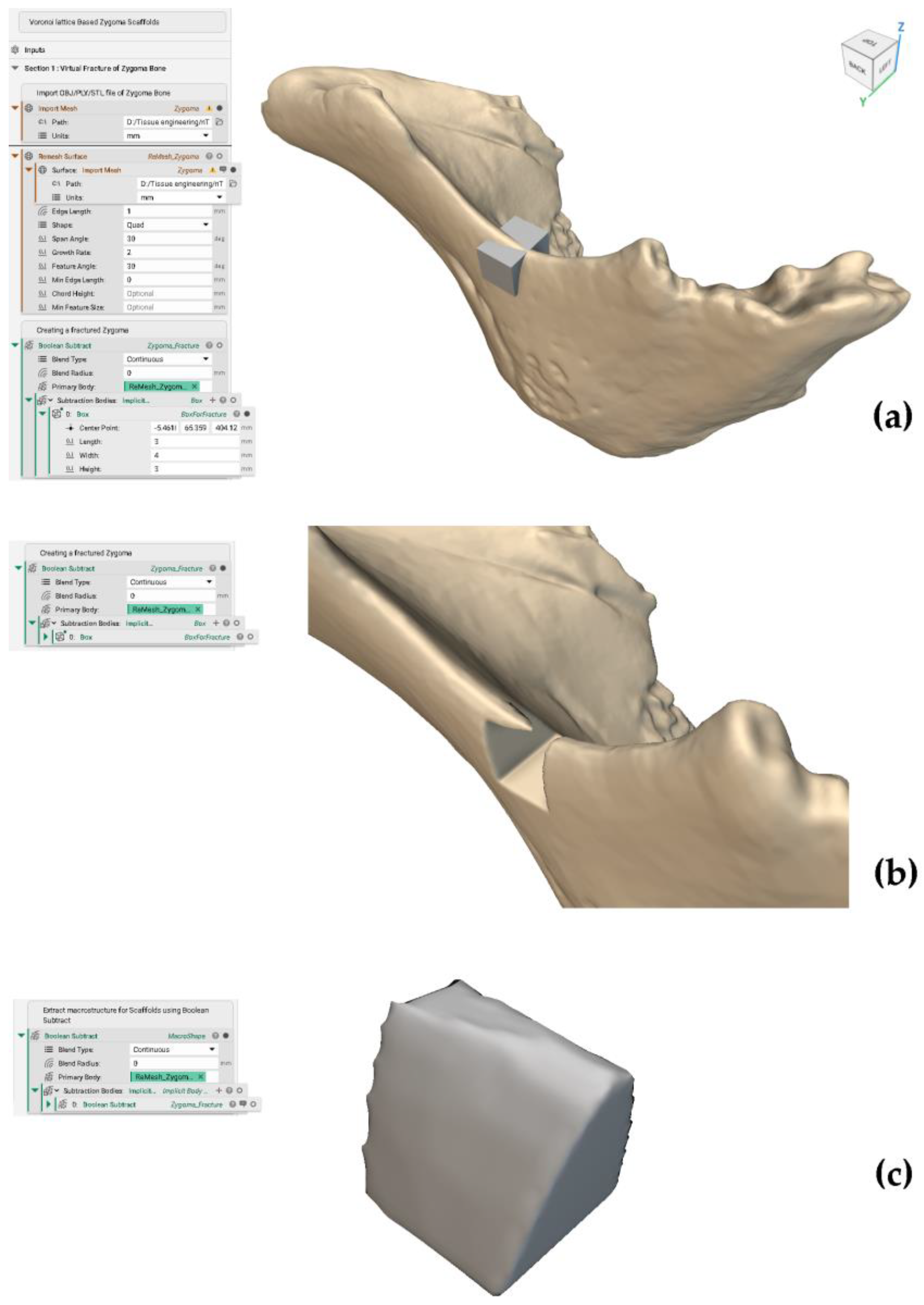
Figure A1. (a) Creation of a virtual fracture of a scaffold using a box. (b) Fractured zygoma region. (c) Extracting the macrostructure or shape of the scaffold from bone defect or fractured region.
Computation 12 00241 g0a1

Appendix B. Creating a Voronoi Lattice (V70 Scaffold)

Figure A2. (a) Creation of random seed points inside the implicit region. (b) Creation of boundary volume for Voronoi cells (Refer to Figure A3c). (c) Creation of Voronoi lattice in boundary volume adding strut thickness of 0.11 mm. (d) V70 Voronoi lattice.
Computation 12 00241 g0a2
Figure A3. (a) Creation of pore diameters using scalar point maps. (b) Formation of spheres inside the Voronoi lattice, and (c) Voronoi cells inside the boundary volume.
Computation 12 00241 g0a3

Appendix C. Calculation of Porosity, Volume Fraction and Surface Area to Volume Ratio

Figure A4. Calculation of porosity, volume fraction and SA: V.
Computation 12 00241 g0a4

References

  1. Torres, A.M.; Trikanad, A.A.; Aubin, C.A.; Lambers, F.M.; Luna, M.; Rimnac, C.M.; Zavattieri, P.; Hernandez, C.J. Bone-inspired microarchitectures achieve enhanced fatigue life. Proc. Natl. Acad. Sci. USA 2019, 116, 24457–24462. [Google Scholar] [CrossRef] [PubMed]
  2. Chinnasami, H.; Dey, M.K.; Devireddy, R. Three-Dimensional Scaffolds for Bone Tissue Engineering. Bioengineering 2023, 10, 759. [Google Scholar] [CrossRef] [PubMed]
  3. Yang, W.; Han, Q.; Chen, H.; Li, Y.; Guo, X.; Zhang, A.; Liu, Y.; Sun, Y.; Wang, J. Additive manufactured trabecular-like Ti-6Al-4V scaffolds for promoting bone regeneration. J. Mater. Sci. Technol. 2024, 188, 116–130. [Google Scholar] [CrossRef]
  4. Sun, L.; Niu, H.; Wu, Y.; Dong, S.; Li, X.; Kim, B.Y.; Liu, C.; Ma, Y.; Jiang, W.; Yuan, Y. Bio-integrated scaffold facilitates large bone regeneration dominated by endochondral ossification. Bioact. Mater. 2024, 35, 208–227. [Google Scholar] [CrossRef] [PubMed]
  5. Laubach, M.; Herath, B.; Bock, N.; Suresh, S.; Saifzadeh, S.; Dargaville, B.L.; McGovern, J.; Wille, M.-L.; Hutmacher, D.W.; Savi, F.M. In vivo characterization of 3D-printed polycaprolactone-hydroxyapatite scaffolds with Voronoi design to advance the concept of scaffold-guided bone regeneration. Front. Bioeng. Biotechnol. 2023, 11, 1272348. [Google Scholar] [CrossRef]
  6. Laubach, M.; Suresh, S.; Herath, B.; Wille, M.-L.; Delbrück, H.; Alabdulrahman, H.; Hutmacher, D.W.; Hildebrand, F. Clinical translation of a patient-specific scaffold-guided bone regeneration concept in four cases with large long bone defects. J. Orthop. Transl. 2022, 34, 73–84. [Google Scholar] [CrossRef]
  7. Yousof, K.; Darwich, M.A.; Darwich, K.; Alassah, G.; Imran, A.; Nazha, H.M. A Case Report of Zygomatic Fracture Reconstruction: Evaluation with Orbital Measurements and Models Registration. Appl. Sci. 2023, 13, 6154. [Google Scholar] [CrossRef]
  8. Liu, G.; Wei, X.; Zhai, Y.; Zhang, J.; Li, J.; Zhao, Z.; Guan, T.; Zhao, D. 3D printed osteochondral scaffolds: Design strategies, present applications and future perspectives. Front. Bioeng. Biotechnol. 2024, 12, 1339916. [Google Scholar] [CrossRef]
  9. Hou, G.; Liu, B.; Tian, Y.; Liu, Z.; Zhou, F. Reconstruction of Ipsilateral Femoral and Tibial Bone Defect by 3D Printed Porous Scaffold Without Bone GraftL: A Case Report. J. Bone Jt. Surg. 2022, 12, e20.00592. [Google Scholar] [CrossRef]
  10. Parisien, A.; ElSayed, M.S.; Frei, H. Mature bone mechanoregulation modelling for the characterization of the osseointegration performance of periodic cellular solids. Materialia 2022, 25, 101552. [Google Scholar] [CrossRef]
  11. Alshammari, A.; Alabdah, F.; Wang, W.; Cooper, G. Virtual Design of 3D-Printed Bone Tissue Engineered Scaffold Shape Using Mechanobiological Modeling: Relationship of Scaffold Pore Architecture to Bone Tissue Formation. Polymers 2023, 15, 3918. [Google Scholar] [CrossRef] [PubMed]
  12. Yamada, S.; Ockermann, P.N.; Schwarz, T.; Mustafa, K.; Hansmann, J. Translation of biophysical environment in bone into dynamic cell culture under flow for bone tissue engineering. Comput. Struct. Biotechnol. J. 2023, 21, 4395–4407. [Google Scholar] [CrossRef] [PubMed]
  13. Ishikawa, K. Bone Substitute Fabrication Based on Dissolution-Precipitation Reactions. Materials 2010, 3, 1138–1155. [Google Scholar] [CrossRef]
  14. Edelmers, E.; Kazoka, D.; Bolocko, K.; Pilmane, M. Different Techniques of Creating Bone Digital 3D Models from Natural Specimens. Appl. Syst. Innov. 2022, 5, 85. [Google Scholar] [CrossRef]
  15. Yoshino, M.; Kato, Y.; Kizu, Y.; Tonogi, M.; Abe, S.; Ide, Y.; Yamane, G.-Y. Study on internal structure of zygomatic bone using micro-finite element analysis model—Differences between dentulous and edentulous dentition in Japanese cadavers. Bull. Tokyo Dent. Coll. 2007, 48, 129–134. [Google Scholar] [CrossRef] [PubMed]
  16. Kato, Y.; Kizuo, Y.; Tonogi, M.; Ide, Y.; Yamane, G.-Y. Observation of the Internal Structure of the Zygomatic Bone by Micro-Computed Tomography. J. Oral Biosci. 2004, 46, 523–529. [Google Scholar] [CrossRef]
  17. Gupta, K.; Meena, K. Artificial bone scaffolds and bone joints by additive manufacturing: A review. Bioprinting 2023, 31, e00268. [Google Scholar] [CrossRef]
  18. Kouhi, M.; Araújo, I.J.d.S.; Asa’ad, F.; Zeenat, L.; Bojedla, S.S.R.; Pati, F.; Zolfagharian, A.; Watts, D.C.; Bottino, M.C.; Bodaghi, M. Recent advances in additive manufacturing of patient-specific devices for dental and maxillofacial rehabilitation. Dent. Mater. 2024, 40, 700–715. [Google Scholar] [CrossRef]
  19. Huang, X.; Lou, Y.; Duan, Y.; Liu, H.; Tian, J.; Shen, Y.; Wei, X. Biomaterial scaffolds in maxillofacial bone tissue engineering: A review of recent advances. Bioact. Mater. 2023, 33, 129–156. [Google Scholar] [CrossRef]
  20. Huo, Y.; Lu, Y.; Meng, L.; Wu, J.; Gong, T.; Zou, J.; Bosiakov, S.; Cheng, L. A Critical Review on the Design, Manufacturing and Assessment of the Bone Scaffold for Large Bone Defects. Front. Bioeng. Biotechnol. 2021, 9, 753715. [Google Scholar] [CrossRef]
  21. Boucetta, A.; Ramtani, S.; Garzón-Alvarado, D.A.; Spadavecchia, J. Computational study promoting engineering biomaterial pre-design to well adapt pores distribution on bone/scaffold assembly section. Comput. Methods Appl. Mech. Eng. 2024, 425, 116934. [Google Scholar] [CrossRef]
  22. Rojas-Rojas, L.; Tozzi, G.; Guillén-Girón, T. A Comprehensive Mechanical Characterization of Subject-Specific 3D Printed Scaffolds Mimicking Trabecular Bone Architecture Biomechanics. Life 2023, 13, 2141. [Google Scholar] [CrossRef] [PubMed]
  23. Garot, C.; Schoffit, S.; Monfoulet, C.; Machillot, P.; Deroy, C.; Roques, S.; Vial, J.; Vollaire, J.; Renard, M.; Ghanem, H.; et al. 3D-Printed Osteoinductive Polymeric Scaffolds with Optimized Architecture to Repair a Sheep Metatarsal Critical-Size Bone Defect. Adv. Health Mater. 2023, 12, e2301692. [Google Scholar] [CrossRef] [PubMed]
  24. Ko, Y.G.; Callahan, L.A.S.; Ma, P.X. Biodegradable Honeycomb-Mimic Scaffolds Consisting of Nanofibrous Walls. Macromol. Biosci. 2024, 24, e2300540. [Google Scholar] [CrossRef]
  25. Li, B.; Hesar, B.D.; Zhao, Y.; Ding, L. Design and additive manufacturing of porous titanium scaffolds for optimum cell viability in bone tissue engineering. Proc. Inst. Mech. Eng. Part B J. Eng. Manuf. 2020, 237, 2015–2024. [Google Scholar] [CrossRef]
  26. Liu, L.; Ma, S.; Zhang, Y.; Zhu, S.; Wu, S.; Liu, G.; Yang, G. Parametric Design of Porous Structure and Optimal Porosity Gradient Distribution Based on Root-Shaped Implants. Materials 2024, 17, 1137. [Google Scholar] [CrossRef]
  27. Li, Z.; Chen, Z.; Chen, X.; Zhao, R. Design and evaluation of TPMS-inspired 3D-printed scaffolds for bone tissue engineering: Enabling tailored mechanical and mass transport properties. Compos. Struct. 2023, 327, 117638. [Google Scholar] [CrossRef]
  28. Yánez, A.; Cuadrado, A.; Martel, O.; Fiorucci, M.P.; Deviaene, S. Mechanical and permeability properties of skeletal and sheet triply periodic minimal surface scaffolds in bone defect reconstruction. Results Eng. 2024, 21, 101883. [Google Scholar] [CrossRef]
  29. Musthafa, H.-S.N.; Walker, J.; Rahman, T.; Bjørkum, A.; Mustafa, K.; Velauthapillai, D. In-Silico Prediction of Mechanical Behaviour of Uniform Gyroid Scaffolds Affected by Its Design Parameters for Bone Tissue Engineering Applications. Computation 2023, 11, 181. [Google Scholar] [CrossRef]
  30. Musthafa, H.-S.N.; Walker, J.; Domagala, M. Computational Modelling and Simulation of Scaffolds for Bone Tissue Engineering. Computation 2024, 12, 74. [Google Scholar] [CrossRef]
  31. Pokojski, W.; Pokojska, P. Voronoi diagrams—Inventor, method, applications. Pol. Cartogr. Rev. 2018, 50, 141–150. [Google Scholar] [CrossRef]
  32. Zhang, L.; Wang, B.; Song, B.; Yao, Y.; Choi, S.-K.; Yang, C.; Shi, Y. 3D Printed Biomimetic Metamaterials with Graded Porosity and Tapering Topology for Improved Cell Seeding and Bone Regeneration. Bioact. Mater. 2022, 25, 677–688. [Google Scholar] [CrossRef] [PubMed]
  33. Castañeda, F.D.; García-Acosta, G.; Garzón-Alvarado, D.A.; Márquez-Flórez, K.; Quexada-Rodriguez, D.A.; Velasco, M.A. Design for the additive manufacturing of structural elements with cellular materials using Voronoi diagrams and Delaunay triangulations: Biological and structural applications. Mech. Adv. Mater. Struct. 2023, 1–21. [Google Scholar] [CrossRef]
  34. Gatto, M.L.; Cerqueni, G.; Furlani, M.; Riberti, N.; Tognoli, E.; Denti, L.; Leonardi, F.; Giuliani, A.; Mattioli-Belmonte, M.; Mengucci, P. Influence of Trabecular Geometry on Scaffold Mechanical Behavior and MG-63 Cell Viability. Materials 2023, 16, 2342. [Google Scholar] [CrossRef] [PubMed]
  35. He, Y.; Chao, L.; Jiao, C.; Wang, H.; Xie, D.; Wu, G.; Wang, L.; Wang, C.; Zhao, J.; Shen, L.; et al. Sub-regional design of the bionic bone scaffolds using macrostructural topology. Int. J. Bioprinting 1970, 9, 222. [Google Scholar] [CrossRef]
  36. Nguyen, D.S. A Method for Generation of Random Lattice Structure for Additive Manufacturing. In Proceedings of the 2019 IEEE International Conference on Industrial Engineering and Engineering Management (IEEM), Macao, China, 15–18 December 2019; pp. 1275–1279. [Google Scholar]
  37. Rezapourian, M.; Hussainova, I. Optimal mechanical properties of Hydroxyapatite gradient Voronoi porous scaffolds for bone applications—A numerical study. J. Mech. Behav. Biomed. Mater. 2023, 148, 106232. [Google Scholar] [CrossRef]
  38. Liu, B.; Feng, J.; Chen, J.; He, Y.; Fu, J. A topology optimisation-based design method for 3D Voronoi porous structures and its application for medical pillows. Virtual Phys. Prototyp. 2023, 18, e2285392. [Google Scholar] [CrossRef]
  39. Xiao, F.; Yin, X. Geometry models of porous media based on Voronoi tessellations and their porosity–permeability relations. Comput. Math. Appl. 2016, 72, 328–348. [Google Scholar] [CrossRef]
  40. Voronoi Diagram Generator. Available online: https://cfbrasz.github.io/Voronoi.html (accessed on 29 October 2024).
  41. Prakoso, A.T.; Basri, H.; Adanta, D.; Yani, I.; Ammarullah, M.I.; Akbar, I.; Ghazali, F.A.; Syahrom, A.; Kamarul, T. The Effect of Tortuosity on Permeability of Porous Scaffold. Biomedicines 2023, 11, 427. [Google Scholar] [CrossRef]
  42. Peng, X.; Huo, Y.; Zhang, G.; Cheng, L.; Lu, Y.; Li, J.; Jin, Z. Controlled mechanical and mass-transport properties of porous scaffolds through hollow strut. Int. J. Mech. Sci. 2023, 248, 108202. [Google Scholar] [CrossRef]
  43. Li, J.; Yang, Y.; Sun, Z.; Peng, K.; Liu, K.; Xu, P.; Li, J.; Wei, X.; He, X. Integrated evaluation of biomechanical and biological properties of the biomimetic structural bone scaffold: Biomechanics, simulation analysis, and osteogenesis. Mater. Today Bio 2023, 24, 100934. [Google Scholar] [CrossRef] [PubMed]
  44. Xu, Y.; Zhang, S.; Ding, W.; Du, H.; Li, M.; Li, Z.; Chen, M. Additively-manufactured gradient porous bio-scaffolds: Permeability, cytocompatibility and mechanical properties. Compos. Struct. 2024, 336, 118021. [Google Scholar] [CrossRef]
  45. Jiao, J.; Hong, Q.; Zhang, D.; Wang, M.; Tang, H.; Yang, J.; Qu, X.; Yue, B. Influence of porosity on osteogenesis, bone growth and osteointegration in trabecular tantalum scaffolds fabricated by additive manufacturing. Front. Bioeng. Biotechnol. 2023, 11, 1117954. [Google Scholar] [CrossRef] [PubMed]
  46. Pires, T.; Dunlop, J.W.C.; Fernandes, P.R.; Castro, A.P.G. Challenges in computational fluid dynamics applications for bone tissue engineering. Proc. R. Soc. A Math. Phys. Eng. Sci. 2022, 478, 20210607. [Google Scholar] [CrossRef]
  47. Liu, W.; Zheng, L.; Wang, C.; Yin, H.; Raffaella, A.; Apicella, A.; Ji, P.; Zhang, H.; Fan, Y. Additively manufactured bioceramic scaffolds with 3D architecture for vertical bone augmentation: A proof-of-concept study. Mater. Des. 2024, 239, 112749. [Google Scholar] [CrossRef]
  48. Ji, G.; Zhang, M.; Lu, Y.; Dong, J. The Basic Theory of CFD Governing Equations and the Numerical Solution Methods for Reactive Flows. In Computational Fluid Dynamics; Chapter 1; Ji, G., Dong, J., Eds.; IntechOpen: Rijeka, Croatia, 2023. [Google Scholar] [CrossRef]
  49. Lintermann, A. Computational Meshing for CFD Simulations. In Clinical and Biomedical Engineering in the Human Nose; Springer: Singapore, 2021; pp. 85–115. [Google Scholar] [CrossRef]
  50. Rice, J.; Schnipke, R. A monotone streamline upwind finite element method for convection-dominated flows. Comput. Methods Appl. Mech. Eng. 1985, 48, 313–327. [Google Scholar] [CrossRef]
  51. Khouya, B.; El-Amrani, M.; Seaid, M. A Conservative and Monotone Characteristic Finite Element Solver for Three-Dimensional Transport and Incompressible Navier-Stokes Equations on Unstructured Grids. Commun. Comput. Phys. 2022, 31, 224–256. [Google Scholar] [CrossRef]
  52. Cengizci, S.; Uğur, Ö.; Natesan, S. A stabilized finite element formulation with shock-capturing for solving advection-dominated convection–diffusion equations having time-fractional derivatives. J. Comput. Sci. 2024, 76, 102214. [Google Scholar] [CrossRef]
  53. Jia, D.; Esmaily, M. A time-consistent stabilized finite element method for fluids with applications to hemodynamics. Sci. Rep. 2023, 13, 19120. [Google Scholar] [CrossRef]
  54. Heydari, S.; Knobloch, P.; Wick, T. Flux-corrected transport stabilization of an evolutionary cross-diffusion cancer invasion model. J. Comput. Phys. 2024, 499, 112711. [Google Scholar] [CrossRef]
  55. Kuzmin, D. On the design of general-purpose flux limiters for finite element schemes. I. Scalar convection. J. Comput. Phys. 2006, 219, 513–531. [Google Scholar] [CrossRef]
  56. Rachowicz, W.; Zdunek, A.; Cecot, W. A discontinuous Petrov-Galerkin method for compressible Navier-Stokes equations in three dimensions. Comput. Math. Appl. 2021, 102, 113–136. [Google Scholar] [CrossRef]
  57. Help|Discretization Method|Autodesk. Available online: https://help.autodesk.com/view/SCDSE/2024/ENU/?guid=GUID-DEE0664D-771B-4446-9ED4-1498267D13FB (accessed on 9 July 2024).
  58. Gensler, M.; Malkmus, C.; Ockermann, P.; Möllmann, M.; Hahn, L.; Salehi, S.; Luxenhofer, R.; Boccaccini, A.R.; Hansmann, J. Perfusable Tissue Bioprinted into a 3D-Printed Tailored Bioreactor System. Bioengineering 2024, 11, 68. [Google Scholar] [CrossRef] [PubMed]
  59. Krasnyakov, I.; Bratsun, D. Cell-Based Modeling of Tissue Developing in the Scaffold Pores of Varying Cross-Sections. Biomimetics 2023, 8, 562. [Google Scholar] [CrossRef]
  60. Wang, S.; Shi, Z.; Liu, L.; Huang, Z.; Li, Z.; Liu, J.; Hao, Y. Honeycomb structure is promising for the repair of human bone defects. Mater. Des. 2021, 207, 109832. [Google Scholar] [CrossRef]
  61. Huang, X.; Zheng, L.; Li, P.; Lin, Z.; Huang, S.; Zhou, C. Fabrication of 3D printed trabecular bone-templated scaffolds modified with rare earth europium (III)-based complex for enhancing mitochondrial function in bone regeneration. Appl. Mater. Today 2024, 37, 102130. [Google Scholar] [CrossRef]
  62. Hussein, N.A.K.; Noordin, M.A.; Saad, A.P.M. Influence of conical graded porous architecture on the mechanical, failure behavior and fluid-flow properties for bone scaffold application. Eng. Fail. Anal. 2024, 157, 107893. [Google Scholar] [CrossRef]
  63. Omar, A.M.; Hassan, M.H.; Daskalakis, E.; Ates, G.; Bright, C.J.; Xu, Z.; Powell, E.J.; Mirihanage, W.; Bartolo, P.J.D.S. Geometry-Based Computational Fluid Dynamic Model for Predicting the Biological Behavior of Bone Tissue Engineering Scaffolds. J. Funct. Biomater. 2022, 13, 104. [Google Scholar] [CrossRef]
  64. Seehanam, S.; Khrueaduangkham, S.; Sinthuvanich, C.; Sae-Ueng, U.; Srimaneepong, V.; Promoppatum, P. Evaluating the effect of pore size for 3d-printed bone scaffolds. Heliyon 2024, 10, e26005. [Google Scholar] [CrossRef]
  65. Seehanam, S.; Chanchareon, W.; Promoppatum, P. Assessing the effect of manufacturing defects and non-Newtonian blood model on flow behaviors of additively manufactured Gyroid TPMS structures. Heliyon 2023, 9, e15711. [Google Scholar] [CrossRef]
  66. Wajihah, S.A.; Sankar, D.S. A review on non-Newtonian fluid models for multi-layered blood rheology in constricted arteries. Arch. Appl. Mech. 2023, 93, 1771–1796. [Google Scholar] [CrossRef] [PubMed]
  67. Hussain, M.A.; Kar, S.; Puniyani, R.R. Relationship between power law coefficients and major blood constituents affecting the whole blood viscosity. J. Biosci. 1999, 24, 329–337. [Google Scholar] [CrossRef]
  68. Marenzana, M.; Arnett, T.R. The Key Role of the Blood Supply to Bone. Bone Res. 2013, 1, 203–215. [Google Scholar] [CrossRef] [PubMed]
  69. Ali, D.; Sen, S. Permeability and fluid flow-induced wall shear stress of bone tissue scaffolds: Computational fluid dynamic analysis using Newtonian and non-Newtonian blood flow models. Comput. Biol. Med. 2018, 99, 201–208. [Google Scholar] [CrossRef] [PubMed]
  70. Mahammod, B.P.; Barua, E.; Deoghare, A.B.; Pandey, K. Permeability quantification of porous polymer scaffold for bone tissue engineering. Mater. Today Proc. 2020, 22, 1687–1693. [Google Scholar] [CrossRef]
  71. Singh, S.; Yadav, S.K.; Meena, V.K.; Vashisth, P.; Kalyanasundaram, D. Orthopedic Scaffolds: Evaluation of Structural Strength and Permeability of Fluid Flow via an Open Cell Neovius Structure for Bone Tissue Engineering. ACS Biomater. Sci. Eng. 2023, 9, 5900–5911. [Google Scholar] [CrossRef]
  72. Li, J.; Guo, D.; Li, J.; Wei, X.; Sun, Z.; Yang, B.; Lu, T.; Ouyang, P.; Chang, S.; Liu, W.; et al. Irregular pore size of degradable bioceramic Voronoi scaffolds prepared by stereolithography: Osteogenesis and computational fluid dynamics analysis. Mater. Des. 2022, 224, 111414. [Google Scholar] [CrossRef]
  73. Lu, T.; Sun, Z.; Jia, C.; Ren, J.; Li, J.; Ma, Z.; Zhang, J.; Li, J.; Zhang, T.; Zang, Q.; et al. Roles of irregularity of pore morphology in osteogenesis of Voronoi scaffolds: From the perspectives of MSC adhesion and mechano-regulated osteoblast differentiation. J. Biomech. 2023, 151, 111542. [Google Scholar] [CrossRef]
  74. Katzenbach, A.; Handschuh, S.; Vettermann, S. JT Format (ISO 14306) and AP 242 (ISO 10303): The Step to the Next Gen-eration Collaborative Product Creation. IFIP Adv. Inf. Commun. Technol. 2013, 411, 41–52. [Google Scholar] [CrossRef]
  75. Bixel, M.G.; Kusumbe, A.P.; Ramasamy, S.K.; Sivaraj, K.K.; Butz, S.; Vestweber, D.; Adams, R.H. Flow Dynamics and HSPC Homing in Bone Marrow Microvessels. Cell Rep. 2017, 18, 1804–1816. [Google Scholar] [CrossRef]
  76. Ali, D.; Ozalp, M.; Blanquer, S.B.; Onel, S. Permeability and fluid flow-induced wall shear stress in bone scaffolds with TPMS and lattice architectures: A CFD analysis. Eur. J. Mech.-B/Fluids 2019, 79, 376–385. [Google Scholar] [CrossRef]
  77. Help|Advection Schemes|Autodesk. Available online: https://help.autodesk.com/view/SCDSE/2024/ENU/?guid=GUID-F691B334-CCE2-47E9-B6C4-21666712C163 (accessed on 9 July 2024).
  78. Loh, Q.L.; Choong, C. Three-Dimensional Scaffolds for Tissue Engineering Applications: Role of Porosity and Pore Size. Tissue Eng. Part B Rev. 2013, 19, 485–502. [Google Scholar] [CrossRef]
  79. Watanabe, S.; Takabatake, K.; Tsujigiwa, H.; Watanabe, T.; Tokuyama, E.; Ito, S.; Nagatsuka, H.; Kimata, Y. Efficacy of Honeycomb TCP-induced Microenvironment on Bone Tissue Regeneration in Craniofacial Area. Int. J. Med. Sci. 2016, 13, 466–476. [Google Scholar] [CrossRef]
  80. Brennan, C.M.; Eichholz, K.F.; A Hoey, D. The effect of pore size within fibrous scaffolds fabricated using melt electrowriting on human bone marrow stem cell osteogenesis. Biomed. Mater. 2019, 14, 065016. [Google Scholar] [CrossRef]
  81. Ai, C.; Liu, L.; Goh, J.C.-H. Pore size modulates in vitro osteogenesis of bone marrow mesenchymal stem cells in fibronectin/gelatin coated silk fibroin scaffolds. Mater. Sci. Eng. C 2021, 124, 112088. [Google Scholar] [CrossRef]
  82. Murphy, C.M.; Haugh, M.G.; O’Brien, F.J. The effect of mean pore size on cell attachment, proliferation and migration in collagen–glycosaminoglycan scaffolds for bone tissue engineering. Biomaterials 2010, 31, 461–466. [Google Scholar] [CrossRef]
  83. Chao, L.; Jiao, C.; Liang, H.; Xie, D.; Shen, L.; Liu, Z. Analysis of Mechanical Properties and Permeability of Trabecular-Like Porous Scaffold by Additive Manufacturing. Front. Bioeng. Biotechnol. 2021, 9, 779854. [Google Scholar] [CrossRef]
  84. Wang, C.; Xu, D.; Lin, L.; Li, S.; Hou, W.; He, Y.; Sheng, L.; Yi, C.; Zhang, X.; Li, H.; et al. Large-pore-size Ti6Al4V scaffolds with different pore structures for vascularized bone regeneration. Mater. Sci. Eng. C 2021, 131, 112499. [Google Scholar] [CrossRef]
  85. Zhou, J.; Xiong, S.; Liu, M.; Yang, H.; Wei, P.; Yi, F.; Ouyang, M.; Xi, H.; Long, Z.; Liu, Y.; et al. Study on the influence of scaffold morphology and structure on osteogenic performance. Front. Bioeng. Biotechnol. 2023, 11, 1127162. [Google Scholar] [CrossRef] [PubMed]
  86. Koushik, T.M.; Miller, C.M.; Antunes, E. Bone Tissue Engineering Scaffolds: Function of Multi-Material Hierarchically Structured Scaffolds. Adv. Heal. Mater. 2023, 12, e2202766. [Google Scholar] [CrossRef] [PubMed]
  87. Nguyen, T.D.; Kadri, O.E.; Sikavitsas, V.I.; Voronov, R.S. Scaffolds with a High Surface Area-to-Volume Ratio and Cultured Under Fast Flow Perfusion Result in Optimal O2 Delivery to the Cells in Artificial Bone Tissues. Appl. Sci. 2019, 9, 2381. [Google Scholar] [CrossRef]
  88. Grimm, M.J.; Williams, J.L. Measurements of permeability in human calcaneal trabecular bone. J. Biomech. 1997, 30, 743–745. [Google Scholar] [CrossRef]
  89. Nauman, E.A.; Fong, K.E.; Keaveny, T.M. Dependence of Intertrabecular Permeability on Flow Direction and Anatomic Site. Ann. Biomed. Eng. 1999, 27, 517–524. [Google Scholar] [CrossRef]
  90. Ma, S.; Tang, Q.; Han, X.; Feng, Q.; Song, J.; Setchi, R.; Liu, Y.; Liu, Y.; Goulas, A.; Engstrøm, D.S.; et al. Manufacturability, Mechanical Properties, Mass-Transport Properties and Biocompatibility of Triply Periodic Minimal Surface (TPMS) Porous Scaffolds Fabricated by Selective Laser Melting. Mater. Des. 2020, 195, 109034. [Google Scholar] [CrossRef]
  91. Santos, J.; Pires, T.; Gouveia, B.P.; Castro, A.P.; Fernandes, P.R. On the permeability of TPMS scaffolds. J. Mech. Behav. Biomed. Mater. 2020, 110, 103932. [Google Scholar] [CrossRef]
  92. Abbasi, N.; Hamlet, S.; Love, R.M.; Nguyen, N.-T. Porous Scaffolds for Bone Regeneration. J. Sci. Adv. Mater. Devices 2020, 5, 1–9. [Google Scholar] [CrossRef]
  93. Kanwar, S.; Al-Ketan, O.; Vijayavenkataraman, S. A novel method to design biomimetic, 3D printable stochastic scaffolds with controlled porosity for bone tissue engineering. Mater. Des. 2022, 220, 110857. [Google Scholar] [CrossRef]
  94. Amini, A.R.; Adams, D.J.; Laurencin, C.T.; Nukavarapu, S.P. Optimally Porous and Biomechanically Compatible Scaffolds for Large-Area Bone Regeneration. Tissue Eng. Part A 2012, 18, 1376–1388. [Google Scholar] [CrossRef]
  95. Lynch, S.; Nama, N.; Figueroa, C.A. Effects of non-Newtonian viscosity on arterial and venous flow and transport. Sci. Rep. 2022, 12, 20568. [Google Scholar] [CrossRef] [PubMed]
  96. Suffo, M.; López-Marín, C.J. A Comparative Study of Turbulence Methods Applied to the Design of a 3D-Printed Scaffold and the Selection of the Appropriate Numerical Scheme to Simulate the Scaffold for Tissue Engineering. Appl. Sci. 2021, 12, 191. [Google Scholar] [CrossRef]
Figure 1. Trabecular bone has interconnected random porous structures of divergent pore sizes and different thicknesses of struts (trabeculae). Modified and reproduced with permission from Ref. [13] CC BY 3.0.
Computation 12 00241 g001
Figure 2. Zygoma and volumes of interest: Jugale (Ju), Middle Point (M.P.) and Zygomaxillare (Zm). Reproduced with permission from Ref. [14] CC BY 4.0. The Ju area has the highest bone volume density (23.2 ± 4.3%) and highest trabecular plate thickness (0.16 ± 0.05 mm) in edentulous maxillae, compared to those at M.P. and Zm areas [15,16].
Computation 12 00241 g002
Figure 3. A Voronoi diagram and its tessellation patterns are based on random points to create the cells or regions in a given space. The boundary of each cell has an equal distance between two or more neighbouring points [40].
Computation 12 00241 g003
Figure 4. A Voronoi lattice-based cuboid biomimetic scaffold with functionally graded pores using a cloud of random seed points to create a stochastic microarchitecture.
Computation 12 00241 g004
Figure 5. Zygoma model (front and back views) obtained from 3D scanning (EinScan-SE V2 3D scanner): Front view (left) and Back view (right) [14].
Computation 12 00241 g005
Figure 6. Virtual fracture process of zygoma: (a) fracture by a box, (b) fractured zygoma, and (c) extraction of the shape of the fracture for the design of Voronoi scaffolds (measurements in mm). (Refer: Appendix A Figure A1).
Computation 12 00241 g006
Figure 7. Random seed points inside the boundary layer with a distance known as point spacing. (Refer to Appendix B Figure A2a).
Computation 12 00241 g007
Figure 8. Conversion of scaffold shape (bone defect region) given on the left side into a volume mesh (Boundary layer) given on the right side. (Refer: Appendix B Figure A2b).
Computation 12 00241 g008
Figure 9. Creation of Voronoi lattices V90, V85, V80, V75 and V70 of porosities 90%, 85%, 80%, 75% and 70%, respectively. (Refer: Appendix B).
Computation 12 00241 g009
Figure 10. JT CAD files of different Voronoi scaffolds (front view) for CFD simulation.
Computation 12 00241 g010
Figure 11. Surface wrapping method to create a fluid domain for the fluid surrounding the Voronoi V90 scaffold (solid domain).
Computation 12 00241 g011
Figure 12. (a) Material properties of fluid and solid domains and (b) boundary conditions (velocity inlet, pressure outlet and no-slip wall condition) on the CFD model of the V90 scaffold.
Computation 12 00241 g012
Figure 13. Generated volume mesh of the given (a) fluid domain and (b) solid domain of V90 scaffold for CFD simulation with tetrahedral elements and nodes.
Computation 12 00241 g013
Figure 14. Scalar point maps and spheres are used to calculate pore sizes and pore numbers of all Voronoi lattice designs V90, V85, V80, V75, and V70. (Pore size calculation was carried out using the Lattice Pore size block of nTopology: Refer: Appendix B).
Computation 12 00241 g014
Figure 15. Graphs of variation of (a) surface area and (b) surface area/volume ratio (SA: V) with variation of porosities of Voronoi scaffolds.
Computation 12 00241 g015
Figure 16. Testing of CFD models at different inlet velocities and the related pressure drops across the given Voronoi scaffolds.
Computation 12 00241 g016
Figure 17. Velocity streamlines for all Voronoi scaffolds (V90, V85, V80, V75, V70) at inlet velocity 0.7 mm/s.
Computation 12 00241 g017
Figure 18. Velocity streamlines for all Voronoi scaffolds (V90, V85, V80, V75, V70) at inlet velocity 0.7 mm/s.
Computation 12 00241 g018
Figure 19. Pressure contours for all Voronoi scaffolds (V90, V85, V80, V75, V70) at inlet velocity of 0.7 mm/s.
Computation 12 00241 g019
Figure 20. Variation of pressure drop and permeability based on the variation of porosity at an inlet velocity of 0.7 mm/s.
Computation 12 00241 g020
Figure 21. Validation of a non-Newtonian model by a Newtonian model (comparison of pressure contours of both models for V90 scaffold) at inlet velocity of 0.7 mm/s.
Computation 12 00241 g021
Figure 22. Shear strain rate contour of CFD model of V90 scaffold.
Computation 12 00241 g022
Table 1. Morphological parameters for Voronoi scaffold designs with constant ST of 0.11 mm.
Voronoi ScaffoldsPorosity (%)Strut Thickness ST (mm)Point Spacing PS (mm)Voronoi
Seed Points
Voronoi Cell NumbersSurface Area *
SA (mm2)
Surface Area/Volume
SA: V (mm−1)
V90900.110.851651170.52.29
V85850.110.668124351023.32
V80800.110.570206701314.26
V75750.110.5023141211605.19
V70700.110.4494331801856.01
* Computation of SA using mass properties block in nTopology; Bounding box Volume V = 30.82 mm3.
Table 2. Range of pore sizes in the Voronoi scaffolds.
Voronoi ScaffoldsNumber of PoresRange of Pore Sizes (µm)
V9021622.2 to 923
V8545520.2 to 669
V8066820.7 to 618
V7598320.1 to 532
V70136219.8 to 482
Table 3. Pressure drop and permeabilities of the Voronoi scaffolds.
Voronoi ScaffoldsPorosity
(%)
Pressure Drop *
(Pa)
Permeability
(×10−9 m2)
V90901.522.17
V85852.421.13
V80803.130.78
V75754.730.44
V70 705.810.33
* Refer to pressure contours for visualisation: Figure 19.
Table 4. Comparison of predicted permeability of Voronoi scaffolds with other research works for trabecular (cancellous) bone applications.
Scaffolds/BonesPermeability (×10−9 m2)References
Voronoi scaffolds *0.33 to 2.17This study *
Human Trabecular bone0.40 to 11Grimm et al. [88]
Trabecular bone specimens0.0268 to 20Nauman et al. [89]
Gyroid scaffolds0.29 to 3.91Ma et al. [90]
Schwartz gyroid/diamond/primitive scaffolds0.431 to 8.44Santos et al. [91]
Sheet and skeletal gyroid scaffolds0.61 to 3.34Yanez et al. [28]
Diamond/Octet/Truncate-Octahedron/Double-Diamond/TPMS Scaffolds1.85 to 5.62Ali et al. [76]
* Refer to Table 3.
Table 5. Validation of non-Newtonian power law models with Newtonian models.
Voronoi
Scaffolds
Newtonian Model
(µ = 0.0045 Pa.s)
Non-Newtonian Power Law Model
(n = 1, K = 0.0045 Pa.s)
Non-Newtonian Power Law Model *
(n = 0.708, K = 0.0017 Pa.s)
Pressure Drop (Pa)Pressure Drop (Pa)Pressure Drop (Pa)
V900.7260.7261.52
V851.261.262.42
V801.751.753.13
V752.882.884.73
V703.763.765.81
* Refer to Table 3 and Figure 19.
Disclaimer/Publisher’s Note: The statements, opinions and data contained in all publications are solely those of the individual author(s) and contributor(s) and not of MDPI and/or the editor(s). MDPI and/or the editor(s) disclaim responsibility for any injury to people or property resulting from any ideas, methods, instructions or products referred to in the content.