Review

Methodological Studies on Estimates of Abundance and Diversity of Heterotrophic Flagellates from the Deep-Sea Floor

Department of General Ecology, Institute for Zoology, Biocenter Cologne, University of Cologne, Zülpicher Straße 47b, Cologne D-50674, Germany
*
Author to whom correspondence should be addressed.
J. Mar. Sci. Eng. 2016, 4(1), 22; https://doi.org/10.3390/jmse4010022
Submission received: 30 September 2015 / Revised: 8 February 2016 / Accepted: 15 February 2016 / Published: 3 March 2016

Abstract

:
Extreme environmental conditions in the deep sea hamper access to protist communities. In combination with the potentially highly diverse species composition, it demands a wide range of methods to be applied at the same time to guarantee a high resolution of quantitative and qualitative studies of deep-sea heterotrophic flagellates (HF). Within this study, we present a possible combination of several culture-independent and culture-dependent methods available for investigating benthic deep-sea HF communities. Besides live-counting and fixation of HF, we refer to cultivation methods and molecular surveys using next generation sequencing. Laboratory ecological experiments under deep-sea conditions (high pressure, low temperature) could allow the approval of the potential deep-sea origin of sampled HF. The combination of different methods offers a unique possibility to receive detailed information on nanofaunal life in the deep sea. Specific fixation techniques to preserve samples directly at the sampling depth must be applied in further studies to reflect the real biodiversity of the largest habitat on earth.

1. Introduction

Although deep-sea ecosystems represent the largest and most remote biome of the Earth [1], only about 5% have been explored so far, even less have been sampled [2]. We lack a firm understanding of species-level distribution (cosmopolitan vs. local) for deep-sea communities [3] as well as the functioning of and the interactions between biodiversity and ecological processes in this vast environment [2]. Marine heterotrophic flagellates contribute not only a major part within the microbial food web and are important nutrient remineralizers in biogeochemical cycles in surface waters [4,5] with densities ranging between 102 and 104 cells mL−1, but are also potentially important regarding material flux and bacterial consumption in the deep sea. Recent studies of microbes have shown that even the deepest parts of our Earth are populated with a large variety of life [6]. Nevertheless, qualitative studies from eukaryotic deep-sea communities concerning diversity, spatial distribution and ecological function are still scarce. Concerning investigations of the bathypelagic deep sea, analysis revealed the occurrence of heterotrophic protists (11±1 cells mL−1) at depths down to 4000 m [7]. A global survey of bathypelagic microbial eukaryote communities identified a few groups as the dominant part of deep-sea communities, whereas the proportional composition of the dominant groups varied on a global scale [8]. Until the end of the last century, besides morphology based studies of foraminiferans [9], only anecdotal reports for other protists existed [10,11] mainly due to methodological issues. While some authors reported a lack of flagellate occurrence in samples from the deep-sea floor [11], other studies revealed densities of HF [10,12,13] up to 105 cells cm−3 [14,15].
There is no standardized protocol for the sampling and analysis of benthic deep-sea protists available at present. Cultivation-based methods miss a majority of taxa since most species require specific cultivation conditions [16]. Molecular barcoding approaches employing PCR introduce significant biases in reported community compositions of marine protists due to the restriction of ‘general’ primers to detect all protist groups [17]. Since molecular surveys cannot yet provide any information on the morphology and abundance of the organisms, culture-dependent and culture-independent investigations are required to gain quantitative and qualitative results concerning deep-sea protist biodiversity.
The aim of this review is the study of benthic flagellated protists, those living in deep-sea sediments/seafloor. We will illustrate the above mentioned methodological problems presenting own recent results and will recommend a combination of methods to get a more reliable estimate of deep-sea benthic nanofauna.

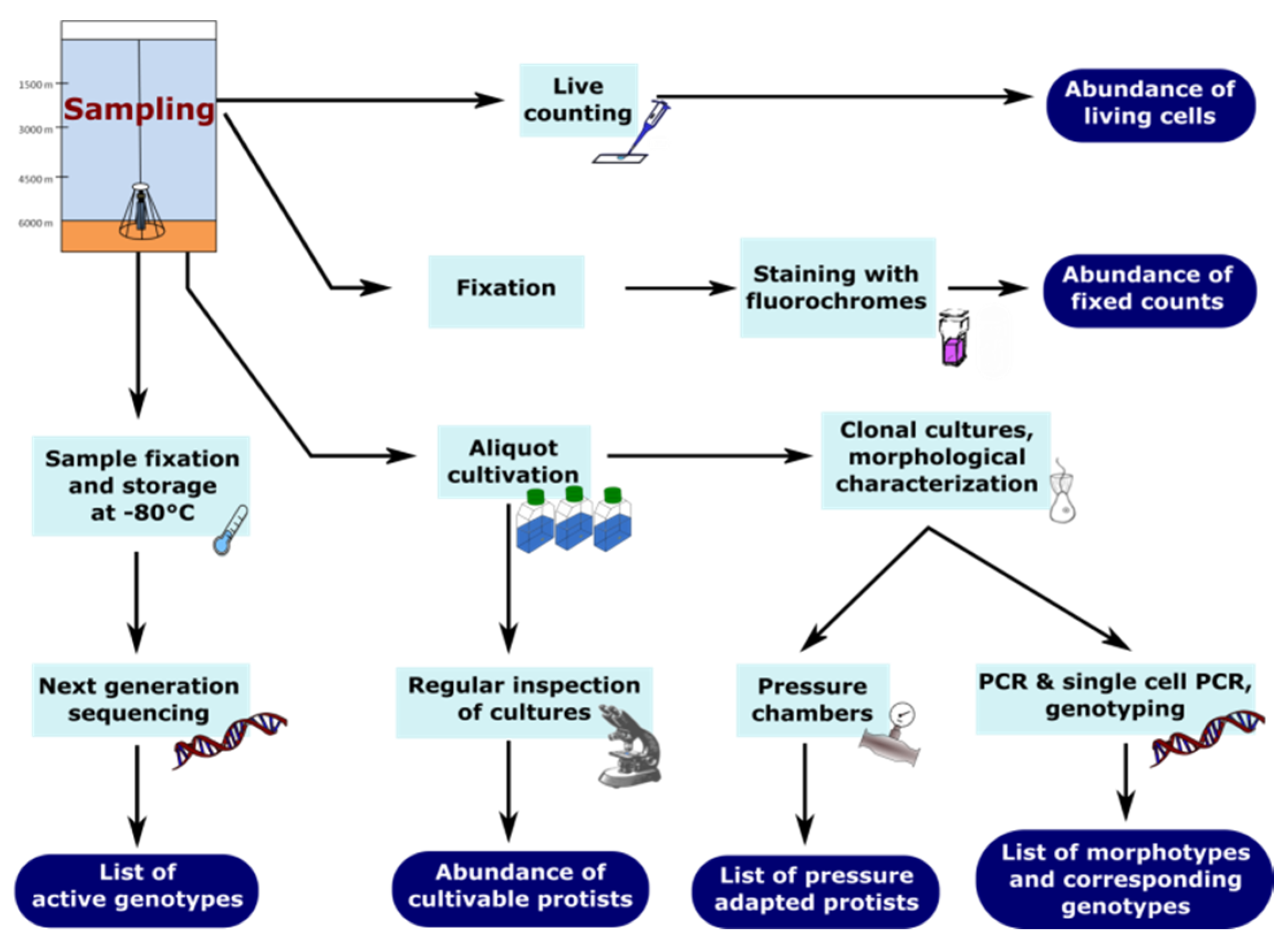
2. Quantification and Qualification of Deep-Sea Protists

Several methods have been applied during the last decades to characterize deep-sea communities. Main procedures for flagellate detection in the past included live-counting of samples immediately after sampling, occurrences in laboratory cultures and molecular surveys using Sanger or next generation sequencing (NGS) (Figure 1). However, the taxonomic identification of protists, especially nanoprotists, in routine samples is difficult due to the general lack of conspicuous morphological features and the selectivity of sampling and counting methods [18,19,20]. The taxonomic identity of heterotrophic flagellates is generally based on cultivated strains, on which ultrastructural, physiological and molecular studies have been performed [16,21]. However, most deep-sea organisms are extremely difficult to cultivate due to their slow in-situ growth rates and their likely strict adaptation to extreme environmental conditions (oligotrophy, low temperatures, high pressure, anoxia) [2]. The role of these cultured strains as representatives within deep-sea protist communities is unclear. Molecular surveys frequently recover novel eukaryotic lineages that have not been recorded from cultures so far [16,22]. Environmental molecular surveys in microbial ecology have revolutionized our knowledge, indicating how far we are from understanding this “untapped reservoir” [23] of microbial diversity in the depth [19,22,24]. A major problem up to now is the assignment of these obtained sequences to species level with existing databases. A better annotation will improve the knowledge that comes from such analysis and sequence libraries. Therefore, such data will become more valuable as better gene annotations become available [25]. Molecular environmental diversity studies of the deep-sea floor have mainly been focusing on assumed “hot spots” of activity (e.g., hydrothermal vents, methane seeps) mostly from the bathyal zone carried out on a local scale [26,27,28]. Our previous studies of deep-sea nanofauna [29,30,31,32,33] indicated the existence of a specific abyssal nanofauna which contains a large number of endemic taxa [31,33].Recent comprehensive studies [34] indicated protists as the most diverse eukaryotic organisms. The diversity of phyla (Figure 1) with their specific differences (e.g., ultrastructure) makes it necessary to consider specifically designed fixatives or molecular techniques.

2.1. How to Sample Deep-Sea Protists

The main tool used up to now to collect benthic deep-sea protist communities is the Multi-Corer system. Due to a closing mechanism at the top and bottom of the cores, the risk of contamination with organisms and cysts from upper water layers is reduced. However, the problem is that samples have to be treated immediately after sampling which means within minutes. We microscopically observed living nanoflagellates within the first 30 min after sampling. Protists are stressed by tremendous physical changes, e.g., varying pressures and temperatures, during sampling. Therefore, it is likely that several flagellate species adapted to deep-sea conditions die, while being raised through the water column. Morgan-Smith et al. [35] sampled deep-sea protists with 200 mL titanium chambers retaining in situ pressure from depths of 2750 and 4000 m to investigate the effect of pressure on protist abundances prior to fixation. Although depressurization pre-versus post fixation did not significantly affect the number of eukaryotes counted, cell physiology might be greatly impacted by changes in pressure. Future methodological studies must be applied to solve these problems. Potential solutions could be the usage of specific fixations of samples already in the depth of sampling. Furthermore, samples might generally be obtained under pressure in special containers to ensure observation of living flagellates under prevailing environmental conditions.

2.2. Live-Counting

Live-counting techniques offer the opportunity to detect living cells up to the morphospecies level in addition to quantitative estimates. Although this method is difficult regarding a limited available time frame for observation and the need of a high amount of expertise, it is useful for obtaining high taxonomic morphotype resolutions [36].
Generally, untreated sediment samples are stored on ice and used to detect living flagellates immediately after sampling. The direct counts can serve as an estimate of deep-sea protistan abundance and as a cultivation-independent record of species. Inspections and counting of 5–10 µL subsamples of sediment suspensions can be conducted using light microscopes (40–63× phase-contrast objectives) combined with video recording [20]. However, it has to be considered that several flagellates die under the microscope during counting, probably caused either by rising temperatures due to microscopic light exposure or exposure to low atmospheric pressure (1 bar). These observations also underline the limitation of culture-dependent studies discussed in more detail in section 2.4. Due to the fact that only a few individuals can be detected within this short time frame after sampling, the low abundances lead to possibly severe underestimations of actual protist abundances as can be seen by comparing the numbers obtained from live-counts with those obtained from the analysis of fixed and stained samples (Figure 2). On the other hand, counts of fixed samples could overestimate real abundances when not fluorescence in-situ hybridization techniques are applied [35], but unspecifically binding fluorochromes are used which may also stain free-floating nuclei and other DNA containing particles. An advantage of live-counting and observation is that the presence of living specimens of genotypes only known from clone libraries and metagenomic studies can be verified. In addition, new taxa can be detected.

2.3. Fixation and Staining

Fixation and staining methods are advantageous due to the possible long-term storage and observation of samples. Generally, glutaraldehyde (1%) or formaldehyde (1%–2%) are used as fixatives combined with staining fluorochromes (e.g., DAPI, FITC, Proflavine) which bind to cell components such as DNA or proteins [38,39,40] to detect potentially eukaryotic cells under epifluorescent microscopes [41]. Morgan-Smith et al [35] suggested the fixation of deep-sea water column samples overnight at room temperature followed by filtration on polycarbonate filters at a vacuum of −200 mbar to ensure the escape of supersaturated gases and, thus, avoid bubble formation on filters. Hondefeld et al. [42] proposed a suitable method to detect protists in marine sediment samples resuspending fixed samples and taking subsamples of the supernatant after a few minutes when the majority of inorganic particles had been settled, alternative methods could be density gradient centrifugation to separate protists from inorganic particles [43,44]. Although fixed counts are up to three orders of magnitude larger than live counts (see Figure 2), no methodological tests of the reliability of this method exist for deep-sea conditions [30]. Quantifying eukaryotic cells in fluorescently stained fixed samples is accompanied by several disadvantages. Critical comparisons of fixed samples of heterotrophic flagellates have found significant discrepancies between parallel counts of each other (cf. Figure 2). One has to keep in mind that obtained abundances might be underestimated due to the fact that cells may not survive the pressure changes during sampling. Several groups of HF are very sensitive to the fixation procedure. A significant part of HF might be disrupted by fixation and difficult to detect [45]. Although several authors emphasize the more accurate identification of protists with DAPI-staining due to the discrimination between the nucleus and cytoplasm and sometimes the display of flagella, a clear identification of all protists is still limited and in several cases doubtful, because large bacteria [7] and free-floating nuclei might also be stained. Thus, staining the nucleus with DAPI in combination with FITC [39,40] or Proflavine [38], which stain entire cell body, seem to be a more accurate way to explicitly identify flagellates.
One improvement in detecting protists is the usage of fluorescence-in-situ-hybridization (FISH). Although FISH is a huge development in identifying microbial eukaryotes as well as prokaryotes, there are still several disadvantages of FISH such as insufficient sensitivity due to the low number of target molecules in cells, low probe permeability of cells and poor probe hybridization efficiency [46]. The catalyzed reporter deposition fluorescence-in-situ-hybridization (CARD FISH) allows characterization of communities in terms of abundance and taxonomy and specifically targets protists, while large bacteria are not confounded [47]. This technique has already been used for analyzing eukaryotic deep-sea microbes together with universal oligonucleotide probes (e.g., EUK516) [35]. The universal probes EUK516 (5′-ACCAGACTTGCCCTCC-3′, [48,49]) and EUK1209 (5′-GGGCATCACAGACCTG-3′, [50,51]) are missing the detection of kinetoplastids within the eukaryotic phylogenetic tree. Thus, the exclusive usage of these two probes would lead to a lack of detection of some free-living protists in at least some marine systems. The overall specificity and reliability of the detection of protists can be increased with a combination of oligonucleotide probes KIN516 (5′-ACCAGACTTGTCCTCC-3′, [52]) and EUK516.

2.4. Cultivation

Cultivation methods offer the possibility of detailed morphological characterizations and the establishment of clonal cultures for molecular studies. Water originating from the sampling depth is autoclaved and bacterial growth is supported by adding organic substances (e.g., yeast extract, glucose) to allow cultivation of bacterivorous species. Generally, not all species appear in cultures due to selective conditions like enrichment of bacteria or the lack of suitable other food sources (e.g., other protists). This results in a support of r-strategists among HF favoring similar genera/species such as Cafeteria, Caecitellus, Rhynchomonas, Neobodo during cultivation [53]. However, sometimes even seldom recorded species may appear, showing that a massive cultivation effort is needed to enhance successful cultivation. To partially overcome this problem, molecular investigations such as next generation sequencing are applied to detect uncultivable organisms.
One suitable method of cultivating protists is the liquid aliquot method (LAM, [54]) inoculating defined aliquots small enough to place one cultivable organism into each culture vessel. Aliquots of deep-sea sediment or overlaying water can be cultivated to estimate the abundance and diversity of cultivable deep-sea nanoprotists (Figure 3B,C).

2.5. Next Generation Sequencing (NGS)

Molecular surveys have revolutionized our understanding of deep-sea protist communities. The methodological spectrum of next-generation sequencing (NGS) and DNA-barcoding for HF has increased significantly in the last years [17,34]. Conserved samples for bulk analysis of RNA (active organisms, metatranscriptomics) and DNA (whole metagenome studies) can be used to analyze the presence of protist genotypes in the deep sea [31,33]. However, there are still some unsolved problems like specific instead of general primers, different rRNA copy numbers for protists, PCR biases, the difficulty of differentiating active from inactive forms (e.g., cysts), and incomplete databases containing incorrect labeled species [45,55,56].
While the usage of quantitative PCR of 18S rRNA genes in conjunction with FISH for marine picoeukaryotes, seemed to be a very promising way to quickly obtain data on the ecological distribution of important phytoplankton groups, primer specificity and varying rRNA gene copy numbers among eukaryotes need to be considered [57]. The potentially selective amplification needs to be incorporated in the interpretation of obtained results concerning species composition and abundances. Thus, the use of multiple sets of primers is required to recover the major part of environmental microbial diversity. Comparison of artificial and environmental 18S rRNA gene libraries revealed, that environmental PCR-based techniques might be sufficient to compare samples, but the total diversity will probably always be underestimated [58]. High amounts of ribosomal sequence data can be obtained by next generation sequencing (e.g., 454-pyrosequencing, Illumina), which has the potential to uncover more organisms including rare species. Both methods, 18S clone libraries and 18S amplicon sequencing, showed significant similarities in protist community composition [59].
An alternative which does not require PCR steps is the metagenomic approach. The analysis of bulk DNA from deep sea-sediments [3] allows for both a qualitative analysis and a rough assignment to trophic functions of deep-sea nanofauna. However, it has to be considered that a large proportion (estimations up to 90%) of the DNA in deep-sea sediments is extracellular [60,61]. Thus, it is uncertain, if detected benthic protist communities are actually thriving under these conditions or are rather an artifact by deposited cells from the upper water column, encysted cells or extracellular DNA [62]. Thus, metagenomics might introduce biases in actual protist biodiversities, because they are accompanied by two major issues, rDNA copy number and extracellular DNA [63]. One major bias of rDNA diversity surveys, the extracellular rDNA, is reduced significantly by rRNA libraries, but such libraries are exclusively recovering the active part of the communities. A solution might be the RNA and DNA extraction from the same sample to assess the composition of the microeukaryotic assemblage by distinguishing between active cells and signals from inactive or even dead organisms [62]. A comparison with transcriptome data from similar sampling sites in the deep sea could help to detect a “passive seed bank” which might contain species which are able to grow in the sampled habitat, but might be inactive due to actually unfavorable conditions. Investigations of sympagic as well as surface protist communities revealed activity patterns of specific groups by comparing rDNA and rRNA libraries [64,65].
The results from our study of deep-sea sediments from the central Pacific indicate the advantages and disadvantages of molecular and cultivation methods. Cultivation recovered only a very minor part in comparison with the diversity obtained by NGS. Cultivation (and Sanger sequencing) allowed the assignment of sequences to species level (Figure 3). Within our studies 91% of all NGS sequences belonged to alveolates (undetermined dinoflagellates) which amplify preferentially with universal primers, whereas they did not occur in cultures. The same was true for cercozoans. Contrary, kinetoplastids were not recovered by NGS due to primer mismatch, but could be detected in cultures (new species). Bicosoecids (new species) occurred in cultures but were obviously too seldom for registration by NGS.

3. Protocol for Detecting Nanofaunal Abundance and Diversity

Estimates of abundance and diversity should be accomplished by culture independent methods such as live-counting of untreated samples as well as counting of fixed and stained samples. Furthermore, cultivation of defined aliquots of the diluted sample (LAM) offer the possibility of morphological characterization and later molecular surveys (PCR, single-cell genomics/transcriptomics) for identifying corresponding genotypes. This addition of known sequences to molecular database is a very important step to increase the knowledge of diversity of protists in the deep ocean. To get an idea regarding the active genotypes in deep-sea samples, NGS applied to RNA is necessary. Clone libraries or next generation sequencing are helpful tools to detect diversity but the results must be verified regarding the origin of the organisms. From an ecological point of view, pressure (>200 bar) and temperature (<4 °C) experiments may confirm the deep-sea origin of sampled HF [66]. At least for some organisms isolated from the deep sea it should be tested in the laboratory whether they are able to survive at deep-sea conditions. Thus, a combination of several methods is recommended when analyzing deep-sea nanofauna (Figure 4).

4. Conclusions

Molecular methods are an appropriate way to investigate deep-sea protistan diversity. With metagenome analysis and 18S rDNA amplicon sequencing, the active as well as inactive fraction of protist communities in the deep sea can be recorded. However, one has to keep in mind, that extracellular DNA is also detected. Thus, it is recommended to add analysis of the RNA (rRNA amplicon sequencing and metatranscriptomics) of the recorded genepool to filter for the active organisms. As DNA is well preserved in this environment and protists may form cysts in the deep sea because of unfavorable conditions, one should consider data from metagenomic analysis as a seed bank analysis. This way, a comparison between spatial and temporal separated samplings in the deep sea can be used to detect theoretically viable protists which were not active during sampling due to environmental factors such as lack of resources. To apply NGS for the analysis of species and hence biodiversity, the need of reliable reference databases is a major hindrance, which has to be overcome. A close combination of NGS together with culture dependent methods, morphological observations, single-cell investigations, as well as ecological studies is a prerequisite for a profound understanding of the diversity and the role of protists in deep-sea food webs. We tried to provide a recommendation of methods for investigating abundance and diversity of deep-sea nanoprotists by combining six different techniques available at present (Figure 4). Each method has its own advantages and disadvantages concerning investigations of HF. The combination of different methods offers a unique possibility to receive detailed information on nanofaunal life in this extreme and hardly accessible environment. However, the fact that protists are usually exposed to high variations in pressure and temperature during sampling procedures compared to their constant original environment may potentially lead to a disruption of flagellates. Future studies must solve these methodological problems. Therefore, the usage of specific fixations of samples already at the depth of sampling should be considered. For investigation concerning diversity and the ecological role of HF, samples should be obtained under pressure from the deep sea to ensure observation of living flagellates.

Acknowledgments

We are extremely grateful to Capt. Oliver Meyer and his crew for valuable help during sampling and the excellent support during the SO223-T (R/V Sonne I) and SO237 (R/V Sonne II) cruise. We thank the whole scientific crew from the research cruise SO237 and SO223-T for a great assistance during all kind of issues. We thank Rosita Bieg, Brigitte Gräfe and Bärbel Jendral for valuable technical support. We thank three anonymous reviewers for valuable suggestions and comments. This work is supported by two grants from BMBF (ProtAbyss-03G0237B & TransGeoBiOc-03G0828A) to H.A.

Author Contributions

All stated authors conceived, designed and carried out the experiments. Schoenle, A. and Arndt, H. wrote the paper.

Conflicts of Interest

The authors declare no conflict of interest.

References

  1. Gage, J.; Tyler, P.A. Deep-Sea Biology: A Natural History of Organisms at the Deep-Sea Floor; Cambridge University Press: Cambridge, UK, 1991. [Google Scholar]
  2. Danovaro, R.; Corinaldesi, C.; Rastelli, E.; Dell’Anno, A. Towards a better quantitative assessment of the relevance of deep-sea viruses, Bacteria and Archaea in the functioning of the ocean seafloor. Aquat. Microb. Ecol. 2015, 75, 81–90. [Google Scholar] [CrossRef]
  3. Bik, H.M.; Sung, W.; De Ley, P.; Baldwin, J.G.; Sharma, J.; Rocha-Olivares, A.; Thomas, W.K. Metagenetic community analysis of microbial eukaryotes illuminates biogeographic patterns in deep-sea and shallow water sediments. Mol. Ecol. 2012, 21, 1048–1059. [Google Scholar] [CrossRef] [PubMed]
  4. Caron, D.A.; Peele, E.R.; Lim, E.L.; Dennett, M.R. Picoplankton and nanoplankton and their trophic coupling in surface waters of the Sargasso Sea south of Bermuda. Limnol. Oceanogr. 1999, 44, 259–272. [Google Scholar] [CrossRef]
  5. Sherr, E.B.; Sherr, B.F. Bacterivory and herbivory: Key roles of phagotrophic protists in pelagic food webs. Microb. Ecol. 1994, 28, 223–235. [Google Scholar] [CrossRef] [PubMed]
  6. Jørgensen, B.B.; Boetius, A. Feast and famine-microbial life in the deep-sea bed. Nat. Rev. Micro. 2007, 5, 770–781. [Google Scholar] [CrossRef] [PubMed]
  7. Pernice, M.C.; Forn, I.; Gomes, A.; Lara, E.; Alonso-Sáez, L.; Arrieta, J.M.; del Carmen Garcia, F.; Hernando-Morales, V.; MacKenzie, R.; Mestre, M.; et al. Global abundance of planktonic heterotrophic protists in the deep ocean. ISME J. 2015, 9, 782–792. [Google Scholar] [CrossRef] [PubMed]
  8. Pernice, M.C.; Giner, C.R.; Logares, R.; Perera-Bel, J.; Acinas, S.G.; Duarte, C.M.; Gasol, J.M.; Massana, R. Large variability of bathypelagic microbial eukaryotic communities across the world’s oceans. ISME J. 2015. [Google Scholar] [CrossRef] [PubMed]
  9. Gooday, A.J.; Rathburn, A.E. Temporal variability in living deep-sea benthic foraminifera: A review. Earth Sci. Rev. 1999, 46, 187–212. [Google Scholar] [CrossRef]
  10. Burnett, B.R. Quantitative sampling of nanobiota (microbiota) of the deep-sea benthos-III. The bathyal San Diego Trough. Deep Sea Res. 1981, 28, 649–663. [Google Scholar] [CrossRef]
  11. Lighthart, B. Planktonic and benthic bacterivorous protozoa at eleven stations in Puget Bay, Australia. J. Fish. Res. Can. 1969, 26, 299–304. [Google Scholar] [CrossRef]
  12. Alongi, D.M. Bacterial growth rates, production and estimates of detrital carbon utilization in deep-sea sediments of the Solomon and Coral Seas. Deep Sea Res. 1990, 37, 731–746. [Google Scholar] [CrossRef]
  13. Burnett, B.R. Quantitative sampling of microbiota of the deep-sea benthos-I. Sampling techniques and some data from the abyssal central North Pacific. Deep Sea Res. 1977, 24, 781–789. [Google Scholar] [CrossRef]
  14. Bak, R.P.M.; Nieuwland, G. Seasonal variation in bacterial and flagellate communities of deep-sea sediments in a monsoonal upwelling system. Deep Sea Res. II 1997, 44, 1281–1292. [Google Scholar] [CrossRef]
  15. Danovaro, R.; Marrale, D.; Della Croce, N.; Dell’Anno, A.; Fabiano, M. Heterotrophic nanoflagellates, bacteria, and labile organic compounds in continental shelf and deep-sea sediments of the Eastern Mediterranean. Microb. Ecol. 1998, 35, 244–255. [Google Scholar] [CrossRef] [PubMed]
  16. Del Campo, J.; Balagué, V.; Forn, I.; Lekunberri, I.; Massana, R. Culturing bias in marine heterotrophic flagellates analyzed through seawater enrichment incubations. Microb. Ecol. 2013, 66, 489–499. [Google Scholar] [CrossRef] [PubMed]
  17. Pawlowski, J.; Audic, S.; Adl, S.M.; Bass, D.; Belbahri, L.; Berney, C.; Bowser, S.S.; Cepicka, I.; Decelle, J.; Dunthorn, M.; et al. CBOL Protist working group: Barcoding eukaryotic richness beyond the animal, plant and fungal kingdoms. PLoS Biol. 2012, 10, e1001419. [Google Scholar] [CrossRef] [PubMed] [Green Version]
  18. Jürgens, K.; Massana, R. Protistan grazing on marine bacterioplankton. In Microbial Ecology of the Oceans, 2nd ed.; Wiley: New York, NY, USA, 2008; pp. 383–441. [Google Scholar]
  19. Massana, R.; Guillou, L.; Diez, B.; Pedrós-Alió, C. Unveiling the organisms behind novel eukaryotic ribosomal DNA sequences from the ocean. Appl. Environ. Microbiol. 2002, 68, 4554–4558. [Google Scholar] [CrossRef] [PubMed]
  20. Arndt, H.; Dietrich, D.; Auer, B.; Cleven, E.-J.; Gräfenhan, T.; Weitere, M.; Mylnikov, A.P. Functional diversity of heterotrophic flagellates in aquatic ecosystems. In The Flagellates—Unity, Diversity and Evolution; Leadbeater, B.S.C., Green, J.C., Eds.; Taylor & Francis Ltd: London, UK, 2000; pp. 240–268. [Google Scholar]
  21. Nitsche, F.; Arndt, H. A new choanoflagellate species from Taiwan: Morphological and molecular biological studies of Diplotheca elongata nov. spec. and D. costata. Eur. J. Protistol. 2008, 44, 220–226. [Google Scholar] [CrossRef] [PubMed]
  22. López-García, P.; Rodríguez-Valera, F.; Pedrós-Alió, C.; Moreira, D. Unexpected diversity of small eukaryotes in deep-sea Antarctic plankton. Nature 2001, 409, 603–607. [Google Scholar] [CrossRef] [PubMed]
  23. Arístegui, J.; Gasol, J.M.; Duarte, C.M.; Herndl, G.J. Microbial oceanography of the dark ocean’s pelagic realm. Limnol. Oceanogr. 2009, 54, 1501–1529. [Google Scholar] [CrossRef]
  24. López-García, P.; Philippe, H.; Gail, F.; Moreira, D. Autochthonous eukaryotic diversity in hydrothermal sediment and experimental microcolonizers at the Mid-Atlantic Ridge. Proc. Natl. Acad. Sci. USA 2003, 100, 697–702. [Google Scholar] [CrossRef] [PubMed]
  25. Adl, S.M.; Simpson, A.G.B.; Lane, C.E.; Lukeš, J.; Bass, D.; Bowser, S.S.; Brown, M.W.; Burki, F.; Dunthorn, M.; Hampl, V.; et al. The revised classification of eukaryotes. J. Eukaryot. Microbiol. 2012, 59, 429–493. [Google Scholar] [CrossRef] [PubMed]
  26. Edgcomb, V.; Orsi, W.; Leslin, C.; Epstein, S.S.; Bunge, J.; Jeon, S.; Yakimov, M.M.; Behnke, A.; Stoeck, T. Protistan community patterns within the brine and halocline of deep hypersaline anoxic basins in the eastern Mediterranean Sea. Extremophiles 2009, 13, 151–167. [Google Scholar] [CrossRef] [PubMed]
  27. Stoeck, T.; Epstein, S. Novel eukaryotic lineages inferred from small-subunit rRNA analyses of oxygen-depleted marine environments. Appl. Environ. Microbiol. 2003, 69, 2657–2663. [Google Scholar] [CrossRef] [PubMed]
  28. Edgcomb, V.P.; Kysela, D.T.; Teske, A.; de Vera Gomez, A.; Sogin, M.L. Benthic eukaryotic diversity in the Guaymas Basin hydrothermal vent environment. Proc. Natl. Acad. Sci. USA 2002, 99, 7658–7662. [Google Scholar] [CrossRef] [PubMed]
  29. Scheckenbach, F.; Wylezich, C.; Weitere, M.; Hausmann, K.; Arndt, H. Molecular identity of strains of heterotrophic flagellates isolated from surface waters and deep-sea sediments of the South Atlantic based on SSU rDNA. Aquat. Microb. Ecol. 2005, 38, 239–247. [Google Scholar] [CrossRef]
  30. Arndt, H.; Hausmann, K.; Wolf, M. Deep-sea heterotrophic nanoflagellates of the Eastern Mediterranean Sea: Qualitative and quantitative aspects of their pelagic and benthic occurrence. Mar. Ecol. Prog. Ser. 2003, 256, 45–56. [Google Scholar] [CrossRef]
  31. Scheckenbach, F.; Hausmann, K.; Wylezich, C.; Weitere, M.; Arndt, H. Large-scale patterns in biodiversity of microbial eukaryotes from the abyssal sea floor. Proc. Natl. Acad. Sci. USA 2010, 107, 115–120. [Google Scholar] [CrossRef] [PubMed]
  32. Hausmann, K.; Selchow, P.; Scheckenbach, F.; Weitere, M.; Arndt, H. Cryptic species in a morphospecies complex of heterotrophic flagellates: The case study of Caecitellus spp. Acta Protozool. 2006, 45, 415–431. [Google Scholar]
  33. Salani, F.S.; Arndt, H.; Hausmann, K.; Nitsche, F.; Scheckenbach, F. Analysis of the community structure of abyssal kinetoplastids revealed similar communities at larger spatial scales. ISME J. 2012, 6, 713–723. [Google Scholar] [CrossRef] [PubMed]
  34. De Vargas, C.; Audic, S.; Henry, N.; Decelle, J.; Mahé, F.; Logares, R.; Lara, E.; Berney, C.; Le Bescot, N.; Probert, I.; et al. Eukaryotic plankton diversity in the sunlit ocean. Science 2015, 348. [Google Scholar] [CrossRef] [PubMed] [Green Version]
  35. Morgan-Smith, D.; Herndl, G.; van Aken, H.; Bochdansky, A. Abundance of eukaryotic microbes in the deep subtropical North Atlantic. Aquat. Microb. Ecol. 2011, 65, 103–115. [Google Scholar] [CrossRef]
  36. Jeuck, A.; Arndt, H. A short guide to common heterotrophic flagellates of freshwater habitats based on the morphology of living organisms. Protist 2013, 164, 842–860. [Google Scholar] [CrossRef] [PubMed]
  37. Schlitzer, R. Ocean Data View. 2012. Available online: http://odv.awi.de (accessed on 28 August 2015).
  38. Tanaka, T.; Rassoulzadegan, F. Full-depth profile (0–2000 m) of bacteria, heterotrophic nanoflagellates and ciliates in the NW Mediterranean Sea: Vertical partitioning of microbial trophic structures. Deep Sea Res. II 2002, 49, 2093–2107. [Google Scholar] [CrossRef]
  39. Fukuda, H.; Sohrin, R.; Nagata, T.; Koike, I. Size distribution and biomass of nanoflagellates in meso-and bathypelagic layers of the subarctic Pacific. Aquat. Microb. Ecol. 2007, 46, 203. [Google Scholar] [CrossRef]
  40. Paffenhöfer, G.-A. Abundance and distribution of nanoplankton in the epipelagic subtropical/tropical open Atlantic Ocean. J. Plankton Res. 2003, 25, 1535–1549. [Google Scholar] [CrossRef]
  41. Sherr, E.B.; Caron, D.A.; Sherr, B.F. Staining of heterotrophic protists for visualization via epifluorescence microscopy. In Handbook of Methods in Aquatic Microbial Ecology; Taylor & Francis: London, UK, 1993; pp. 213–228. [Google Scholar]
  42. Hondeveld, B.J.; Nieuwland, G.; Van Duyl, F.C.; Bak, R.P. Temporal and spatial variations in heterotrophic nanoflagellate abundance in North Sea sediments. Mar. Ecol. Prog. Ser. 1994, 109, 235–235. [Google Scholar] [CrossRef]
  43. Epstein, S.S. Simultaneous enumeration of protozoa and micrometazoa from marine sandy sediments. Aquat. Microb. Ecol. 1995, 9, 219–227. [Google Scholar] [CrossRef]
  44. Scherwass, A.; Wickham, S.A.; Arndt, H. Determination of the abundance of ciliates in highly turbid running waters—An improved method tested for the River Rhine. Arch. Hydrobiol. 2002, 156, 135–143. [Google Scholar] [CrossRef]
  45. Jeuck, A.; Nitsche, F.; Wylezich, C.; Wirth, O.; Hennemann, M.; Nopper, N.; Bergfeld, T.; Monir, S.; Scherwass, A.; Arndt, H. A comparison of some methods to quantify heterotrophic flagellates of different taxonomic groups. In Proceedings of the VII ECOP-ISOP joint meeting, Sevilla, Spain, 5–10 October 2015.
  46. Kubota, K. CARD-FISH for environmental microorganisms: Technical advancement and future applications. Microbes Environ. 2013, 28, 3–12. [Google Scholar] [CrossRef] [PubMed]
  47. Pernthaler, A.; Pernthaler, J.; Amann, R. Fluorescence in situ hybridization and catalyzed reporter deposition for the identification of marine bacteria. Appl. Environ. Microbiol. 2002, 68, 3094–3101. [Google Scholar] [CrossRef] [PubMed]
  48. Amann, R.I.; Ludwig, W.; Schleifer, K.-H. Phylogenetic identification and in situ detection of individual microbial cells without cultivation. Microb. Rev. 1995, 59, 143–169. [Google Scholar]
  49. Beardsley, C.; Knittel, K.; Amann, R.I.; Pernthaler, J. Quantification and distinction of aplastidic and plastidic marine nanoplankton by fluorescence in situ hybridization. Aquat. Microb. Ecol. 2005, 41, 163–169. [Google Scholar] [CrossRef]
  50. Biegala, I.C.; Not, F.; Vaulot, D.; Simon, N. Quantitative assessment of picoeukaryotes in the natural environment by using taxon-specific oligonucleotide probes in association with tyramide signal amplification-fluorescence in situ hybridization and flow cytometry. Appl. Environ. Microbiol. 2003, 69, 5519–5529. [Google Scholar] [CrossRef] [PubMed]
  51. Giovannoni, S.J.; De Long, E.F.; Olsen, G.J.; Pace, N.R. Phylogenetic group-specific oligodeoxynucleotide probes for identification of single microbial cells. J. Bact. 1988, 170, 720–726. [Google Scholar] [PubMed]
  52. Bochdansky, A.B.; Huang, L. Re-Evaluation of the EUK516 Probe for the Domain Eukarya Results in a Suitable Probe for the Detection of Kinetoplastids, an Important Group of Parasitic and Free-Living Flagellates. J. Eukaryot. Microbiol. 2010, 57, 229–235. [Google Scholar] [CrossRef] [PubMed]
  53. Patterson, D.J.; Lee, W.J. Geographic distribution and diversity of free-living heterotrophic flagellates. In The Flagellates - Unity, Diversity and Evolution; Taylor & Francis Ltd: London, UK, 2000; pp. 269–287. [Google Scholar]
  54. Butler, H.; Rogerson, A. Temporal and spatial abundance of naked amoebae (Gymnamoebae) in marine benthic sediments of the Clyde Sea Area, Scotland. J. Eukaryot. Microbiol. 1995, 42, 724–730. [Google Scholar] [CrossRef]
  55. Weber, A.A.-T.; Pawlowski, J. Can abundance of protists be inferred from sequence data: A case study of foraminifera. PLoS ONE 2013, 8, e56739. [Google Scholar] [CrossRef] [PubMed]
  56. Prosdocimi, E.M.; Novati, S.; Bruno, R.; Bandi, C.; Mulatto, P.; Giannico, R.; Casiraghi, M.; Ferri, E. Errors in ribosomal sequence datasets generated using PCR-coupled “panbacterial” pyrosequencing, and the establishment of an improved approach. Mol. Cell. Probes 2013, 27, 65–67. [Google Scholar] [CrossRef] [PubMed]
  57. Zhu, F.; Massana, R.; Not, F.; Marie, D.; Vaulot, D. Mapping of picoeucaryotes in marine ecosystems with quantitative PCR of the 18S rRNA gene. FEMS Microbiol. 2005, 52, 79–92. [Google Scholar] [CrossRef] [PubMed]
  58. Potvin, M.; Lovejoy, C. PCR-based diversity estimates of artificial and environmental 18S RNA gene libraries. J. Eukaryot. Microbiol. 2009, 56, 174–181. [Google Scholar] [CrossRef] [PubMed]
  59. Wolf, C.; Kilias, E.S.; Metfies, K. Evaluating the potential of 18S rDNA clone libraries to complement pyrosequencing data of marine protists with near full-length sequence information. Mar. Biol. Res. 2014, 10, 771–780. [Google Scholar] [CrossRef] [Green Version]
  60. Dell’Anno, A. Extracellular DNA plays a key role in deep-sea ecosystem functioning. Science 2005, 309, 2179–2179. [Google Scholar] [CrossRef] [PubMed]
  61. Torti, A.; Lever, M.A.; Jørgensen, B.B. Origin, dynamics, and implications of extracellular DNA pools in marine sediments. Mar. Genom. 2015, 24, 185–196. [Google Scholar] [CrossRef] [PubMed]
  62. Stoeck, T.; Zuendorf, A.; Breiner, H.-W.; Behnke, A. A molecular approach to identify active microbes in environmental eukaryote clone libraries. Microb. Ecol. 2007, 53, 328–339. [Google Scholar] [CrossRef] [PubMed]
  63. Not, F.; Gausling, R.; Azam, F.; Heidelberg, J.F.; Worden, A.Z. Vertical distribution of picoeukaryotic diversity in the Sargasso Sea. Environ. Microbiol. 2007, 9, 1233–1252. [Google Scholar] [CrossRef] [PubMed]
  64. Stecher, A.; Neuhaus, S.; Lange, B.; Frickenhaus, S.; Beszteri, B.; Kroth, P.G.; Valentin, K. rRNA and rDNA based assessment of sea ice protist biodiversity from the central Arctic Ocean. Eur. J. Phycol. 2016, 51. [Google Scholar] [CrossRef] [Green Version]
  65. Logares, R.; Audic, S.; Bass, D.; Bittner, L.; Boutte, C.; Christen, R.; Claverie, J.-M.; Decelle, J.; Dolan, J.R.; Dunthorn, M.; et al. Patterns of rare and abundant marine microbial eukaryotes. Curr. Biol. 2014, 24, 813–821. [Google Scholar] [CrossRef] [PubMed] [Green Version]
  66. Schoenle, A.; Werner, J.; Nitsche, F.; Arndt, H. Ciliates in the abyss: Occurrence and survival at high hydrostatic pressures. University of Cologne, Cologne, Germany, Unpublished work. 2015. [Google Scholar]
Figure 1. Taxonomic composition of heterotrophic flagellate groups (according to [25]) known from the deep sea. Colors indicate different kinds of quantification and/or qualification methods used for detection: Live-counting (blue), cultures (green), molecular surveys (red) (compilation of literature and own results; scheme derived from [33]).
Jmse 04 00022 g001
Figure 2. Comparison of methods applied for investigations of deep-sea heterotrophic flagellates in the VEMA fracture zone, southern North Atlantic. (A) Station map (created with Ocean Data View [37]) of the research cruise with R/V Sonne II (SO 237, 14.12.2014–26.01.2015). Sampling stations are indicated by black dots and station labelling (A1–A4, B1, B2). (B) Mean heterotrophic flagellates (HF) abundance (n = 3) of live, fixed and cultivation (liquid aliquot method, LAM) counts (ind./mL sediment). LAM counts were plotted with a separate y-axis. (C) Percentage of taxonomic HF group composition for stations A1–A4 revealed with LAM within the first 2–4 weeks. Live-counting: Inspections and counting of 5–10 µL subsamples of sediment suspensions was conducted using light microscopes (40–63× phase-contrast objectives) combined with video recording. Fixation: Sediment subsamples were fixed with formaldehyde (2%), stained with DAPI (4′,6-Diamidin-2-phenylindol, Sigma-Aldrich, Munich, Germany) and filtered on 0.2 µm membrane filters. Following criteria were defined for the detection of flagellates: roundish shape, larger than 1.5 µm and clear blue coloration. Cultivation (LAM): Subsamples of 2 mL of the sediment suspension were cultivated in 50 mL tissue-culture flasks (Sarstedt, Nümbrecht, Germany) filled with 30 mL autoclaved sea water. Additionally, 650 mL culture flasks were filled with 400–500 mL overlaying water.
Jmse 04 00022 g002
Figure 3. Comparison of methods applied for investigations of deep-sea heterotrophic flagellates in the Mariana Basin, Central Northern Pacific. (A) Position of the sampling station (2686 m depth), R/V Sonne I (SO223T, 09.09.2012–10.10.2012). Created with Ocean Data View [37]. (B) Percentage contribution of sequence reads of HF groups obtained by 454 sequencing. (C) Percentage contribution of HF groups revealed with the liquid aliquot method. NGS: Whole genomic DNA extracted from sediment samples using the PowerSoil® DNA isolation kit (Mo Bio Laboratories, Inc., Carlsbad, CA, USA) and quantified using a spectrophotometer. The highly variable V4 region of the 18S rRNA gene was directly amplified from the samples using the eukaryotic specific primers 590F (5′-CGGTAATTCCAGCTCCAATAGC-3′) and 1300R (5′-CACCAACTAAGAACGGCCATGC-3′). Each sample was PCR’ed in triplicate and pooled to a final concentration of 20 ng/µL to reduce possible PCR biases. NGS using the GS-FLX Titanium sequencer (Roche, Mannheim, Germany) was performed by GATC Biotech AG, (Cologne, Germany). Sequencing was done as from adaptor A (forward primer or 5′-end). Obtained sequences (100% query coverage) were clustered in OTUs using a pairwise identity of 80% on the “class” level, since most reads did not yet have hits in public databases. Cultivation: see Figure 2.
Jmse 04 00022 g003
Figure 4. Proposed protocol for diversity and abundance estimates of deep-sea nanoprotists including molecular surveys (such as environmental RNA), fixation, live-counting and aliquot cultivation.
Jmse 04 00022 g004

