Article

Morphological and Immunohistochemical Study of Ventral Photophores of Ichthyococcus ovatus (Cocco, 1838) (Fam: Stomiidae)

1
Zebrafish Neuromorphology Lab, Department of Veterinary Sciences, University of Messina, Polo Universitario dell’ Annunziata, 98168 Messina, Italy
2
Department of Chemical, Biological, Pharmaceutical and Environmental Sciences, University of Messina, Viale Ferdinando Stagno D’Alcontres 31, 98166 Messina, Italy
*
Authors to whom correspondence should be addressed.
These authors contributed equally to this work.
J. Mar. Sci. Eng. 2025, 13(8), 1534; https://doi.org/10.3390/jmse13081534
Submission received: 25 June 2025 / Revised: 5 August 2025 / Accepted: 7 August 2025 / Published: 10 August 2025
(This article belongs to the Section Marine Biology)

Abstract

Photophores are light-producing organs found in many fish species living in the mesopelagic, bathypelagic, and abyssal layers of the ocean. They function to attract prey, confuse predators, and communicate with other individuals of the same species. Understanding the structure and function of photophores is crucial to exploring bioluminescence and the ecological adaptations of marine life in deep-sea environments. The present study is the first to investigate the photophore anatomy of the mesopelagic fish Ichthyococcus ovatus (Cocco, 1838), using specimens naturally stranded along the coast of the Strait of Messina. The morphology of the ventral photophores of I. ovatus includes four functional parts: a tank containing photogenic cells, a lens filter, a reflector surrounding the entire organ, and a pigmented layer. An immunohistochemical assay was conducted using anti-nNOS and anti-S100p antibodies. The presence of nNOS/NOS type I immunolabeling the pigmented layer surrounding the photophores and the nerve fibers reaching the lens suggests a potential role of neuronal nitric oxide signaling in modulating light shielding by the pigment sheath, controlling light exposure, and adjusting light focusing though the lens-associated nerves. S100p immunostaining was observed in the nerve fibers reaching the photophores, highlighting its potential involvement in regulating neuronal calcium levels and, consequently, influencing signal transmission to control bioluminescence output. A sensory feedback pathway from the photophore to the CNS is suggested. Within the lens and in the irregularly shaped cells located in the photophore’s lens, S100p immunolabeling could indicate active signaling and differentiation processes. These findings expand our understanding of light-emitting systems in mesopelagic fishes and offer a valuable foundation for future studies on the functional and evolutionary significance of photophores.

1. Introduction

Bioluminescence, the emission of light by living organisms, is a widespread and ecologically relevant phenomenon, particularly in marine environments. The light production arises through endogenous processes, mediated by specialized light organs that contain photogenic cells (photocytes), or through symbiotic relationships with luminous bacteria [1]. Both glandular and bacterially mediated mechanisms have evolved multiple times, demonstrating the versatility and ecological value of this adaptation [2]. Bioluminescence is commonly employed for communication, camouflage, prey attraction, and predator deterrence, especially in the dimly lit mesopelagic zone [1]. At the biochemical level, bioluminescence is an efficient form of chemiluminescence, in which molecular oxygen oxidizes the substrate luciferin through the action of the enzyme luciferase, generating a high-energy intermediate that decays to the ground state while emitting light [3]. The diversity of chemical substrates and mechanisms underlying bioluminescence reflects its multiple independent evolutionary origins, estimated to have arisen in at least 40–50 lineages across bacteria, fungi, dinoflagellates, insects, and marine organisms [4]. In abyssal fish, the enzyme neuronal nitric oxide synthase (nNOS) plays a significant modulatory role in bioluminescence. Although it does not directly generate light, nNos optimizes the timing, intensity, and coordination of light emission in response to stimuli such as hormones or nerve signals [5,6,7]. nNOS is activated by the calcium/calmodulin complex; therefore, calcium-binding proteins such as S100p can modulate its activity by interacting with other proteins involved in its activation or by competing with calmodulin itself [8,9,10]. S100p, a calcium-binding protein, participates in numerous cellular functions, including differentiation, cell survival, and intracellular transport. In vertebrates, including fish, S100p is expressed in rib cartilage and in neuroectodermal cells. In species such as zebrafish (Danio rerio), it is localized in both the central and peripheral nervous systems, including sensory cells [11,12,13,14]. Currently, no direct studies have addresses S100p expression in the photophores of deep-sea fishes; however, the S100p family in teleosts comprises at least 14 subfamilies, with highly specific expression patterns across different cell types [15].
Although bioluminescent organisms inhabit various ecosystems, the majority populate the ocean either as nocturnal shallow-water species or, more commonly, as deep-sea organisms where dim light conditions favor communication via luminous signals. Among vertebrates, elasmobranch and teleost fishes are the only groups capable of producing bioluminescence, representing approximately 37% of all bioluminescent marine genera [2,16]. Their light-emitting organs are remarkably diverse and highly specialized, enabling fine-tuned light modulation for distinct ecological functions such as counter-illumination (to minimize their silhouette against downwelling light), sexual and intraspecific signaling, and predator avoidance [2]. Despite their ecological relevance, bioluminescent fishes are rarely exploited commercially due to the difficulty of accessing their deep habitats [17].
Within this context, light fishes represent a key component of the mesopelagic nekton, with only three species recorded in the Mediterranean Sea: Ichthyococcus ovatus, Vinciguerria attenuata, and Vinciguerria poweriae. They are widely distributed across the Atlantic, Indian, and Pacific Oceans. A recent study significantly revised the taxonomy of the mesopelagic species [18]. In fact, I. ovatus, previously classified within the Phosichthydae family, which included approximately 24 species across seven genera [19], has been reassigned, following the genetic and taxonomic study by Smith W. L. et al., to the larger and more complex family of Stomiidae. The Stomiidae family, in its newly revised and expanded classification, now represents an exceptionally rich and diverse group comprising 35 genera and 344 species. This taxonomic update has led to the inclusion not only of the traditional members of the subfamily Stomiinae but also of groups previously regarded as distinct, such as the former representatives of the Phosichthyidae family, and the genus Triplophos.
As a result of these taxonomic revisions, the redefined Stomiidae family is now recognized as the largest group of deep-sea vertebrates, playing a key role in the biodiversity of deep marine ecosystems. More broadly, it currently ranks as the third-largest family of predominantly marine fishes and the ninth-largest fish family worldwide [20]. Despite their small body size, lightfishes make a substantial contribution to oceanic trophic dynamics. As abundant mesopelagic biomass components, they play a central role in marine ecosystem by transferring energy from shallow to deep waters [21]. These species perform one of the largest diel vertical migrations in the biosphere: at night, they ascend from the mesopelagic zone (200–1000 m depth) to the epipelagic waters (0–200 m) to feed on zooplankton and micronekton, while during daylight they return to deeper waters to avoid visual predators. This daily migration promotes the vertical transfer of energy, carbon, and nutrients, contributing to the so-called “biological pump,” a crucial process for global carbon cycle regulation [22,23].
Among the distinct features of lightfishes, the morphology of photophores represents a distinctive characteristic [19,24,25]. Photophores are specialized cutaneous glandular organs involved in light production, found across various marine organisms [2,26,27,28,29]. They are present in both elasmobranch and teleost fishes, spanning several taxonomic classes [2,26,27,28,29]. The emitted light, known as bioluminescence, results from a tightly regulated chemical reaction [29,30,31,32,33].
Photophores are widely distributed among mesopelagic fish families, including Gonostomatidae, Myctophidae, Sternoptychidae, and Stomiidae, many of which frequently strand along of the Strait of Messina coast [34,35,36]. Studies on their biology, ecology and light emission mechanisms are essential to understanding deep-sea ecological dynamics. In some species, photophores can represent up to 12% of the body surface and 15% of the total body volume [21,37,38,39,40,41,42,43,44,45]. While their gross organization appears similar across taxa, microscopic analyses reveal significant morphological differences. Among lightfish families, Stomiidae are often underrepresented in catches, limiting opportunities for detailed anatomical and molecular studies. In this context, the unique hydrographic conditions of the Strait of Messina, characterized by strong upwelling currents, intense tidal flows, turbulence, vertical shear, internal waves, and mesoscale vortices, occasionally bring deep-sea species to the surface [46]. These conditions provided an opportunity to examine specimens belonging to this family, including Ictiococcus ovatus (Cocco, 1838). The Strait’s environment therefore contributes to a better understanding of light-organ morphology in this species, highlighting the ecological significance of such dynamic and heterogeneous habitats.
The recent taxonomic reclassification of I. ovatus within Stomiidae underscores the importance of comparing its photophore structure with other Stomiidae species, such as Cyclothone braueri and Chauliodus sloani [1,47,48]. In all three species, bioluminescence is of glandular origin; however, photophore structure and morphology are not entirely comparable. In fact, while the tank containing photocytes is largely conserved, the organization of the lens filter varies considerably among species. In contrast, the dioptric appendages (reflector and pigmented layer) show strong structural similarities. This study represents the first investigation into the structural organization of the ventral photophores in I. ovatus (Cocco, 1838): one of seven species (see Table 1, [49,50]) within the genus Ichthyococcus and the only one native to the Mediterranean Sea [38,51]. Considering the known role of nNOS in the abyssal fish photophores and the potential involvement of S100 protein (S100p) in modulating enzymatic activity and signal transduction related to bioluminescent emission, this research aimed, for the first time, to perform an immunohistochemical analysis to localize nNOS and S100p in the photophores of I. ovatus. This approach provides a foundation for future studies exploring the functional contributions of these molecules to the cellular mechanisms underlying bioluminescence.

2. Materials and Methods

2.1. Sampling and Tissue Treatment

Specimens used in this study were obtained from natural stranding events, which provide valuable material, although often partially damaged due to the strong wave action. For this reason, the analysis focused on ventral photophores, as they are better preserved and less affected by external damage. Among these, the best-preserved photophores—and therefore those selected for this study—were those belonging to series IV.
A total of ten I. ovatus specimens were collected along the Sicilian coast of the Strait of Messina (central Mediterranean Sea). At the time of collection, the fish were still either alive or recently deceased, and their photophores were still active. The ventral photophores were isolated and promptly fixed in 4% paraformaldehyde in phosphate-buffered saline (PBS) 0.1 mol/L (pH 7.4) for 24 h. The fixed samples were then dehydrated through an increasing alcohol series and paraffin-embedded using lateral and transversal orientations. Serial sections, 7 µm in thickness, were cut from the embedded tissue and mounted on gelatin-coated microscope slides [52]. Sections were dried in a stove for 12 h, deparaffinized in xylene, and rehydrated through a descending alcohol series. Toluidine Blue (pH ~ 4) (Sigma-Aldrich, Saint Louis, MO, USA cat#T3260) was used for staining [11]. Finally, slides were dehydrated, mounted, and examined under a Leica DMRB light microscope equipped with a Leica DFC7000 T camera (Leica Microsystems GmbH, Wetzlar, Germany). Photomicrographs were acquired using the Leica Application Suite LAS V4.7 software.

2.2. Immunohistochemistry

Immunofluorescence staining was carried out on the ventral body region of I. ovatus, corresponding to the main area of preserved photophores. Serial sections were deparaffinized, rehydrated, repeatedly rinsed in PBS/0.3% Triton X-100, and blocked in 2.5% bovine serum albumin (BSA) for 1 h. primary antibody incubation was carried out overnight at 4 °C in a humid chamber, using polyclonal rabbit anti-nNOS/NOS type I (BD Biosciences, Franklin Lakes, NJ, USA Cat. 610311) (dilution 1:250) and polyclonal rabbit S100 (Dako Glostrup, Denmark Cat. Z0311) (dilution 1:100). After incubation, the slides were washed and incubated for 2 h at room temperature with Alexa Fluor® 594 goat anti-rabbit IgG secondary antibody (Invitrogen, Carlsbad, CA, USA Cat. A32754) (dilution 1:300). Slides were subsequently washed and mounted using Fluoromount aqueous mounting medium to prevent photobleaching and cover-slipped [53]. Each image was rapidly acquired in order to minimize photodegradation. The slides were analyzed and images acquired using a Zeiss LSM DUO confocal laser scanning microscope equipped with a META module (Carl Zeiss MicroImaging GmbH, Jena, Germany) as quickly as possible to minimize fluorescence fading [54,55]. To provide negative controls, representative sections were incubated without primary antibodies with the same procedure described above. Under these conditions, no positive immunostaining was observed (Supplementary Materials, Figure S1).

3. Results

3.1. Morphological Analysis

The photogenic system of Ichthyococcus ovatus consists of 85–92 photophores distributed along each side of the body. These include 2 orbital (ORB) and 3 opercular photophores (OP) in the head region; 11–12 branchiostegal photophores (BR) along the mandible; 24–25 (23–26) photophores forming the lateral series (OA); 25 (24–26) photophores constitute the ventral series located anterior to the pelvic-fin base (IV). A further 21–24 photophores are arranged along the ventral–caudal surface (VAV + AC), where VAV denotes the ventral series between the pelvic-fin base and the anal-fin origin, and AC represents the ventral series posterior to the anal-fin origin (Figure 1), according to Whitehead et al. [56].
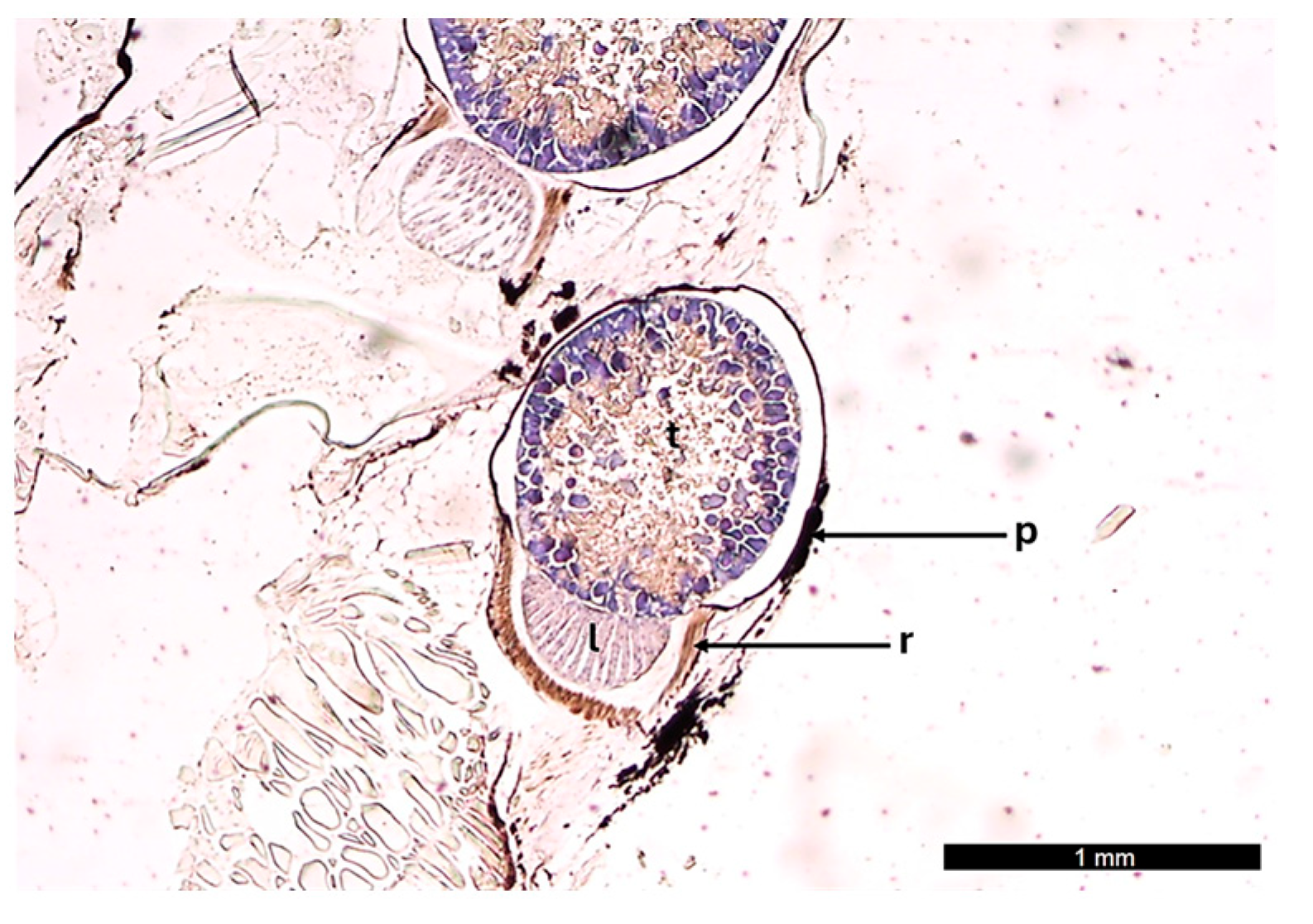
The ventral photophores of I. ovatus consist of a photogenic chamber (the tank) containing photocytes, a lens filter, a reflector, and a pigmented layer (Figure 2).
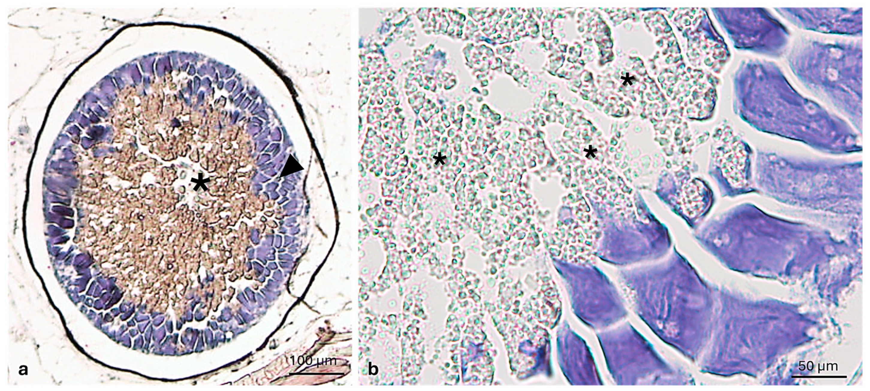
The tank is circular in shape, with photocytes aligned in an orderly manner along the periphery of the tank. The photocytes show accumulations of secretory granules in the apical portion of their body, oriented towards the tank lumen (Figure 3).
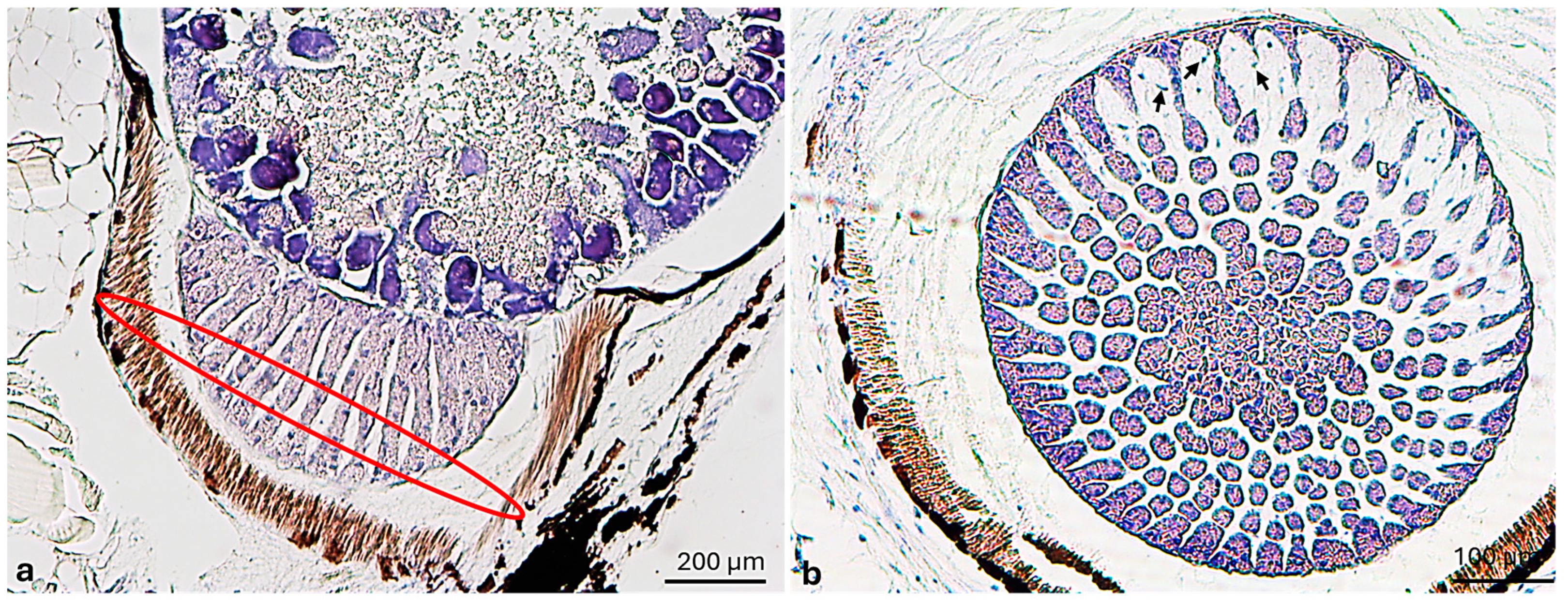
The lens filter has the shape of a semilunar cap and is arranged in correspondence with the lower part of the photogenic chamber. It consists of star-shaped cells embedded within an amorphous extracellular matrix arranged in radially oriented cords (Figure 4).
The reflector envelops the entire organ; however, its organization varies depending on whether it wraps the photogenic chamber or the lens filter. The portion surrounding the lens filter is characterized by an enlarged and less compact thickness, composed of parallel filamentary subunits. Additionally, a pigmented layer envelops the entire ventral photophore (Figure 5).

3.2. Immunohistochemical Analysis

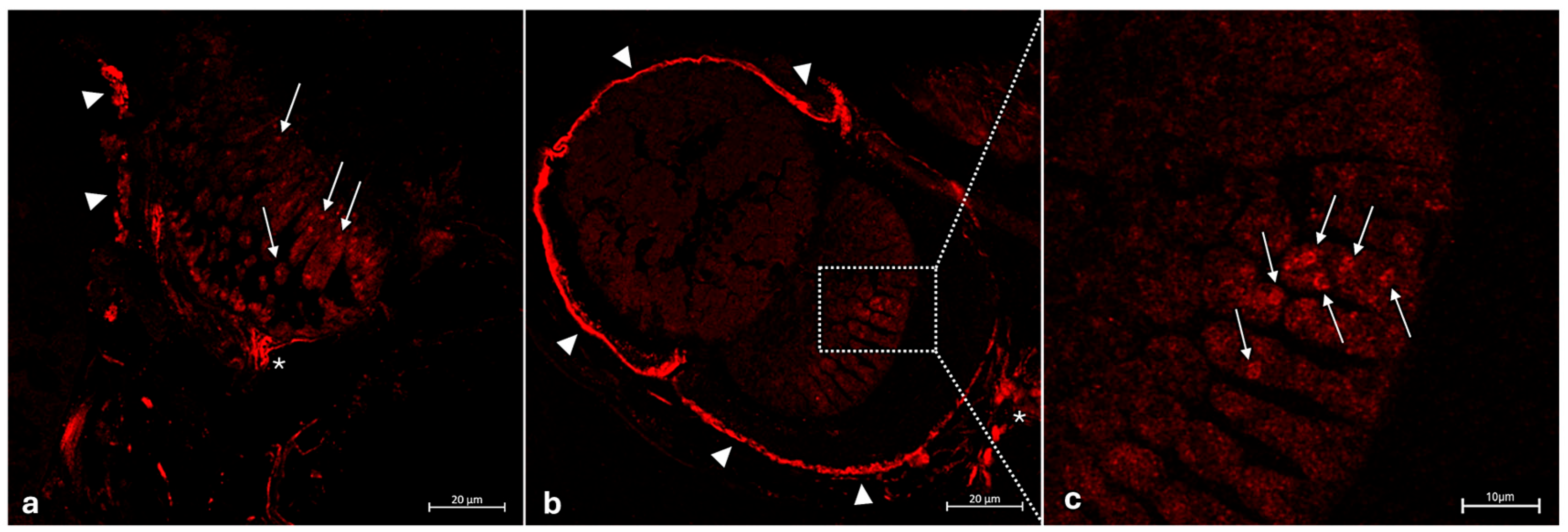
Specific anti-type I nitric oxide synthase (nNOS/NOS type I) immunoreactivity was observed in the pigmented layer around the ventral photophores (Figure 6a,b). Moreover, an intense and specific immunoreaction for nNOS/NOS type I was found in the innervation reaching the lens (Figure 6a,b). Irregularly shaped cells were found within the inner part of the lens, arranged in columns and oriented radially. Immunohistochemical staining revealed immunopositivity limited to the cytoplasmic granules of these cells (Figure 6a,c).
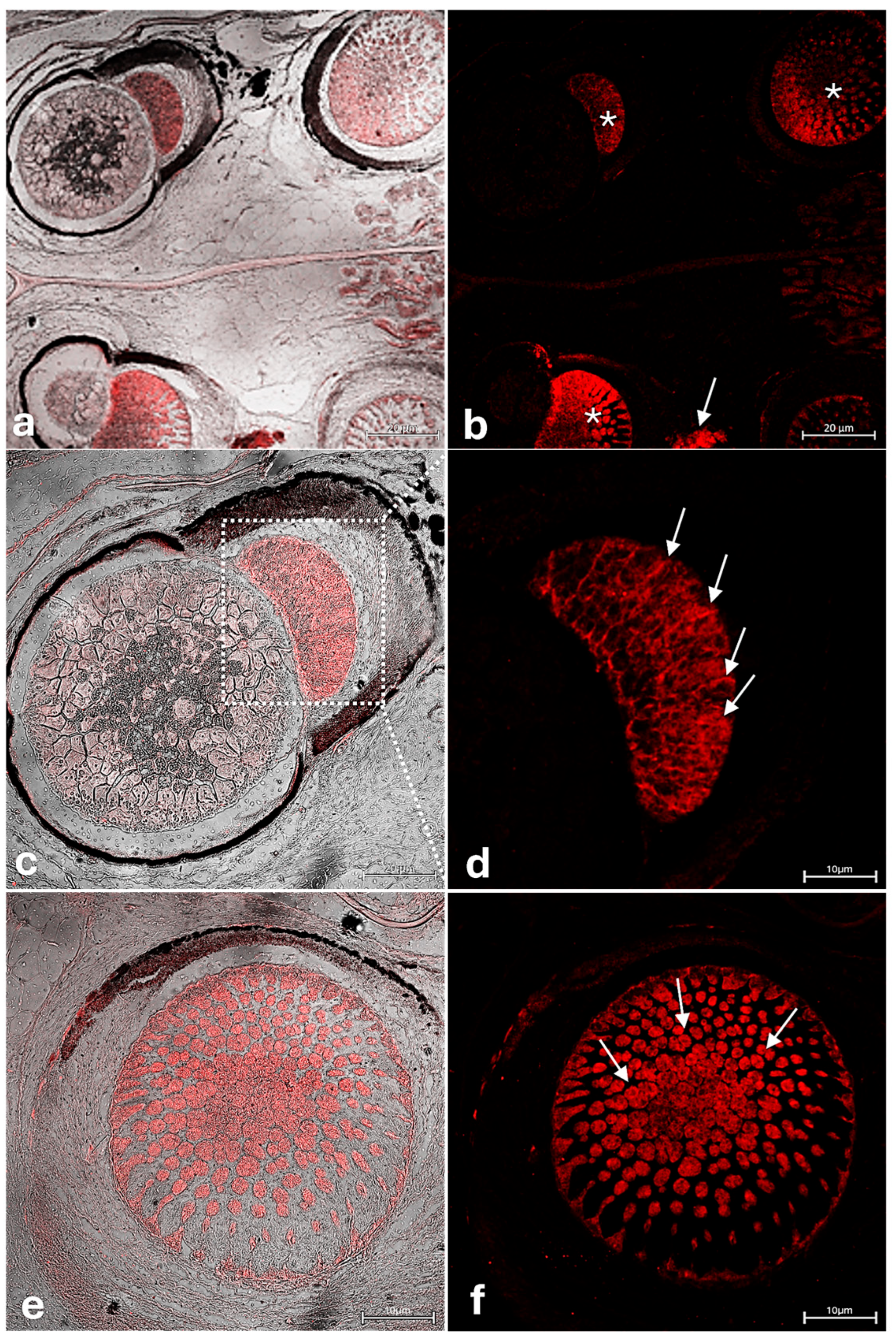
In addition, S100 protein (S100p) immunoreactivity was observed in the nerve fibers reaching the ventral photophores and within the lens of I. ovatus. The typical irregularly shaped cylindrical cells present in the lens of the ventral photophores also showed positive S100p immunoreactivity (Figure 7).
Control experiments showed no immunopositivity for nNOS/NOS Type I and S100p, thus supporting the validity of the results obtained (see Figure S1 in the Supplementary Materials).

4. Discussion

This study provides the first comprehensive morphological and immunohistochemical characterization of the ventral photophore in Ichthyococcus ovatus, revealing its glandular architecture and functional specialization. The structural organization, including the peripheral arrangement of polarized photocytes and the presence of distinct optical components such as the lens filter and reflector, supports a bioluminescence model based on targeted secretion and controlled light emission. Furthermore, the identification of dioptric accessory structures, combined with the absence of a gelatinous body observed in other taxa, highlights unique morphological adaptations in I. ovatus likely associated with the optimized directional light projection.
The structure of the ventral photophore of I. ovatus is described here for the first time. The analysis confirms its glandular nature, consistent with previous studies performed on other mesopelagic fish species [32,39,57,58,59,60,61,62,63,64]. In members of the Stomiidae family, the photophore tank exhibits a comparable structural organization [41], with a rounded shape and photocytes arranged in an orderly manner along the periphery, adhering to the tank walls. The characteristics of these cells are related to their mode of secretion. Indeed, the accumulation of granules exclusively in their apical portion facing the tank lumen induces a sort of polarization of the cell itself as occurs in apo-merocrine glands. This secretion product is poured into the tank lumen, from where it is directed to the lens filter for further processing. This step is presumed to trigger the energy transformation that ultimately results in light emission. The lens filter, shaped like a semi-lunar cap, consists of cells arranged in cords interspersed with areas containing cells that, based on their morphological feature, resemble mesenchymal-like cells. The role of the lens filter in I. ovatus is probably to re-elaborate the secretion products for the subsequent light emission [1]. Although the primary function of the lens filter is to modulate and process the contents of the secretory granules, thereby triggering the biochemical cascade responsible for bioluminescent emission, the ultrastructural organization of this key region exhibits species-specific variability within the Stomiidae family. A comparative analysis between I. ovatus and C. sloani shows that the lens filter of C. sloani consists of less orderly and more loosely arranged cells compared to the highly organized cellular architecture observed in I. ovatus. However, C. sloani exhibits a distinct collector channel that appears to function in the transport of the secretum toward the filtering chamber, a structural feature absent in I. ovatus. Conversely, in the Cyclothone braueri [43], the lens tissue is formed by tightly adherent but disorganized cells, lacking defined morphological boundaries. These cells are stratified into multiple layers and display rounded nuclei, suggesting a divergent cellular differentiation pattern [1,43]. As reported in other Stomiidae species [18] possessing photophores, morphological analysis also confirmed for I. ovatus the presence of two key dioptric annexes: the reflector and the pigmented layer. These structures play a dual role, both protecting the luminous organ and contributing to its dioptric function. Specifically, the reflector appears fibrillar, compact, and uniform, based on morphological analysis, consistent with its role in directing light beams along a defined axis and preventing dispersion. Also, for this species, the pigmented layer seems to be composed of melanin granules of variable abundance [61]. The dioptric function of this layer is supported by its structure: the orderly and continuous arrangement of melanin granules prevents uncontrolled light radiation, ensuring efficient light guidance out of the organ and modulation of intensity and color. Similar dioptric roles have been demonstrated in other deep-sea fishes, such as Argyropelecus aculeatus, where the melanin layer surrounding the photophores acts as a light shutter, regulating both the amount and direction of emitted bioluminescence [65,66]. Together, the pigmented layer and reflector function synergistically to enhance photophore efficiency. Interestingly, in I. ovatus, the dioptric annex known as the gelatinous body, commonly reported in other taxa [21], was not observed in this species.
Immunohistochemical analysis using (nNOS/NOS type I) was also conducted. Nitric oxide synthase (NOS) is a cytosolic protein responsible for the synthesis of nitric oxide (NO) from L-arginine. NO functions as a neurotransmitter and/or modulator of the adrenergic control of bioluminescence [5,6]. The anti-type I nitric oxide synthase (nNOS/NOS type I) immunolabeling was detected in the cytoplasmic granules of lens cells. This immunoreactivity suggests a neuronal nitric oxide signaling potential implication in the pigment sheath light shielding or exposure modulation or the adjustment of light focusing via the lens-associated nerves. A similar role has been already described in other species. Indeed, in Lantern shark (Etmopterus spinax), NOS-like immunoreactivity was found in photocytes, the pigmented sheath, lens cells, and associated nerves [6]. This pathway seems to be preserved through evolution since nNOS immunoreactivity was reported as well in photophore nerve fibers and pigmented cells of Argyropelecus hemigymnus [5].
The conservation of the nNOS protein sequence across species suggests that its biological functions are preserved between fish and mammals [67,68]. The constitutive calcium-dependent form of NOS (cNOS) provides a basal NO release and is localized to the plasma membranes of Golgi bodies [69], known to be involved in post-translational sorting and packaging of novel proteins [70]. In this context, the calcium-binding protein S100p was also found to be immunoreactive in the cylindrical cells radially arranged within the photophore lens. S100 proteins (S100p) constitute a large subfamily of EF-hand calcium-binding proteins, highly conserved during evolution and expressed in both neuronal and non-neuronal tissues of vertebrates. These proteins, found in the cytoplasm and/or nucleus of a wide range of cells, are involved in intracellular Ca2+ homeostasis, functioning as trigger or activator proteins. S100p, in particular, can reversibly bind calcium and zinc ions under physiological pH and ionic strength, inducing conformational changes that regulate diverse cellular functions. In particular, S100p has been implicated in the regulation of cell growth, enhancement of membrane permeability to cations under physiological conditions, stimulation of RNA polymerase activity, and transport of proteins and free fatty acids in adipocytes [12,13,14,71]. As for nNOS, S100p immunoreactivity was detected in the nerve fibers reaching the ventral photophores, suggesting the potential role for NO in mediating the adrenergic control of bioluminescence. Moreover, the detection of S100p in lens cells supports their role as differentiating cells. In this context, the mesenchymal-like cells identified in the lens may play a pivotal role in maintaining the organ’s homeostatic balance, particularly under the extreme conditions of mesopelagic zones. These cells may represent a regenerative reservoir potentially activated in response to physiological stimuli such as growth-related remodeling or mechanical stress, contributing to the repair processes by differentiating into photocytes or other structural components of the photophore (e.g., pigmented cells, or supportive fibers). This functional adaptation may preserve the physiological plasticity and integrity of the organ over time via cellular renewal, the secretion of trophic factors (e.g., VEGF, TGF-β, Wnt), and the modulation of the extracellular environment. Moreover, these cells appear to serve another structural and mechanical role, providing physical support to photocytes and contributing to the tissue resilience of the photophore, particularly under conditions of high hydrostatic pressure, low temperatures, and limited energy availability, which are typical of deep-sea ecosystems. They seem to be a dynamic and interactive component of the photophore microenvironment, sharing the same properties of epithelial or neural tissue stem cells [72,73]. Future immunohistochemical and transcriptomic studies assessing specific markers like CD90, CD105, vimentin, and Sox9 may clarify their degree of pluripotency or induced differentiation in response to environmental stressors [74,75,76]. Overall, the detection of nNOS and S100p within the ventral photophore, particularly in lens-associated cells and innervating neural structures, suggests a regulatory network involving calcium-mediated signaling and nitric oxide pathways. There are currently no direct studies of S100p expression in the photophores of deep-sea fish; however, the S100p family in teleosts comprises at least 14 subfamilies with highly specific expressions in different cell types [15]. Data on related sensory tissues suggest that S100p may be involved in intracellular calcium regulation and/or may serve as a marker of specialized or nerve support cells [13,14,71,77,78,79,80]. Based on these observations, S100p may contribute to the regulation of intracellular calcium transients in photogenic cells, influencing the dynamics of the bioluminescent response, which is often Ca2-dependent. Similarly, S100B and S100A1 modulate calcium homeostasis in neurons and muscle cells [81,82,83,84,85]. Given the presence of S100p in fish neuroepithelial tissues (such as in olfactory cells or peripheral glia), it may also support maturation, maintenance, or turnover of photophore cells, which exhibit intense metabolic activity. Furthermore, photophores are innervated [7,86,87] and regulated by both central and environmental signals (e.g., light, stress, circadian rhythms); therefore, S100p could mediate nerve signaling or the functional plasticity of the organ, similarly to the role known in the CNS [15,82,83,84,85]. These findings indicate a potential role of NO in the adrenergic modulation of light emission and point to the presence of differentiating cell populations, as marked by S100p immunoreactivity. The involvement of nitric oxide in regulating light emission, already reported in the photophores of other mesopelagic species, suggests that these systems are modulated by complex neuroendocrine interactions. This points to their relevance for future studies in neurobiology and comparative physiology [88,89].
Future investigations are needed to clarify whether the observed nNOS immunostaining in the pigmented epithelium acts through autocrine and/or paracrine signaling mechanisms. These insights open new avenues for studying bioluminescence in deep-sea organisms, particularly with regard to the highly specialized glandular and dioptric structures that have evolved to optimize directional light projection [86]. From a functional perspective, the behavior of I. ovatus, including diel vertical migration and counter-illumination camouflage, makes it a valuable model for exploring how bioluminescence shapes trophic interactions and contributes to biogeochemical cycles such as carbon flux and the biological pump [16,87,88]. From an evolutionary standpoint, the species-specific optical structures and the widespread photophore polymorphism within Stomiidae and both related and unrelated taxa support the hypothesis of multiple adaptive origins of bioluminescence in meso- and bathypelagic fish lineages [34]. Understanding the evolutionary significance of this structural diversity will require further investigation.
Overall, the identification and characterization of the cellular and molecular mechanisms involved in photophore function lay the groundwork for innovative biotechnological applications. This expanding body of knowledge may foster the development of novel biosensors and optical systems based on luciferases or other photoproteins, as well as environmental technologies designed to monitor or influence biological processes in the deep sea [89]. Future research should aim to clarify the precise molecular interactions governing photophore activation and control, thereby deepening our grasp of bioluminescent regulation and its broader implications in neurobiology, evolutionary biology, and biotechnology.

5. Conclusions

The findings of this study significantly enhance our understanding of the cellular architecture and neurochemical regulation of the bioluminescent system in mesopelagic fishes such as Ichthyococcus ovatus. The immunohistochemical investigation indicated a potential role of NO in adrenergic modulation of light emission, while the detection of S100p, reported here for the first time, points to the presence of differentiating cell populations. The analogy with calcium-modulated sensory tissues further suggests a role for S100p in the molecular and functional control of photophores. The absence of previous data opens up promising avenues for future studies aimed at clarifying the cellular and molecular mechanisms that regulate light activity, with potential implications for neurobiology, evolutionary biology, and biotechnology. Future research should focus on elucidating the precise molecular interactions underlying photophore activation and control, as well as exploring the evolutionary significance of structural diversity within Stomiidae. Overall, these results advance our knowledge of bioluminescent systems in mesopelagic fishes and provide a solid foundation for future investigations.

Supplementary Materials

The following supporting information can be downloaded at https://www.mdpi.com/article/10.3390/jmse13081534/s1, Figure S1. Photomicrograph of Ichthyococcus ovatus photophores nNOS/NOS type I (a) and s100 (b) immunofluorescences performed without primary antibodies. No immunostaining was observed.

Author Contributions

Conceptualization, M.C., M.A. and M.C.G.; methodology, M.C., M.A., K.M., L.P. and M.C.G.; software, M.C., M.A., K.M., L.P. and M.C.G.; validation, M.C., M.A., K.M., L.P., R.L., M.L., G.M., F.A. and M.C.G.; formal analysis, M.C., M.A., K.M., L.P. and M.C.G.; investigation, M.C., M.A., K.M., L.P. and M.C.G.; resources, M.C., M.A., K.M., L.P., R.L., M.L., G.M., F.A. and M.C.G.; data curation, M.C., M.A., K.M., L.P. and M.C.G.; writing—original draft preparation M.C., M.A. and M.C.G.; writing—review and editing, M.C., M.A., K.M., L.P., R.L., M.L., G.M., F.A. and M.C.G.; visualization, M.C., M.A., K.M., L.P., R.L., M.L., G.M., F.A. and M.C.G.; supervision, M.C., M.A., L.P., R.L., M.L., G.M., F.A. and M.C.G.; project administration, M.C., M.A., L.P. and M.C.G.; funding acquisition, G.M. and M.C.G. All authors have read and agreed to the published version of the manuscript.

Funding

This research received no external funding.

Data Availability Statement

All data presented in this study are available from the corresponding author upon responsible request.

Acknowledgments

The authors thank Sergio De Matteo for providing the photo of the I. ovatus collected after stranding from the Sicilian coast of the Strait of Messina.

Conflicts of Interest

The authors declare no conflicts of interest.

References

  1. Cavallaro, M.; Pansera, L.; Mhalhel, K.; Abbate, F.; Levanti, M.; Guerrera, M.C.; Montalbano, G.; Briglia, M.; Aragona, M.; Laurà, R. The Skin Photophores of Chauliodus sloani Bloch & Schneider, 1801 (Pisces: Stomiidae): A Morphological, Ultrastructural and Immunohistochemical Study. Animals 2025, 15, 1738. [Google Scholar] [CrossRef]
  2. Herring, P.J. Systematic distribution of bioluminescence in living organisms. J. Biolumin. Chemilumin. 1987, 1, 147–163. [Google Scholar] [CrossRef]
  3. Schramm, S.; Weiß, D. Bioluminescence—The Vibrant Glow of Nature and its Chemical Mechanisms. ChemBioChem 2024, 25, e202400106. [Google Scholar] [CrossRef]
  4. Kaskova, Z.M.; Tsarkova, A.S.; Yampolsky, I.V. 1001 lights: Luciferins, luciferases, their mechanisms of action and applications in chemical analysis, biology and medicine. Chem. Soc. Rev. 2016, 45, 6048–6077. [Google Scholar] [CrossRef]
  5. Krönström, J.; Holmgren, S.; Baguet, F.; Salpietro, L.; Mallefet, J. Nitric oxide in control of luminescence from hatchetfish (Argyropelecus hemigymnus) photophores. J. Exp. Biol. 2005, 208, 2951–2961. [Google Scholar] [CrossRef][Green Version]
  6. Claes, J.M.; Aksnes, D.L.; Mallefet, J. Phantom hunter of the fjords: Camouflage by counterillumination in a shark (Etmopterus spinax). J. Exp. Mar. Biol. Ecol. 2010, 388, 28–32. [Google Scholar] [CrossRef]
  7. Zaccone, G.; Abelli, L.; Salpietro, L.; Zaccone, D.; Macrì, B.; Marino, F. Nervous control of photophores in luminescent fishes. Acta Histochem. 2011, 113, 387–394. [Google Scholar] [CrossRef]
  8. Fleming, I.; Busse, R. Chapter 38—Activation of NOS by Ca2+-Dependent and Ca2+-Independent Mechanisms. In Nitric Oxide; Ignarro, L.J., Ed.; Academic Press: San Diego, CA, USA, 2000; pp. 621–632. [Google Scholar] [CrossRef]
  9. Wright, N.T.; Prosser, B.L.; Varney, K.M.; Zimmer, D.B.; Schneider, M.F.; Weber, D.J. S100A1 and Calmodulin Compete for the Same Binding Site on Ryanodine Receptor*. J. Biol. Chem. 2008, 283, 26676–26683. [Google Scholar] [CrossRef]
  10. Rebbeck, R.T.; Nitu, F.R.; Rohde, D.; Most, P.; Bers, D.M.; Thomas, D.D.; Cornea, R.L. S100A1 Protein Does Not Compete with Calmodulin for Ryanodine Receptor Binding but Structurally Alters the Ryanodine Receptor Calmodulin Complex. J. Biol. Chem. 2016, 291, 15896–15907. [Google Scholar] [CrossRef]
  11. Aragona, M.; Mhalhel, K.; Cometa, M.; Franco, G.A.; Montalbano, G.; Guerrera, M.C.; Levanti, M.; Laurà, R.; Abbate, F.; Vega, J.A.; et al. Piezo 1 and Piezo 2 in the Chemosensory Organs of Zebrafish (Danio rerio). Int. J. Mol. Sci. 2024, 25, 7404. [Google Scholar] [CrossRef]
  12. Aragona, M.; Porcino, C.; Guerrera, M.C.; Montalbano, G.; Laurà, R.; Levanti, M.; Abbate, F.; Cobo, T.; Capitelli, G.; Calapai, F.; et al. Localization of BDNF and Calretinin in Olfactory Epithelium and Taste Buds of Zebrafish (Danio rerio). Int. J. Mol. Sci. 2022, 23, 4696. [Google Scholar] [CrossRef]
  13. Germanà, A.; Guerrera, M.C.; Laurà, R.; Levanti, M.; Aragona, M.; Mhalhel, K.; Germanà, G.; Montalbano, G.; Abbate, F. Expression and Localization of BDNF/TrkB System in the Zebrafish Inner Ear. Int. J. Mol. Sci. 2020, 21, 5787. [Google Scholar] [CrossRef]
  14. Germanà, A.; Marino, F.; Guerrera, M.C.; Campo, S.; de Girolamo, P.; Montalbano, G.; Germanà, G.P.; Ochoa-Erena, F.J.; Ciriaco, E.; Vega, J.A. Expression and distribution of S100 protein in the nervous system of the adult zebrafish (Danio rerio). Microsc. Res. Tech. 2008, 71, 248–255. [Google Scholar] [CrossRef]
  15. Kraemer, A.M.; Saraiva, L.R.; Korsching, S.I. Structural and functional diversification in the teleost S100 family of calcium-binding proteins. BMC Evol. Biol. 2008, 8, 48. [Google Scholar] [CrossRef]
  16. Haddock, S.H.D.; Moline, M.A.; Case, J.F. Bioluminescence in the Sea. Annu. Rev. Mar. Sci. 2010, 2, 443–493. [Google Scholar] [CrossRef]
  17. Paitio, J.; Oba, Y. Luminous fishes: Endocrine and neuronal regulation of bioluminescence. Aquac. Fish. 2024, 9, 486–500. [Google Scholar] [CrossRef]
  18. Smith, W.L.; Girard, M.G.; Walker, H.J., Jr.; Davis, M.P. The phylogeny of bristlemouths, lightfishes, and portholefishes with a revised family-level classification of the dragonfishes (Teleostei: Stomiiformes). Prof. Pap. NMFS 2024, 24, 167–184. [Google Scholar] [CrossRef]
  19. Nelson, J.S.; Grande, T.C.; Wilson, M.V. Fishes of the World; John Wiley & Sons: Hoboken, NJ, USA, 2016. [Google Scholar]
  20. Fricke, R.; Eschmeyer, W.N.; van der Laan, R. (Eds.) Eschmeyer’s Catalog of Fishes: Genera, Species, References; California Academy of Sciences: San Francisco, CA, USA, 2024. [Google Scholar]
  21. Cavallaro, M.; Guerrera, M.C.; Abbate, F.; Levanti, M.B.; Laurà, R.; Ammendolia, G.; Malara, D.; Stipa, M.G.; Battaglia, P. Morphological, ultrastructural and immunohistochemical study on the skin ventral photophores of Diaphus holti Tåning, 1918 (Family: Myctophidae). Acta Zool. 2020, 102, 405–411. [Google Scholar] [CrossRef]
  22. Gjøsæter, J.; Kawaguchi, K. A Review of the World Resources of Mesopelagic Fish; Food and Agriculture Organization of the United Nations: Rome, Italy, 1980. [Google Scholar]
  23. Stukel, M.R.; Ducklow, H.W. Stirring Up the Biological Pump: Vertical Mixing and Carbon Export in the Southern Ocean. Glob. Biogeochem. Cycles 2017, 31, 1420–1434. [Google Scholar] [CrossRef]
  24. Clardy, T.R.; Gopalan, J.; Rabaoui, L.J. Morphology of photophores in juvenile Vinciguerria mabahiss (Stomiiformes: Phosichthyidae). Ichthyol. Res. 2025. [Google Scholar] [CrossRef]
  25. Martin, R.P.; Carr, E.M.; Sparks, J.S. Variation in lanternfish (Myctophidae) photophore structure: A comprehensive comparative analysis. PLoS ONE 2024, 19, e0310976. [Google Scholar] [CrossRef]
  26. Harvey, E.N. The luminous organs of fishes. In The Physiology of Fishes; Brown, M.E., Ed.; Elsevier: New York, NY, USA, 1957; Volume 3, pp. 345–366. [Google Scholar]
  27. Haygood, M.G.; Edwards, D.B.; Mowlds, G.; Rosenblatt, R.H. Bioluminescence of myctophid and stomiiform fishes is not due to bacterial luciferase. J. Exp. Zool. 1994, 270, 225–231. [Google Scholar] [CrossRef]
  28. Baguet, F. Les Photophores des Poissons Lumineux. Rev. Quest. Sci. 1977, 148, 21–41. [Google Scholar]
  29. Herring, P.J. Bioluminescence of marine organisms. Nature 1977, 267, 788–793. [Google Scholar] [CrossRef]
  30. Anctil, M. Stimulation of bioluminescence in lanternfishes (Myctophidae). II. Can. J. Zool. 1972, 50, 233–237. [Google Scholar] [CrossRef]
  31. Anctil, M.; Case, J.F. The caudal luminous organs of lanternfishes: General innervation and ultrastructure. Am. J. Anat. 1977, 149, 1–21. [Google Scholar] [CrossRef]
  32. Edwards, A.; Herring, P. Observations on the comparative morphology and operation of the photogenic tissues of myctophid fishes. Mar. Biol. 1977, 41, 59–70. [Google Scholar] [CrossRef]
  33. Herring, P.J. How to survive in the dark: Bioluminescence in the deep sea. Symp. Soc. Exp. Biol. 1985, 39, 323–350. [Google Scholar]
  34. Davis, M.P.; Sparks, J.S.; Smith, W.L. Repeated and widespread evolution of bioluminescence in marine fishes. PLoS ONE 2016, 11, e0155154. [Google Scholar] [CrossRef]
  35. Paitio, J.; Oba, Y.; Meyer-Rochow, V.B. Bioluminescent fishes and their eyes. In Luminescence—An Outlook on the Phenomena and Their Applications; Intech Open: London, UK, 2016. [Google Scholar]
  36. Paitio, J.; Yano, D.; Muneyama, E.; Takei, S.; Asada, H.; Iwasaka, M.; Oba, Y. Reflector of the body photophore in lanternfish is mechanistically tuned to project the biochemical emission in photocytes for counterillumination. Biochem. Biophys. Res. Commun. 2020, 521, 821–826. [Google Scholar] [CrossRef]
  37. Altun, T.; Celi, F.; Danabas, D. Bioluminescence in aquatic organisms. J. Anim. Vet. Adv. 2008, 7, 841–846. [Google Scholar]
  38. Battaglia, P.; Ammendolia, G.; Cavallaro, M.; Consoli, P.; Esposito, V.; Malara, D.; Rao, I.; Romeo, T.; Andaloro, F. Influence of lunar phases, winds and seasonality on the stranding of mesopelagic fish in the Strait of Messina (Central Mediterranean Sea). Mar. Ecol. 2017, 38, e12459. [Google Scholar] [CrossRef]
  39. Cavallaro, M. Studio Morfologico ed Ultrastrutturale dei Fotofori in Diverse Specie di Pesci Mesopelagici Dello Stretto di Messina. Ph.D. Thesis, Università degli Studi di Messina, Messina, Italy, 2017. [Google Scholar]
  40. Cavallaro, M.; Aragona, M.; Germanà, A. Morphological and immunohistochemical study on photophores of Gonostoma denudatum Rafinesque, 1810 (Fam: Gonostomatidae). Atti Accad. Peloritana Pericolanti Cl. Sci. Med. Biol. 2021, 109, 1–7. [Google Scholar] [CrossRef]
  41. Cavallaro, M.; Battaglia, P.; Guerrera, M.C.; Abbate, F.; Levanti, M.B.; Ammendolia, G.; Andaloro, F.; Germanà, A.; Laurà, R. Structure and ultrastructure study on photophores of the Madeira lanternfish, Ceratoscopelus maderensis (Lowe, 1839), Pisces: Myctophidae. Acta Zool. 2019, 100, 89–95. [Google Scholar] [CrossRef]
  42. Cavallaro, M.; Battaglia, P.; Guerrera, M.C.; Abbate, F.; Levanti, M.B.; Andaloro, F.; Germanà, A.; Laurà, R. New data on morphology and ultrastructure of skin photophores in the deep-sea squid Histioteuthis bonnellii (Férussac, 1834), Cephalopoda: Histioteuthidae. Acta Zool. 2017, 98, 271–277. [Google Scholar] [CrossRef]
  43. Cavallaro, M.; Battaglia, P.; Laurà, R.; Guerrera, M.C.; Abbate, F.; Germanà, A. The morphology of photophores in the garrick, Cyclothone braueri (Family: G onostomatidae): An ultrastructure study. Acta Zool. 2015, 96, 296–300. [Google Scholar] [CrossRef]
  44. Cavallaro, M.; Mammola, C.; Verdiglione, R. Structural and ultrastructural comparison of photophores of two species of deep-sea fishes: Argyropelecus hemigymnus and Maurolicus muelleri. J. Fish Biol. 2004, 64, 1552–1567. [Google Scholar] [CrossRef]
  45. Robison, B.H. Deep pelagic biology. J. Exp. Mar. Biol. Ecol. 2004, 300, 253–272. [Google Scholar] [CrossRef]
  46. Battaglia, P.; Pagano, L.; Consoli, P.; Esposito, V.; Granata, A.; Guglielmo, L.; Pedá, C.; Romeo, T.; Zagami, G.; Vicchio, T.M.; et al. Consumption of mesopelagic prey in the Strait of Messina, an upwelling area of the central Mediterranean Sea: Feeding behaviour of the blue jack mackerel Trachurus picturatus (Bowdich, 1825). Deep Sea Res. Part I Oceanogr. Res. Pap. 2020, 155, 103158. [Google Scholar] [CrossRef]
  47. Brauer, A. Die Tiefsee-Fische. II. Anatomischer Teil. Wiss. Ergebn. Dtsch. Tiefsee Exped. “Valdivia” 1908, 15, 1–266. [Google Scholar]
  48. Froese, R.; Pauly, D. FishBase. World Wide Web Electronic PublicationFishBase. 2019. Available online: http://www.fishbase.org (accessed on 24 June 2025).
  49. Board, W.E. World Register of Marine Species. 2024. Available online: https://www.marinespecies.org (accessed on 24 June 2025).
  50. Van der Land, J.; Costello, M.; Zavodnik, D.; Santos, R.; Porteiro, F.; Bailly, N.; Eschmeyer, W.; Froese, R. Pisces. Collect. Patrim. Nat. 2001, 357–374. [Google Scholar]
  51. Abbate, F.; Guerrera, M.C.; Levanti, M.; Laurà, R.; Montalbano, G.; Cavallaro, M.; Germanà, A. The tongue of Leopard Gecko (Eublepharis macularius): LM, SEM and confocal laser study. Anat. Histol. Embryol. 2020, 49, 51–59. [Google Scholar] [CrossRef] [PubMed]
  52. Abbate, F.; Guerrera, M.C.; Levanti, M.; Laurà, R.; Aragona, M.; Mhalhel, K.; Montalbano, G.; Germanà, A. Morphological characteristics of the blackspot seabream (Pagellus bogaraveo) tongue: A structural and immunohistochemical study. Anat. Histol. Embryol. 2022, 51, 103–111. [Google Scholar] [CrossRef] [PubMed]
  53. Alesci, A.; Albano, M.; Savoca, S.; Mokhtar, D.M.; Fumia, A.; Aragona, M.; Lo Cascio, P.; Hussein, M.M.; Capillo, G.; Pergolizzi, S.; et al. Confocal Identification of Immune Molecules in Skin Club Cells of Zebrafish (Danio rerio, Hamilton 1882) and Their Possible Role in Immunity. Biology 2022, 11, 1653. [Google Scholar] [CrossRef]
  54. Zaccone, G.; Maina, J.; Germanà, A.; Montalbano, G.; Capillo, G.; Aragona, L.; Kuciel, M.J.; Lauriano, E.R.; Icardo, J.M. First demonstration of the neuroepithelial cells and their chemical code in the accessory respiratory organ and the gill of the sharptooth catfish, Clarias gariepinus: A preliminary study. Acta Zool. 2019, 100, 160–166. [Google Scholar] [CrossRef]
  55. Whitehead, P.J.P.; Bauchot, M.-L.; Hureau, J.-C.; Nielsen, J.; Tortonese, E. Fishes of the North-Eastern Atlantic and the Mediterranean; UNESCO: Paris, France, 1984; Volume 1. [Google Scholar]
  56. Cavallaro, M.; Ammendolia, G.; Andaloro, F.; Battaglia, P. First record of the mesopelagic fish Diaphus dumerilii (Bleeker, 1856) in the Mediterranean Sea. Mar. Biodivers. 2017, 47, 585–588. [Google Scholar] [CrossRef]
  57. Denton, E.; Gilpin-Brown, J.; Roberts, B. On the organization and function of the photophores of Argyropelecus. J. Physiol. 1969, 204, 38P–39P. [Google Scholar]
  58. Denton, E.; Gilpin-Brown, J.; Wright, P. On the ‘filters’ in the photophores of mesopelagic fish and on a fish emitting red light and especially sensitive to red light. J. Physiol. 1970, 208, 72P–73P. [Google Scholar]
  59. Denton, E.; Herring, P.J. On the filters in the ventral photophores of mesopelagic animals [proceedings]. J. Physiol. 1978, 284, 42P. [Google Scholar] [PubMed]
  60. Denton, E.J.; Herring, P.; Widder, E.; Latz, M.; Case, J. The roles of filters in the photophores of oceanic animals and their relation to vision in the oceanic environment. Proc. R. Soc. London. Ser. B. Biol. Sci. 1985, 225, 63–97. [Google Scholar] [CrossRef]
  61. Mallefet, J.; Duchatelet, L.; Hermans, C.; Baguet, F. Luminescence control of Stomiidae photophores. Acta Histochem. 2019, 121, 7–15. [Google Scholar] [CrossRef]
  62. Mallefet, J.; Shimomura, O. Presence of coelenterazine in mesopelagic fishes from the Strait of Messina. Mar. Biol. 1995, 124, 381–385. [Google Scholar] [CrossRef]
  63. Davis, A.L.; Sutton, T.T.; Kier, W.M.; Johnsen, S. Evidence that eye-facing photophores serve as a reference for counterillumination in an order of deep-sea fishes. Proc. Biol. Sci. 2020, 287, 20192918. [Google Scholar] [CrossRef]
  64. Krönström, J.; Dupont, S.; Mallefet, J.; Thorndyke, M.; Holmgren, S. Serotonin and nitric oxide interaction in the control of bioluminescence in northern krill, Meganyctiphanes norvegica (M. Sars). J. Exp. Biol. 2007, 210, 3179–3187. [Google Scholar] [CrossRef]
  65. Coccimiglio, M.L.; Jonz, M.G. Serotonergic neuroepithelial cells of the skin in developing zebrafish: Morphology, innervation and oxygen-sensitive properties. J. Exp. Biol. 2012, 215, 3881–3894. [Google Scholar] [CrossRef]
  66. Jonz, M.G.; Nurse, C.A. Peripheral chemoreceptors in air-versus water-breathers. In Arterial Chemoreception: From Molecules to Systems; Springer: Berlin/Heidelberg, Germany, 2012; pp. 19–27. [Google Scholar] [CrossRef]
  67. Percival, J.M.; Anderson, K.N.; Huang, P.; Adams, M.E.; Froehner, S.C. Golgi and sarcolemmal neuronal NOS differentially regulate contraction-induced fatigue and vasoconstriction in exercising mouse skeletal muscle. J. Clin. Investig. 2010, 120, 816–826. [Google Scholar] [CrossRef]
  68. Förstermann, U.; Sessa, W.C. Nitric oxide synthases: Regulation and function. Eur. Heart J. 2012, 33, 829–837. [Google Scholar] [CrossRef]
  69. Aragona, M.; Mhalhel, K.; Pansera, L.; Montalbano, G.; Guerrera, M.C.; Levanti, M.; Laurà, R.; Abbate, F.; Vega, J.A.; Germanà, A. Localization of Piezo 1 and Piezo 2 in Lateral Line System and Inner Ear of Zebrafish (Danio rerio). Int. J. Mol. Sci. 2024, 25, 9204. [Google Scholar] [CrossRef]
  70. Liu, J.; Gao, J.; Liang, Z.; Gao, C.; Niu, Q.; Wu, F.; Zhang, L. Mesenchymal stem cells and their microenvironment. Stem Cell Res. Ther. 2022, 13, 429. [Google Scholar] [CrossRef] [PubMed]
  71. Lv, F.-J.; Tuan, R.S.; Cheung, K.M.C.; Leung, V.Y.L. Concise Review: The Surface Markers and Identity of Human Mesenchymal Stem Cells. Stem Cells 2014, 32, 1408–1419. [Google Scholar] [CrossRef]
  72. Consentius, C.; Mirenska, A.; Jurisch, A.; Reinke, S.; Scharm, M.; Zenclussen, A.C.; Hennig, C.; Volk, H.-D. In situ detection of CD73+ CD90+ CD105+ lineage: Mesenchymal stromal cells in human placenta and bone marrow specimens by chipcytometry. Cytom. Part A 2018, 93, 889–893. [Google Scholar] [CrossRef]
  73. Fonseca, L.N.; Bolívar-Moná, S.; Agudelo, T.; Beltrán, L.D.; Camargo, D.; Correa, N.; Del Castillo, M.A.; Fernández de Castro, S.; Fula, V.; García, G.; et al. Cell surface markers for mesenchymal stem cells related to the skeletal system: A scoping review. Heliyon 2023, 9, e13464. [Google Scholar] [CrossRef]
  74. Spooner-Harris, M.; Kerns, K.; Zigo, M.; Sutovsky, P.; Balboula, A.; Patterson, A.L. A re-appraisal of mesenchymal-epithelial transition (MET) in endometrial epithelial remodeling. Cell Tissue Res. 2023, 391, 393–408. [Google Scholar] [CrossRef]
  75. Abbate, F.; Catania, S.; Germana, A.; González, T.; Diaz-Esnal, B.; Germana, G.; Vega, J. S-100 protein is a selective marker for sensory hair cells of the lateral line system in teleosts. Neurosci. Lett. 2002, 329, 133–136. [Google Scholar] [CrossRef]
  76. Germana, A.; Abbate, F.; González-Martínez, T.; Del Valle, M.; De Carlos, F.; Germanà, G.; Vega, J. S100 protein is a useful and specific marker for hair cells of the lateral line system in postembryonic zebrafish. Neurosci. Lett. 2004, 365, 186–189. [Google Scholar] [CrossRef]
  77. Germana, A.; Montalbano, G.; Laura, R.; Ciriaco, E.; Del Valle, M.; Vega, J.A. S100 protein-like immunoreactivity in the crypt olfactory neurons of the adult zebrafish. Neurosci. Lett. 2004, 371, 196–198. [Google Scholar] [CrossRef]
  78. Germanà, A.; Paruta, S.; Germanà, G.P.; Ochoa-Erena, F.J.; Montalbano, G.; Cobo, J.; Vega, J.A. Differential distribution of S100 protein and calretinin in mechanosensory and chemosensory cells of adult zebrafish (Danio rerio). Brain Res. 2007, 1162, 48–55. [Google Scholar] [CrossRef]
  79. Donato, R. Intracellular and extracellular roles of S100 proteins. Microsc. Res. Tech. 2003, 60, 540–551. [Google Scholar] [CrossRef]
  80. Donato, R. S100: A multigenic family of calcium-modulated proteins of the EF-hand type with intracellular and extracellular functional roles. Int. J. Biochem. Cell Biol. 2001, 33, 637–668. [Google Scholar] [CrossRef]
  81. Donato, R.; Geczy, C.L.; Weber, D.J. S100 Proteins. In Encyclopedia of Metalloproteins; Kretsinger, R.H., Uversky, V.N., Permyakov, E.A., Eds.; Springer: New York, NY, USA, 2013; pp. 1863–1874. [Google Scholar] [CrossRef]
  82. Donato, R.; Cannon, B.R.; Sorci, G.; Riuzzi, F.; Hsu, K.; Weber, D.J.; Geczy, C.L. Functions of S100 proteins. Curr. Mol. Med. 2013, 13, 24–57. [Google Scholar] [CrossRef]
  83. Nicol, J.A.C. Observations on Photophores and Luminescence in the Teleost Porichthys. J. Cell Sci. 1957, 3, 179–188. [Google Scholar] [CrossRef]
  84. Krönström, J. Control of Bioluminescence. Operating the Light Switch in Photophores from Marine Animals. Ph.D. Thesis, University of Gothenburg, Göteborg, Sweden, 2009. [Google Scholar]
  85. Claes, J.M.; Krönström, J.; Holmgren, S.; Mallefet, J. Nitric oxide in the control of luminescence from lantern shark (Etmopterus spinax) photophores. J. Exp. Biol. 2010, 213, 3005–3011. [Google Scholar] [CrossRef] [PubMed]
  86. Davis, M.P.; Holcroft, N.I.; Wiley, E.O.; Sparks, J.S.; Leo Smith, W. Species-specific bioluminescence facilitates speciation in the deep sea. Mar. Biol. 2014, 161, 1139–1148. [Google Scholar] [CrossRef] [PubMed]
  87. Irigoien, X.; Klevjer, T.A.; Røstad, A.; Martinez, U.; Boyra, G.; Acuña, J.L.; Bode, A.; Echevarria, F.; Gonzalez-Gordillo, J.I.; Hernandez-Leon, S.; et al. Large mesopelagic fishes biomass and trophic efficiency in the open ocean. Nat. Commun. 2014, 5, 3271. [Google Scholar] [CrossRef]
  88. Davison, P.C.; Checkley, D.M.; Koslow, J.A.; Barlow, J. Carbon export mediated by mesopelagic fishes in the northeast Pacific Ocean. Prog. Oceanogr. 2013, 116, 14–30. [Google Scholar] [CrossRef]
  89. Roda, A.; Pasini, P.; Mirasoli, M.; Michelini, E.; Guardigli, M. Biotechnological applications of bioluminescence and chemiluminescence. Trends Biotechnol. 2004, 22, 295–303. [Google Scholar] [CrossRef]
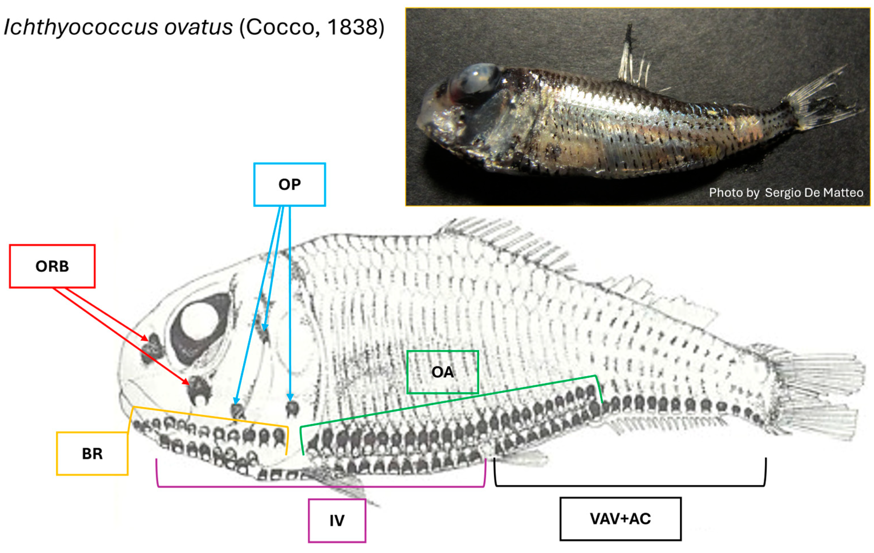
Figure 1. Pattern illustrating the distribution of photophores on the body of I. ovatus, modified from Whitehead et al. [56]. The insert shows a specimen of the I. ovatus collected after stranding along the Sicilian coast of the Strait of Messina (photo by Sergio De Matteo).
Jmse 13 01534 g001
Figure 2. Longitudinal section of ventral photophore (ventral series IV) of I. ovatus. The photomicrograph shows (t) tank, (l) lens filter, (r) reflector, and (p) pigmented layer. Toluidine Blue stain.
Jmse 13 01534 g002
Figure 3. (a) The photomicrograph shows a transversal section of the isolated tank of ventral photophore (ventral series IV) of I. ovatus. Arrowhead indicates photocytes, asterisk indicates the tank lumen. (b) High magnification of a photocyte with apo-merocrine secretion mode. The secretion mode can be stabilized by the presence of secretory spheroidal particles (asterisks). Toluidine Blue stain.
Jmse 13 01534 g003
Figure 4. The photomicrograph shows: (a) Longitudinal section of the lens filter of a ventral photophore (ventral series IV) in I. ovatus. The red oval shape indicates the area shown in cross-section in picture. (b) In the cross section of the lens filter of I. ovatus ventral photophore, mesenchymal-like cells (arrows) were identified by their morphological features. Toluidine Blue stain.
Jmse 13 01534 g004
Figure 5. The photomicrograph shows a longitudinal section of lens filter of ventral photophore (ventral series IV) of I. ovatus. The asterisks indicate the reflector and the arrow designs the pigmented layer. Toluidine Blue stain.
Jmse 13 01534 g005
Figure 6. Longitudinal section of I. ovatus ventral photophores (ventral series IV). Immunofluorescence of nNOS/NOS type I. (a) nNOS/NOS type I immunoreactivity is visible in the pigmented layer (arrowheads), in granules within the lens (arrows), and in lens innervation (asterisk). (b) nNOS/NOS type I immunoreactivity in the pigmented layer (arrowheads) and in lens innervation (asterisk). (c) High magnification of nNOS immunoreactive granules in the lens (arrows).
Jmse 13 01534 g006
Figure 7. Longitudinal view of I. ovatus. Ventral photophores (ventral series IV) S100p immunoreactive. (a) Transmitted light of S100p immunostaining in longitudinal and transverse ventral photophores sections. (b) S100p immunoreactivity observed in the lens (asterisk) and in the nerve fibers reaching the ventral photophores (arrow). (c) Transmitted light image of S100p immunostaining in longitudinal photophores section. (d) High magnification of the ventral photophores lens, highlighting S100p-positive cylindrical cells arranged radially (arrows). (e) Transmitted light image of S100p immunostaining in a cross-section of ventral photophores lens. (f) S100p immunoreactivity in transversal sections of ventral photophores lens (arrows).
Jmse 13 01534 g007
Table 1. The seven species of the genus Ichthyococcus.
Scientific NameAuthor
Ichthyococcus australisMukhacheva, 1980
Ichthyococcus elongatusImai, 1941
Ichthyococcus intermediusMukhacheva, 1980
Ichthyococcus irregularisRechnitzer and Böhlke, 1958
Ichthyococcus ovatusCocco, 1838
Ichthyococcus pariniMukhacheva, 1980
Ichthyococcus polliBlache, 1963
Disclaimer/Publisher’s Note: The statements, opinions and data contained in all publications are solely those of the individual author(s) and contributor(s) and not of MDPI and/or the editor(s). MDPI and/or the editor(s) disclaim responsibility for any injury to people or property resulting from any ideas, methods, instructions or products referred to in the content.