Article

Does the Elemental Composition of Rock Surfaces Affect Marine Benthic Communities of Diatoms and Cyanobacteria?

by
Anastasiia Blaginina
1,
Daria Balycheva
1,
Ekaterina Miroshnichenko
1,
Larisa Ryabushko
1,
Sergey Kapranov
1,
Sophia Barinova
2,* and
Denis Lishaev
1
1
A.O. Kovalevsky Institute of Biology of the Southern Seas of RAS, 299011 Sevastopol, Russia
2
Institute of Evolution, University of Haifa, Mount Carmel, Haifa 498838, Israel
*
Author to whom correspondence should be addressed.
J. Mar. Sci. Eng. 2023, 11(8), 1569; https://doi.org/10.3390/jmse11081569
Submission received: 24 June 2023 / Revised: 20 July 2023 / Accepted: 5 August 2023 / Published: 9 August 2023
(This article belongs to the Section Marine Environmental Science)

Abstract

:
Rocky seabeds, as an integral part of ecotopes in marine ecosystems, are actively inhabited by diatoms and cyanobacteria. It is currently unknown whether the element contents in the surface layer of seabed rocks affect the epilithon species composition and abundance in microphytobenthos communities in the sea. The results of this study on the rock surface element composition and correlation analysis of the element contents with the abundance of epilithon diatoms and cyanobacteria in three bays in Sevastopol (Black Sea) are presented. Ca, Fe, and Si were the major elements with the largest weight fraction in the rock surface layer. Using cluster analysis, the differentiation of samples in the content and distribution of these three elements was shown. In total, 63 taxa of diatoms and 20 species of cyanobacteria were found, with their abundance ranging from 14,000 to 17,6000 cells/cm2 and from 12,000 to 1,198,000 cells/cm2, respectively. In general, it was found that the elemental composition of the rock surface is not a decisive factor affecting the total abundance of the benthic diatom and cyanobacterial communities as no strong correlations with any element contents were observed. However, when analyzing the abundance of populations of certain largely non-dominant species, the majority of diatoms showed noticeable (r = 0.5–0.7) to very high (r = 0.9–0.99) correlations with Fe. The highest positive correlations were noted for the diatoms Bacillaria paxillifer and Navicula directa with Fe. For the cyanobacteria Chroococcus minutus, Pseudanabaena minima, and Spirulina subsalsa, strong positive correlations with Ca and negative correlations with Si were observed. The correlations with Fe were very strong and negative for Lyngbya confervoides and strong and positive for Kamptonema laetevirens and Phormidium holdenii.

1. Introduction

Biogeochemical cycles in the sea, i.e., the cyclic fluxes of elements and inorganic compounds between the community and the ecotope, in which biogenic and chemogenic processes are coupled, are responsible for both physicochemical phenomena, such as mineralization and sedimentation, and for bioproduction processes [1]. Communities of cyanobacteria and diatoms dominate marine epilithon [2,3,4,5,6] and are actively involved in marine biogeochemical cycles, especially in biomineralization on substrates [7,8,9,10,11,12,13]. Some cyanobacterial species can biomineralize carbonates intracellularly [14].
The rocky seabed is an integral part of biotopes of marine ecosystems. According to the granulometric composition in shallow waters in the coastal zone, clastic sedimentary rocks of the psephitic structure predominate, which are represented mainly by monomineral conglomerates and loose rocks: crushed stone, pebbles, gravel, etc. In cemented rocks, the binders are carbonates (calcite, dolomite), silicon oxide (opal, chalcedony, quartz), iron oxides, clay minerals, and a number of others [15]. The shores of Sevastopol Bay are made up of the Neogene, mainly Sarmatian, limestone and are categorized as resistant to abrasion. The lithological composition of the coast of the Sevastopol Bay is represented by layered, weakly recrystallized limestone with marl interlayers [16] predominantly of carbonate and quartz-carbonate composition [17]. The nature of rocky seabeds is one of the important abiotic factors in aquatic ecosystems for the life of attached hydrobionts, which, in turn, are involved in the formation of sedimentary rocks. From the geosphere, the biota receives the necessary minerals and returns the products of vital activity there. Organisms in unity with the natural geochemical environment are the object of study in geochemical ecology [18], and from this point of view, benthic communities are of particular interest. Species living on rocky substrates in the coastal area can be affected by many stress factors, such as intense solar radiation, drying and rehydration, extreme temperature fluctuations, lack of nutrients, etc. [19]. Geochemical inhomogeneity is one of the most important factors responsible for the variability in the metabolism and synthesis of biologically active compounds in living organisms. It affects the species diversity in communities, the structure of populations [18], and the benthic community formation, which are directly dependent on the structure and surface area of the inhabited substrates [4,13,20,21,22]. Biogeochemical cycles primarily involve nutrients such as Si, NH3, H2S, P, Fe, Mn, and Zn [23,24] that are necessary for the organisms’ lives. Diatoms are the main consumers of silicate [25,26,27] and belong to “iron-loving” organisms [28,29,30]. Such an element as calcium promotes the secretion of polysaccharides, which ensure the attachment of cells to the substrate and the formation of colonies [31].
The chemical composition of marine rocks reflects the chemical composition of the sedimentation environment, i.e., sea water. On the other hand, vice versa, the seawater composition depends on the leaching of element ions from the rocky bed [25,29]. Thus, these factors are inextricably linked and make an important contribution to the state of the habitats of aquatic organisms. While the influence of hydrochemical conditions on microorganisms is a matter of continuous research, the effects of the chemical composition of the seabed are still obscure [26]. The impact of the substrate on benthic organisms is as significant as their participation in the processes of biomineralization and biodeposition. Biofilms and their constituents, extracellular polymeric substances, slowly change the substrate properties, including the pH at the surface (chemical vulnerability), grain cohesion (physical vulnerability), and the hydrophobicity [19]. Despite the abiotic stresses, cyanobacteria tend to populate even bare rock surfaces [32]. Diatoms and cyanobacteria play an important role in the biogeochemical cycles in marine coastal ecosystems. But the effects of substrate chemistry on the microphytobenthos communities on rocky beds in the seas and, in particular, in the Black Sea have not yet been studied. Given that the leaching processes are intensified under the action of attached communities [33], the chemical composition of the surface can be crucial in these processes.
The aim of this work is to study diatom and cyanobacterial communities on rocky seabeds in relation to the elemental composition of the rock surface in three bays in the Black Sea.

2. Materials and Methods

2.1. Sampling Area

The sampling of rocks for studying their elemental composition and the epilithon communities of diatoms and cyanobacteria on them was carried out in three bays: Martynova Bay (MB) (44°37′01.52″ N 33°30′21.37″ E), Yuzhnaya Bay (YuB) (44°36′24.57″ N 33°31′45.99″ E), and Inkerman Bay (IB) (44°36′50.22″ N 33°36′00.64″ E), shown in Figure 1. These are marginal areas of Sevastopol Bay, a typical semi-enclosed brackish water area of estuarine type in the Black Sea. It is located in the southwestern part of the Crimean peninsula (45°24′ N 33°00′ E), indented with a number of small bays containing well-developed port infrastructure, and involved in different economic activities in the city of Sevastopol. It is these water areas that are most prone to environmental disturbance due to anthropogenic pressure. In the eastern part (IB), the bay receives the waters of the Chernaya River, which belong to the hydrocarbonate class, the calcium group. From the west (MB), sea waters enter the bay. The main factor that drives the water circulation in the bay is wind. Under its action, the change in the spatial distribution of temperature and salinity in the bay occurs within a few hours. Thus, the intensity of water exchange is determined mainly by surge phenomena [34]. The average silicate fluctuations in Sevastopol Bay are between 0.6 and 13.1 μM and the highest concentrations of silicon and other nutrients are typical for YuB. The pH value of the bay water is 8.3–8.4. In the IB area, the water contains high concentrations of iron.
Seabed rock samples were collected on 2 September 2019 simultaneously from the three bays at the average water temperature in Sevastopol Bay of 22 °C and salinity ranging from 16 (IB) to 18 psu (MB). In MB and IB, rock samples were taken randomly from a depth of 1–2 m, and in YuB, the sampling was carried out at a depth of 6 m by diving. From each sampling station, one liter of seawater was taken for the sample processing.

2.2. Geological Samples

The characteristics of sedimentary rocks in the sampling areas are as follows. The rocky bed of Inkerman Bay is represented by nummulite and bryozoan limestones. Oolitic spongy limestone is typical for Yuzhnaya Bay, and nummulite limestone is typical for Martynova Bay [35].
The rock samples differed in surface area, ranging from 18 to 620 cm2, and in surface structure, as shown in Figure 2a–f for the samples from Martynova Bay. The surface area of rocks was calculated using the equation [36] S = π/3 (xy + yz + xz), where x, y, and z are the linear sizes of the substrate. The surface areas of each rock sample were as follows: SIB = 25, 19, 29, 18, and 23 cm2; SMB = 102, 69, and 77 cm2; and SYuB = 620, 330, and 280 cm2, with the determination error being ±5%.
To characterize the elemental composition and determine the origin of the rocky beds, an energy-dispersive X-ray spectroscopy (EDX) system incorporated into a scanning electron microscope (SEM) Hitachi SU3500 (Japan) was used. Gold-palladium coatings were sputtered on rock samples in a Leica EM ACE200. The element contents in the rock surface layer were measured in 10 spots on each rock sample.
The origin of the rocky substrates was identified according to the method for determining the genesis of marine sedimentary deposits [15]. To this end, the rocks were placed in containers and stored in a dry dark room for 3–6 months. In the process of the evaporation of solutions coming to the surface of rocks from the pores, seasonal minerals are formed [37]. Then, the element composition of seasonal minerals formed on the rock surface was analyzed, and it was the criterion for deciding on the marine or terrestrial genesis of the rock surface. The element abundance order Na > Mg > Ca in the seasonal minerals, similar to that in seawater, suggests the marine origin of the rocks under investigation [15].

2.3. Microphytobenthos Sampling

Diatoms and cyanobacteria were sampled from the rock surface by scrubbing with a plastic brush and washing out with filtered seawater into containers for analysis. The collected samples were fixed with 4% formalin for further treatment. All samples were examined under the light microscopes (LMs) Bresser Bio Science Trino and Axioskop 40 C. Zeiss at the appropriate magnifications of 10 × 20, 10 × 40, and 10 × 100. For the more accurate identification of diatoms, scanning electron microscopy (SEM, Hitachi SU3500, Japan) was applied. The sample preparation for the examination was carried out according to [38]. The diatom suspension was cleaned of organic matter by keeping it in KMnO4 for 24 h, which was followed by adding HCl and heating this mixture to remove insoluble salts (e.g., carbonates). Then, the samples were rinsed with distilled water using repeated centrifugations to remove acid. Dried preparations of diatom valves were coated with gold-palladium for the SEM visualization.
The identification of diatoms and cyanobacteria was carried out using identification guides and atlases [39,40,41,42,43,44,45,46,47,48,49]. The occurrence of cyanobacterial species in a sample was assessed visually and evaluated using a 5-point scale from 1 (very rare) to 5 (very frequent) [50].
Cells of diatoms and cyanobacteria were counted under the microscope in a hemocytometer (Goryaev’s chamber) in three replicates for each sample. The abundance (N, cell/cm2) was calculated using the following relationship [4]:
N = n · V S · V k
where n is the number of cells in Goryaev’s chamber; V is the sample volume in mL; Vk = 0.9 mm3 is the volume of Goryaev’s chamber; and S is the surface area of the rock.

2.4. Statistical Analysis

Statistical data processing was performed using Microsoft Office Excel software. The GRAPHS software module was used to perform cluster analysis with the Sörensen coefficient [51] as the species similarity measure. Correlation analysis was carried out in the R programming language in the open-access R-studio development environment.
The degree of association between the average abundances of diatoms and cyanobacteria was based on the linear correlation coefficient (r). The linear correlation coefficient was also calculated for the total abundance of diatom and cyanobacterial communities and the elemental composition. To assess the relationship between the abundance of individual species and the element contents, Spearman’s correlation coefficient (rs) was used. Only values falling within the 95% confidence interval were taken into account. The degree of association was defined according to the following correlation coefficient grades: 0.1–0.3 (weak), 0.3–0.5 (moderate), 0.5–0.7 (noticeable), 0.7–0.9 (strong), and 0.9–0.99 (very strong).

3. Results

3.1. Abiotic Data Analysis

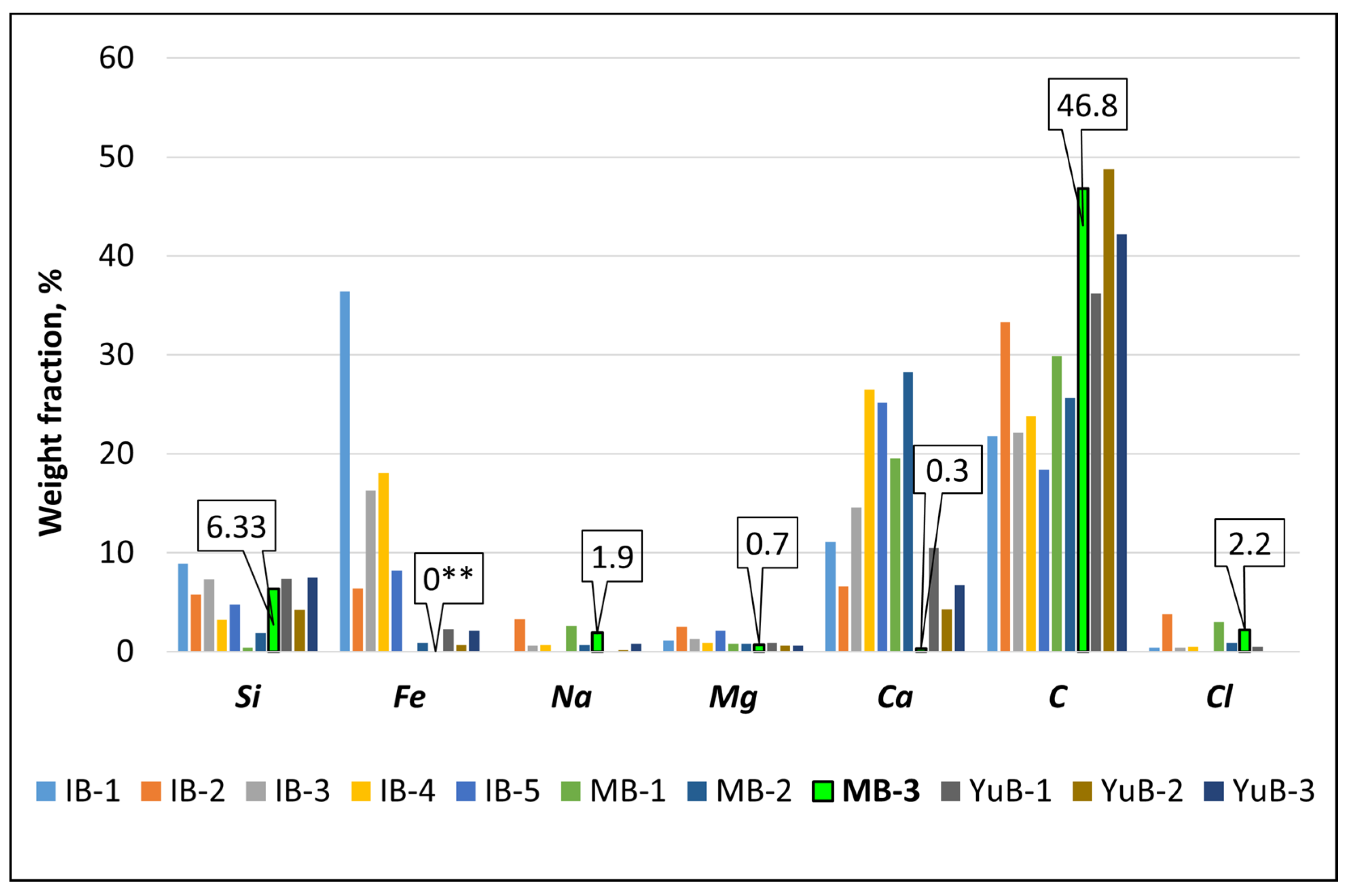
3.1.1. Elemental Composition of Rock Surfaces

The elemental composition and distribution in the rock surface layer was studied. The results of the EDX study, shown in Figure 2 and Figure 3 and in Table 1, allowed us to establish that elements are unevenly distributed in the rock surface layer. The major elements with the highest weight fraction were Ca, Fe, and Si (Figure 3). The example of MB shows that the content (in %) of these three elements on the rock surfaces in this bay varied in wide ranges: Si from 0.4 to 6.3, Ca from 0.3 to 19.5, and Fe from 0 to 0.9 (Table 1, Figure 3 and Figure 4). In IB, the Fe content was 3–18 times higher than in YuB, and in MB rocks, this element was virtually absent. The weight fraction of O and C was high, too (Figure 3). These elements are constituents of compounds, including such biogenic components of sedimentary rocks as carbonates (CaCO3) and oxides (SiO2).
Differences in the Fe, Ca, and Si content on the rocks’ surfaces were present not only in different bays but even within the same station and may be due to the physical and geographical characteristics of Sevastopol Bay, which is characterized by an indented coastline, as shown in Figure 1. Based on the elemental composition of the sampled rocks, the following conclusion was made about their origin: out of eleven samples under study, ten were of continental origin, and only one MB-3* rock sample was of marine origin (Table 1) since the inequality Na > Mg > Ca was observed in the elemental composition of its surface, namely 1.9 > 0.7 > 0.3 (Figure 4 and Table 1).

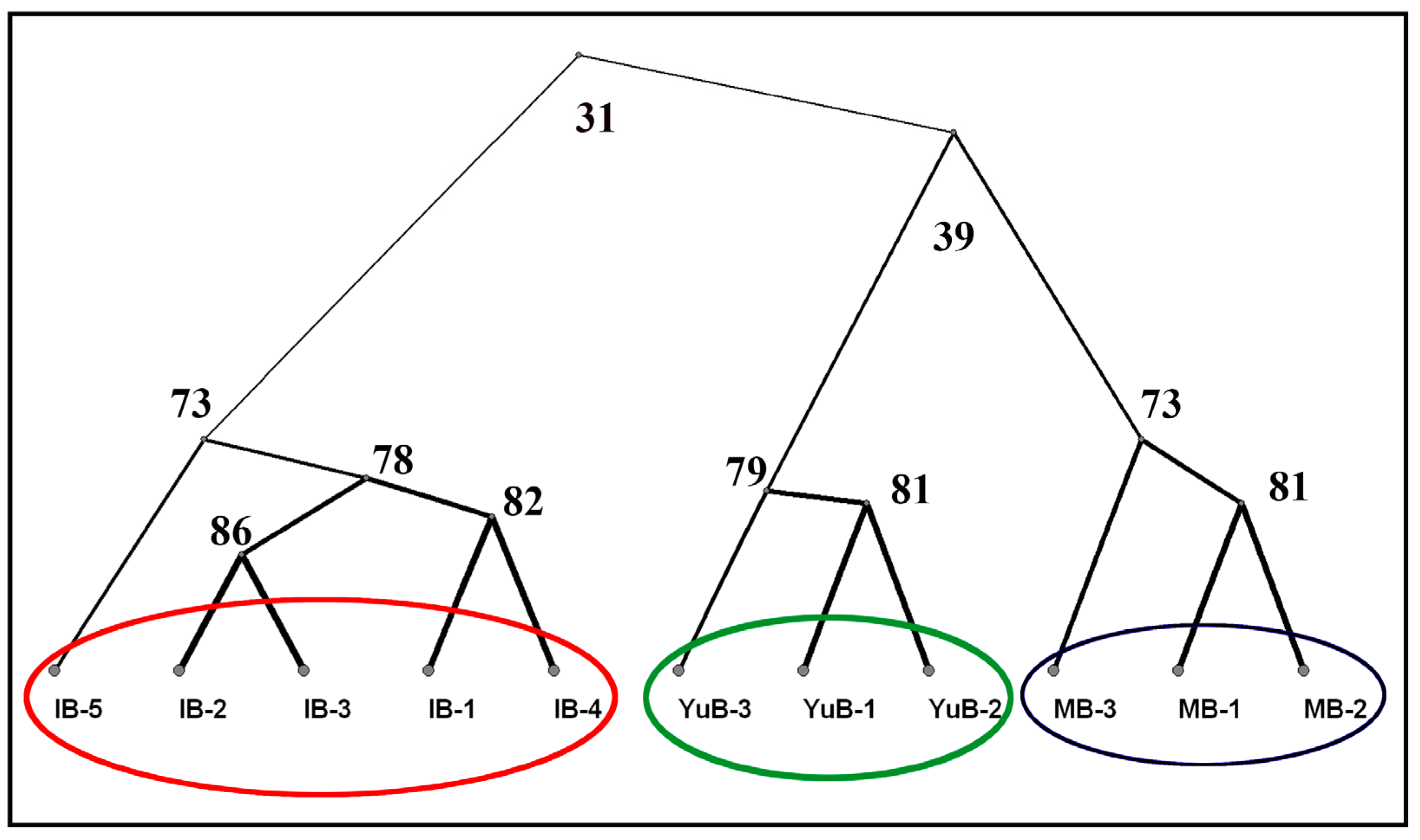
3.1.2. Cluster Analysis of the Rocks Similarity in Terms of Composition and Content of Chemical Elements on Their Surface

Cluster analysis (Figure 5a) of the element contents in the rock surface layer demonstrated a similarity of more than 68% among the clusters irrespective of the sampling area. Rocks from the same sampling stations were combined in clusters with a similarity of more than 80%, which reflects the influence of similar environmental conditions.
A dendrogram of similarity in the element contents and distribution in rocks is presented in Figure 5b. Ca and Fe are isolated in a separate cluster with a high similarity. Another separate cluster that combined C and O indicates the presence of either a biotic component or carbonates on the rock surface [48,49,50]. It should be noted that Si formed its own singleton cluster, which is 93% similar to that of other elements and has a similarity of 68% with the cluster of Ca and Fe. The high similarity and contents of these lithogenic elements determined the need for evaluating their impact on the diatom and cyanobacterial communities in the rock epilithon. The data obtained show the geochemical inhomogeneity of the habitat of benthic organisms, which inevitably affects the flows of matter, metabolism, and adaptation of organisms to specific geochemical conditions and is reflected in the mosaic pattern of epilithic communities.

3.2. Biotic Data Analysis

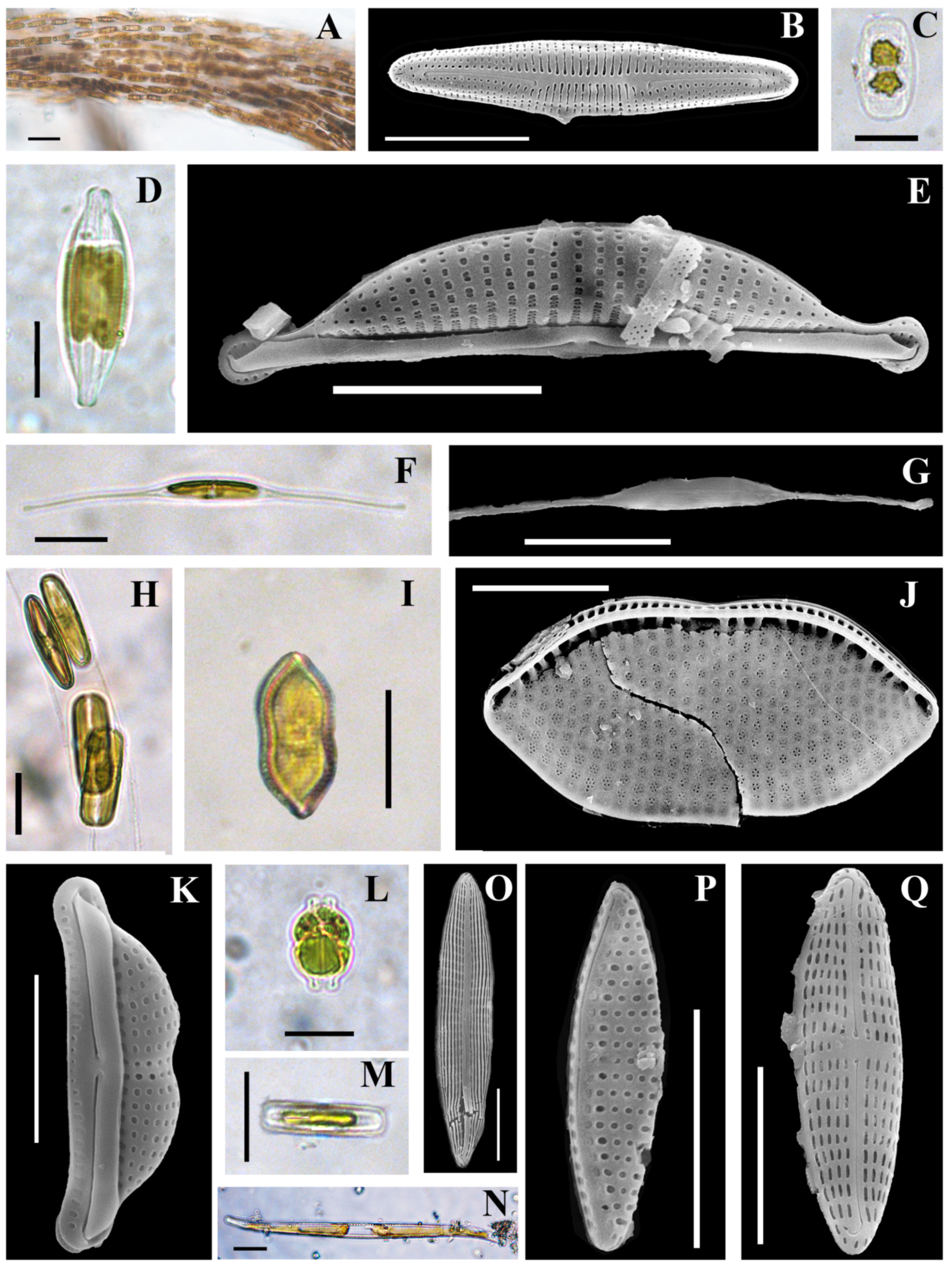
3.2.1. Composition of the Diatom and Cyanobacterial Communities on the Rock Surface

The species composition and abundance of diatoms and cyanobacteria were estimated in the epilithon of rocks from the three bays. In Table 2, a total of 63 identified diatom taxa are presented, with 31 of them being in Martynova Bay, 48 in Inkerman Bay, and 19 in Yuzhnaya Bay. Among them, eight species were common. Marine (26) and brackish-marine (18) species were dominant among diatoms. There were a few freshwater (seven) and freshwater-brackish (three) species, and twenty-eight cosmopolite species were detected. In MB, the abundance varied from 50,000 to 176,000 cells/cm2; in IB, it varied from 51,000 to 149,000 cells/cm2; and in YuB, it ranged from 14,000 to 28,000 cells/cm2 (Table 3).
Among cyanobacteria, 20 species were found which belong to 6 orders, 10 families, and 12 genera. Of these, fifteen species were found in Martynova Bay, seven in Inkerman Bay, and four in Yuzhnaya Bay. The highest species richness saturation was noted for the family Oscillatoriaceae (nine). Most of the species were marine (six) and halotolerant (eight), with the cosmopolite (ten) and boreal–tropical–notal (six) occurrence (Table 2). Some species of diatoms and cyanobacteria examined under the optical and scanning electron microscopes are presented in Figure 6 and Figure 7.
Eight diatom taxa, Berkeleya rutilans (Figure 6A,B), Undatella lineolata (Figure 6C), Halamphora coffeiformis (Figure 6D,E), Cylindrotheca closterium (Figure 6G), Psammodictyon panduriforme var. minor (Figure 6I,J), Navicula ramosissima (Figure 6M), Nitzschia sigma (Figure 6N), and Haslea subagnita (Figure 6O), and one cyanobacterium, Leptolyngbya fragilis (Figure 7C), were common to all bays. The numbers of species of diatoms/cyanobacteria that occurred in only one of the bays were 10/0 in YuB, 23/5 in IB, and 8/10 in MB.
The total abundance of diatoms varied from 14,000 to 176,000, and the total abundance of cyanobacteria varied from 12,000 to 1,198,000 cells/cm2 (Table 2 and Table 3). On the rock of marine origin (MB-3*), the highest abundance and number of marine diatom species (12) were noted. However, as shown in Table 3, the abundance of cyanobacteria on MB-3* was low (249,000 cells/cm2), and the number of cyanobacterial species was the smallest. The maximum abundance of cyanobacteria was noted on rocks of continental origin in Inkerman Bay (IB-3). The minimum abundance of diatoms and cyanobacteria was found on all rocks from Yuzhnaya Bay.
In the epilithon of MB, 31 diatom taxa were found, which belong to 23 genera (Table 2). The average abundance was 97,000 cells/cm2, with the dominance of the bentoplanktonic diatom Cylindrotheca closterium (Figure 6F,G) with N = 18,000 cells/cm2. The subdominant species were the small-celled Berkeleya rutilans (Figure 6A,B) and Parlibellus delognei (Figure 6H) with 12,000 and 14,000 cells/cm2, respectively. Cyanobacteria were represented by 15 species, with the dominance of Lyngbya confervoides and Potamolinea aerugineocaeruleum (Table 2). The total abundance of cyanobacteria varied from 74,760 to 380,740 cells/cm2.
In IB, diatoms were represented by 48 taxa from 30 genera. Their average abundance was 112,000 cells/cm2, with the dominance of Undatella lineolata (Figure 6C) at 22,000 cells/cm2. The subdominants were Berkeleya rutilans and Trachyneis aspera, with abundances of 15,000 and 14,000 cells/cm2, respectively. Here, seven cyanobacterial species were found, with the dominance of Kamptonema laetevirens and Phormidium holdenii (Figure 7B). The average total abundance was 628,000 cells/cm2, ranging from 158,000 to 119,8000 cells/cm2 (Table 3).
In YuB, 19 taxa of diatoms belonging to 14 genera were found. The diatom abundance was 19,000 cells/cm2, with the dominance of Cylindrotheca closterium (4000 cells/cm2), Amphora bigibba (Figure 6K,L), and Berkeleya rutilans (N = 2000 cells/cm2). There were only four species of cyanobacteria in YuB, with the average total abundance of 29,780 cells/cm2, which was the smallest in comparison with the other bays.
The quantitative characteristics of the benthic communities of diatoms and cyanobacteria were used in a cluster analysis of the structure of communities on different rocks, shown in Figure 8. Communities on rocks from the same station were allocated in clusters with a similarity above 80%, and the similarity of the species composition and abundance between the areas was 31–39%.
Consequently, the dendrogram of the rock similarity in terms of the elemental composition (Figure 5a) is almost identical to that of the community composition on the same rocks (Figure 8). However, the similarity of rocks between water areas was high, from 68 to 80%, and the similarity of benthic communities between water areas was low, only 31–39%.

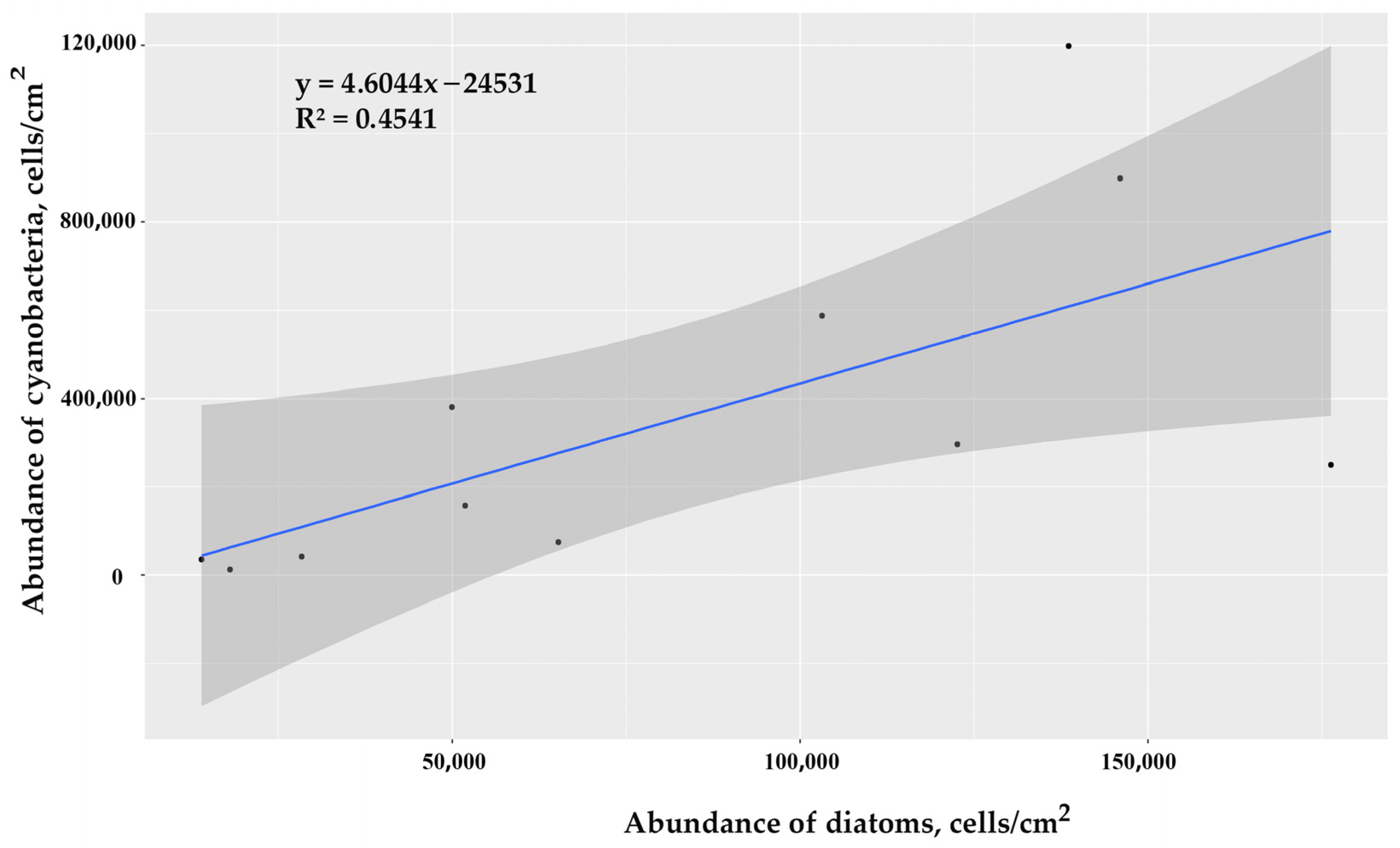
3.2.2. Correlations between the Abundance of Diatoms and Cyanobacteria in Epilithon Communities

Since diatoms and cyanobacteria form the basis of benthic communities, the biocenotic relationships between them are of particular interest. The correlation analysis showed a positive significant relationship between their abundances, shown in Figure 9. Similar responses of the photosynthetic microorganisms to environmental factors indicate their joint participation in the formation of benthic communities.

3.3. Biotic Factors in Relation to the Abiotic Variables

In the correlation analysis of the total average abundance of the diatom and cyanobacterial communities and the element contents in the rock surface layer for each rock, the correlation coefficient, shown in the Table 4, varied mostly in the ranges characteristic of weak and moderate associations (from −0.01 to ±0.5), which indicate the absence of a reliable correlation. When assessing the correlations of the population abundances with all detected elements, they showed considerable variability among sampling areas. The elemental composition of the rocks’ surfaces was similar across regions (Figure 5a), and therefore, the abundance of diatoms and cyanobacteria was more strongly influenced by other factors, e.g., substrate surface structure, water temperature, depth, illuminance, and grazing by phytophages [4].
Since the cluster analysis of elements in the surface layer of rocks demonstrated the greatest unevenness in the distribution of Si, Ca, and Fe, these three elements were selected to test the effect of their contents on the diatom and cyanobacterial populations’ abundances. The results of the correlation analysis for diatoms are shown in Figure 10a. The assessment of correlations showed that the largest number of diatom species (25 out of 39 included in the calculations) had noticeable to very high correlations with the Fe content. The highest association was noted for Bacillaria paxillifer and Navicula directa. However, despite this, the correlation coefficient between the iron content and the total number of diatoms was only 0.1 (Table 4). Only fourteen species of diatoms showed significant correlations with Si, with nine of them being negative. Almost no significant correlations were found for Ca. The results of correlation analysis of the element content and the abundance of cyanobacteria are shown in Figure 10b. The cyanobacterial species S. subsalsa, C. minutus, and P. minima from MB had strong positive correlations with Ca (rs = 0.71–0.73) and negative correlations with Si (rs = 0.71–0.73). Strong negative correlations were found between Fe and the dominant species L. confervoides (rs = −0.94) and P. aeruginaocaeruleum (rs = −0.60) (Figure 7M) from MB, and positive correlations were found with the dominants P. retzii (rs = 0.62), K. laetevirens (rs = 0.71), and P. holdenii (rs = 0.81) (Figure 7K) from IB. Despite the fact that these species were dominant, their dependence on Fe did not affect the results of the correlation analysis of the abundance of the entire cyanobacterial community. As shown in Table 4, the correlation coefficient between iron content and the abundance of the cyanobacterial community was r = −0.2.
For 11 diatom species, Achnanthes brevipes, Berkeleya rutilans, Caloneis liber, Halamphora coffeiformis (Figure 6D), Navicula palpebralis, Navicula ramosissima (Figure 6M), Nitzschia sigma (Figure 6N), Seminavis ventricosa, Tabularia tabulata, Trachyneis aspera, and Undatella lineolata, and one cyanobacterium, Leptolyngbya fragilis (Figure 7B,C), no correlations of the abundance with the element contents were noted (rs < ±0.5)

4. Discussion

Our study is based on the hypothesis of the significant effect of the rock surface elemental composition on the formation of benthic diatoms and cyanobacterial communities and the biocenotic relationships that arise in this interaction. It is known that the rock surface composition reflects the chemical regime of the aquatic environment, and the chemical composition of the seabed, in turn, affects the seawater composition [29], which is consistent with the data obtained. For example, the main mass of carbonates in water is associated with the dissolution of carbonate rocks. In our study, it was shown that the elemental composition of the rock surfaces in three bays differed from each other mainly in the distribution of such elements as Ca, Fe, and Si, which had the highest weight and were dependent on the aquatic environment. This may be due to the physiographic features of Sevastopol Bay, which has a rugged coastline which provides zoning and differences in the thermohaline and hydrochemical characteristics [34].
The registered composition of diatom and cyanobacterial communities in the epilithon of Sevastopol Bay was typical for the Black Sea. Cyanobacteria on rock substrates were not very diverse, and all the species had been found on the Crimean coast earlier [13,52]. The most abundant cyanobacteria in the epilithon samples under consideration were those of the Lyngbya and Phormidium genera, which are common on rocks [32,53]
For most of the diatom species abundance, noticeable to very high correlations with Fe were found, which are due to the “iron-loving” nature of these microalgae. From data from the literature, the presence of ferritin in diatoms is known, which gives them an advantage over algae that do not contain ferritin [8,30]. It is a very interesting property for biotechnology. In the conditions of Fe deficiency, diatoms have several strategies of reducing the need for this element [54,55,56,57,58], e.g., by forming siderophores—the iron-storing compounds [8]. In addition, adaptations at the genome level may evolve under some circumstances, e.g., in relation to iron bioavailability [59]. In a review paper on the effect of Fe on phytoplankton [60], the authors point out that an increase in the content of iron in water in natural habitats and in cultures positively affects the growth rate, species diversity, and abundance of diatoms. Many diatom species contain high-affinity ferric reductases that dissociate iron III from ligands, multicopper oxidases that oxidize Fe2+ to Fe3+, and permease systems that receive iron (II) for translocation across membranes [8,61].
The strong negative correlation of Fe with the dominant cyanobacteria from MB that is deficient in iron and the strong positive correlation with the dominants from the iron-rich IB indicate the sensitivity of certain cyanobacterial species to Fe availability. It is known that both the excess and deficiency of iron result in severe abnormal changes in microbial physiology through the generation of reactive oxygen species and free hydroxyl radicals, which ultimately cause cell death [62]. The color of cyanobacteria in the epilithon of MB (Figure 7C,E,F,J,L,M) was more bluish than in species from IB. This may be related to the decrease in cellular pigmentation during iron deprivation causing a blue shift in the main red absorption band of chlorophyll a and inducing the iron acquisition process [63]. Iron is also crucial for other essential processes like photosynthesis and respiration [62]. This apparently demonstrates the ability of the species to deposit Fe compounds when iron is deficient in the environment by regulating their metabolism through reducing the need for iron or forming siderophores [64] similar to those in diatoms [8,30]. Apart from siderophores, ferric citrate is another simple compound that has iron-chelating properties and is transported into the cells of cyanobacteria [65]. On the other hand, in the iron homeostasis in K. laetevirens, P. holdenii, and P. retzii, the dominant cyanobacteria in IB, where there is a high concentration of Fe in the water and sediments, the central role is likely played by bacterioferritin, similar to that in the model cyanobacteria Synechocystis sp. [66]. In recent experiments with cultures of cyanobacteria, it was shown that they were able to extract Fe from the solid magnetite phase [67]. Thus, the epilithic cyanobacteria found in IB, K. laetevirens, P. holdenii, and P. retzii [47], may also be able to extract iron from the rock surface.
Diatoms are one of the main consumers of Si in seawater [7,10,26]. However, the correlation coefficients of the diatom abundance and the content of this element in the rock surface layer did not exceed 0.7, and in many species, there was no correlation at all. This can be explained by the fact that diatoms can consume only an aqueous solution of silicic acid Si(OH)4, which is transformed to SiO2 after accumulation in special organelles [7]. It follows from this that diatoms do not consume silicon from the surface of rocks but only from the aquatic environment.
Diatoms are organisms that concentrate Si and Fe [8,27], which are cofactors of many enzymes and affect the formation of nucleic acids [16]. This may be the reason for the largest abundance of diatoms in Inkerman Bay, which is characterized by a high concentration of iron ions in the water [47] and, consequently, the high levels of this element in the seabed [10]. It is known that diatoms are the only microalgae capable of assimilating iron from colloidal forms [10]. It is a very important property for biotechnology.
Ca promotes the secretion of polysaccharides, which ensure the adhesion of attached diatoms to the substrate and the formation of colonies [31]. However, no significant correlation was found between Ca and the diatom abundance. In cyanobacteria, a strong correlation was found between the abundance of S. subsalsa, C. minutus, and P. minima on rocks of continental origin in Martynova Bay and Ca content in the rock surface, which may indicate the ability of these species to biocalcify.
The lack of significant correlations between the element contents and the average abundance of diatoms and cyanobacterial communities can be explained by the stronger influence of other factors on the formation of microphytobenthos communities. For example, there is information about the absence of any significant effect of the chemical composition of rocks on the biomass of benthic cyanobacteria and diatoms in river mouths [68]. As the author points out, the position of the substrates relative to the flow and the flow intensity are much more important factors, and the insignificance of the rock composition effect is accounted for by the low rate of dissolution of the rocks in the experiment. In addition, there are not only individual, but also synergistic effects of elements on the development of diatoms and cyanobacteria. For example, it was found in [69,70] that under Fe limitation, specific growth rates and cell sizes of cultivated diatoms decreased, and after an increase in the Cu content, their growth rates again increased. In the work [71] on four cultivated diatoms, it was shown that by varying the ratios of Na, Ca, and Mg, one can tune the growth rate of a species and achieve its optimum.
In the literature, there are data on the effect of the substrate area and surface roughness on the colonization by benthic organisms. It was shown that diatoms and cyanobacteria prefer substrates with high roughness and an uneven surface [22]. Wahl (1989) described in detail the model of substrate colonization by benthic organisms, in which it was noted that the colonization of surfaces by diatoms may be preceded by the growth of bacteria [72]. Therefore, the elemental composition of rock substrates can have both direct and indirect effects on communities since biofilms always contain organisms capable of leaching metal ions [33].
In our study, the similarity of the elemental composition of rocks in the same area was as high as above 80%, and exactly the same result was obtained when comparing communities in the same area. At the same time, the similarity of rocks from different regions was also quite strong, from 68 to 80%, whereas communities from different areas were similar only by 31–39%. This suggests that the elemental composition was not a decisive factor, and communities are strongly affected by other environmental factors specific to each area. The data obtained show the geochemical inhomogeneity of the habitat of benthic organisms, which is expressed in the mosaic pattern of the benthic communities but is closely associated with a multitude of other abiotic factors inherent to the study areas. In [73], the authors emphasize that for coastal estuarine ecosystems with high environmental variability, multivariate analysis is required, which would help achieve an integral view on the strength and direction of changes of the environmental variables. At this stage, we have revealed only the effect of the elemental composition of the substrate surface, but in the future, multivariate studies of the influence of environmental conditions on microphytobenthos communities are needed. This is especially relevant to estuarine ecosystems, where one of the main factors affecting microphytobenthos is freshwater runoff, whose effect can be uneven, as demonstrated in the above-mentioned work. Perhaps for this reason, the similarity of communities across the areas was very low: it could be affected by the Chernaya river inflow in IB, sewage discharge in YuB, and seawater recharge in MB.
As for other factors affecting microphytobenthos communities, authors often single out such a factor as seasonality [74,75,76,77]. The effect of seasonality was noted also in our previous works on the Black Sea [4,78,79]. This factor encompasses the totality of environmental conditions, such as temperature, salinity, illumination, storms, and many others. Earlier, for the microphytobenthos of the Sevastopol coast, we established a strong correlation between the abundance of microalgae and water temperature [80].
The effect of salinity on microphytobenthos is clear in the example of Sivash Bay in Crimea, in which salinity varied from 30 to 100 psu [81]. It was revealed that the salinity variation is not the only or major factor that determines the species composition of the microphytobenthos. Other factors (total suspended matter concentration, etc.) can play an equally important role. However, quantitative indicators of microphytobenthos may positively correlate with salinity.
Most of the species found on the rocks are cosmopolites, i.e., they are common in the world and live under a wide range of environmental conditions. As for diatoms, for the majority of dominants and species encountered in all regions, a correlation with the elemental composition of the rocks’ surfaces was not found. These facts apparently explain why our hypothesis about the effect of the rock surface composition on benthic communities has not been confirmed to as large an extent as expected. It is interesting to note that the number of species found on rocks from YuB in this study was 1.5–2-fold lower than those in IB and MB. The chemogenic origin of the rocks in Yuzhnaya Bay is known, while the limestones in Martynova and Inkerman bays are of biogenic origin [35]. It is also known that more than 80% of carbon-bearing rocks are of precisely biogenic origin. Often, these rocks are formed as a result of the vital activity of cyanobacterial communities and other organisms. Among them are deposits of phytogenic origin—formed by carbonate excretions of microalgae [35]. This may explain the high content of carbon in the elemental composition of rock surfaces and the species-rich communities of microphytobenthos in IB and MB, where the geology of rocks indicates historically more favorable conditions for the development of communities than in YuB, where conditions favored chemogenic processes of rock formation. This study highlights the manifestations of the biogeoecology laws [18] at the biocenotic and population levels. The data obtained show that the mosaic pattern of the rock epilithon is associated with the inhomogeneity of the geochemical environment in the water area. The equal involvement of both cyanobacteria and diatoms was noted in the formation of fouling communities, which may indicate their mutually beneficial interactions and similar response to environmental factors. For example, cyanobacteria produce cellulose, which serves as a defense against stress factors for other organisms [82]. Such connections may have developed historically owing to co-evolution [30]. The work [83] shows the ability of microcommunities to leach metal ions from rock, which are then accumulated in cellulose biofilm, and the effects of rock on the metabolism of microorganisms. These biochemical features emphasize the interrelation between the structure of fouling communities and biomineralization. This means that geomicrobiological studies are important not only in studying ecology and biodiversity but also for solving problems of the impact of microorganisms on the geological environment [84]. We suggest relying on a combination of biology and geoecology methodologies to study different benthic communities to isolate members of the community for different operational purposes (in biotechnology, biomining, etc.).

5. Conclusions

As part of the present study, the geochemical inhomogeneity of the habitat of benthic organisms and the mosaic pattern of epilithic communities on rocky beds have been shown. The data obtained have proven the close connection between the formation of sedimentary rocks and the complex of abiotic factors in the aquatic (marine) environment, which affect the elemental composition of the rock surface. It was shown that the rock surface elemental composition lacked influence on the epilithon communities but was present for populations of some diatom and cyanobacterial species. Perhaps this result was due to the fact that most of the species found on the rocks are cosmopolites. At the same time, we have found several diatom and cyanobacterial species whose population abundance significantly correlated with the iron and calcium contents in the rock surfaces. This expands our knowledge of species that can be used in cultivation and in search of other biotechnologically valuable microbial species. The results have shown the mutually beneficial interactions of cyanobacteria and diatoms in the formation of communities on rocks and proven the relationship between species diversity and the predominance of chemogenic or biogenic processes in the rock formation in different areas.

Author Contributions

A.B. and L.R. developed the concept and methodology of this study; D.L. conducted sampling; A.B. and E.M. provided microalgae sample processing; A.B. and D.L. prepared diatom samples and obtained SEM images; L.R. and D.B. identified diatom species; E.M. identified cyanobacterial species; A.B., E.M., D.B. and S.B. conducted data formal analysis; A.B. and S.K. conducted statistical analysis. All authors took part in writing, reviewing, and editing this manuscript. All authors have read and agreed to the published version of the manuscript.

Funding

This research was funded by state assignment No. 121030300149-0 from A.O. Kovalevsky Institute of Biology of the Southern Seas of RAS.

Institutional Review Board Statement

Not applicable.

Informed Consent Statement

Not applicable.

Data Availability Statement

All data used in this study are available upon request from the corresponding author.

Acknowledgments

We would like to express our gratitude to V.N. Lishaev for the assistance in obtaining SEM images and A.A. Pugach for the assistance in processing the results of this research using modern software. We also thanks to Israeli Ministry of Aliyah and Integration for support.

Conflicts of Interest

The authors declare no conflict of interest.

References

  1. Vernadsky, V.I. Living Matter; Nauka: Moskow, Russia, 1978; 358p. (In Russian) [Google Scholar]
  2. Cattaneo, A. Periphyton. In Bitton, Encyclopedia of Environmental Microbiology; John Wiley & Sons: Hoboken, NJ, USA, 2002; pp. 2357–2367. [Google Scholar]
  3. Durr, S.; Thomason, J.C. Biofouling, 1st ed.; Wiley-Blackwell: Singapore, 2010; 429p. [Google Scholar]
  4. Ryabushko, L.I. Microphytobenthos of the Black Sea; Gaevskaya, A.V., Ed.; EKOSI-Gidrofizica: Sevastopol, Ukraine, 2013; 416p. (In Russian) [Google Scholar]
  5. Ryabushko, L.I.; Begun, A.A. Diatoms of Microphytobenthos of the Sea of Japan; N. Orianda: Simferopol, Ukraine, 2015; Volume 1, 288p. (In Russian) [Google Scholar]
  6. Anderson, O.R. Marine and estuarine natural microbial biofilms: Ecological and biogeochemical dimensions. AIMS Microbiol. 2016, 2, 304–331. [Google Scholar] [CrossRef]
  7. Epple, M. Biomineralization: From Biology to Biotechnology and Medical Application; Baeuerlein, E., Ed.; Wiley-VCH: Weinheim, Germany, 2001; 337p. [Google Scholar]
  8. Groussman, R.D.; Parker, M.S.; Armbrust, E.V. Diversity and evolutionary history of iron metabolism genes in diatoms. PLoS ONE 2015, 10, e0129081. [Google Scholar] [CrossRef] [Green Version]
  9. Sigler, W.; Bachofen, R.; Zeyer, J. Molecular characterization of endolithic cyanobacteria inhabiting exposed dolomite in central Switzerland. Environ. Microbiol. 2003, 5, 618–627. [Google Scholar] [CrossRef] [PubMed] [Green Version]
  10. Tréguer, P.J.; De La Rocha, C.L. The world ocean silica cycle. Ann. Rev. Mar. Sci. 2013, 5, 477–501. [Google Scholar] [CrossRef] [PubMed]
  11. Gebeshuber, I.C. Biomineralization in marine organisms. Chapter 58, Part XI (Marinebiotechnological products in industrial application). In Springer Handbook of Marine Biotechnology; Kim, S.-K., Ed.; Springer: Berlin/Heidelberg, Germany, 2015; pp. 1279–1300. ISBN 978-3-642-53970-1. [Google Scholar] [CrossRef]
  12. Dang, H.; Lovell, C.R. Microbial surface colonization and biofilm development in marine environments. Microbiol. Mol. Biol. Rev. 2016, 80, 91–138. [Google Scholar] [CrossRef] [Green Version]
  13. Ryabushko, L.; Miroshnichenko, E.; Blaginina, A.; Shiroyan, A. Diatom and cyanobacteria communities on artificial polymer substrates in the Crimean coastal waters of the Black Sea. Mar. Pollut. Bull. 2021, 169, 112521. [Google Scholar] [CrossRef] [PubMed]
  14. Benzerara, K.; Skouri-Panet, F.; Li, J.; Férard, C.; Gugger, M.; Laurent, T.; Couradeau, E.; Ragon, M.; Cosmidis, J.; Menguy, N.; et al. Intracellular Ca-carbonate biomineralization is widespread in cyanobacteria. Proc. Natl. Acad. Sci. USA 2014, 111, 10933–10938. [Google Scholar] [CrossRef]
  15. Terekhov, E.P.; Mozherovsky, A.V.; Barinov, N.N. Patent. 2665152 Russian Federation, IPC G01N 33/24 Method for Determining the Genesis of Marine Sedimentary Deposits. Available online: https://i.moscow/patents/ru2665152c1_20180828 (accessed on 21 June 2021). (In Russian).
  16. Agarkova-Lyakh, I.V.; Lyakh, A.M. Coast state and the exogenous geological processes from Cape Konstantinovkiy to Cape Vinogradny on the south-western coast of Crimea. Sci. Notes V.I. Vernadsky Crime. Fed. Univ. 2019, 5, 118–133. (In Russian) [Google Scholar]
  17. Romanyuk, O.S. Compile a Cadastre of the Surface part of the Crimean Coast in Relation to a Scale of 1:200,000: Report. Crimean Hydrogeological Expedition; Institute of Mineral Resources: Simferopol, Ukraine, 1988. (In Russian) [Google Scholar]
  18. Kovalsky, V.V. Geochemical Ecology; Nauka: Moscow, Russia, 1974; 300p. (In Russian) [Google Scholar]
  19. Gorbushina, A.A. Life on the rocks. Environ. Microbiol. 2007, 9, 1613–1631. [Google Scholar] [CrossRef]
  20. Stephenson, W. Experimental studies on the ecology of intertidal environments at Heron Island. II. The effect of substratum. Aust. J. Mar. Freshw. Res. 1961, 12, 132–188. [Google Scholar] [CrossRef]
  21. Ledger, M.E.; Hildrew, A.G. Temporal and spatial variation in the epilithic biofilm of an acid stream. Freshw. Biol. 1998, 40, 655–670. [Google Scholar] [CrossRef]
  22. Hutchinson, N.; Nagarkar, S.; Aitchison, J.C.; Williams, G.A. Microspatial variation in marine biofilm abundance on intertidal rock surfaces. Aquat. Microb. Ecol. 2006, 42, 187–197. [Google Scholar] [CrossRef] [Green Version]
  23. Rozhanskaya, L.I. The metals and its radionuklids composition in the hydrobionts of pelagial. In Hemoradioecologia of Pelagial and Bental; Naukova Dumka: Kiev, Ukraine, 1974; pp. 40–55. (In Russian) [Google Scholar]
  24. Volcani, B.E. Role of Silicon in Diatom Metabolism and Silicification. In Biochemistry of Silicon and Related Problems, Nobel Foundation Symposia, Lidingo, 1977; Bendz, G., Lindqvist, I., Runnström-Reio, V., Eds.; Springer: Boston, MA, USA, 1978; Volume 40. [Google Scholar] [CrossRef]
  25. Ktitorova, E.N.; Lukyanov, Y.S.; Ivanova, K.K. Mass Concentration of Silicon in Sea Water Photometric Measurement Technique in the form of a Blue Form of Molybdosilicic Acid; Federal Service for Hydrometeorology and Environmental Monitoring: Moscow, Russia, 2010; 14p. (In Russian)
  26. Paasche, E.; Kristiansen, S. Silicon and ecology of marine plankton diatoms. II. Silicate-uptake kinetics in five diatom species. Mar. Biol. 1973, 19, 262–269. [Google Scholar] [CrossRef]
  27. Gordova, V.S.; Sapozhnikov, S.P.; Sergeeva, V.E.; Karyshev, P.B. The basics of biosilicification (literature review). Bull. Chuvash Univ. 2013, 3, 401–409. (In Russian) [Google Scholar]
  28. Dyakov, Y.T. (Ed.) Botany: Algology and Mycology Course; Moscow State University Press: Moscow, Russia, 2007; 559p. (In Russian) [Google Scholar]
  29. Alekin, O.A.; Lyahin, Y.I. Himiya Okeana; Gidrometeoizdat: Leningrad, Russia, 1984; 344p. (In Russian) [Google Scholar]
  30. Falciatore, A.; Jaubert, M.; Bouly, J.-P.; Bailleul, B.; Mockc, T. Diatom molecular research comes of age: Model species for studying phytoplankton biology and diversity. Plant Cell 2020, 32, 547–572. [Google Scholar] [CrossRef]
  31. Ohgai, M.; Matsui, T.; Okuda, T.; Tsukahara, H. Fine structure of adhesive parts of diatoms (Bacillariaceae) and adhesive mechanism. J. Shimonoseki Univ. Fish. 1984, 33, 27–35. [Google Scholar]
  32. Kala, R.; Pandey, V.D. Variable growth patterns of epilithic cyanobacteria on different rocks. In Recent Advancements in Sciences with Special Reference to Himalaya; Gautam, A.S., Ed.; Ancient Publishing House: Delhi, India, 2020; pp. 117–121. [Google Scholar]
  33. Zaets, I.; Burlak, O.; Rogutskyy, I.; Vasilenko, A.; Mytrokhyn, O.; Lukashov, D.; Foing, B.; Kozyrovska, N. Bioaugmentation in growing plants for lunar bases. Adv. Space Res. 2011, 47, 1071–1078. [Google Scholar] [CrossRef]
  34. Ivanov, V.A.; Ovsyany, E.I.; Repetin, L.N. Hydrological and Hydrochemical Regime of the Sevastopol Bay and Its Changes under the Influence of Climatic and Anthropogenic Factors; MGI NAS of Ukraine: Sevastopol, Ukraine, 2006; 90p. (In Russian) [Google Scholar]
  35. Moiseev, A.S. Hydrogeological sketch of the city of Sevastopol and its environs. In All-Union Geological Exploration Association of People’s Commissariat for Heavy Industry of the USSR; State Scientific and Technical Geological Exploration Publishing House: Moscow, Russia, 1932; Volume 137, 48p. [Google Scholar]
  36. Graham, A.A.; McCaughan, D.J.; McKee, F.S. Measurement of surface area of stones. Hydrobiologia 1988, 157, 85–87. [Google Scholar] [CrossRef]
  37. Srebrodolsky, B.I. Secrets of Seasonal Minerals; Nauka: Moscow, Russia, 1989; 144p. [Google Scholar]
  38. Simonsen, R. Untersuchungen zur Systematik und Okologie der Bodendiatomeen der Westlichcn Ostsee. Int. Rev. Ges. Hydrobiol. Beih. 1962, 1, 9–144. [Google Scholar]
  39. Smith, W. A Synopsis of the British Diatomaceae; Smith and Beck, Pub.: London, UK, 1853; Volume 1, 89p. [Google Scholar]
  40. Proschkina-Lavrenko, A.I. (Ed.) Diatom Analysis. Guide to Fossil and Modern Diatoms; Book. 3; Gosgelioizdat: Leningrad, Russia, 1950; 398p. (In Russian) [Google Scholar]
  41. Proschkina-Lavrenko, A.I. Diatoms of the Black Sea Benthos; USSR Academy of Sciences Publishing House: Leningrad, Russia, 1963; 243p. (In Russian) [Google Scholar]
  42. Hendey, N.I. An Introductory Account of the Smaller Algae of British Coastal Waters, Part V: Bacillariophyceae (Diatoms); Ministry of Agriculture, Fisheries and Food, Fishery Investigations: London, UK, 1964; 317p. [Google Scholar]
  43. Guslyakov, N.E.; Zakordonets, O.A.; Gerasimyuk, V.P. Atlas of Diatoms of the Benthos of the Northwestern Part of the Black Sea and Adjacent Water Bodies; Naukova Dumka: Kyiv, Ukraine, 1992; 112p. (In Russian) [Google Scholar]
  44. Ryabushko, L.I.; Begun, A.A. Diatoms of Microphytobenthos of the Sea of Japan (Synopsis and Atlas); PK “KIA”: Sevastopol, Ukraine, 2016; Volume 2, 324p. (In Russian) [Google Scholar]
  45. Komárek, J.; Anagnostidis, K. Cyanoprokaryota. 1 Teil: Chroococcales. Süßwasserflora von Mitteleuropa; Bd 19/1; Spektrum Akademischer Verlag: Heidelberg, Germany; Berlin, Germany, 1999; 523p. [Google Scholar]
  46. Witkowski, A.; Lange-Bertalot, H.; Metzeltin, D. Diatom Flora of Marine Coasts I. Iconographia Diatomologica, Vol. 7: Diversity—Taxonomy—Identification; Lange-Bertalot, H., Ed.; A. R. G. Gantner Verlag K. G.: Koningstein, Germany, 2000; 925p. [Google Scholar]
  47. Komárek, J.; Anagnostidis, K. Cyanoprokaryota. 2 Teil: Oscillatoriales. Süßwasserflora von Mitteleuropa; Bd 19/2; Elsevier GmbH: München, Germany, 2005; 759p. [Google Scholar]
  48. Komárek, J. Cyanoprokaryota. 3 Teil: Heterocytous genera. Süßwasserflora von Mitteleuropa; 19/3; Springer: Berlin/Heidelberg, Germany, 2013; 1132p. [Google Scholar] [CrossRef]
  49. Guiry, M.D.; Guiry, G.M. AlgaeBase; World-Wide Electronic Publication, National University of Ireland: Galway, Ireland, 2021; Available online: http://www.algaebase.org (accessed on 21 June 2021).
  50. Whitton, B.A.; Roth, E.; Friedrich, G. Use of Algae for Monitoring Rivers; Institut für Botanik Univ. Press: Innsbruck, Austria, 1991; 196p. [Google Scholar]
  51. Sörensen, T. A new method of establishing group of equal amplitude in plant sociology based on similarity of a species content and its application to analysis of the vegetation on Dannish commons. K. Dan. Vidensk. Selsk. Biol. Meddelelser 1948, 85, 1–34. [Google Scholar]
  52. Vinogradova, O.; Bryantseva, Y. Taxonomic revision of the species composition of Cyanobacteria/Cyanoprokaryota of the Ukrainian coast of the Black Sea. Algologia 2017, 27, 436–457. [Google Scholar] [CrossRef] [Green Version]
  53. Whitton, B.A. Ecology of Cyanobacteria II: Their Diversity in Space and Time; Springer Science+Business Media, B.V.: Berlin, Germany, 2012; 760p. [Google Scholar]
  54. Strzepek, R.F.; Hunter, K.A.; Frew, R.D.; Harrison, P.J.; Boyd, P.W. Iron-Light Interactions Differ in Southern Ocean Phyto-plankton. Limnol. Oceanogr. 2012, 57, 1182–1200. [Google Scholar] [CrossRef] [Green Version]
  55. Strzepek, R.F.; Boyd, P.W.; Sunda, W.G. Photosynthetic Adaptation to Low Iron, Light, and Temperature in Southern Ocean Phytoplankton. Proc. Natl. Acad. Sci. USA 2019, 116, 4388–4393. [Google Scholar] [CrossRef] [Green Version]
  56. Moreno, C.M.; Lin, Y.; Davies, S.; Monbureau, E.; Cassar, N.; Marchetti, A. Examination of Gene Repertoires and Physiological Responses to Iron and Light Limitation in Southern Ocean Diatoms. Polar Biol. 2018, 41, 679–696. [Google Scholar] [CrossRef]
  57. Alderkamp, A.-C.; van Dijken, G.L.; Lowry, K.E.; Lewis, K.M.; Joy-Warren, H.L.; van de Poll, W.; Laan, P.; Gerringa, L.; Delmont, T.O.; Jenkins, B.D.; et al. Effects of Iron and Light Availability on Phytoplankton Photosynthetic Properties in the Ross Sea. Mar. Ecol. Prog. Ser. 2019, 621, 33–50. [Google Scholar] [CrossRef] [Green Version]
  58. Coale, T.H.; Bertrand, E.M.; Lampe, R.H.; Allen, A.E. Molecular Mechanisms Underlying Micronutrient Utilization in Marine Diatoms. In The Molecular Life of Diatoms; Falciatore, A., Mock, T., Eds.; Springer International Publishing: Cham, Switzerland, 2022; pp. 567–604. [Google Scholar]
  59. Peers, G.; Price, N.M. Copper-containing plastocyanin used for electron transport by an oceanic diatom. Nature 2006, 441, 341–344. [Google Scholar] [CrossRef] [PubMed]
  60. Bazzani, E.; Lauritano, C.; Saggiomo, M. Southern Ocean Iron Limitation of Primary Production between Past Knowledge and Future Projections. J. Mar. Sci. Eng. 2023, 11, 272. [Google Scholar] [CrossRef]
  61. Street, J.H.; Paytan, A. Iron, Phytoplankton Growth, and the Carbon Cycle. In Metal Ions in Biological Systems, Vol. 43—Biogeochemical Cycles of Elements; CRC Press: Boca Raton, FL, USA, 2005; pp. 153–193. [Google Scholar]
  62. Kaushik, M.S.; Singh, P.; Tiwari, B.; Mishra, A.K. Ferric uptake regulator (FUR) protein: Properties and implications in cyanobacteria. Ann. Microbiol. 2015, 66, 61–75. [Google Scholar] [CrossRef]
  63. Cheng, D.; He, Q. PfsR is a key regulator of iron homeostasis in Synechocystis PCC 6803. PLoS ONE 2014, 9, e101743. [Google Scholar] [CrossRef] [Green Version]
  64. Chakraborty, S.; Verma, E.; Singh, S.S. Cyanobacterial siderophores: Ecological and biotechnological significance. Ch. 19. In Cyanobacteria. From Basic Science to Applications; Mishra, A.K., Tiwari, D.N., Rai, A.N., Eds.; Academic Press, Elsevier Inc.: Cambridge, MA, USA, 2019; pp. 383–397. [Google Scholar] [CrossRef]
  65. Yue, W.W.; Grizot, S.; Buchanan, S.K. Structural evidence for iron-free citrate and ferric citrate binding to the TonB-dependent outer membrane transporter FecA. J. Mol. Biol. 2003, 332, 353–368. [Google Scholar] [CrossRef]
  66. Keren, N.; Aurora, R.; Pakrasi, H.B. Critical roles of bacterioferritins in iron storage and proliferation of cyanobacteria. Plant Physiol. 2004, 135, 1666–1673. [Google Scholar] [CrossRef] [Green Version]
  67. Huang, W.; Wang, T.; Perez-Fernandez, C.; DiRuggiero, J.; Kisailus, D. Iron acquisition and mineral transformation by cyanobacteria living in extreme environments. Mater. Today Bio 2022, 17, 100493. [Google Scholar] [CrossRef]
  68. Bergey, E.A. Does rock chemistry affect periphyton accrual in streams? Hydrobiologia 2008, 614, 141–150. [Google Scholar] [CrossRef] [Green Version]
  69. Amber, A.; Lapi, S.; Ruth, T.; Maldonado, M. The Effects of Cu and Fe Availability on the Growth and Cu:C Ratios of Marine Diatoms. Limnol. Oceanogr. 2008, 53, 2451–2461. [Google Scholar] [CrossRef] [Green Version]
  70. Mora, K.; Echeveste, P. Physiological responses of Humboldt current system diatoms to Fe and Cu co-limitation. Mar. Environ. Res. 2023, 187, 105937. [Google Scholar] [CrossRef]
  71. Rao, V.N.R.; Kannan, V.; Duraisamy, A. Relative concentrations of Na, Ca and Mg for growth of some diatoms. Proc. Indian Acad. Sci. 1981, 90, 169–175. [Google Scholar] [CrossRef]
  72. Wahl, M. Marine epibiosis. I. Fouling and antifouling: Some basic aspects. Mar. Ecol. Prog. Ser. 1989, 58, 175–189. [Google Scholar] [CrossRef] [Green Version]
  73. López-Mejía, D.; Gutiérrez-Mendieta, F.J.; Zavala-Hurtado, J.A.; Siqueiros-Beltrones, D.A.; Varona-Cordero, F.; Herrera-Moro Chao, I.R. Physicochemical gradients in a coastal lagoon from the southern Gulf of Mexico; a multivariate approach. Reg. Stud. Mar. Sci. 2022, 52, 102284. [Google Scholar] [CrossRef]
  74. McIntire, C.D.; Overton, W.S. Distributional patterns in assemblages of attached diatoms from Yaquina Estuary, Oregon. Ecology 1971, 52, 758–777. [Google Scholar] [CrossRef]
  75. Cibic, T.; Blasutto, O.; Falconi, C.; Umani, F. Microphytobenthic biomass, species composition and nutrient availability in sublittoral sediments of the Gulf of Trieste (northern Adriatic Sea). Estuar. Coast. Shelf Sci. 2007, 75, 50–62. [Google Scholar] [CrossRef]
  76. Farhadian, O.; Pouladi, M.; Vazirizadeh, A.; Sedaghat, R. A Study of Diatoms Seasonal Distribution and Biodiversity in Helleh River Estuary, Persian Gulf. Environ. Stud. Persian Gulf 2015, 2, 32–44. [Google Scholar]
  77. Maraşlıoğlu, F.; Donmez, M. Seasonal Distribution of Epipelic Diatom Assemblages and Relations to Environmental Variables in a Mesotrophic Pond. Oxid. Commun. 2016, 39, 2466–2475. [Google Scholar]
  78. Balycheva, D.S. Species Composition, Structure and Function Characteristics of Microalgae of Antropogenic Substrates Periphyton in the Crimean Coastal Waters of the Black Sea. Ph.D. Thesis, Kovalevsky Institute of Biology of Southern Seas of Russian Academy of Sciences, Sevastopol, Ukraine, 2014; 193p. (In Russian). [Google Scholar]
  79. Ryabushko, L.I.; Lishaev, D.N.; Kovrigina, N.P. Species Diversity of Epilithon Diatoms and the Quality of the Waters of the Donuzlav Gulf Ecosystem (Crimea, the Black Sea). Diversity 2019, 11, 114. [Google Scholar] [CrossRef] [Green Version]
  80. Ryabushko, L.I.; Pospelova, N.V.; Balycheva, D.S.; Kovrigina, N.P.; Troshchenko, O.A.; Kapranov, S.V. Epizoon microalgae of the cultivated mollusk Mytilus galloprovincialis Lam. 1819, phytoplankton, hydrological and hydrochemical characteristic in the mussel-and-oyster farm area (Sevastopol, the Black Sea). Mar. Biol. J. 2017, 2, 67–83. (In Russian) [Google Scholar]
  81. Shadrin, N.; Balycheva, D.; Anufriieva, E. Microphytobenthos in the Hypersaline Water Bodies, the Case of Bay Sivash (Crimea): Is Salinity the Main Determinant of Species Composition? Water 2021, 13, 1542. [Google Scholar] [CrossRef]
  82. Nobles, D.R.; Brown, R.M. The pivotal role of cyanobacteria in the evolution of cellulose synthases and cellulose synthase-like proteins. Cellulose 2004, 11, 437–448. [Google Scholar] [CrossRef]
  83. Zaets, I.; Podolich, O.; Kukharenko, O.; Reshetnyak, G.; Shpylova, S.; Sosnin, M.; Khirunenko, L.; Kozyrovska, N.; de Vera, J.-P. Bacterial cellulose may provide the microbial-life biosignature in the rock records. Adv. Space Res. 2014, 53, 828–835. [Google Scholar] [CrossRef]
  84. Sorokovikova, E.G. Cyanobacteria of Thermal Sources of the Baical Rift Zone and Their Role in the Deposition of Silica as Vodel Objects for the Study of Microfossils. Ph.D. Thesis, Paleontological Institute of RAS, Moscow, Russia, 2008; 17p. (In Russian). [Google Scholar]
Figure 1. Map of the sampling area (Sevastopol Bay, Black Sea): MB = Martynova Bay, YuB = Yuzhnaya Bay, IB = Inkerman Bay.
Jmse 11 01569 g001
Figure 2. Rocky substrates from Martynova Bay (MB-1; MB-2; MB-3): (ac) general appearance; (df) SEM image of the rock surface; (gi) backscattered electron images (SEM) of the elemental composition of rock surface.
Jmse 11 01569 g002
Figure 3. Analysis of the elemental composition (%) of three rock surfaces from Martynova Bay (MB-1, MB-2, MB-3) using EDX: (a,e,i) EDX spectra; (bd, fh, jl) backscattered electron images (SEM) of distribution of the major elements in the rock surface layer ((b,f,j) = C, (c,g,k) = Ca, (d,h,l) = Si).
Jmse 11 01569 g003
Figure 4. Element distribution in the surface layer of rocks from three bays in Sevastopol. IB: Inkerman Bay, YuB: Yuzhnaya Bay, MB: Martynova Bay. The MB-3* rock sample is of marine origin. ** Below limit of detection.
Jmse 11 01569 g004
Figure 5. Dendrograms of the similarity (in %) of the element contents in rocky beds based on the Sörensen coefficient: (a) elemental composition similarity, red—Inkerman Bay, green—Martynova Bay, blue—Yuzhnaya Bay; (b) similarity of the distribution of elements in the rock surface layer.
Jmse 11 01569 g005
Figure 6. Common and frequently occurring diatoms in epilithon in three bays in Sevastopol. (A,B) Berkeleya rutilans; (C) Undatella lineolata; (D,E) Halamphora coffeiformis; (F,G) Cylindrotheca closterium; (H) Parlibellus delognei; (I,J) Psammodictyon panduriforme var. minor; (K,L) Amphora bigibba; (M) Navicula ramosissima; (N) Nitzschia sigma; (O) Haslea subagnita; (P) Nitzschia inconspicua; (Q) Navicula perminuta. (A,C,D,F,H,I,LN) = light microscopy; other photos = SEM. Scale bar: (A,C,FI,M,N) = 20 μm; (D,L) = 10 μm; (B,E,J,O,P) = 5 μm; (K,Q) = 4 μm.
Jmse 11 01569 g006
Figure 7. Cyanobacteria of epilithon in three bays in Sevastopol. (A) Phormidium nigroviride; (B,C) Leptolyngbya fragilis (colony and single filament); (D) Gloeocapsopsis crepidinum; (E) Chroococcus minutus; (F) Spirulina subtilissima; (G) Oscillatoria corallinae; (H) Lyngbya martensiana; (I,J) Oscillatoria bonnemaisonii; (K) Phormidium holdenii; (L) Leptolyngbya subtilis; (M) Potamolinea aerugineacaeruleum. Scale bar = 10 μm.
Jmse 11 01569 g007
Figure 8. Dendrogram of the rock similarity (in %) according to the structure of diatom and cyanobacteria communities; red—Inkerman Bay, green—Martynova Bay, blue—Yuzhnaya Bay.
Jmse 11 01569 g008
Figure 9. Correlation between the total abundance of diatoms and cyanobacteria in the epilithon of three bays in Sevastopol.
Jmse 11 01569 g009
Figure 10. Correlations of the abundance of diatoms (a) and cyanobacteria (b) with the element contents in the rock surface layer.
Jmse 11 01569 g010
Table 1. Contents of major elements, W, % of rock surfaces of continental (C) and marine (M) origin in three bays in Sevastopol.
Rock SampleSiFeNaMgCaCClOrigin
Inkerman Bay (IB)
IB-18.9 ± 0.0536.4 ± 0.11.1 ± 0.0111.1 ± 0.0521.8 ± 0.10.4 ± 0.001C
IB-25.8 ± 0.056.4 ± 0.053.3 ± 0.012.5 ± 0.016.6 ± 0.0133.3 ± 0.13.8 ± 0.01C
IB-37.3 ± 0.0116.3 ± 0.10.6 ± 0.0011.3 ± 0.0114.6 ± 0.122.1 ± 1.20.4 ± 0.001C
IB-43.2 ± 0.118.1 ± 0.050.7 ± 0.010.9 ± 0.0526.5 ± 0.123.8 ± 0.10.5 ± 0.001C
IB-54.8 ± 0.18.2 ± 0.12.1 ± 0.0125.2 ± 0.0518.4 ± 0.05C
Martynova Bay (MB)
MB-10.4 ± 0.0012.6 ± 0.010.8 ± 0.00119.5 ± 0.129.9 ± 0.053.0 ± 0.03C
MB-21.87 ± 0.10.9 ± 0.010.7 ± 0.010.8 ± 0.00128.3 ± 0.125.7 ± 0.10.9 ± 0.01C
MB-36.33 ± 0.11.9 ± 0.010.7 ± 0.030.3 ± 0.0346.8 ± 0.12.2 ± 0.1M
Yuzhnaya Bay (YuB)
YuB-17.4 ± 0.12.3 ± 0.10.9 ± 0.110.5 ± 0.136.2 ± 0.20.5C
YuB-24.2 ± 0.10.7 ± 0.10.2 ± 0.10.6 ± 0.14.3 ± 0.148.8 ± 1.2C
YuB-37.5 ± 0.12.1 ± 0.10.8 ± 0.10.6 ± 0.16.7 ± 0.142.2 ± 0.01C
“–“ element contents below limit of detection.
Table 2. Species composition and abundance (N, 103 cells/cm2) of Bacillariophyta and occurrence (F) of Cyanobacteria in epilithon of the three bays and their ecological (Ecol) and phytogeographical (Phyt) characteristics with abbreviations.
TaxaMBIBYuBEcolPhytAbbreviated Name
Phylum Bacillariophyta
Achnanthes brevipes C.A. Agardh 1824 var. brevipes0.598BMCAchBr
Achnanthes brevipes var. intermedia (Kützing) P.T. Cleve 18950.9243.654BMCAchBrI
Amphora bigibba Grunow ex A. Schmidt 18752.6432.311M/FBT notAmBig
Amphora helenensis Giffen 1973+MB not
Amphora ovalis (Kützing) Kützing 18440.990FBCAmOv
Amphora pediculus (Kützing) Grunow 1875+FABT not
Aulacoseira granulata (Ehrenberg) Simonsen 19792.815FBCAuGr
Bacillaria paxillifer (O.F. Müller) N. Hendey 19545.8450.916BMC
Berkeleya rutilans (Trentepohl ex Roth) Grunow 188012.35214.8752.093BMCBerRu
Caloneis liber (W. Smith) P.T. Cleve 18940.8810.335MCCaLib
Carinasigma rectum (Donkin) G. Reid 2012+MBT not
Cocconeis peltoides Hustedt 1939+MB not
Cocconeis placentula var. euglypta (Ehrenberg) P.T. Cleve 1895+MC
Cocconeis scutellum Ehrenberg 1838+6.594BMCCoScu
Cocconeis neodiminuta Krammer 1990+FBT not
Cocconeis neothumensis Krammer 1990+M/FB not
Coscinodiscus jonesianus (Greville) Ostenfeld 19155.7930.491MBCosJo
Cylindrotheca closterium (Ehrenberg) Reimer et Lewin 196418.3801.0214.449MCCyClo
Cymbella helvetica Kützing 1844+FBT not
Diploneis smithii (Brébisson) P.T. Cleve 18942.7460.916BMC
Fallacia tenera (Hustedt) D.G. Mann 1990+FB not
Gomphonemopsis pseudexigua (Kützing) L.K. Medlin 19861.262+MABT not
Grammatophora marina (Lyngbye) Kützing 18441.728MCGrMa
Gyrosigma tenuissimum (W. Smith) J.W. Griffith et Henfrey 1856+MBT not
Halamphora coffeiformis (C. Agardh) Levkov 20093.2637.1160.131BMCHaCof
Halamphora hyalina (Kützing) Rimet et R. Jahn 2018++BMABT not
Haslea ostrearia (Gaillon) Simonsen 19747.6160.654MBTHOst
Haslea subagnita (Proschkina-Lavrenko) I.V. Makarova et N.I. Karajeva 19854.275+0.218BCHSub
Licmophora abbreviata C. Agardh 18311.332MCLiAb
Licmophora communis (Heiberg) Grunow 18802.910MAB notLicCom
Licmophora paradoxa (Lyngbye) C. Agardh 1828+MC
Nanofrustulum shiloi (J.J. Lee, Reimer et McEnery) Round, Hallsteinsen et Paasche 1999+MB not
Navicula cancellata Donkin 18733.047BC
Navicula directa (W. Smith) Ralfs ex Pritch. 18612.8620.436BMCNavDir
Navicula palpebralis Brébisson ex W. Smith 18530.990 + 0.3742.991MABT notNavPal
Navicula pennata A.W.F. Schmidt 18763.167+BMBT notNavPen
Navicula perminuta Grunow 1880+M/FC
Navicula ramosissima (C. Agardh) P.T. Cleve 18953.2913.8151.047BMABT notNavRam
Navicula salinicola Hustedt 1939+F/BMBT not
Nitzschia aequorea Hustedt 1939+MBT not
Nitzschia dissipata (Kützing) Rabenhorst 1860+FABT not
Nitzschia frustulum (Kützing) Grunow 1880+M/FC
Nitzschia hybrida f. hyalina Proschkina-Lavrenko1963+BMB
Nitzschia inconspicua Grunow 1862+FC
Nitzschia sigma (Kützing) W. Smith 18530.7610.3630.523BCNitSigm
Nitzschia sigmoidea (Nitzsch) W. Smith 18530.218FBBT notNitSigd
Parlibellus delognei (Van Heurck) E.J. Cox 198813.889+MCParDel
Plagiogramma staurophorum (W. Gregory) Heiberg 18630.452MBT notPlSta
Plagiotropis lepidoptera (W. Gregory) Kuntze 1898++MABT not
Pleurosigma elongatum W. Smith 1852+BMC
Psammodictyon panduriforme var. minor (Grunow) L.I. Ryabushko 20060.163+1.221MBT notPsPM
Rhopalodia gibba (Ehrenberg) O.F. Müller 18951.782FABT not
Rhopalodia gibberula (Ehrenberg) O.F. Müller 18950.2591.919M/FBT notRapGib
Rhoicosphenia marina (Kützing) M. Schmidt 18990.335MB notRhMar
Seminavis ventricosa (Gregory) M. Garcia-Baptista 19930.1140.920MCSemVen
Striatella unipunctata (Lyngbya) C.A. Agardh 18322.174MBT notStUni
Tabularia fasciculata (C.A. Agardh) D.M. Williams et Round 19860.6480.393BMCTabFa
Tabularia parva (Kützing) D.M. Williams et Round 1986+BMABT not
Tabularia tabulata (C.A. Agardh) Snoeijs 19900.480BMCTabTa
Tetramphora decussata (Grunow) Stepanek et Kociolek 2016+MBT not
Trachyneis aspera (Ehrenberg) P.T. Cleve 18949.96714.341MCTrAsp
Tryblionella coarctata (Grunow) D.G. Mann 19905.6750.436BMBTTryCoar
Undatella lineolata (Ehrenberg) L.I. Ryabushko 20065.18921.6150.305BMABT notUnLi
Average number of species314819
Average abundance of species, 103 cells/cm29711219
Phylum Cyanobacteria
Calothrix parva Ercegovic 19251MABCp
Chroococcus minutus (Kützing) Nägeli 18492FCCm
Gloeocapsopsis crepidinum (Thuret) Geitler ex Komárek 19931M/FCGc
Kamptonema laetevirens (H.M. Crouan et P.L. Crouan ex Gomont) Strunecký, Komárek et J. Smarda 201434M/FBT notKl
Komvophoron breve (N. Carter) Anagnostidis 20013BBTKb
Leptolyngbya fragilis (Gomont) Anagnostidis et Komárek 1988211MCLpf
Leptolyngbya subtilis (West) Anagnostidis 200122FBT notLps
Lyngbya confervoides C. Agardh ex Gomont 189241MCLc
Lyngbya martensiana Meneghini ex Gomont 18923EuHCLm
Lyngbya semiplena J. Agardh ex Gomont 18921M/FBT notLs
Oscillatoria bonnemaisonii P. Crouan et H. Crouan ex Gomont 18921MBT notOb
Oscillatoria corallinae Gomont ex Gomont 18901MBT notOc
Phormidium breve (Kützing ex Gomont) Anagnostidis et Komárek 19881FCPhb
Phormidium holdenii (Forti) Branco, Sant’Anna, Azevedo et Sormus 19974M/FABT notPhh
Phormidium nigroviride (Thwaites ex Gomont) Anagnostidis et Komárek 19883MCPhn
Phormidium retzii Kützing ex Gomont 18922FCPhr
Potamolinea aerugineacaeruleum (Gomont) M.D. Martins et L.H.Z. Branco 201641FCPa
Pseudanabaena minima (G.S. An) Anagnostidis 20012FABT notPsm
Spirulina subsalsa Oersted ex Gomont 18921M/FCSs
Spirulina subtilissima Kützing ex Gomont 18922M/FBT notSsm
Total number of species: 20, of which in bays:1574
Note: (–) the species was absent; (+) the species was present in sample, but was not counted. Relation to water salinity (Ecol): M = marine, B = brackish, BM = brackish-marine, FW = freshwater, FB = freshwater-brackish, EuH = euryhaline species. Phytogeographical characteristics (Phyt): B = boreal, AB = arctic–boreal, BT = boreal–tropical, ABT = arctic–boreal–tropical, C = cosmopolite, not = notal species found in the southern hemisphere.
Table 3. Rock origin and abundances (N, 103 cells/cm2) of diatoms and cyanobacteria in the rock epilithon from three bays.
Rock SampleRock OriginDiatomsCyanobacteria
IB-1C104588
IB-2C149899
IB-3C1401198
IB-4C118297
IB-5C51158
MB-1C50381
MB-2C6575
MB-3M176249
YuB-1C2842
YuB-2C1435
YuB-3C1812
Table 4. Correlation coefficients (r) for the total abundance of diatom and cyanobacteria communities of epilithon and the element contents in the rock surface layers.
ElementsDiatom CommunitiesCyanobacteria Communities
O0.670.21
C−0.450.04
Ca0.480.04
Fe0.11−0.23
Si−0.28−0.27
Cl0.110.37
Na−0.010.32
Mg0.19−0.23
K−0.46−0.47
Al−0.42−0.55
S−0.14−0.27
Mn−0.17−0.16
Zn0.06−0.32
Pb0.10−0.43
Disclaimer/Publisher’s Note: The statements, opinions and data contained in all publications are solely those of the individual author(s) and contributor(s) and not of MDPI and/or the editor(s). MDPI and/or the editor(s) disclaim responsibility for any injury to people or property resulting from any ideas, methods, instructions or products referred to in the content.

