Article

Morphology, Molecular Genetics and Potential Importance for Mucilage Events of the New Coccolithophorid Ochrosphaera neapolitana in the Sea of Marmara

by
Elif Eker-Develi
1,
Dilek Tekdal
2,
Atıf Emre Demet
3,
Hüseyin Bekir Yıldız
4,5 and
Ahmet Erkan Kideys
6,*
1
Department of Mathematics and Science Education, Faculty of Education, Mersin University, 33343 Mersin, Turkey
2
Department of Biotechnology, Faculty of Science, Mersin University, 33343 Mersin, Turkey
3
Department of Energy Systems Engineering, Faculty of Engineering, Necmettin Erbakan University, 42090 Konya, Turkey
4
Department of Metallurgical and Materials Engineering, Faculty of Engineering and Natural Sciences, KTO Karatay University, 42020 Konya, Turkey
5
Department of Mechanical Engineering, Faculty of Engineering, Architecture, and Design, Bartin University, 74100 Bartin, Turkey
6
Institute of Marine Sciences, Middle East Technical University, 33731 Erdemli, Turkey
*
Author to whom correspondence should be addressed.
J. Mar. Sci. Eng. 2023, 11(3), 468; https://doi.org/10.3390/jmse11030468
Submission received: 19 January 2023 / Revised: 11 February 2023 / Accepted: 20 February 2023 / Published: 22 February 2023
(This article belongs to the Section Marine Biology)

Abstract

:
The coccolithophorid Ochrosphaera neapolitana was reported for the first time from samples obtained during a large-scale mucilage event in the Sea of Marmara in May 2022 in a previous study. We also found this species in our samples obtained about a year ago (i.e., in June 2021). In our study, O. neapolitana was further isolated and produced in the laboratory as a monoculture for further investigations using electron microscopy and molecular methods. Ochrosphaera neapolitana was identified using a small sub-unit (SSU) rRNA sequence and subsequent phylogenetic analysis. During the laboratory experiments, O. neapolitana was surprisingly observed to produce conspicuous levels of mucilage as a skim layer in mono- or multi-species cultures, mainly comprising other diatom species. This observation could be a significant milestone in understanding the reasons and mechanisms of mucilage events that occur in the Sea of Marmara.

1. Introduction

Marine mucilage is a type of gelatinous organic (mainly polysaccharides) material produced by marine organisms such as phytoplankton, presumably to protect them from fluctuations in environmental conditions [1,2,3]. Phytoplankton excretes heteropolysaccharides [4] and their fibrils bond to each other, gradually forming larger macroaggregates such as marine snow [5]. As a result of these macroaggregates sticking to detritus and suspended particles, mucilage formation may be extended [5,6,7]. Marine mucilage may also contain specific microbial flora [7,8]. Mucilage formation has adverse effects on fisheries, tourism, maritime operations [9], and benthos [10]. Its occurrence frequency is reported to increase with elevated nutrient loads and sudden alteration in the ratios of limiting nutrients in coastal areas, which could be also associated with rising temperatures and irradiances [1,11,12].
The Marmara Sea has a surface area of 11,500 km2 and is located at the intersection point between the Black Sea and the Mediterranean Sea. Together with its two narrow channels, it forms the Turkish strait system. The Marmara Sea has a brackish surface layer (0–25 m) with a salinity of 22 originating from the Black Sea and a higher salinity (~38) of deep water inflowing from the Mediterranean Sea [13]. This enclosed sea has been exposed to extreme levels of municipal and industrial pollutants and nutrients throughout its drainage basins associated with high population, especially in Istanbul (about 15 million inhabitants), and inflow of particulate/dissolved pollutants from the Black Sea since the 1970s [14,15]. Approximately five million cubic meters of wastewater including high levels of dissolved nutrients (such as nitrogen and phosphorus) discharge into the Marmara Sea daily [16]. Vardar et al. [17] reported that on a daily basis almost 3 billion microplastic particles were released from the effluent of a single wastewater treatment plant (i.e., Ambarlı) to the Sea of Marmara. Such a high number of microplastics could also act as a substrate for the bottom-dwelling or other pelagic algae and for their dormant stages in addition to the provision of a larger area of surface for the attachment of the mucilage layer.
The first reports of large-scale marine mucilage events in the Marmara Sea correspond to the 2007 period [18,19,20,21]. The mucilage phenomenon reappeared in the Marmara Sea in November 2020 and intensified during April–June 2021, causing serious environmental problems [22,23] and with public attention in the national media continuing for weeks. Although neither the organisms responsible nor the triggering mechanism is known, one or more species of diatoms and dinoflagellates were thought of as responsible organisms for the events in that period [18,19,20,24].
Since 2021, ecological studies trying to determine organisms responsible for producing mucilage in the Sea of Marmara have been intensified, resulting in the finding of new plankton species in this sensitive ecosystem [25,26,27]. Very recently, Durmuş et al. [25] reported a total of new 65 taxa of microalgae for the first time in the Sea of Marmara during and after an intense mucilage period from March 2021 to June 2022. The coccolithophorid Ochrosphaera neapolitana was also included in the list of new records for the Sea of Marmara obtained from samples in May 2022, which is the first reporting of this species [25]. The genus Ochrosphaera (most probably O. neapolitana) was also mentioned in İmamoğlu et al. [28]’s study as a species in the culture collection of microalgae at Ege University in Ege, Izmir, Turkey.
The littoral coccolithophorid genus Ochrosphaera Schussnig is not frequently reported to form visible blooms [29] and an insignificant role is attributed to this genus in the biogeochemical context of long-term carbonate fixation. It has been reported in the coastal waters of the North Atlantic, the Indian Ocean, the Mediterranean Sea [30], and Japan [31].
Since the other two species (i.e., O. verrucosa Schussnig and O. rovignensis Schussnig) were suppressed [29], O. neopolitana appears as the only species of this genus. Typical coccoliths of Ochrosphaera are named tremaliths (these are vase-shaped coccoliths calcified only on the rim of the baseplate scale). Ochrosphaera has a heteromorphic life cycle [31] comprising diploid coccolith-bearing cells and a haploid non-calcifying phase, which produces mucilage layers [29]. This genus has the specific feature of progressive extracellular calcification of the old coccoliths instead of their renewal [29].
Using Ochrosphaera neapolitana samples obtained from the Kalamis region of Istanbul city, the aims of the present study are (1) the description of its detailed morphology using scanning electron microscopy (in addition to light microscopy), (2) disclosing its molecular genetics, and (3) the evaluation of its potential role in mucilage formation events in the Sea of Marmara.

2. Materials and Methods
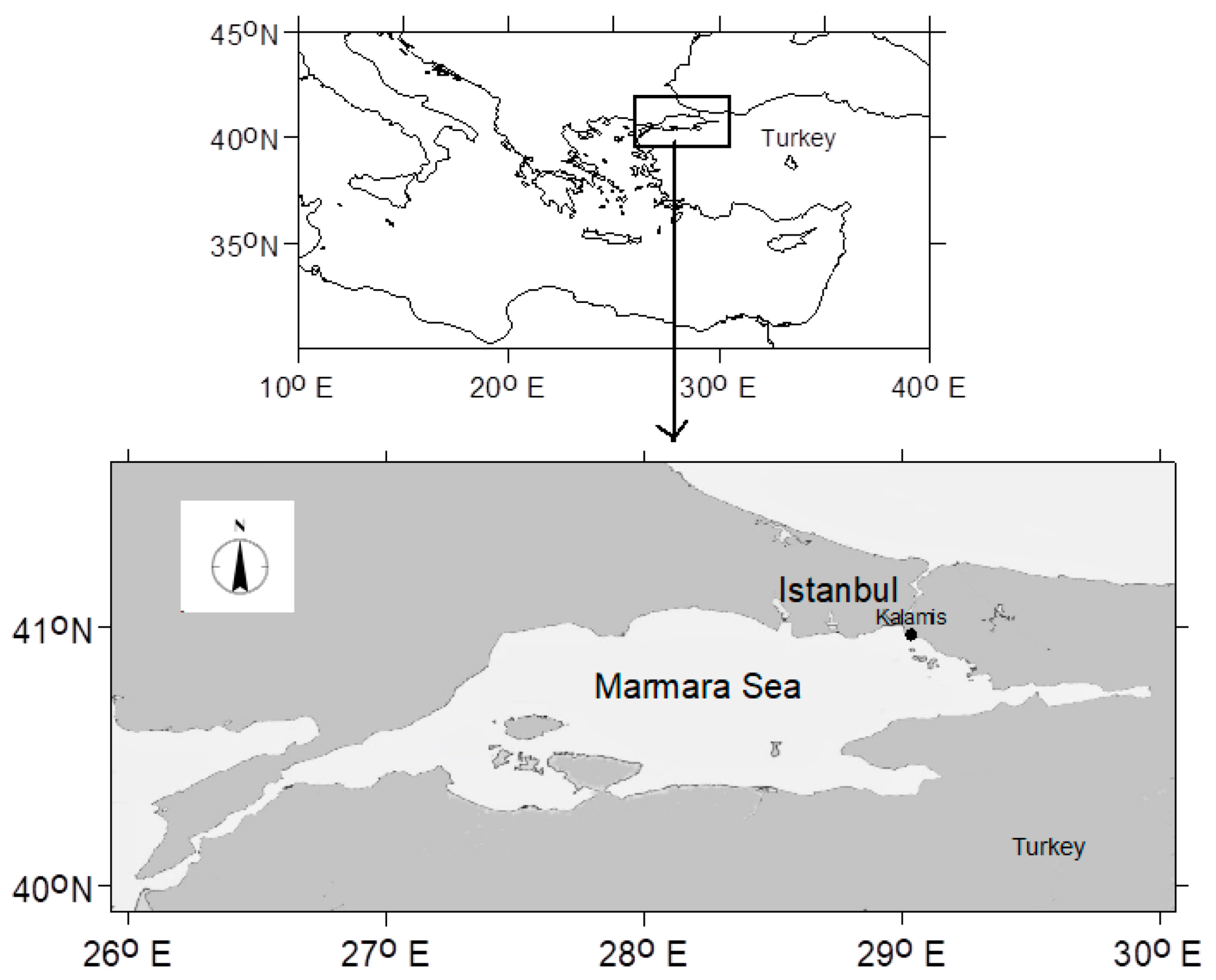
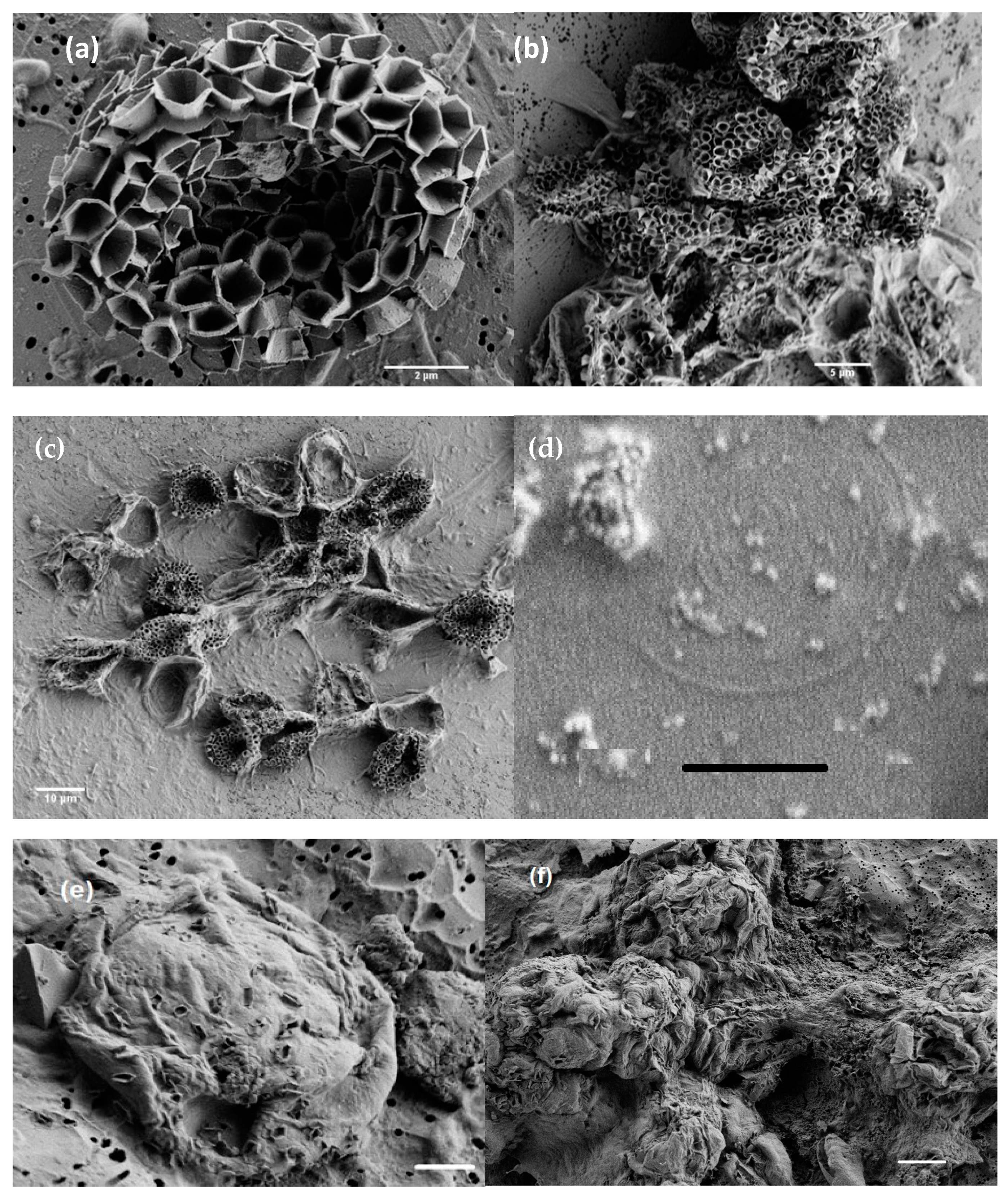
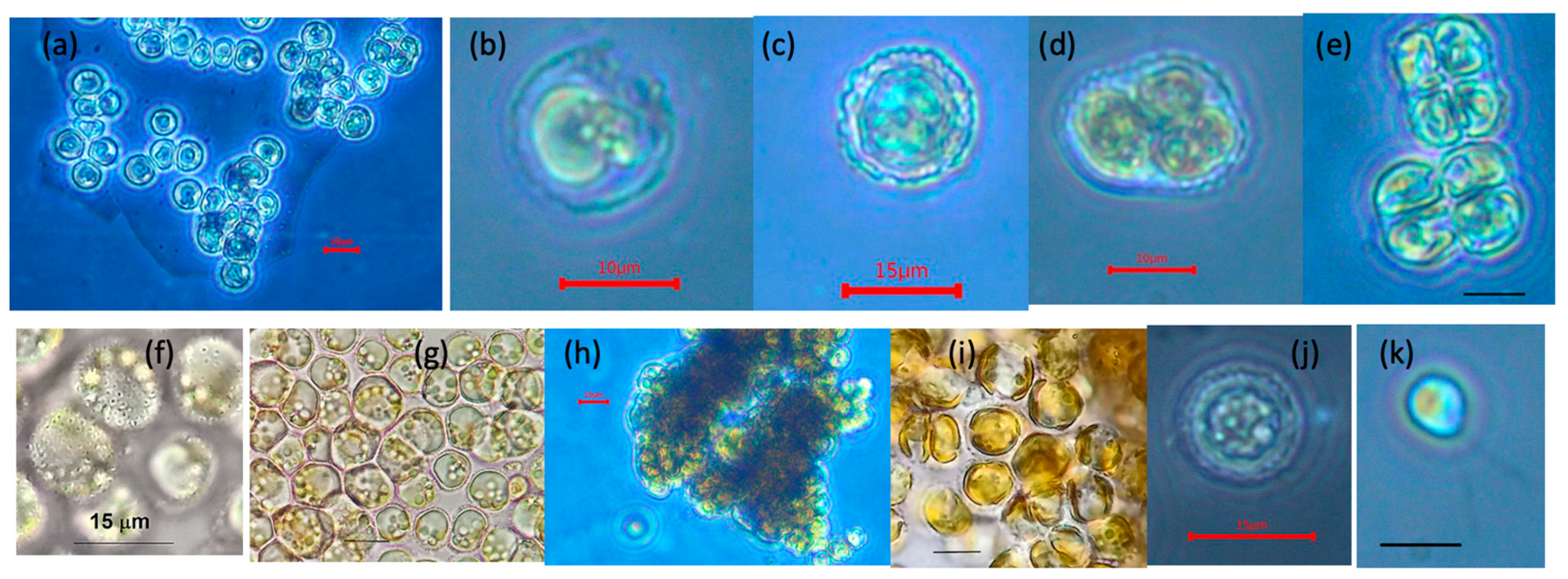
A seawater sample was obtained using a polyethylene bottle on 11 June 2021 from the surface, which had a thick mucilage layer, from the Sea of Marmara, located at Kalamis, Istanbul (40°58′39.99″ N, 29°02′18.37″ E, Figure 1). The temperature and salinity of the seawater were 22 °C and 23 PSU, respectively (Hanna HI98319 salinity tester).
Seawater samples were transferred into 100 mL Erlenmeyer flasks in the laboratory and enriched with F/2 Medium. Cells that increased in the top layer of the flask were chosen with thinned Pasteur Pipettes and grown in tubes. Then, they were transferred into 100 mL flasks and kept under 30–40 µmol photon m−2 s−1 irradiance, 20 °C temperature, and 12:12 light–dark cycle conditions in a climate chamber.

2.1. Microscopy

Live cells of Ochrosphaera neapolitana were investigated under the light microscopes Nikon/Eclipse E100 and Nikon/Eclipse TS100 with 1000× and 400× magnifications. For the scanning electron microscopy, culture cells were fixed with 2% glutaraldehyde solution (Grade I, Sigma G-5882, St. Louis, MO, USA) for a few minutes at 22 °C temperature and filtered on 0.2 µm pore size polycarbonate filters. It is worth noting that we have used high-grade glutaraldehyde (SEM grade), which is always kept in the deep freeze when not used. We believe that we obtained high-quality SEM photographs because of this well-preserved SEM-grade glutaraldehyde. This glutaraldehyde was added to the liquid sample directly, and after a few minutes, the sample was filtered and then covered with iridium without additional washing with distilled water or ethanol for photographing using the SEM. After coating the filters with iridium using the Leica EM ACE600 Sputter Coater instrument, samples were examined with a Zeiss GeminiSEM 500 field emission scanning electron microscope, in Germany. In pursuing good quality photography, cells were also separately fixed with 1% osmium tetraoxide and passed through an ethanol series of 25%, 50%, 75%, and 90% with polycarbonate syringe filters and investigated with SEM as above.

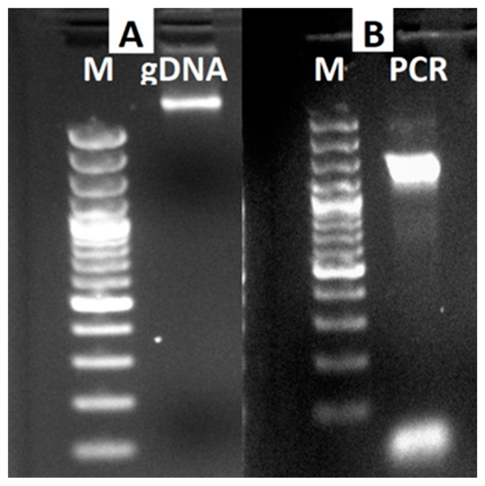
2.2. DNA Isolation, PCR Analysis, and Phylogenetic Tree Construction

Following the manufacturer’s instructions, DNA was extracted from about 300 mg of the material using a commercially available kit (QIAamp DNA Minikit (Cat#51304, Qiagen, Valencia, CA, USA). For this, the algal cell culture in a 15 mL falcon tube was centrifuged and then the supernatant was discarded, obtaining an algal cell pellet (~300 mg). Two-milliliter tubes containing the ~300 mg sample with three-millimeter tungsten carbide beads were then frozen in liquid nitrogen. The tubes were put into the adapter sets, with the Tis-sueLyser’s clamps fastened. TissueLyser was used to disturb frozen plant tissue samples to homogenize them. Steps of 30 Hz shaking lasting two minutes were used to create the disturbance. Pellets were washed with PBS to a final volume of 200 μL. Then, 20 μL QIAGEN proteinase K (Qiagen Inc., Valencia, CA, USA) and 200 μL Buffer AL were added into the microcentrifuge tube and mixed by pulse-vortexing for 15 s. The mixed sample was incubated at 56 °C for 10 min. An aliquot of 200 μL ethanol (96–100%) was added to the sample and remixed by pulse-vortexing for 15 s before being centrifuged at 6000× g for 1 min. The supernatant was transferred to the QIAamp Mini spin column in a clean 2 mL collection tube, and the tube containing the filtrate was discarded. A 500 μL Buffer AW1 was added to the collection tube, well mixed, and centrifuged at 6000× g for 1 min. Afterward, 500 μL Buffer AW2 was added to the collection tube and centrifuged at 20,000× g for 3 min. The QIAamp Mini spin column was placed in a clean 1.5 mL microcentrifuge tube. In total, 200 μL Buffer AE was added and incubated at room temperature (15–25 °C) for 1 min, and then centrifuged at 6000× g for 1 min.
The integrity of isolated gDNA was checked by visualization of ethidium bromide-stained DNA separated on 1.5% (w/v) agarose gel. The DNA sample was stored at −80 °C until it was used. The isolated gDNA was used as the template for PCR-based amplification using Small Sub-Unit (SSU) primers to identify the algal species isolated from marine mucilage events. According to the manufacturer’s protocol, PCR was performed using the Solis BioDyne 5× FIREPol® Master Mix kit (Solis BioDyne, Tartu, Estonia). The SSU rRNA gene was amplified using the forward primer 5’ CTGCCCTATCAGCTTTGGATGG -3’ (18SF) [32] and the reverse primer 5’ CCATTCAATCGGTAGGTGCG -3’ (18SR). For PCR amplification, thermal cycler (Applied Biosystems™ MiniAmp™ Thermal Cycler, Thermo Fisher Scientific Inc., Waltham, MA, USA) conditions were as follows: initial denaturation at 94 °C for 4 min followed by 35 cycles of denaturation at 94 °C for 1 min., annealing at 58 °C for 1 min., and extension at 72 °C for 90 s. These cycles were followed by a final extension step at 72 °C for 5 min. The DNA band around the predicted size was removed from the agarose gel (1%) using the Qiagen Gel Extraction Kit according to the manufacturer’s protocol. PCR products were sequenced by Sentebiolab, Ankara, Turkey (https://sentebiolab.com.tr/; accessed on 29 December 2022). The raw DNA sequence data were manually examined. Using the submission portal (https://submit.ncbi.nlm.nih.gov/subs/genbank/; accessed on 11 January 2023), the identified O. neapolitana SSU rRNA sequence was deposited in the NCBI GeneBank (https://www.ncbi.nlm.nih.gov/; accessed on 11 January 2023) under accession number OQ220476. Phylogenetic analysis was performed by MEGA7 software (https://www.megasoftware.net/; accessed on 12 January 2023) [33]. The evolutionary history was inferred using the Maximum Likelihood method based on the Jukes–Cantor model [34]. Initial tree(s) for the heuristic search were obtained by applying the Neighbor-Joining method to a matrix of pairwise distances estimated using the Maximum Composite Likelihood (MCL) approach. The tree is drawn to scale, with branch lengths measured in the number of substitutions per site. The proportion of sites where at least one unambiguous base is present in at least 1 sequence for each descendent clade is shown next to each internal node in the tree. The analysis involved eight nucleotide sequences. The codon positions included were 1st+2nd+3rd+Noncoding. All positions with less than 95% site coverage were eliminated. Fewer than 5% alignment gaps, missing data, and ambiguous bases were allowed at any position. There was a total of 78 positions in the final dataset.

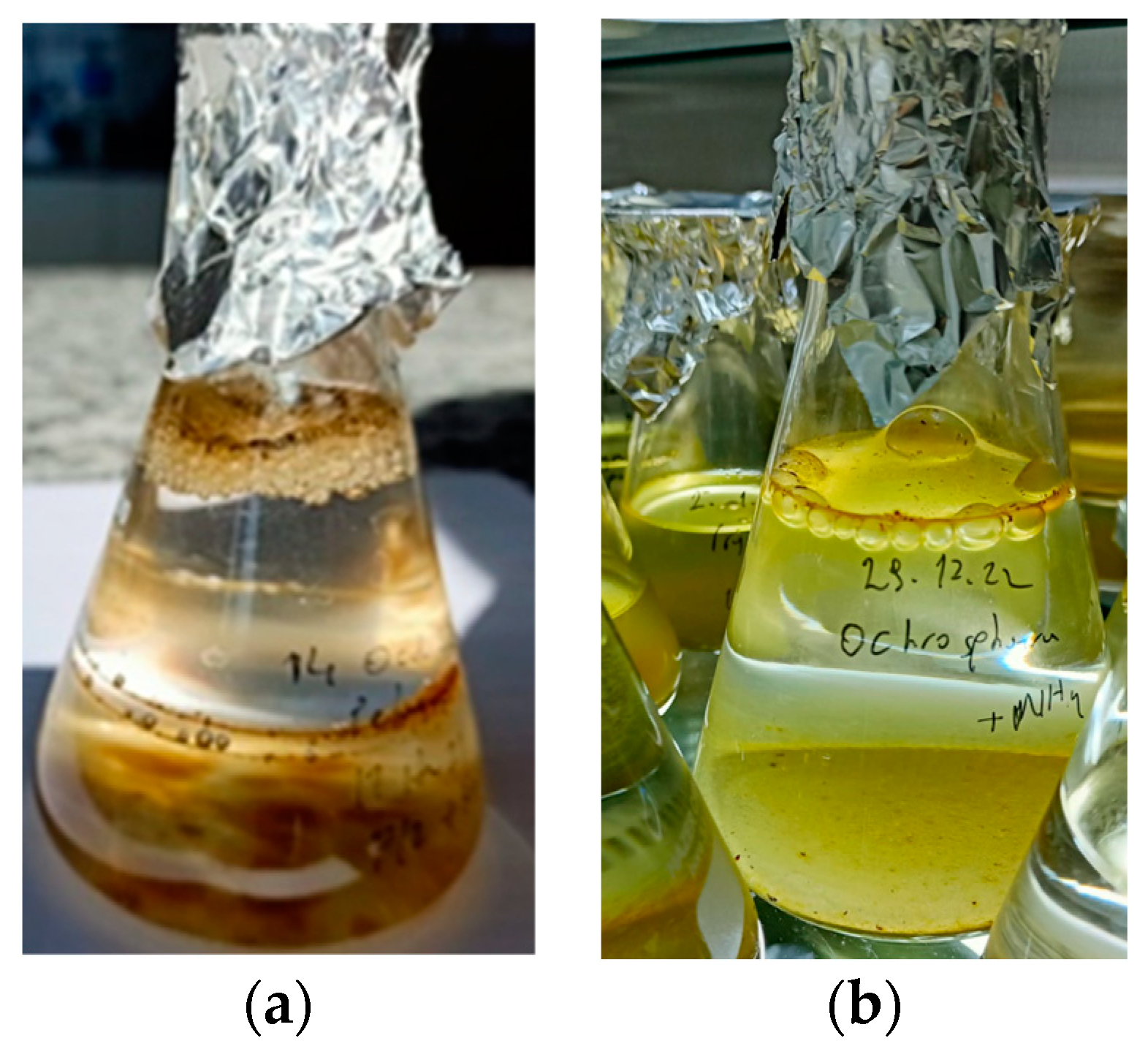
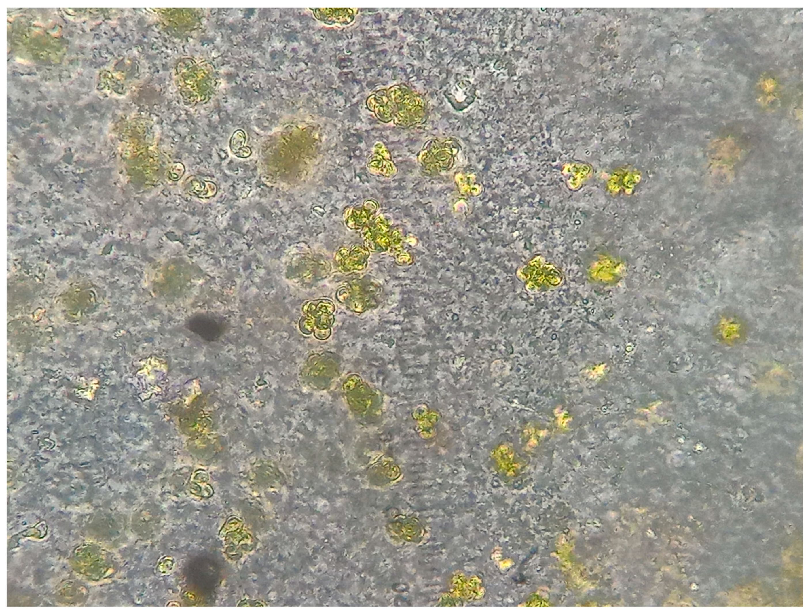
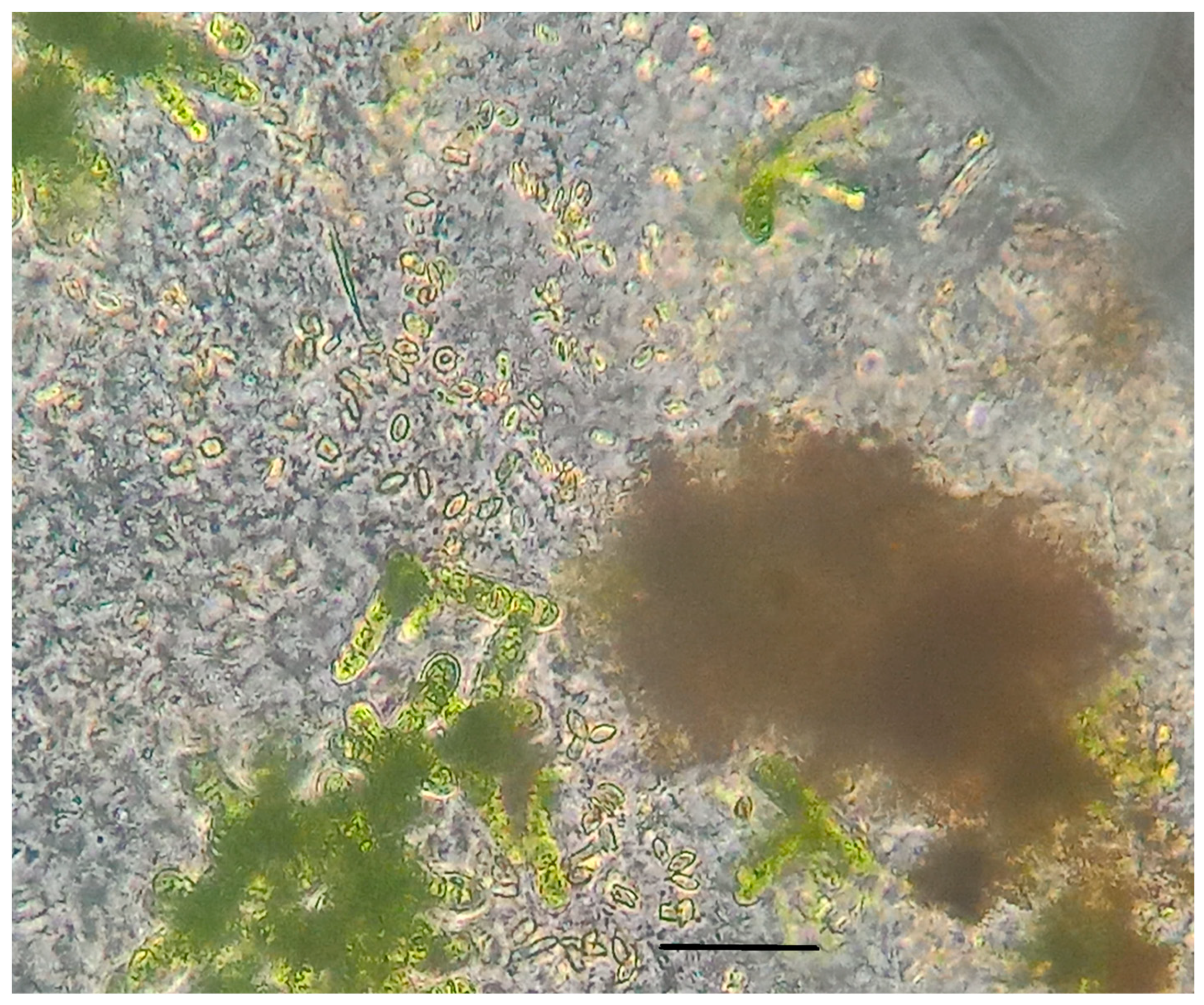
2.3. Mucilage Observation

Observations were performed on the extracellular foamy organic material (mucilage produced) by both single-cell cultures of Ochrosphaera neapolitana and co-cultures with two other diatom species. These two diatom species are Nitzschia sp. and N. amabilis, which were observed to occur together with O. neapolitana in the original sample. Cultures were made daily following inoculation in the culturing flasks and lasted for at least 3–4 months (>10 inoculations). The same mucilage production observations were observed in all inoculations consistently (i.e., more than 10 inoculations) for both mono- and multi-species cultures with Ochrosphaera neapolitana.

3. Results

3.1. Morphology from Light and SEM Microscopy

O. neapolitana shows a heteromorphic life cycle with an alternation between diploid coccolith-bearing cells and a haploid non-calcifying phase. The coccolith-bearing phase can be either non-motile or motile with two slightly unequal flagella. Coccolith-bearing flagellated cells do not have haptonema. The size range of the cells was 6–15 µm (Figure 2 and Figure 3).
Ochrosphaera coccoliths are found in the typical tremalith structure (Figure 2a–c). Tremaliths are vase-shaped muroliths that have a rim with a sub-vertical wall. Unfortunately, we do not have a close-up picture of the liths. Pulley-shaped coccoliths have not been observed in the samples. Unlike other coccolithophore species, scales are not shed in this species (Figure 2a–c). Another type of scale belonging to motile S-cell (different from C cells) is shown in Figure 2d.
It is worth noting that the osmium tetraoxide fixation method used here damaged the cells, causing deformation or loss of the majority of the scales. The damage could also be partly due to the pressure in the syringe filtration or the dehydration process with ethanol series (Figure 2e).
Mucilaginous extracts of the non-calcifying cells are observed under the phase contrast microscope (Figure 3a). Calcifying cells under the light microscope are also seen (Figure 3b–k). These calcifying cells are also observed in high quantities in the mucilage layer at the top of the flasks, indicating that in addition to non-calcifying cells, they also contribute to mucilage formation. Meiocytes are observed in Figure 3d,e. While there is a cover of coccoliths surrounding the quadrate cells, no coccoliths surround each meiocyte. Coccolith-bearing colonies are seen under low magnification (Figure 3b) and high magnification (Figure 3f) with light microscopy. Chloroplasts are observed under the light microscope (Figure 3i). A calcareous ‘pseudocyst’ is formed due to the extracellular over-calcification of coccoliths (Figure 3j). A coccolith-bearing motile cell with two flagella is seen in Figure 3k. Culturing flasks of O. neapolitana with the two diatom species or as the sole species are seen in Figure 4a,b, respectively.

3.2. Molecular Analysis

In the present study, axenic and living cultures were analyzed for gDNA isolation; gDNA was isolated successfully and the integrity of extracted gDNA isolated with agarose gel (Figure 5A) was checked, at which time it was determined that they were of high quality for PCR analysis. Isolated gDNA was used as the template for PCR-based amplification using 18S primers to identify the algal species isolated from marine mucilage events. The PCR product obtained after PCR analysis was visualized on agarose gel (1.5%) (Figure 5B), and it was isolated and directly sequenced by the Sanger method.
The SSU sequence of the mucilage-isolated algal species was compared to the SSU rRNA sequencing identified in the GenBank database using the BLAST algorithm for searching sequences. The sequence obtained through PCR was verified to be 205 bp in length. Using BLAST and phylogenetic analyses (Figure 6), the species was verified, and the identified sequence were observed to have a high similarity to the SSU gene from other strains of Ochrosphaera. To investigate the relationships among the Ochrosphaera species based on their shared SSU rRNA sequences, the sequence data for the isolate were also employed to build a phylogenetic tree. By comparing the sequences discovered from the SSU rRNA PCR profiles, the 29 isolates received from the database at NCBI as a result of the comparison of SSU rRNA sequence similarities were clustered into four branches. In total, 22 isolates were pooled together into the same branch, but seven isolates were grouped into different branches (Figure 6). Pairwise sequence alignment of the identified sequence from O. neapolitana isolate TR2 (OQ220476) with the known 18S rDNA gene indicated that the maximum sequence identity (98.04%) was between O. neapolitana isolate TR2 (OQ220476) and O. neapolitana culture collection CCAP 932/1 (FR865767). Although defined as different species in different studies, all the species of Ochrosphaera are now suppressed as O. neapolitana by Fresnel et al. [29].

3.3. Mucilage Observation

In the original field sample, a dense presence of Ochrosphaera neapolitana cells within the mucilage was noted (Figure 7).
In the laboratory, the mono-species O. neapolitana culture, (including both calcifying and non-calcifying phases) was observed to produce conspicuous amounts of amorphous mucilage following 2 weeks from its inoculation in culture flasks. The mucilage layer at the surface of the culture flasks was visible for several weeks in both the O. neapolitana monoculture as well as mixed culture with Nitzschia sp. and Nitzschia amabilis. They were also observed to occur together in the sea when they were sampled (Figure 8). They also lived together and produced a dense layer of mucilage in these culture flasks, consistently lasting several months (Figure 4a,b). The density of the mucilage produced by O. neapolitana in the monoculture was visibly lower than that when it was cultured with Nitzschia sp. and Nitzschia amabilis (both species together). These two species, Nitzschia sp., and N. amabilis, did not produce any mucilage after their isolation from O. neapolitana.
Except for the diatom Thalassiosira sp. and the cyanobacteria Spirulina sp., the coccolithophore Ochrosphaera neapolitana was seen as the only species causing the production of mucilaginous organic matter among the 40 different phytoplankton species (belonging to diatoms, dinoflagellates, prasinophytes, haptophytes, dictyochophytes, pelagophytes, cryptophytes, and eustigmatophytes) cultured (monoculture or multi-species culture) in our laboratory through the years (personal observation by Elif Eker Develi). Spirulina sp. also produced a gelatinous structure that generally sticks to the glass surface of the flask. However, its density was not high in the samples collected from the field samples on 11 June 2021 from Kalamis, Istanbul, in the northern Sea of Marmara, wherein Ochrosphaera neapolitana was quite dense (Figure 7).

4. Discussion

According to Guiry and Guiry [35], Ochrosphaera neapolitana belongs to the order Coccolithales of the Class Prymnesiophyceae under the Phylum Haptophyta. The species Ochrosphaera neapolitana was first described by Schussnig [36]. About ten years later, Schussnig [36] described two more species as O. verrucosa and O. rovignensis, and several investigators in later years supposedly reported the former from different regions [29,37]. Identification of Ochrosphaera cells is a particular challenge for the non-calcifying phase of this species. However, Fresnel and Probert [29] suggested that most of these observations may be O. neapolitana and further proposed suppression of the two other species in this genus. The range in cell diameter from the Sea of Marmara strain (6–15 µm) is larger than that reported from the cultured organisms in France (5–12 µm, [29]).
O. neapolitana is reported as the sole coccolithophorid that does not continually renew the coccosphere, and due to extracellular over-calcification of coccoliths, a calcareous ‘pseudocyst’ has been observed in cultures [29]. O. neapolitana is known to have a heteromorphic life cycle involving an alternation between (diploid) coccolith-bearing cells and a (haploid) non-calcifying phase [29]. The coccolith-bearing phase may be non-motile or possess flagella as shown in Figure 3k. Although not visible in this figure, this motile phase (S-cells) also bears scales. These types of scales are called base-plate scales, which are non-calcified and different from those of non-motile C-cells, which are mineralized. Fresnel et al. [29] mentioned that haploid S-cells were mainly responsible for mucilage production. In addition to these haploid cells, calcifying non-motile cells observed in our study accumulated in the mucilage layer at the top of the culture flasks, presumably contributing to mucilage formation. Similar to the life cycle of Ochrosphaera, another haptophyte genus, Phaeocytsis, also has haploid and diploid life cycles, both of which are suggested to be responsible for mucilage events in different regions of the world, such as the Adriatic Sea, Ligurian Sea, North Sea, Japan, and Brazilian coasts [2,11,12,38,39,40,41,42,43,44]. Haptophytes generally have a high proportion of lipids in their biomass [45], enabling the floatation of the cells. We have also observed another haptophyte species, Chrysochromulina alifera, generally concentrated at the surface layer of the flask in the laboratory culture (personal observation by Elif Eker-Develi).
This is the second report on the haptophyte coccolithophorid Ochrosphaera neapolitana in the Marmara Sea. Recently, along with 65 other new species of phytoplankton from the Sea of Marmara, Durmuş et al. [1] published the first presence of O. neapolitana from two stations in the Sea of Marmara in samples obtained about one year after (i.e., May 2022) compared to our samples.
This species is reported in different regions along the coastal waters of the North Atlantic, Indian Ocean, Mediterranean Sea [29,46,47], and Japan [29]. However, it is either infrequent or occurs in low abundance in the Mediterranean samples. For example, Bonomo et al. [48] did not find O. neapolitana collected at 24 marine stations in a coastal area (Gulf of Gaeta, central Tyrrhenian Sea) during a three-year survey (2012–2014). During their intense sampling at the four stations along a north-south transect in the area receiving the inflowing surface Black Sea Water (BSW) in front of the Dardanelles during October 2013, March 2014, and July 2014 within the photic layer (3–75 m), Karatsolis et al. [49] found a total of 95 species of coccolithophores using a scanning electron microscope (SEM); however, none of the Ochrosphaera species was present. Similarly, no Ochrosphaera was noted among the 98 taxa of coccolithophores identified using SEM from the monthly samples obtained from February 2018–January 2019 in two stations located on the coastal shelf and in the open sea of the ultra-oligotrophic South-East Levantine Basin, Eastern Mediterranean Sea, during 2018–2019.
The small subunit (SSU) rRNA gene, a critical marker for random target PCR in environmental diversification detection, is one of the most commonly used genes in phylogenetic analysis [50]. For this reason, the SSU rRNA gene was studied in the presented study, and an attempt was made to identify the isolated algae species. There are 58 nucleotide sequences registered in the NCBI database belonging to the genus Ochrosphaera. Two species belonging to the genus Ochrosphaera were registered: (1) O. verrucosa and (2) O. neapolitana. Other sequence information was registered as Ochrosphaera sp. There are seven records of the O. verrucosa species, and the lengths of the sequences entered range from 255 bp to 1722 bp. For O. verrucosa, the 18S (SSU), LSU, and tufA genes were identified. In addition, for O. neapolitana, 13 pieces of sequence information is available for plastids atpA and atpB, ITS, 16S, 18S, 28S, and LSU genes, and the lengths of these sequences are between 255 bp and 4396 bp. Another 38 registered sequences are 18S, 28S, 16S, and LSU gene sequences and belong to Ochrosphaera sp. The lengths of these sequences range from 255 bp to 1.56 bp. LSU sequences are the shortest registered sequences (255 bp) in the database. When the information of the Ochrosphaera genus registered in the NCBI database is examined, it is understood that the sources from which the O. verrucosa species were isolated are (1) Tuticorin coastal waters, Tamil Nadu and (2) the Atlantic Ocean (Gulf of Mexico, Ewing Bank). Likewise, when the information on O. neapolitana is examined, it is seen that the species is isolated from (1) the Mediterranean Sea (French coast) and (2) the Atlantic Ocean (North Atlantic–East Channel waters). O. neapolitana isolated from the present study was first isolated from the Marmara Sea, and a mucilage sample was formed. O. neapolitana with the SSU gene sequence in the NCBI database is the only strain found in the CCAP932/1 coded culture collection. In the present study, when the phylogenetic tree was performed with the species whose SSU gene sequence was examined, it is seen that the species isolated from the Sea of Marmara mucilage sample shows a high similarity with O. neapolitana CCAP932/1.
Although cell numbers were not counted in the original samples, high cell numbers of this haptophyte species were observed in the original samples within the mucilage. They have continued to produce mucilage in the cultures at the top layer of the flasks, especially when they are together with other diatoms. It is most probable that Ochrosphaera cells uptake organic matter produced by these diatoms, sustaining further mucilage production. Ochrosphaera neapolitana is among the species of haptophytes that utilises organic compounds through osmotrophy [51]. The single-cell cultures were observed to form bubbles at the top of the flasks. A similar finding of mucilage production in the laboratory for the haploid non-calcifying stage is also reported by Fresnel and Probert [29]. This observation is very important as this species could potentially be a strong candidate as the responsible agent for the large-scale mucilage events in the Sea of Marmara observed both in 2007 and 2020/2021. So far, Phaeocystis pouchetii (haptophyte), Skeletonema costatum, Cylindrotheca closterium, Thalassiosira rotula, Proboscia alata, Rhizosolenia sp., Pseudosolenia calcar-avis, Ditylum brightwellii, Coscinodiscus spp., Leptocylindrus minimus, Chaetoceros spp., Cerataulina pelagica, Pseudo-nitzschia cf. seriata (diatom), and Gonyaulax hyalina or G. fragilis (dinoflagellate) are suggested as potentially responsible organisms for mucilage production during the events of 2007 and 2020/2021 in the Marmara Sea [18,19,22]. Among these phytoplankton species, Cylindrotheca closterium and Gonyaulax fragilis were also suggested as organisms causing mucilage events that occurred in the northern Adriatic Sea during the summers between 1999–2002 [12,52,53]. Recently, the cyanobacteria Trichodesmium erythraeum has been also suggested to produce mucilaginous blooms in the Bay of Bengal [54]. However, except for Phaeocyctis antarctica [55] and G. fragilis [52,56], none of the species were shown to produce mucilage in laboratory conditions. In our mono- or mixed-species cultures without O. neapolitana, we did not notice conspicuous mucilage production in the laboratory, except on a very small scale in the culture flasks with Thalassiosira sp., Cylotella sp., and Spirulina sp.
Except for the haptophyte Phaeocystis pouchetii, all species reported during the mucilage events in the field samples are common and at times abundant organisms in the Sea of Marmara or other regions of the Mediterranean when/where no mucilage events are observed. Due to its small size (6–15 µm), O. neapolitana is difficult to identify in field samples using traditional microscopy; therefore, we believe that it never appeared as a responsible organism in the studies published so far. Additionally, it could be suggested that this species somehow found the ideal conditions to contribute to the unprecedented levels of mucilage formation in its new environment, the Sea of Marmara. In any case, following its more detailed morphological description and observation of its conspicuous mucilaginous material in this study, further investigations, we believe, will help in better elucidating its role in the past and in possible future mucilage events in the Sea of Marmara.

Author Contributions

Conceptualization, E.E.-D. and A.E.K.; methodology, E.E.-D.; software, D.T.; validation, A.E.K., E.E.-D. and D.T.; formal analysis, E.E.-D., D.T., A.E.D. and H.B.Y.; investigation, E.E.-D. and D.T.; data curation, D.T.; writing—original draft preparation, E.E.-D. and A.E.K.; writing—review and editing, E.E.-D., A.E.K. and D.T.; supervision, A.E.K.; project administration, H.B.Y.; funding acquisition. All authors have read and agreed to the published version of the manuscript.

Funding

This research was funded by TUBITAK, grant number 121G114.

Institutional Review Board Statement

Not applicable.

Informed Consent Statement

Not applicable.

Data Availability Statement

The molecular data presented in this study are openly available in the NCBI GeneBank (https://www.ncbi.nlm.nih.gov/; released on 16 January 2023), under accession number OQ220476.

Acknowledgments

We thank Tülay Çokacar from İstanbul University for the sampling. We also appreciate helps of Robert Anderson and Bente Edvardsen from University of Oslo in identification of the haptophyte species.

Conflicts of Interest

The authors declare no conflict of interest.

References

  1. Degobbis, D.; Fonda-Umani, S.; Franco, P.; Malej, A.; Precali, R.; Smodlaka, N. Changes in the northern Adriatic ecosystem and hypertrophic appearance of gelatinous aggregates. Sci. Total Environ. 1995, 165, 43–58. [Google Scholar] [CrossRef]
  2. Fonda-Umani, S.; Milani, L.; Borme, D.; de Olazabal, A.; Parlato, S.; Precali, R.; Kraus, R.; Lučić, D.; Njire, J.; Totti, C.; et al. Inter-annual variations of planktonic food webs in the northern Adriatic Sea. Sci. Total Environ. 2005, 353, 218–231. [Google Scholar] [CrossRef]
  3. Laroche, C. Exopolysaccharides from microalgae and cyanobacteria: Diversity of strains, production strategies, and applications. Mar. Drugs 2022, 20, 336. [Google Scholar] [CrossRef]
  4. Svetličić, V.; Žutić, V.; Radić, T.M.; Pletikapić, G.; Zimmermann, A.H.; Urbani, R. Polymer networks produced by marine diatoms in the Northern Adriatic Sea. Mar. Drugs 2011, 9, 666–679. [Google Scholar] [CrossRef] [PubMed] [Green Version]
  5. Ricci, F.; Penna, N.; Capellaci, S.; Penna, A. Potential environmental factors influencing mucilage formation in the northern Adriatic Sea. Chem. Ecol. 2014, 30, 364–375. [Google Scholar] [CrossRef]
  6. Ergul, H.A.; Balkis-Ozdelice, N.; Koral, M.; Aksan, S.; Durmus, T.; Kaya, M.; Kayal, M.; Ekmekci, F.; Canli, O. The early stage of mucilage formation in the Marmara Sea during spring. J. Black Sea/Mediterr. Environ. 2021, 27, 232–257. [Google Scholar]
  7. Turk, V.; Hagström, A.; Kova, N.; Faganeli, J. Composition and function of mucilage 42. macroaggregates in the northern Adriatic. Aquat. Microb. Ecol. 2010, 61, 279–289. [Google Scholar] [CrossRef] [Green Version]
  8. Danovaro, R.; Umani, S.F.; Pusceddu, A. Climate change and the potential spreading of marine mucilage and microbial pathogens in the Mediterranean Sea. PLoS ONE 2009, 4, e7006. [Google Scholar] [CrossRef] [Green Version]
  9. Uflaz, E.; Akyüz, E.; Bolat, F.; Bolat, P.; Arslan, Ö. Investigation of the effects of mucilage on maritime operation. J. Black Sea/Mediterr. Environ. 2021, 27, 140–153. [Google Scholar]
  10. Topçu, N.E.; Öztürk, B. The impact of the massive mucilage outbreak in the Sea of Marmara on gorgonians of Prince Islands: A qualitative assessment. J. Black Sea/Mediterr. Environ. 2021, 27, 270–278. [Google Scholar]
  11. Lancelot, C. The mucilage phenomenon in the continental coastal waters of the North Sea. Sci. Total Environ. 1995, 165, 83–102. [Google Scholar] [CrossRef]
  12. Najdek, M.; Blazina, M.; Djakovac, T.; Kraus, R. The role of the diatom Cylindrotheca closterium in a mucilage event in the northern Adriatic Sea: Coupling with high salinity water intrusions. J. Plankton Res. 2005, 27/9, 851–862. [Google Scholar] [CrossRef]
  13. Besiktepe, S.T.; Sur, H.İ.; Ozsoy, E.; Latif, M.A.; Oguz, T.; Unluata, U. The circulation and hydrography of the Marmara Sea. Prog. Oceanogr. 1994, 34, 285–334. [Google Scholar] [CrossRef]
  14. Orhon, D.; Uslu, O.; Meriç Salihoğlu, S.; Filibeli, I. Waste-water management for Istanbul: Basis for treatment and disposal. Environ. Pollut. 1994, 84, 167–178. [Google Scholar] [CrossRef]
  15. Burak, S. Evaluation of pollution abatement policies in the Marmara Sea with water quality monitoring. Asian J. Chem. 2008, 20, 4117–4128. [Google Scholar]
  16. TUIK. Municipality Wastewater Statistics 2018. 2018. Available online: https://biruni.tuik.gov.tr/medas/?kn=120&locale=tr (accessed on 14 August 2021).
  17. Vardar, S.; Onay, T.T.; Demirel, B.; Kideys, A.E. Evaluation of microplastics removal efficiency at a wastewater treatment plant discharging to the Sea of Marmara. Environ. Pollut. 2021, 289, 117862. [Google Scholar] [CrossRef] [PubMed]
  18. Aktan, Y.; Dede, A.; Çiftçi, P.S. Mucilage event associated with diatoms and dinoflagellates in Sea of Marmara, Turkey. Harmful Algae News 2008, 36, 1–3. [Google Scholar]
  19. Tüfekçi, V.; Balkıs, N.; Beken, C.P.; Ediger, D.; Mantıkçı, M. Phytoplankton composition and environmental conditions of a mucilage event in the Sea of Marmara. Turk. J. Biol. 2010, 34, 199–210. [Google Scholar] [CrossRef]
  20. Balkis, N.; Atabay, H.; Türetgen, I.; Albayrak, S.; Balkis, H.; Tüfekçi, V. Role of single-celled organisms in mucilage formation on the shores of Büyükada Island (the Marmara Sea). J. Mar. Biol. Assoc. United Kingd. 2011, 91, 771–781. [Google Scholar] [CrossRef]
  21. İşinibilir-Okyar, M.; Üstün, F.; Orun, D.A. Changes in abundance and community structure of zooplankton population during the 2008 mucilage event in the northeastern Marmara Sea. Turk. J. Zool. 2015, 39, 28–38. [Google Scholar] [CrossRef] [Green Version]
  22. Balkis-Ozdelice, N.; Durmus, T.; Balci, M. A preliminary study on the intense pelagic and benthic mucilage phenomenon observed in the Sea of Marmara. Int. J. Environ. Geoinformatics 2021, 8, 414–422. [Google Scholar] [CrossRef]
  23. Karadurmuş, U.; Sarı, M. Marine mucilage in the Sea of Marmara and its effects on the marine ecosystem: Mass deaths. Turk. J. Zool. 2022, 46, 6. Available online: https://journals.tubitak.gov.tr/zoology/vol46/iss1/6 (accessed on 18 January 2023). [CrossRef]
  24. Taş, S.; Kus, D.; Yilmaz, N. Temporal variations in phytoplankton composition in the northeastern Sea of Marmara: Potentially toxic species and mucilage event. Mediterr. Mar. Sci. 2020, 21, 668–683. [Google Scholar] [CrossRef]
  25. Durmuş, T.; Balkıs-Ozdelıce, N.; Taş, S.; Bayram Partal, F.; Balcı, M.; Dalyan, C.; Sarı, M. New Records for Microalgae Species of the Turkish Seas Under the Effect of Intense Mucilage in the Sea of Marmara. Eur. J. Biol. 2022, 81, 144–162. [Google Scholar] [CrossRef]
  26. Eker-Develi, E.; Kiydes, A.E. First record of the diatom Nitzschia navis-varingica in the Sea of Marmara. Mar. Sci. Tech. Bull. 2022, 11, 231–235. [Google Scholar]
  27. Svetlichny, L.; Isinibilir, M.; Kideys, A.E. First finding of live specimens of non-native copepod Calanus finmarchicus (Gunnerus, 1770) in the Sea of Marmara. Aquat. Sci. Eng. 2023. [Google Scholar] [CrossRef]
  28. İmamoğlu, E.; Demirel, Z.; Dalay, M.C. Evaluation of different microalgae strains for biomass production. In Proceedings of the First National Workshop on Marine Biotechnology and Genomics, Bodrum, Mugla, Turkey, 24–25 May 2012; Turan, C., Ed.; Turkish Marine Research Foundation (TUDAV): Istanbul, Turkey, 2012; Volume 36, pp. 107–112. [Google Scholar]
  29. Fresnel, J.; Probert, I. The ultrastructure and life cycle of the coastal coccolithophorid Ochrosphaera neapolitana (Prymnesiophyceae). Eur. J. Phycol. 2005, 40, 105–122. [Google Scholar] [CrossRef] [Green Version]
  30. Inouye, I.; Pienaar, R.N. Light, and electron microscope observations of the type species of Syracosphaera, S. pulchra (Prymnesiophyceae). Br. Phycol. J. 1988, 23, 205–217. [Google Scholar] [CrossRef] [Green Version]
  31. Schwartz, E. Beitra ge zur Entwicklungsgeschichte der Protophyten. Arch. Protistenk. 1932, 77, 434–462. [Google Scholar]
  32. Gastineau, R.; Hansen, G.; Davidovich, N.A.; Davidovich, O.; Bardeau, J.F.; Kaczmarska, I.; Mouget, J.L. A new blue-pigmented haploid diatom, Haslea provincialis, from the Mediterranean Sea. Eur. J. Phycol. 2016, 51, 156–170. [Google Scholar] [CrossRef] [Green Version]
  33. Kumar, S.; Stecher, G.; Tamura, K. MEGA7: Molecular evolutionary genetics analysis version 7.0 for bigger datasets. Mol. Biol. Evol. 2016, 33, 1870–1874. [Google Scholar] [CrossRef] [Green Version]
  34. Jukes, T.H.; Cantor, C.R. Evolution of Protein Molecules. In Mammalian Protein Metabolism; Munro, H.N., Ed.; Academic Press: New York, NY, USA, 1969; pp. 21–132. [Google Scholar]
  35. Guiry, M.D.; Guiry, G.M. AlgaeBase. World-wide electronic publication, National University of Ireland, Galway (taxonomic information republished from AlgaeBase with permission of M.D. Guiry). Ochrosphaera Schussnig, 1930. 2023. Available online: https://www.marinespecies.org/aphia.php?p=taxdetails&id=601148 (accessed on 12 January 2023).
  36. Schussnig, B. Über einige neue Protophyten aus der Adria. Arch. Protistenk. 1940, 93, 317–330. [Google Scholar]
  37. Norris, R.E. Microalgae in enrichment cultures from Puerto Penasco, Sonora, Mexico. Bull. South Calif. Acad. Sci. 1967, 66, 233–250. [Google Scholar]
  38. Degobbis, D.; Malej, A.; Fonda Umani, S. The mucilage phenomenon in the northern Adriatic Sea: A critical review of the present scientific hypotheses. Ann. Dell’istituto Super. Di Sanità 1999, 35, 373d381. [Google Scholar]
  39. McKenzie, L.; Sims, I.; Beuzenberg, V.; Gillespie, P. Mass accumulation of mucilage caused by dinoflagellate polysaccharide exudates in Tasman Bay, New Zeland. Harmful Algae 2002, 1, 69–83. [Google Scholar] [CrossRef]
  40. Malej, A.; Mozetič, P.; Turk, V.; Terzič, S.; Ahel, M.; Cauwet, G. Changes in particulate and dissolved organic matter in nutrient-enriched enclosures from an area influenced by mucilage: The northern Adriatic Sea. J. Plankton Res. 2003, 25, 949–966. [Google Scholar] [CrossRef] [Green Version]
  41. Cozzi, S.; Ivancic, I.; Catalano, G.; Djakovac, T.; Degobbis, D. Dynamics of the oceanographic properties during mucilage appearance in the Northern Adriatic Sea: Analysis of the 1997 event in comparison to earlier events. J. Mar. Syst. 2004, 50, 223–241. [Google Scholar] [CrossRef]
  42. Fukao, T.; Kimoto, K.; Yamatogi, T.; Yamamoto, K.; Yoshida, Y.; Kotani, Y. Marine mucilage in Ariake Sound, Japan, is composed of transparent exopolymer particles produced by the diatom Coscinodiscus granii. Fish. Sci. 2009, 75, 1007e1014. [Google Scholar] [CrossRef]
  43. Schiaparelli, S.; Castellano, M.; Povero, P.; Sartoni, G.; Cattaneo-Vietti, R. A benthic mucilage event in North-Western Mediterranean Sea and its possible relationships with the summer 2003 European heatwave: Short term effects on littoral rocky assemblages. Mar. Ecol. 2007, 28, 1–13. [Google Scholar] [CrossRef]
  44. Fernandes, L.F.; Frassão-Santos, E.K. Mucilaginous species of Thalassiosira Cleve emend. Hasle (Diatomeae) in South Brazilian waters. Acta Bot. Bras. 2011, 25, 31–42. [Google Scholar] [CrossRef] [Green Version]
  45. Lowenstein, D.P.; Mayers, K.; Fredricks, H.F.; Van Mooy, B.A.S. Targeted and untargeted lipidomic analysis of haptophyte cultures reveals novel and divergent nutrient-stress adaptations. Org. Geochem. 2021, 161, 104315. [Google Scholar] [CrossRef]
  46. Dimiza, M.D.; Triantaphyllou, M.V.; Malinverno, E.; Psarra, S.; Karatsolis, B.T.; Mara, P.; Lagaria, A.; Gogou, A. The composition and distribution of living coccolithophores in the Aegean Sea (NE Mediterranean). Micropaleontology 2015, 61, 521–540. [Google Scholar] [CrossRef]
  47. Fresnel, J. Les Coccolithophorides (Prymnesiophyceae) du Littoral. Genres: Cricosphaera, Cruciplacolithus, Hymenomonas et Ochrosphaera, Ultrastructure, Cycle Biologique, Syste´Matique. Ph.D. Thesis, Universite´ de Caen, Caen, France, 1989; p. 281. [Google Scholar]
  48. Bonomo, S.; Cascella, A.; Alberico, I.; Lirer, F.; Vallefuoco, M.; Marsella, E.; Ferraro, E. Living and thanatocoenosis coccolithophore communities in a neritic area of the central Tyrrhenian Sea. Mar. Micropaleontol. 2018, 142, 67–91. [Google Scholar] [CrossRef]
  49. Karatsolis, B.-T.; Triantaphyllou, M.; Dimiza, M.; Malinverno, E.; Lagaria, A.; Mara, P.; Archontikis, O.; Psarra, S. Coccolithophore assemblage response to Black Sea Water inflow into the North Aegean Sea (NE Mediterranean). Cont. Shelf Res. 2017, 149, 138–150. [Google Scholar] [CrossRef]
  50. Meyer, A.; Todt, C.; Mikkelsen, N.T.; Lieb, B. Fast evolving 18S rRNA sequences from Solenogastres (Mollusca) resist standard PCR amplification and give new insights into mollusk substitution rate heterogeneity. BMC Evol. Biol. 2010, 10, 70. [Google Scholar] [CrossRef] [PubMed] [Green Version]
  51. Godrijan, J.; Drapeau, D.; Balch, W.M. Mixotrophic uptake of organic compounds by coccolith-ophores. Limnol. Oceanogr. 2020, 65, 1410–1421. [Google Scholar] [CrossRef]
  52. Pompei, M.; Mazziotti, C.; Guerrini, F.; Cangini, M.; Pigozzi, S.; Benzib, M.; Palamidesic, S.; Bonic, L.; Pistocchi, R. Correlation between the presence of Gonyaulax fragilis (Dinophyceae) and the mucilage phenomena of the Emilia-Romagna coast (northern Adriatic Sea). Harmful Algae 2003, 2, 301–316. [Google Scholar] [CrossRef]
  53. Pistocchi, R.; Cangini, M.; Totti, C.; Urbani, R.; Guerrini, F.; Romagnoli, T.; Sist, P.; Palamidesi, S.; Boni, L.; Pompei, M. Relevance of the dinoflagellate Gonyaulax fragilis in mucilage formations of the Adriatic Sea. Sci. Total Environ. 2005, 353, 307–316. [Google Scholar] [CrossRef]
  54. Shaika, N.A.; Alhomaidi, E.; Sarker, M.M.; An Nur, A.; Sadat, M.A.; Awal, S.; Mostafa, G.; Hasan, S.J.; Mahmud, Y.; Khan, S. Winter bloom of marine cyanobacterium, Trichodesmium erythraeum and its relation to environmental factors. Sustainability 2023, 15, 1311. [Google Scholar] [CrossRef]
  55. Bolinesi, F.; Saggiomo, M.; Aceto, S.; Cordone, A.; Serino, E.; Valoroso, M.C.; Mangoni, O. On the relationship between a novel Prorocentrum sp. and colonial Phaeocystis antarctica under iron and vitamin B12 limitation: Ecological implications for Antarctic waters. Appl. Sci. 2020, 10, 6965. [Google Scholar] [CrossRef]
  56. Escalera, L.; Italiano, A.; Pistocchi, R.; Montresor, M.; Zingone, A. Gonyaulax hyalina and Gonyaulax fragilis (Dinoflagellata), two names associated with ‘mare sporco’, indicate the same species. Phycologia 2018, 57, 453–464. [Google Scholar] [CrossRef]
Figure 1. Sampling region for the coccolithophorid Ochrosphaera neapolitana in the Sea of Marmara on 11 June 2021.
Jmse 11 00468 g001
Figure 2. Ochrosphaera neapolitana images from the SEM: (a) a coccolith-bearing diploid cell scale bar; scale is 2 µm, (b) a group of coccolith-bearing cells; scale is 5 µm, (c) an aged colony, scale 10 µm, (d) an organic base-plate scale of an S-cell; scale is 300 nm, (e) a coccolith-bearing cell fixed with 1% osmium tetraoxide; scale 1 µm, and (f) deformed cells after fixing with osmium tetraoxide; scale 4 µm.
Jmse 11 00468 g002
Figure 3. Ochrosphaera neapolitana images from the light microscope: (a) light microscope view of non-calcifying cells; scale 10 µm (b) a coccolith-bearing cell; scale 10 µm, (c) another coccolith-bearing cell; scale 15 µm, (d) a divided coccolith-bearing cell; scale 10 µm, (e) tetrad (meiocytes) before the reappearance of S-cells; scale 10 µm, (f) colony of coccolith-bearing cells under the light microscope; scale 15 µm, (g) image showing insides of coccolith-bearing cells; scale 15 µm, (h) a colony under the light microscope; scale 10 µm, (i) two chloroplasts seen in each cell; scale 10 µm, (j) over-calcified pseudocysts; scale 15 µm, and (k) a coccolith-bearing flagellated cell; scale 15 µm.
Jmse 11 00468 g003
Figure 4. (a) Mucilage formation at the surface layer of the flask when Ochrosphaera neapolitana was cultured together with two different diatom species (2 weeks old) and (b) mucilage formation in monospecific Ochrosphaera neapolitana culture (2 weeks old).
Jmse 11 00468 g004
Figure 5. (A) Agarose gel (1.5%) electrophoresis results of genomic DNA of O. neapolitana isolated from mucilage (M: Marker, DNA: gDNA of O. neapolitana isolate TR2 (OQ220476)) and (B) agarose gel (1%) electrophoresis result in which 18S primers were used (M: Marker, PCR: amplification product of O. neapolitana isolate TR2 (OQ220476)).
Jmse 11 00468 g005
Figure 6. Phylogenetic tree based on SSU rRNA gene sequence, showing the position of the O. neapolitana isolate TR2 (OQ220476) (shown in the filled square). The evolutionary history was inferred using the Maximum Likelihood method based on the Jukes–Cantor model [34]. As stated in the NCBI GenBank, names and accession numbers are provided. The percentage of trees in which the associated taxa clustered together in the bootstrap test (1000 replicates) is shown next to the branches. The diatom Ulnaria acus was used as an outgroup.
Jmse 11 00468 g006
Figure 7. High concentration of Ochrosphaera neapolitana cell groups in the original samples taken from Kalamis, Istanbul, on 11 June 2021; the scale is 50 µm.
Jmse 11 00468 g007
Figure 8. A section of the mucilage sample taken from Kalamis, Istanbul, on 11 June 2021, shows green microalgal species and the pennate diatom Nitzschia amabilis (small oval cells); scale is 50 µm.
Jmse 11 00468 g008
Disclaimer/Publisher’s Note: The statements, opinions and data contained in all publications are solely those of the individual author(s) and contributor(s) and not of MDPI and/or the editor(s). MDPI and/or the editor(s) disclaim responsibility for any injury to people or property resulting from any ideas, methods, instructions or products referred to in the content.

