Article

Reproductive Cycle of the Sea Urchin Paracentrotus lividus (Lamarck, 1816) on the Central West Coast of Portugal: New Perspective on the Gametogenic Cycle

1
MARE—Marine and Environmental Sciences Centre/ARNET, ESTM, Polytechnic of Leiria, 2520-641 Peniche, Portugal
2
ICBAS—Instituto de Ciências Biomédicas de Abel Salazar, Universidade do Porto, 4050-313 Porto, Portugal
3
MARE—Marine and Environmental Sciences Centre/ARNET, Department of Life Sciences, Faculty of Sciences and Technology, University of Coimbra, 3004-517 Coimbra, Portugal
4
MARE—Marine and Environmental Sciences Centre/ARNET, Faculdade de Ciências, Universidade de Lisboa, 1749-016 Lisboa, Portugal
*
Author to whom correspondence should be addressed.
J. Mar. Sci. Eng. 2023, 11(12), 2366; https://doi.org/10.3390/jmse11122366
Submission received: 29 September 2023 / Revised: 6 December 2023 / Accepted: 13 December 2023 / Published: 14 December 2023

Abstract

:
A population of sea urchins, Paracentrotus lividus, from the central west coast of Portugal was studied to characterise their reproductive biology and possible relationships with environmental factors. An annual gametogenic cycle was found, with a broad spawning season, from May to November, according to a relatively synchronous gamete maturation process. Depending on the environmental factors (temperature, photoperiod), two separate periods could be distinguished, with more individuals maturing and spawning at the same time. When this happened, the first event evolved when temperature rose to a critical point, and the second occurred afterwards, when temperature decreased significantly. Notwithstanding, it was found that individuals matured later than previously described for other populations (e.g., north of Portugal), mostly in late spring, with a higher gonadosomatic index in May. A new classification scale was proposed for identifying the stages of P. lividus gametogenic cycle, based on new findings. It contributed to its simplification and easier comprehension. This study provides useful information for a differentiated sustainable management of P. lividus, according to local conditions. Establishing a closed harvesting season might be considered, based on the differences observed between Portuguese populations and other European ones.

1. Introduction

Sea urchins are known as a delicacy due to their reddish-orange gonads, commonly named as roe in the food industry [1]. This seafood product was first appreciated in Asia, but nowadays it is also a delicacy and has a high commercial value in North and South America, as well as in Europe [1,2,3,4,5]. In Portugal, the harvest of sea urchins is focused on Paracentrotus lividus, which is one of the numerous species of echinoderms that inhabit the intertidal rocky habitats [6]. The consumption of P. lividus is related to traditional customs, but the demand for this product has been growing mainly in northern coastal regions of Portugal, where sea urchins have been sold mainly to the Spanish and French markets [3].
Sea urchins present a high tolerance to physiological pressure, enduring high salinity levels, temperature variations, desiccation, and food shortage for long periods of time [7,8,9]. The existing populations determine the functioning of the biological communities where they live due to their grazing activity [10,11,12], which contributes to controlling the growth and excessive propagation of some macroalgae [6,13,14]. In addition, adult sea urchins often serve as shelter for small invertebrate organisms [15].
Some studies have been carried out in the north [2,12,16,17,18,19] and south [17,20,21] of Portugal related to the harvest, biology and ecology of sea urchins. Nevertheless, research on the central west coast of the country is very scarce [11] and not recent. In this region, there is a sea urchin harvesting tradition in specific periods of the year related to an international sea urchin festival and other seafood festivals. The nominal catches of sea urchins in Portugal increased from 2013 (3 tons) to 2022 (456 tons) [22,23], meaning that the demand for this resource has been increasing in a very significant way. The overexploitation of the natural populations can lead to a severe stock depletion, as has already been reported in other countries [1,24]. In this context, aquaculture may emerge as a solution to decrease the impact of harvesting natural stocks of sea urchins. Moreover, it will contribute to a controlled production of P. lividus, which allows the market demand to be supplied all year round and for overexploited areas to be restocked. Therefore, it is imperative to acquire knowledge on the P. lividus reproductive cycle, by characterising and understanding all stages of gametogenesis and the factors influencing it.
Most echinoderms, including P. lividus, are gonochoric organisms, devoid of sexual dimorphism with external fertilisation. Different studies have shown that, even within the same population, individuals do not synchronise their reproductive cycle and, thus, mature individuals are found all year round [5,25,26,27,28,29,30,31,32,33,34,35,36]. Seasonal changes in environmental parameters (such as photoperiod and temperature) influence reproduction and gametogenesis within and between populations, as well as the nutritional condition of the individuals [4,17,18,19,37,38].
Some studies have been performed to attempt to describe the gametogenic cycle of P. lividus in other European populations [25,28]. However, there are some differences and discrepancies in the characterisation made by [25] and the one performed by [28]. Thus, there is a need to make some changes and adjustments for a better description and classification of the gametogenic stages, creating a new, simplified and comprehensive scale.
In this context, the aim of this study was to analyse the reproductive biology of the sea urchin P. lividus from a natural population in the central west coast of Portugal. Moreover, it also intended to understand how seasonal factors, such as temperature and photoperiod, affect its gametogenic cycle. Thus, biometric parameters and the gonadosomatic index were assessed for 18 months. Sea urchins’ gonads were histologically analysed for determining sex ratio and surveying the gametogenic cycle. A readjustment of the characterisation of the different gametogenic stages was proposed based on the descriptions of previous works [25,28] in order to simplify and more accurately analyse the cycle. It intends to better identify each stage and to have a more comprehensive perception of the morphology of P. lividus gonads. The results of this study can be useful for a better understanding of the reproductive cycle of P. lividus in function of the environmental factors. Its results were compared with others obtained in previous studies at different geographical locations to contribute to a better decision-making in future management plans for this resource.

2. Materials and Methods

2.1. Study Site and Sampling Method

The present study was performed on an Atlantic rocky shore at Praia do Abalo (39°22′12.69″ N; 009°23′7.07″ W) in Peniche (central west coast of Portugal). This rocky shore has low hydrodynamics and a diverse morphology, also containing a small sandy area. Tides are semidiurnal and mesotidal tides (tidal range between 2 and 3.5 m). A total of 30 sea urchins, with test diameters between 30 and 50 mm, were collected monthly for 18 months (July 2015 to December 2016) and transported to the laboratory of Aquaculture of MARE—Marine and Environmental Sciences Centre of the Polytechnic of Leiria—in 5 L containers filled with local sea water. Sea urchins smaller than 30 mm of diameter were disregarded, as they were most probably sexually immature [39].

2.2. Biometric Measurements

The sea urchins were weighed (TW) with an analytical balance (AE ADAM PGL 3002, Milton Keynes, England, ±0.01 g accuracy), and test diameter was measured with a Vernier Calliper (Insize, code 1205-150S, INSIZE Co., Ltd., Zamudio, Spain; ±0.05 mm accuracy). The sampled sea urchins were then dissected to remove and weigh the gonads of all individuals.

2.3. Histology

The sea urchin gonads (n = 24 to 30 individuals per month) were fixed and preserved in buffered formaldehyde at 4% for 24 h; then, they were dehydrated and embedded in paraffin using a Leica® TP1020 Automatic Tissue Processor (Leica Microsystems GmbH, Wetzlar, Germany). Afterwards, sections of 5 μm were obtained using an Accu-Cut® SRM™ 200 Rotary Microtome (Sakura Finetek Europe BV, Alphen aan den Rijn, The Netherlands) and stained with hematoxylin (Scharlab S.L., Sentmenat, Barcelona, Spain) and eosin (VWR International, Leuven, Belgium). After the staining procedure, the slides were analysed using a Leica® DM 2000 LED light optical microscope equipped with a Leica® MC170 5MP HD Microscope Camera and the LAS V4.4.0 software (Leica Application Suite, Wetzlar, Germany). The different stages of gametogenesis were then determined and characterised as described in the next subsection, the sex ratio was determined and the oocyte diameter (DO, n = 30 per female) was measured by its major axis.

Development of the Gametogenic Stage Scale

The study [25] identified stage I as the phase in which the resorption of relict gametes occurs. On the contrary, in the present study, this event was considered to be the last stage—stage VII (reabsorption). Therefore, in this study the characterisation of the gametogenic cycle stages were adapted from the ones described in [25,28], with modifications that allowed the following sequence of stages to be created.
  • Stage I—Initial phase
The gametogenesis begins, in which the first previtellogenic gametes start to appear along the ascinal wall of the ovary/testis. Nutritive phagocytes (NPs) increase in size and number, filling the lumen of the gonad.
  • Stage II—Growth
Previtellogenic gametes are surrounded by NPs, which provide the nutrients for their development. In females, the oocytes increase in size as they absorb the nutrients.
  • Stage III—Premature stage
The first mature gametes begin to appear. Once the gametes are mature, they start to move from the ascinal wall of the ovary/testis towards the gonad lumen. However, the gametes do not mature all at the same time. Thus, many previtellogenic gametes remain along the ascinal wall. This stage is manly characterised by the presence of gametes in different growing phases.
  • Stage IV—Mature
The gonad lumen is fully occupied by mature gametes. The number of NPs is very low, and they create a very thin layer along the ascinal wall, where some growing gametes can still be found.
  • Stage V—Spawning
As sea urchins start the spawning event, mature gametes are less concentrated and voids begin to appear in the lumen. These voids are better seen between the oocytes in females than spermatozoa in males.
  • Stage VI—Spent
The spent stage is essentially characterised by the loss of nutritive material and internal structural organisation. The previtellogenic oocytes detach from the ascinal wall and become loose, together with mature ova, demonstrating the internal disorganisation of the gonad. In females, the spent stage may be difficult to identify and may even be confused with a premature spawning. They can be distinguished by the presence of voids in the spent stage rather than nutritive material between the oocytes, as in premature spawning. In males, this stage is easier to identify, since only empty spaces are observed, left by the released sperm, leaving the testis with a tracery aspect.
  • Stage VII—Reabsorption
The internal structure of the ovaries/testis begins to reorganise and the thickness of the ascinal wall is reduced in both sexes. In females, relict oocytes accumulate in the lumen and are surrounded by an eosinophilic mesh of NPs. Here, the NPs are active as phagocytes of relict gametes, gametogenic cells and cellular debris (Pearse and Cameron, 1991). These will promote the absorption of nutritive material. The concept is the same for males, with the lyses of relict spermatozoa and primary spermatocytes.

2.4. Temperature and Photoperiod

Data on mean seawater temperature (°C), monitored via oceanographic buoys closest to the sampling site and during photoperiod (L:D hours), were provided by the Portuguese Hydrographic Institute (IH). These data were used to analyse the associations between these parameters and the gonadosomatic index from July 2015 to December 2016.

2.5. Data Analysis

The gonadosomatic index of P. lividus was determined as GI = gonadal weight/wet weight × 100. The sex ratio of the population was calculated as sex ratio = number of males/number of females.
To compare individual total wet weight (TW), gonadosomatic index (GI), gonadal weight (GW) and oocyte diameter (DO) during 18 months of sampling (July 2015 to December 2016), normality and homogeneity of data were assessed. Then, when these assumptions were met, data were subjected to a one-way ANOVA to search for the existence of statistically significant differences [40]. When differences were found, the Tukey HSD test was performed. When no normality and/or homogeneity of data were met, non-parametric Kruskal–Wallis tests were performed, and when differences were found, the Dunn test was performed (when the normality of the data was not met and the sample size was unequal) [40]. In order to verify whether there was a significant association between the sex ratio and the months of sampling, a Chi-square (χ2) test was performed [40]. To evaluate the relationship between environmental variables (temperature and photoperiod), gonadal weight (GW) and gonadosomatic index (GI), Pearson’s correlation tests (r) were performed. When applicable, the values were given as mean ± standard deviation. The confidence level considered in the statistical tests was p ≤ 0.05. Statistical tests were performed using the statistical program SigmaStat 4.0 (Systat Software Inc., San Jose, CA, USA).

3. Results

3.1. Biometric Data: Wet Weight and Gonadal Weight

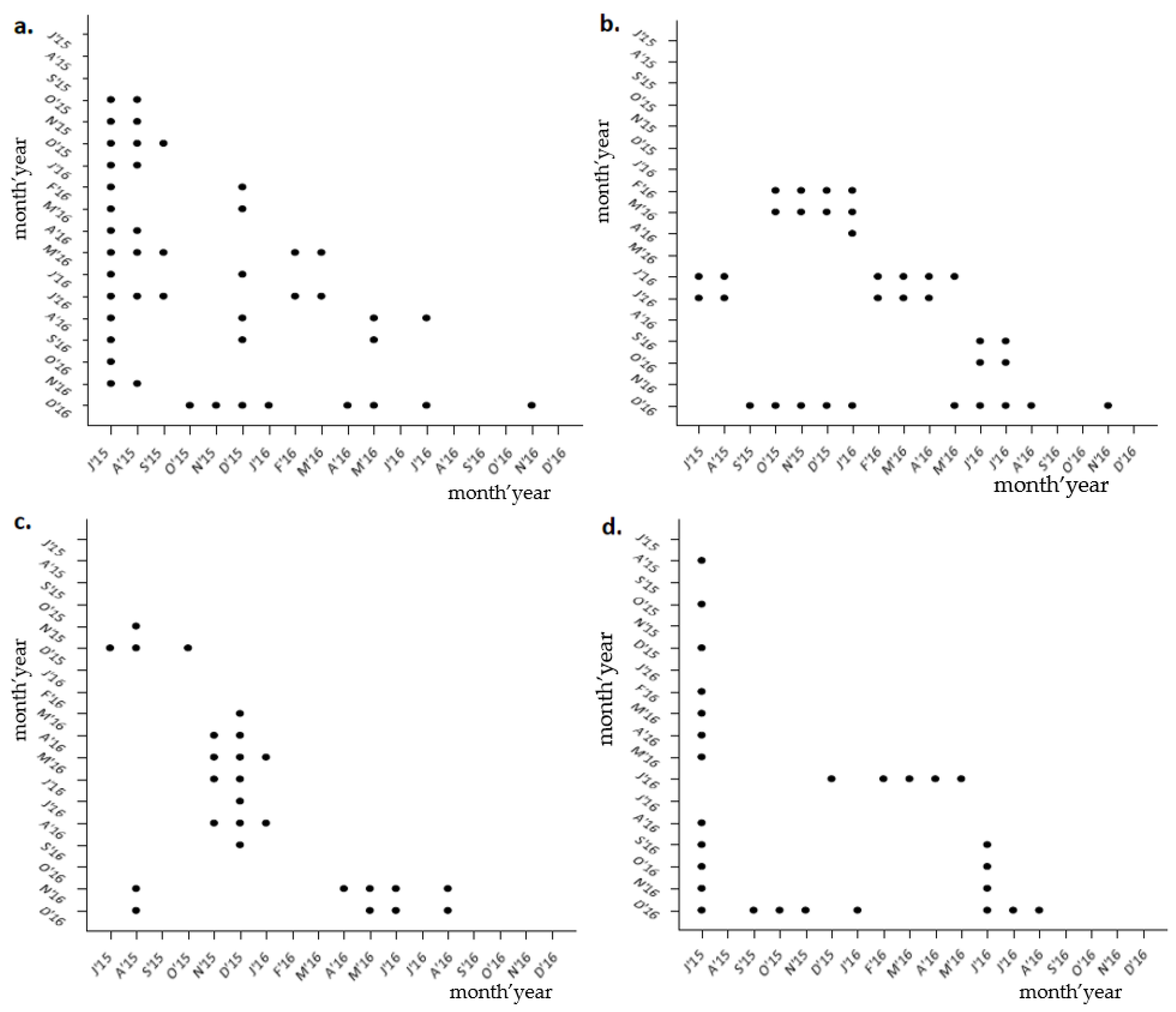
The individual total wet weight (TW) of sea urchins varied significantly with time (Kruskal–Wallis, H17 = 229,257; p < 0.001). Sea urchins had the highest weights in July 2015 (53.19 ± 12.72 g, Figure 1), which was statistically different from the other months (Dunn test; p < 0.05, Figure 2), and the lowest weights in December 2015 (17.99 ± 3.43 g).
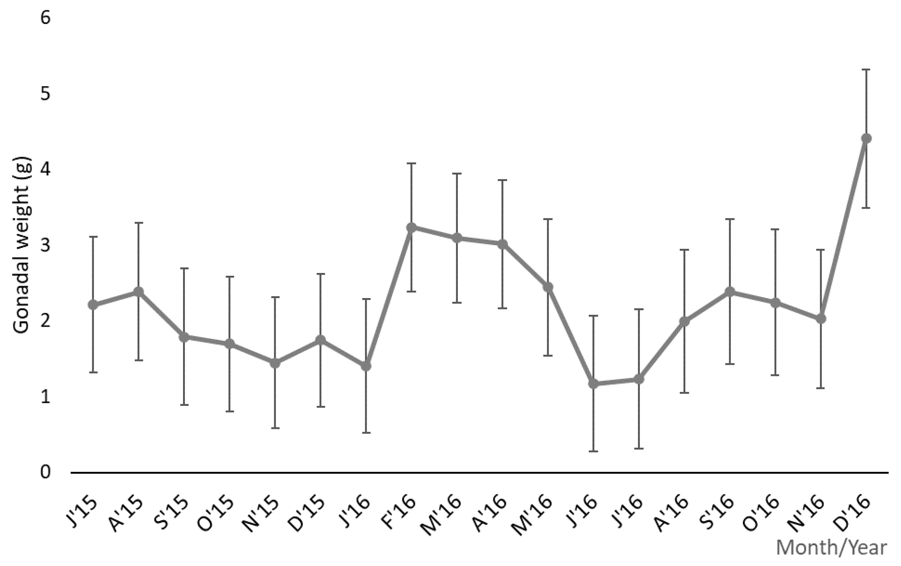
Gonadal weight values were also found to be statistically different over the 18 months (Kruskal–Wallis H17 = 136,073; p < 0.05). The individuals had heavier gonads in the month of December 2016 (4.41 ± 2.27 g, Figure 3), which was statistically different from June 2016 (Dunn test; p < 0.05, Figure 2), when sea urchins had the lowest values (1.17 ± 0.4 g).

3.2. Variation in Gonadosomatic Index, Temperature and Photoperiod

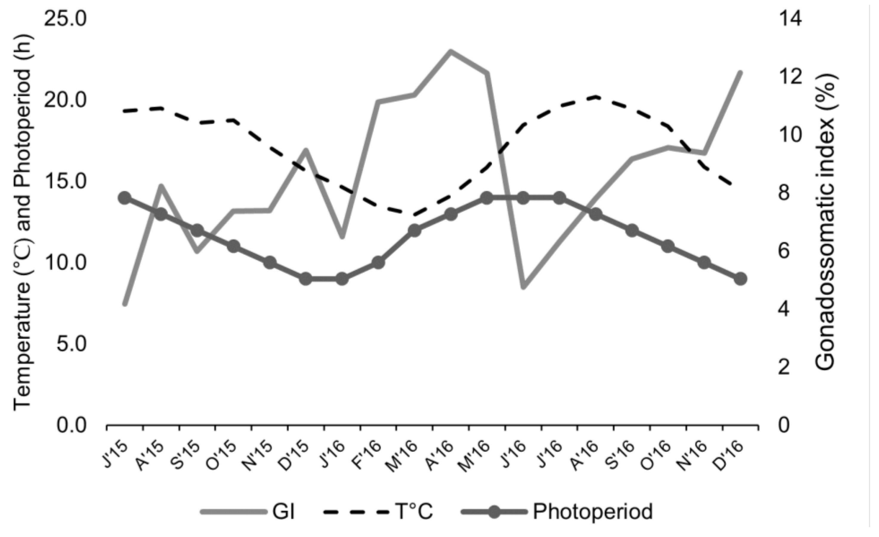
The gonadosomatic index (GI) was found to be different over time (Kruskal–Wallis, H17 = 155.234; p < 0.05). The highest GI values were recorded in April 2016 (12.87 ± 6.43%, Figure 4), and were significantly different from July 2015 and June 2016 (Dunn test; p < 0.05, Figure 2). The month with the lowest GI values was July 2015 (4.18 ± 1.3%), which was significantly different from the other months (Dunn’s Method, p < 0.05), with the exception of September 2015, November 2015, January 2016, June 2016 and July 2016 (Dunn’s Method, p > 0.05).
Seawater temperature and average number of daylight hours followed a similar pattern (Figure 4)—although with a time offset of two months—and both showed a positive correlation (Pearson correlation r = 0.544; p < 0.05). The lowest temperatures were recorded in February 2016 and March 2016 (13 °C in both months, plus 10 and 12 h of light, respectively), and the highest temperature in July 2016 and August (19 °C in both months, plus 14 and 13 h of light, respectively). There was a strong negative correlation between the GI and temperature (Pearson correlation r = −0.678; p < 0.05), although no significant correlation was established between the GI and photoperiod (Pearson correlation r = −0.114; p > 0.05), or between either of these environmental variables and the gonadal weight (temperature, Pearson correlation r = −0.542; p > 0.05; photoperiod, Pearson correlation r = −0.218; p > 0.05).

3.3. Variation in Oocyte Diameter

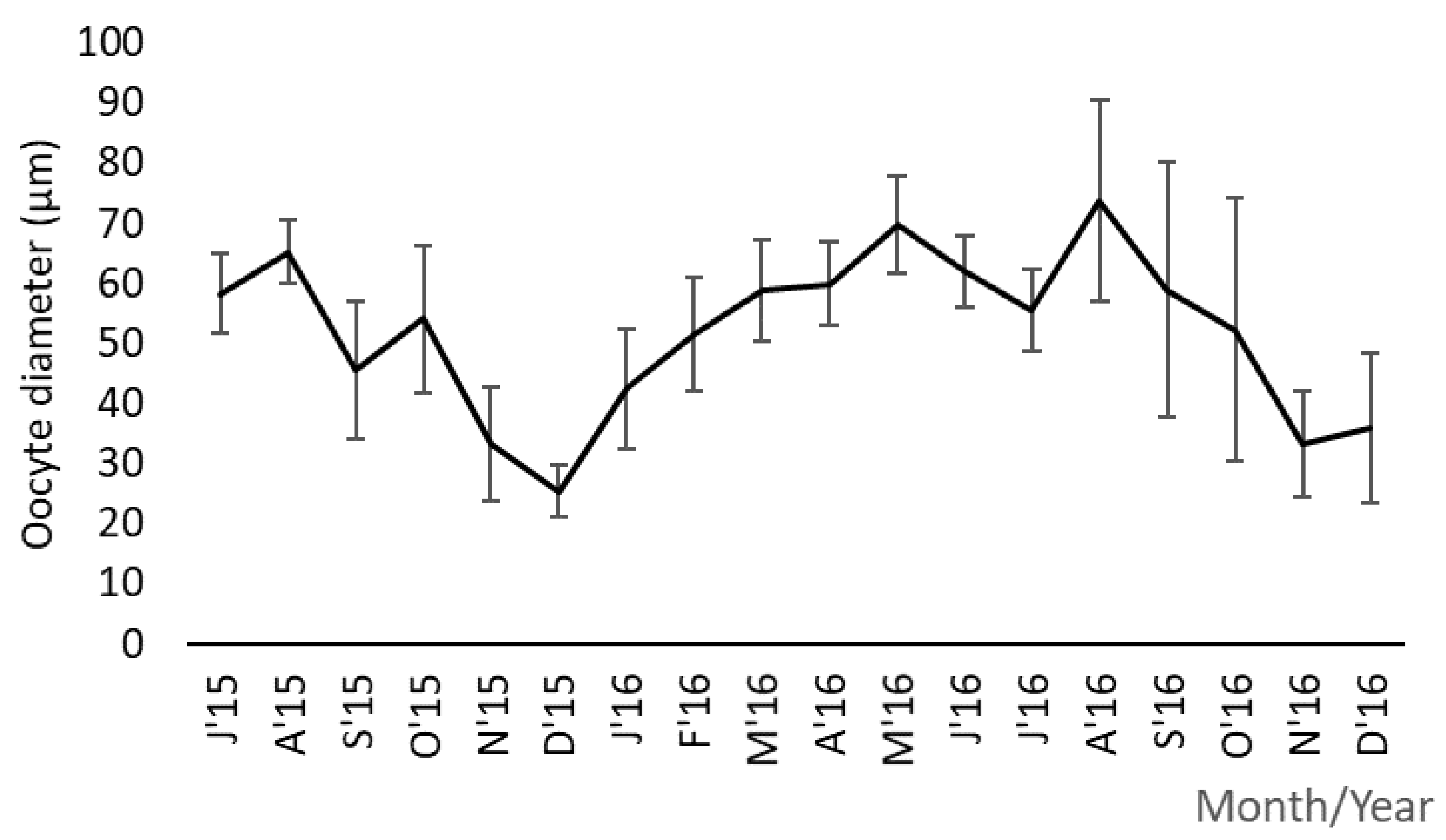
The oocyte diameter values showed statistically significant differences over the sampling time (Kruskal–Wallis H (17) = 105.589; p < 0.05). This biological parameter decreased gradually from the summer of 2015, reaching the lowest mean value in December 2015 (25.45 ± 4.22 μm, Figure 5). The oocyte diameter increased, reaching the highest values in August 2016 (73.69 ± 16.61 μm), and decreasing again until November 2016, reaching a minimum of 33.27 ± 8.76 μm. Overall, there was a statistically significant difference in the oocyte diameter between the summer months and the winter months (Dunn’s Method, p < 0.05, Figure 2).

3.4. Population’s Sex Ratio

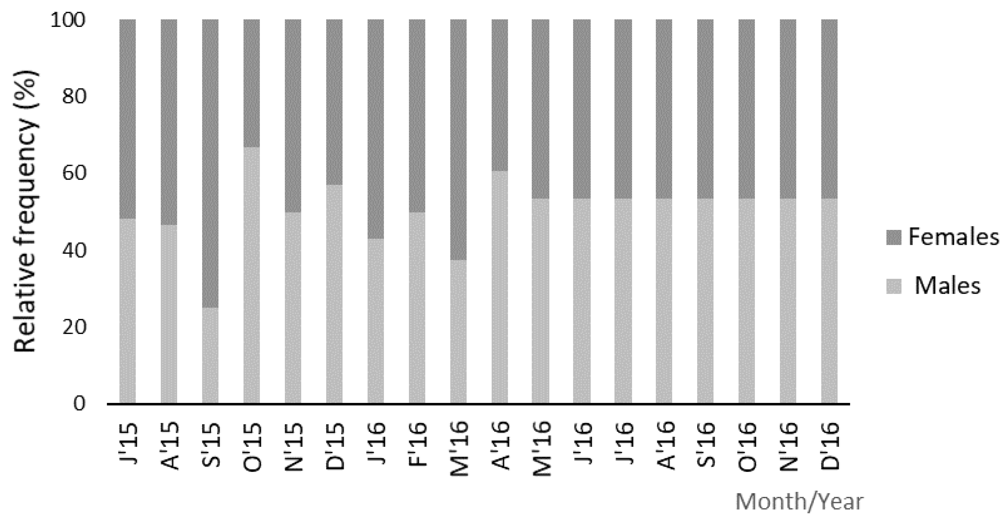
Regarding the population’s sex ratio, the lowest proportion was observed in September 2015 (1:3) and the highest value (2:1) in October 2015 (Figure 6). However, there were no significant fluctuations in this parameter (χ2 (11) = 7.242; p > 0.05).

3.5. Gametogenic Cycle

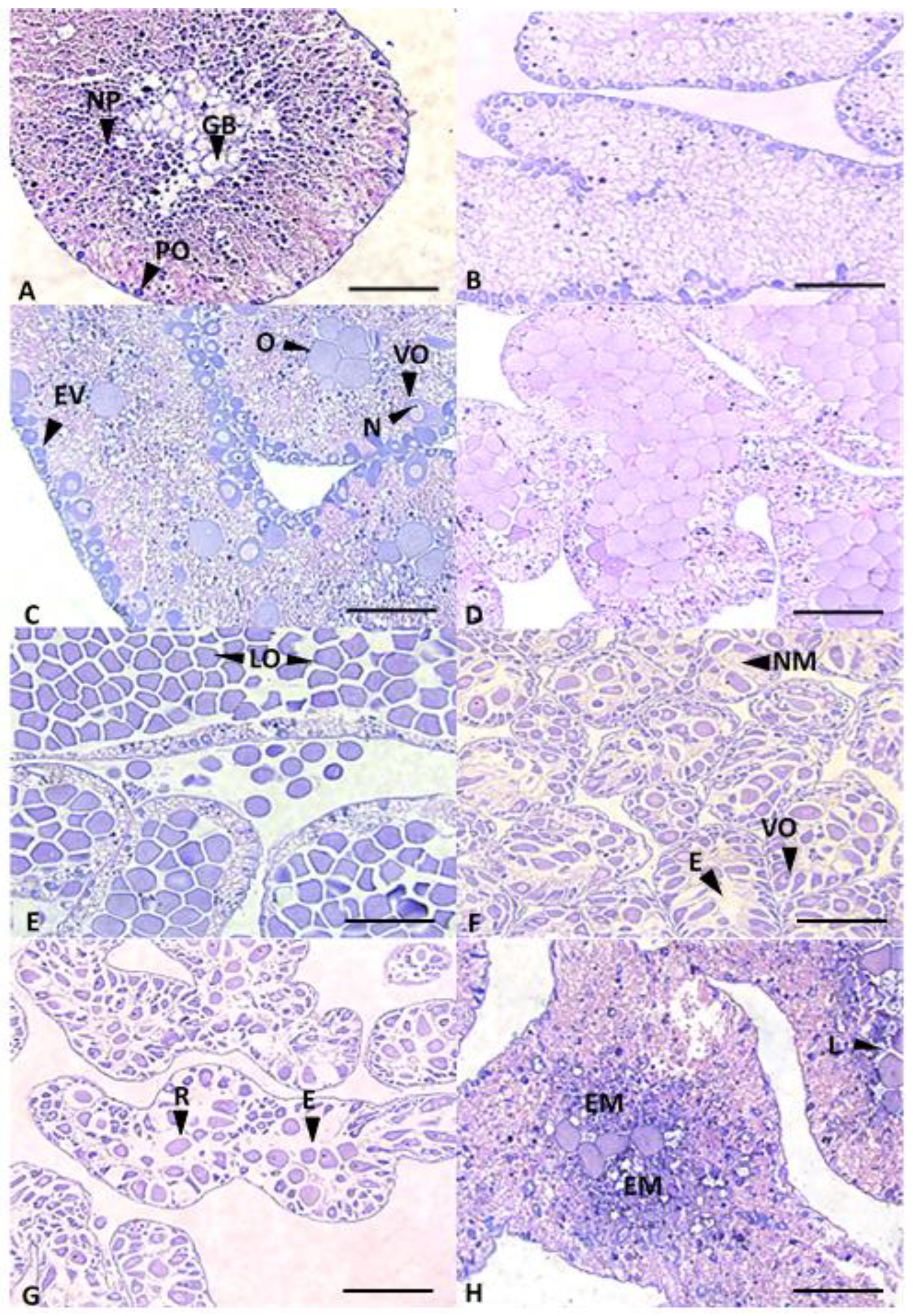
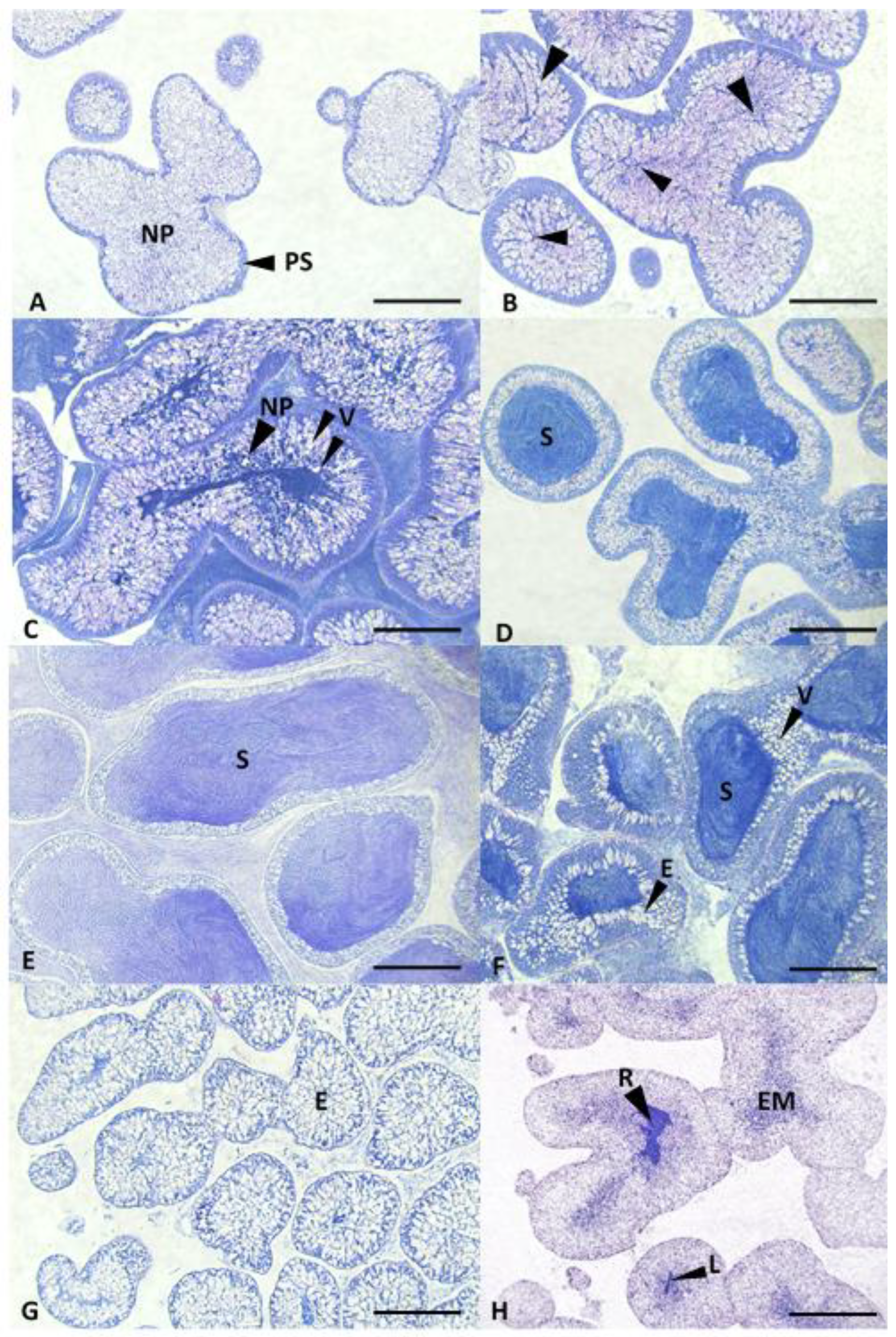
All stages of the gametogenic cycle were observed during the sampled period (Figure 7 and Figure 8).
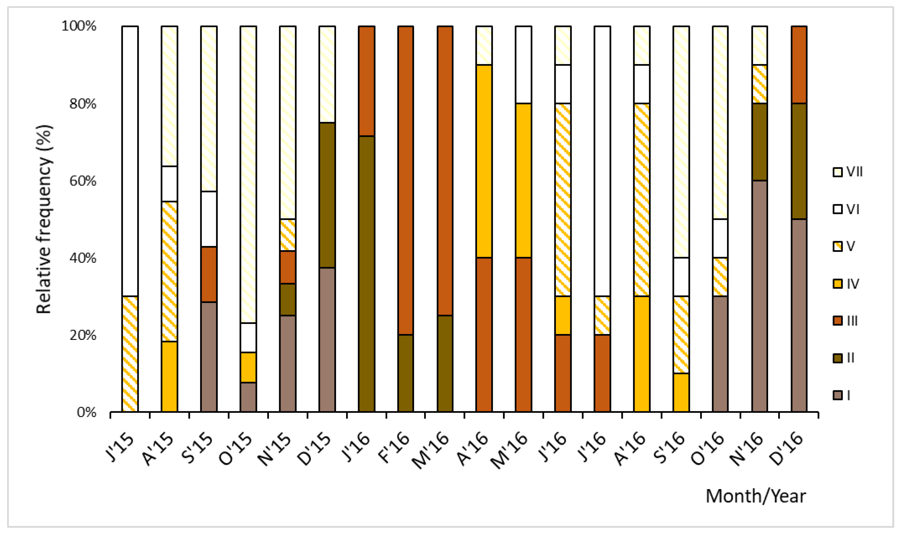
In July 2015, sea urchins were mostly at the end of the reproductive season, with gonads predominantly in stages V and VI (Figure 9). Stage V (spawning) was recorded in the months of July 2015 (30%), August 2015 (36.4%) and November 2015, (8.3%). Stage VI was the most observed in July 2015, in which 70% of the individuals presented spent gonads. In the following months (September, October and November 2015), sea urchins were mainly in stage VII (reabsorption) (42.9%, 76.9% and 50%, respectively), in which the relict gametes were reabsorbed. In December 2015 and January 2016, the great majority of the sea urchins were in the beginning of the gametogenic cycle (December 2015, 37.5% in stage I and stage II; January 2016, 71.4% in stage II). In February and March 2016, stage III was the most observed, with 80% and 75% of P. lividus being in premature stage, respectively. In the following months, April and May 2016, sea urchins were 50% and 40% mature, respectively. In the year of 2016, there was one extended spawning event, which started in June 2016 (with 50% of P. lividus in stage V) and lasted until November 2016 (with 10% of P. lividus in stage V). In November 2016 and December 2016, individuals were mainly in stage I, with 60% and 50% of P. lividus reinitiating their gametogenic cycle, respectively.

4. Discussion

Seawater temperature was found to be a preponderant factor for the gonadal growth of P. lividus, influencing the gonadosomatic index and gametogenic development. These relationships have also been described for other P. lividus populations in France [28,41], Ireland [25] and Portugal [11,17,18], as well as for Loxechinus albus (Molina, 1782) in Argentina [42]. Temperature induced a reverse response on the sea urchins’ reproductive biology indicators. When temperature started to decrease, gonadal weight and gonadosomatic index tended to increase, concomitant with the beginning of a new gametogenic cycle. As the temperature increased, the gametogenic cycle came to its end, culminating with spawning, which contributed to lowering the gonadosomatic index and decreasing gonadal weight.
According to [28], the highest gonadosomatic index for French P. lividus populations was recorded in early spring (March), but it suddenly declined as the temperature increased during summer. Moreover, ref. [25] monitored two Irish populations of P. lividus for two years (1986–1988), and detected a similar pattern. Likewise, ref. [5] verified a similar situation for a population of P. lividus in the north of Spain from 2004 to 2005. In the present study, the relationship between temperature and gonadosomatic index resembled the trend described by those previous authors, being lowest in July 2015 and June 2016, increasing during the following months, and reaching its maximum values in the spring (April 2016). The same relationship applied for the gonadal weight and seawater temperature. In a northern Portuguese population of P. lividus, ref. [18] observed that the gonadal weight decreased to almost half of its value from March to June 2016 (10.3 ± 2.7 g to 5.5 ± 2.3 g, respectively), and then slowly increased until March 2017 (15.0 ± 3.3 g). A similar pattern was described for P. lividus on the southern coast of Portugal [17], as well as the population from Brittany (France) studied by [28]. Accordingly, the present study registered the highest gonadal weight during the transition from winter to spring and lowest in the transition from summer to autumn.
In addition to temperature, photoperiod also seemed to influence the gonadal growth, gonadosomatic index and gametogenic development of P. lividus. In this study, it was noticeable that gonadosomatic index and gonadal weight tended to increase with shorter daylength, although no statistically significant association was found between photoperiod and those two parameters. These relationships have been demonstrated in previous studies on other sea urchins such as Strongylocentrotus purpuratus (Stimpson, 1857) [43], Strongylocentrotus droebachiensis (O.F. Müller, 1776) [44] and Loxechinus albus [42]. Previous laboratory studies [37,45] found that temperatures above 20 °C, combined with short daylength promoted the gonadal growth of P. lividus. Moreover, ref. [46] also found that P. lividus from the Gulf of Tunisia tended to increase the gonadal weight, in its natural environment during the colder months of the year with shorter daylength.
Seasonal abiotic factors, such as water temperature and photoperiod, play an important role in gametogenic development, influencing the maturation periods and spawning events. Thus, when variations in gonadal growth and/or gonadosomatic index are observed, they are usually related to the stage of gametogenic development of the sea urchin gonads. Accordingly, a decrease in gonadal weight was observed in the months of September, October and November 2015, which was related to the absence of gametes. Those individuals were mostly in the reabsorption stages (with 42.9%, 76.9% and 50% of the sea urchins in stage VII, respectively, for each of those months). The same occurred in June 2016, when 50% of sea urchins were in the spawning stage (stage V), and in July 2016, when 70% of P. lividus were in the spent stage (stage VI). This means that the sea urchins were depleted of mature gametes in both months. P. lividus started the gametogenic cycle in December of 2015 and in December of 2016. This month includes days with fewer light hours (9 h), when the highest percentage of P. lividus was observed in the early stages of gametogenic development (stage I—37.5% and 50% and stage II—37.5% and 30%, in each year, respectively). In January 2016, the gonadal weight started to increase, reaching a maximum value in February 2016 (3.24 ± 1.62 g), in which 80% of sea urchins were in stage III (premature). The same pattern was observed by [5] in P. lividus from Cantabria (Spain). At this stage, nutritive phagocytes provide the nutrients necessary for the maturation of the gametes. This happens in the transition from stage III (premature) to stage IV (mature), as verified by [46]. This means that the breeding season also coincides with the months in which the temperature and photoperiod increased.
P. lividus spawning can occur once or twice a year (Table A1), and usually, it does not involve all individuals at the same time. Still, the released gametes can serve as a stimulus for other adult individuals initiate the spawning process [6]. Moreover, spawning events may present interannual differences in terms of time and frequency at the same geographical location (Table A1). For instance, ref. [6] noticed a difference of up to four weeks for the spawning season, in distinct years, for the same population. When two spawning events occur, the first usually happens when the temperature rises to a critical point, and the second occurs afterwards, when the temperature decreases significantly [6,31].
In Portugal, previous studies [11,17,18,19] recorded only one spawning event per year, whilst in this work, there seemed to be a trend towards two spawning events, depending on the environmental conditions and based on the observations of 2015. Spawning occurred mainly in July and August of that year, and no individuals were found in stage V for the next two months (September and October 2015). Sea water temperature did not present significant changes until then. But, 8.3% of the individuals reappeared in the spawning (stage V) in November 2015, coinciding with a significant decrease in the sea water temperature. Although stage V sea urchins constituted a lesser percentage than the individuals observed during the summer, this record can be considered a second spawning event. The sudden drop in temperature could have acted as a stress signal, which may have triggered sea urchins with a delayed gametogenic cycle to spawn as a last chance to reproduce and start a new cycle. In fact, there were still P. lividus at the premature stage (III) in September 2015 and at mature stage (IV) in October 2015 to support this theory. In the following year, the temperature started to decrease steadily from September to December 2016, leading to a single spawning event extended for a long period of time (until October 2016, but accentuated in June–August 2016). This observation reinforced the fact that spawning is an unpredictable event and highly subordinated to environmental factors—in this case, temperature. It appears to be a fast phenomenon, as some sea urchins were at the spent (VI) and reabsorption (VII) stages at the same time as the first mature (stage IV) and spawning (stage V) individuals. As in the present study, ref. [5] observed the same pattern for the appearance of P. lividus in the reabsorption (stage VII—replacement of nutritional reserves—from September–November 2004), in the Bay of Biscay (north of Spain). Moreover, they also observed some P. lividus individuals in stages I and III, between two spawning periods, as in September–November 2015 from the present work. These observations indicate the beginning of a new gametogenic/reproductive cycle. The study from [47] suggested that two reproductive cycles can occur, since gonads can develop a new gametogenic cycle and present early stages of maturation between the two spawning events.
Echinoderms are known to release their gametes as a response to stress factors, which may be the reason why they partially empty their gonads and produce a new spawning event later. Thus, this reinforces the idea that P. lividus produced a single gametogenic cycle per year. But, under specific environmental conditions, many sea urchins may undergo simultaneous spawning, allowing distinct events in time to be distinguished, which may result in the recognition and follow up of two distinct cohorts in future population dynamics studies. In addition to temperature and photoperiod, hydrodynamic conditions may have been another important factor. Although this factor is seldom monitored and taken into account, it may have induced stress periods, in which many sea urchins may have been forced to release their gametes, even if partially, at the same time. This could account for the differences observed regarding other northern and southern Portuguese populations, in which a single spawning event was registered [11,17,18,19]. Therefore, hydrodynamics should be considered in future studies concerning the population dynamics and reproductive biology of sea urchins.
In terms of oocyte size, ref. [25] measured diameters from 10 to 50 μm in the early stages of the gametogenic development and maximum sizes up to 90 μm in the premature and spawning stages. This indicated that in the early stages, previtellogenic oocytes increase over time during the development of the gametogenic cycle. The present study showed that P. lividus were mostly at the early gametogenic stages (I and II) in December 2015, and their oocytes had a 25.45 ± 4.22 μm diameter. Then, there was a progressive increase in the number and diameter of the oocytes up to 69.79 ± 8.13 μm until May 2016. This value coincided with the highest percentage of mature individuals (stage IV = 40%). From May 2016 onwards, the mean oocyte diameter decreased, probably due to the permanence of relict oocytes after a yearly partial spawning, since some sea urchins had already undergone the spent stage (VI). However, a maximum mean oocyte diameter was recorded (73.59 ± 16.62 μm) in August 2016, coinciding with the highest percentage of spawning individuals (V). These measurements were lower than those found by [25]. Nevertheless, they fit into the values published by the previous authors, especially within the initial stages I and II. Also, the presence of small oocytes was observed in all stages of the gametogenic cycle, throughout the sampling period, as previously reported for this species [25,48].
It was decided to readjust the characterisation of the different stages of P. lividus gonads, adapted from the descriptions of [25,28], as some discrepancies were found between them, and new observations/discoveries emerged during this work. Regarding the order of events and identification of the different gametogenic stages, those authors followed the same reasoning. Ref. [25] described the recovery stage I as the stage in which gametes not released during spawning were reabsorbed and new ones started to emerge. In the present study, it was decided that the gametogenic stages should be described beginning with the emergence of the first gametes and ending with the absorption of relict oocytes/spermatozoa. The fact that these two events have distinct histological characteristics supports the opinion that they should not be considered as the same stage, but as two different ones. Consequently, this work reports stage I (initial phase) as the surge of the first immature gametes, whilst the reabsorption of gametes corresponds to the last stage—stage VII (reabsorption). Moreover, as more histological sections were being observed, new findings were noticed, not yet described by other authors, especially those corresponding to post-spawning events. The gonads undergo various transformations from spawning to the start of a new gametogenic cycle. Thus, it was decided to characterise and identify them for a better understanding of gametogenic development, proposing the spent stage, stage VI, prior to the reabsorption stage (now stage VII). Post spawning is essentially characterised by the loss of both nutritional material and internal structural organisation. In females, the spent stage (VI) can be difficult to identify, and can even be confused with premature spawning, as the oocytes detach from the wall and become loose, demonstrating the loss of the gonads’ internal structure. This stage can only be distinguished from premature spawning due to the existence of voids and absence of nutritional material between the oocytes. In males, this stage is easier to identify, as only empty spaces left by the released sperm are visible, granting a lacy appearance to the testis. The objective of reorganising the gametogenic stages was to simplify the characterisation of the P. lividus gametogenic cycle and contribute to its better understanding, facilitating the identification of each stage and a more comprehensive perception of the morphology of its gonads.
Considering the information gathered by several authors [32,48,49,50], the high number of nutritive phagocytes in the early stages of the gametogenic cycles gives a firmer structure to the gonads. At this point, P. lividus gonads are dense, firm and pleasant-tasting, whereas the higher number of mature gametes in the gonads (mature stage—IV) makes them very fragile, lacking firmness and unpleasant-tasting. Therefore, it is best to harvest sea urchins in the beginning of spring in Portuguese territory, when the average seawater temperature begins to increase and sea urchins have premature gonads. Thus, sea urchin harvesting is characterised by being seasonal. But, it may differ slightly from year to year, depending on the influence of environmental factors (mostly temperature and photoperiod, but hydrodynamics as well) over the population dynamics, reproductive biology and secondary production of P. lividus. On the other hand, the fact that most captures happen in spring brings up another issue: if harvesting coincides with the spawning period, it can have a severe impact on the species reproductive output [17,48]. Then, it endangers the stock, with possible consequences for biological communities (altering their structure), contributing to biodiversity loss and impairing ecosystem functioning. Therefore, effective legal regulations for harvesting sea urchins are important. In Portugal, there is a legislation that stipulates a maximum of 50 kg of sea urchins per person [51] and a minimum conservation reference size of 50 mm for test diameter [52]. In the study [17], additional management measures were proposed, namely an eventual establishment of a closed season (no-take period) during spring, near the end of the harvesting season, comprising the peak of gamete release by P. lividus. This will allow spawning and promote recruitment, with limited impact on the harvest activity. On the central west coast of Portugal, the closed season should be extended to the summer, as this study observed spawning throughout July and August. This suggestion seems to be the best alternative to complement the current legislation. In addition, juvenile reseeding strategies could be another management measure for achieving sustainable stock, and are already being tested in Portugal, using shelters and a calcein tagging method [53].
Nonetheless, further studies are required to assess the P. lividus distribution, population dynamics, migration patterns, growth models, diet and trophic relationships during the different seasons of the year. Moreover, knowledge on different environmental parameters that influence P. lividus reproduction is needed to fully understand its reproductive strategies and to define the best stock management measures. In this way, it would be possible to contribute quality data for a regulatory legislation update in order to minimise the effect of sea urchin harvesting.

Author Contributions

Conceptualisation, A.P. and S.M.F.F.; methodology, A.R.; software, A.R., A.P. and S.M.F.F.; validation, A.P., S.M.F.F. and S.C.G.; investigation, A.R., R.R., C.A. and T.B.; data curation, A.P., S.M.F.F., P.M.S., J.L.C. and S.C.G.; writing—original draft preparation, A.R.; writing—review and editing, A.P., S.M.F.F., P.M.S. and S.C.G.; supervision, A.P. and S.M.F.F.; funding acquisition, J.L.C. and A.P. All authors have read and agreed to the published version of the manuscript.

Funding

This project has the financial support of Operational Programme MAR2020 through the project 16-02-01-FMP-0004: Ouriceira Aqua: Aquaculture and Enhancement of Gonad Production in the Sea Urchin (Paracentrotus lividus). It received further financial support from Fundação para a Ciência e a Tecnologia (project UIDB/04292/2020) and the project LA/P/0069/2020 granted to the Associate Laboratory ARNET and A. Pombo through the Scientific Employment Stimulus Programme—Institutional Call—CEECINST/00051/2018. A. Raposo is currently engaged in the SANFEED doctoral programme (Grant PD/BDE/150525/2019) supported by Fundação para a Ciência e Tecnologia, and SPAROS Lda, Portugal.

Institutional Review Board Statement

Not applicable.

Informed Consent Statement

Not applicable.

Data Availability Statement

The data presented in this study are available on reasonable request from the corresponding author.

Conflicts of Interest

The authors declare no conflict of interest.

Appendix A

Table A1. Records of spawning events of Paracentrotus lividus by different authors in several geographical locations.
SiteYearNº of Spawning EventsSpawning PeriodsReferences
Bantry Bay, Ireland19722January–March
August–September
[54]
Ballynahown, Ireland1986–19881May–July[25]
Glinsk, Ireland1986–19881June–July[25]
North of Brittany, France19681March–September[7]
Brittany, France1993–19951May–August[28]
Villefranche, France-2June
October–November
[41]
Villefranche, France1984–19882April–May
September–October
[26]
Marseille, France-2June
September–November
[55]
Corsica, France1991–19922March–June
September–October
[29]
Bergeggi, Italy20042January–May
September–December
[47]
Tavolara island, Italy2013–20142June–July
October
[36]
Sínis peninsula, Italy2013–20141March–April[43]
Bistrina Bay, Croatia2002–20031March–July[33]
Bay of Biscay, Spain2004–20052March–April
July–September
[5]
Bay of Biscay, Spain2008–20091April-May[56]
Gallize, Spain1988–19931April–July[27]
Torregorda, Spain20031April–July[34]
Pagasitikos Gulf, Greece2008–20102March–April
October–November
[48]
Viana do Castelo, Portugal2016–20171May–July[18]
Carreço, Portugal2010–20121May–Jun[17]
Cascais, Portugal1999–20001May–Jun[11]
Aljezur, Portugal2010–20121May–Jun[17]
Gulf of Tunis, Tunisia2004–20051April–July[46]
Casablanca, Morocco1999–20001March–June[31]

References

  1. McBride, S.C. Sea urchin aquaculture. In American Fisheries Society Symposium; American Fisheries Society: Bethesda, MD, USA, 2005; p. 179. [Google Scholar]
  2. Bertocci, I.; Dominguez, R.; Freitas, C.; Sousa-Pinto, I. Patterns of variation of intertidal species of commercial interest in the Parque Litoral Norte (north Portugal) MPA: Comparison with three reference shores. Mar. Environ. Res. 2012, 77, 60–70. [Google Scholar] [CrossRef] [PubMed]
  3. Bertocci, I.; Dominguez, R.; Machado, I.; Freitas, C.; Godino, J.D.; Sousa-Pinto, I.; Gonçalves, M.; Gaspar, M.B. Multiple effects of harvesting on populations of the purple sea urchin Paracentrotus lividus in north Portugal. Fish. Res. 2014, 150, 60–65. [Google Scholar] [CrossRef]
  4. Carboni, S. Research and Development of Hatchery Techniques to Optimise Juvenile Production of the Edible Sea Urchin, Paracentrotus lividus. Ph.D Thesis, University of Stirling, Stirling, UK, 2013. [Google Scholar]
  5. González-Irusta, J.M.; Goñi de Cerio, P.; Canteras, J.C. Reproductive cycle of the sea urchin Paracentrotus lividus in the Cantabrian Sea (northern Spain): Environmental effects. J. Mar. Biol. Assoc. U. K. 2010, 90, 699–709. [Google Scholar] [CrossRef]
  6. Boudouresque, C.F.; Verlaque, M. Ecology of Paracentrotus lividus. Dev. Aquac. Fish. Sci. 2007, 37, 243–285. [Google Scholar]
  7. Allain, J.Y. Structure des populations de Paracentrotus lividus (Lamarck) (Echinodermata, Echinoidea) soumises à la peche sur les côtes nord de Bretagne. Rev. Trav. L’institut Pêches Marit. 1975, 39, 171–212. [Google Scholar]
  8. Le Gall, P.; Bucaille, D.; Dutot, P. Résistance aux variations de salinité chez Paracentrotus et Psammechinus. Vie Mar. HS 1989, 10, 83–84. [Google Scholar]
  9. Hereu, B. Movement patterns of the sea urchin Paracentrotus lividus in a marine reserve and an unprotected area in the NW Mediterranean. Mar. Ecol. 2005, 26, 54–62. [Google Scholar] [CrossRef]
  10. Fernandez, C. Effect of Diet on the Biochemical Composition of Paracentrotus lividus (Echinodermata: Echinoidea) Under Natural and Rearing Conditions (Effect of Diet on Biochemical Composition of Urchins). Comp. Biochem. Physiol. 1997, 118A, 1377–1384. [Google Scholar] [CrossRef]
  11. Gago, J.; Range, P.; Luís, O. Growth, reproductive biology and habitat selection of the sea urchin Paracentrotus lividus in the coastal waters of Cascais, Portugal. In Echinoderm Research 2001; Féral, J.P., David, B., Eds.; AA Balkema: Lisse, NL, USA, 2003; pp. 269–276. [Google Scholar]
  12. Torres, A.C.; Rubal, M.; Sousa-Pinto, I.; Veiga, P. Effects of Paracentrotus lividus (Lamark, 1816) harvesting on benthic assemblages. An experimental approach. Mar. Ecol. 2019, 40, e12569. [Google Scholar] [CrossRef]
  13. Abraham, E.R. Sea-urchin feeding fronts. Ecol. Complex. 2007, 4, 161–168. [Google Scholar] [CrossRef]
  14. Agnetta, D.; Bonaviri, C.; Badalamenti, F.; Scianna, C.; Vizzini, S.; Gianguzza, P. Functional traits of two co-occurring sea urchins across a barren / forest patch system. J. Sea Res. 2012, 76, 170–177. [Google Scholar] [CrossRef]
  15. Brusca, R.C.; Brusca, G.J. Invertebrates, 2nd ed.; Sinauer Associatespxix: Sunderland, MA, USA, 2003; p. 936. [Google Scholar]
  16. Domínguez, R.; Godino, J.D.; Freitas, C.; Machado, I.; Bertocci, I. Habitat traits and patterns of abundance of the purple sea urchin, Paracentrotus lividus (Lamarck, 1816), at multiple scales along the north Portuguese coast. Estuar. Coast. Shelf Sci. 2015, 155, 47–55. [Google Scholar] [CrossRef]
  17. Machado, I.; Moura, P.; Pereira, F.; Vasconcelos, P.; Gaspar, M.B. Reproductive cycle of the commercially harvested sea urchin (Paracentrotus lividus) along the western coast of Portugal. Invertebr. Biol. 2019, 138, 40–54. [Google Scholar] [CrossRef]
  18. Rocha, F.; Baião, L.F.; Moutinho, S.; Reis, B.; Oliveira, A.; Arenas, F.; Maia MR, G.; Fonseca AJ, M.; Pintado, M.; Valente, L.M. The effect of sex, season and gametogenic cycle on gonad yield, biochemical composition and quality traits of Paracentrotus lividus along the North Atlantic coast of Portugal. Sci. Rep. 2019, 9, 2994. [Google Scholar] [CrossRef] [PubMed]
  19. Rocha, F.; Rocha, A.C.; Baião, L.F.; Gadelha, J.; Camacho, C.; Carvalho, M.L.; Arenas, F.; Oliveira, A.; Maia, M.R.G.; Cabrita, A.R.; et al. Seasonal effect in nutritional quality and safety of the wild sea urchin Paracentrotus lividus harvested in the European Atlantic shores. Food Chem. 2019, 282, 84–94. [Google Scholar] [CrossRef] [PubMed]
  20. Jacinto, D.; Cruz, T. Paracentrotus lividus (Echinodermata: Echinoidea) attachment force and burrowing behavior in rocky shores of SW Portugal. Zoosymposia 2012, 7, 231–240. [Google Scholar] [CrossRef]
  21. Jacinto, D.; Bulleri, F.; Benedetti-Cecchi, L.; Cruz, T. Patterns of abundance, population size structure and microhabitat usage of Paracentrotus lividus (Echinodermata: Echinoidea) in SW Portugal and NW Italy. Mar. Biol. 2013, 160, 1135–1146. [Google Scholar] [CrossRef]
  22. Instituto Nacional de Estatística. Estatísticas da Pesca—2013; Instituto Nacional de Estatística e Direção Geral de Recursos Naturais, Segurança e Serviços Marítimos (DGRM): Lisboa, Portugal, 2014; p. 60.
  23. Instituto Nacional de Estatística. Estatísticas da Pesca—2022; Instituto Nacional de Estatistica e Direção Geral de Recursos Naturais, Segurança e Serviços Marítimos (DGRM): Lisboa, Portugal, 2023.
  24. Andrew, N.L.; Agatsuma, Y.; Ballesteros, E.; Bazhin, A.G.; Creaser, E.P.; Barnes, D.K.; Botsford, L.W.; Bradbury, A.; Campbell, A.; Dixon, J.D.; et al. Status and management of world sea urchin fisheries. In Oceanography and Marine Biology; An Annual Review; CRC Press: Boca Raton, FL, USA, 2002; Volume 40, pp. 351–438. [Google Scholar]
  25. Byrne, M. Annual reproductive cycles of the commercial sea urchin Paracentrotus lividus from an exposed intertidal and a sheltered habitat on the west coast of Ireland. Mar. Biol. 1990, 104, 275–289. [Google Scholar] [CrossRef]
  26. Pedrotti, M.L. Spatial and temporal distribution and recruitment of echinoderm larvae in the Ligurian Sea. J. Mar. Biol. Assoc. U. K. 1993, 73, 513–530. [Google Scholar] [CrossRef]
  27. Catoira, J.L. Spatial and temporal evolution of the gonad index of the sea urchin Paracentrotus lividus (Lamarck) in Galicia, Spain. Echinoderm Res. 1995, 1995, 295. [Google Scholar]
  28. Spirlet, C.; Grosjean, P.; Jangoux, M. Reproductive cycle of the echinoid Paracentrotus lividus: Analysis by means of maturity index. Invertebr. Reprod. Dev. 1998, 34, 69–81. [Google Scholar] [CrossRef]
  29. Fernandez, C. Seasonal changes in the biochemical composition of the edible sea urchin Paracentrotus lividus (Echinodermata: Echinoidea) in a Lagoonal Environment. Mar. Ecol. 1998, 19, 1–11. [Google Scholar] [CrossRef]
  30. Sánchez-España, A.I.; Martínez-Pita, I.; García, F.J. Gonadal growth and reproduction in the commercial sea urchin Paracentrotus lividus (Lamarck, 1816) (Echinodermata: Echinoidea) from southern Spain. Hydrobiologia 2004, 519, 61–72. [Google Scholar] [CrossRef]
  31. Bayed, A.; Benrha, A.; Guillou, M. The Paracentrotus lividus populations from the northern Moroccan Atlantic coast: Growth, reproduction and health condition. J. Mar. Biol. Assoc. U. K. 2005, 85, 999–1007. [Google Scholar] [CrossRef]
  32. Walker, C.W.; Unuma, T.; Lesser, M.P. Gametogenesis and Reproduction of Sea Urchins. In Edible Sea Urchins: Biology and Ecology; Lawrence, J.M., Ed.; Elsevier Science: Amsterdam, The Netherlands, 2007; pp. 11–33. [Google Scholar]
  33. Tomšić, S.; Conides, A.; Dupčić Radić, I.; Glamuzina, B. Growth, size class frequency and reproduction of purple sea urchin, Paracentrotus lividus (Lamarck, 1816) in Bistrina Bay (Adriatic Sea, Croatia). Acta Adriat. 2010, 51, 67–77. [Google Scholar]
  34. Martinez-Pita, I.; Garcia, F.J.; Pita, M.L. The effect of seasonality on gonad fatty acids of the sea urchins Paracentrotus lividus and Arbacia lixula (Echinodermata:Echinoidea). J. Shellfish. Res. 2010, 29, 517–525. [Google Scholar] [CrossRef]
  35. Ouréns, R.; Fernández, L.; Freire, J. Geographic, population, and seasonal patterns in the reproductive parameters of the sea urchin Paracentrotus lividus. Mar. Biol. 2011, 158, 793–804. [Google Scholar] [CrossRef]
  36. Siliani, S.; Melis, R.; Loi, B.; Guala, I.; Baroli, M.; Sanna, R.; Uzzau, S.; Roggio Addis, M.F.; Anedda, R. Influence of seasonal and environmental patterns on the lipid content and fatty acid profiles in gonads of the edible sea urchin Paracentrotus lividus from Sardinia. Mar. Environ. Res. 2016, 113, 124–133. [Google Scholar] [CrossRef]
  37. Shpigel, M.; McBride, S.C.; Marciano, S.; Lupatsch, I. The effect of photoperiod and temperature on the reproduction of European sea urchin Paracentrotus lividus. Aquaculture 2004, 232, 342–355. [Google Scholar] [CrossRef]
  38. Dincer, T.; Cakli, S. Chemical composition and biometrical measurements of the Turkish sea urchin (Paracentrotus lividus, Lamarck, 1816). Crit. Rev. Food Sci. Nutr. 2007, 47, 21–26. [Google Scholar] [CrossRef]
  39. Cellario, C.; Fenaux, L. Paracentrotus lividus (Lamarck) in culture (larval and benthic phases): Parameters of growth observed during two years following metamorphosis. Aquaculture 1990, 84, 173–188. [Google Scholar] [CrossRef]
  40. Zar, J.H. Biostatistical Analysis, 5th ed.; Practice Hall: Hoboken, NJ, USA, 2010; pp. 226–244. [Google Scholar]
  41. Fenaux, L. Maturation des gonades et cycle saisonnier des larves chez A. lirula, P. lividus et P. microtuberculatus (Echinides) à Villefranche-sur-Mer. Vie Milieu 1968, 19, 1–52. [Google Scholar]
  42. Pérez, A.F.; Boy, C.; Morriconi, E.; Calvo, J. Reproductive cycle and reproductive output of the sea urchin Loxechinus albus (Echinodermata: Echinoidea) from Beagle Channel, Tierra del Fuego, Argentina. Polar Biol. 2010, 33, 271–280. [Google Scholar] [CrossRef]
  43. Pearse, J.S.; Pearse, V.B.; Davis, K.K. Photoperiod regulation of gametogenesis and growth in the sea urchin Strongylocentrus purpuratus. J. Exp. Zool. 1986, 237, 107–118. [Google Scholar] [CrossRef]
  44. Walker, C.W.; Lesser, M.P. Manipulation of food and photoperiod promotes out-of-season gametogenesis in the green sea urchin, Strongylocentrotus droebachiensis: Implications for aquaculture. Mar. Biol. 1998, 132, 663–676. [Google Scholar] [CrossRef]
  45. Spirlet, C.; Grosjean, P.; Jangoux, M. Optimization of gonad growth by manipulation of temperature and photoperiod in cultivated sea urchins, Paracentrotus lividus (Lamarck) (Echinodermata). Aquaculture 2000, 185, 85–99. [Google Scholar] [CrossRef]
  46. Arafa, S.; Chouaibi, M.; Sadok, S.; El Abed, A. The influence of season on the gonad index and biochemical composition of the sea urchin Paracentrotus lividus from the Golf of Tunis. Sci. World J. 2012, 2012, 815935. [Google Scholar] [CrossRef]
  47. Barbaglio, A.; Sugni, M.; Di Benedetto, C.; Bonasoro, F.; Schnell, S.; Lavado, R.; Porte, C.; Candia Carnevali, D.M. Gametogenesis correlated with steroid levels during the gonadal cycle of the sea urchin Paracentrotus lividus (Echinodermata: Echinoidea). Comp. Biochem. Physiol. 2007, 147 Pt A, 466–474. [Google Scholar] [CrossRef]
  48. Vafidis, D.; Antoniadou, C.; Kyriakouli, K. Reproductive Cycle of the Edible Sea Urchin Paracentrotus lividus (Echinodermata: Echinoidae) in the Aegean Sea. Water 2019, 11, 1029. [Google Scholar] [CrossRef]
  49. Walker, C.W.; Harrington, L.M.; Lesser, M.P.; Fagerberg, W.R. Nutritive phagocyte incubation chambers provide a structural and nutritive microenvironment for germ cells of Strongylocentrotus droebachiensis, the green sea urchin. Biol. Bull. 2005, 209, 31–48. [Google Scholar] [CrossRef]
  50. Sun, J.; Chiang, F.S. Use and Exploitation of Sea Urchins. In Echinoderm Aquaculture; John Wiley & Sons: Hoboken, NJ, USA, 2015; pp. 25–45. [Google Scholar]
  51. Regulamento da Apanha; Portaria N° 1228/2010 de 6 de Dezembro. Diário da República, 1ª Série, N° 235; Ministério da Agricultura, do Desenvolvimento Rural e das Pescas: Lisbon, Portugal, 2010; pp. 5471–5477.
  52. Tamanhos Mínimos de Desembarque; Portaria N° 82/2011 de 22 de Fevereiro. Diário da República, 1ª Série, N° 37; Ministério da Agricultura, do Desenvolvimento Rural e das Pescas: Lisbon, Portugal, 2011; pp. 886–887.
  53. Correia, M.J.; Lopes, P.M.; Santos, P.M.; Jacinto, D.; Mateus, D.; Maresca, F.; Quintella, B.R.; Cruz, T.; Lourenço, S.; Pombo, A.; et al. Pilot studies for stock enhancement of purple sea urchins (Paracentrotus lividus, Lamarck, 1816): Usefulness of refuges and calcein marking for the monitoring of juveniles released into the natural environment. Aquat. Living Resour. 2023, 36, 12. [Google Scholar] [CrossRef]
  54. Crapp, G.B.; Willis, M.E. Age determination in the sea urchin Paracentrotus lividus (Lamarck), with notes on the reproductive cycle. J. Exp. Mar. Biol. Ecol. 1975, 20, 157–178. [Google Scholar] [CrossRef]
  55. Régis, M.B. Analyses des fluctuations des indices physiologiques chez deux échinoïdes (Paracentrotus lividus (Lmk) et Arbacia lixula (L.) du Golfe de Marseille. Théthys 1979, 9, 167–181. [Google Scholar]
  56. Garmendia, J.M.; Menchaca, I.; Belzunce, M.J.; Franco, J.; Revilla, M. Seasonal variability in gonad development in the sea urchin (Paracentrotus lividus) on the Basque coast (Southeastern Bay of Biscay). Mar. Pollut. Bull. 2010, 61, 259–266. [Google Scholar] [CrossRef] [PubMed]
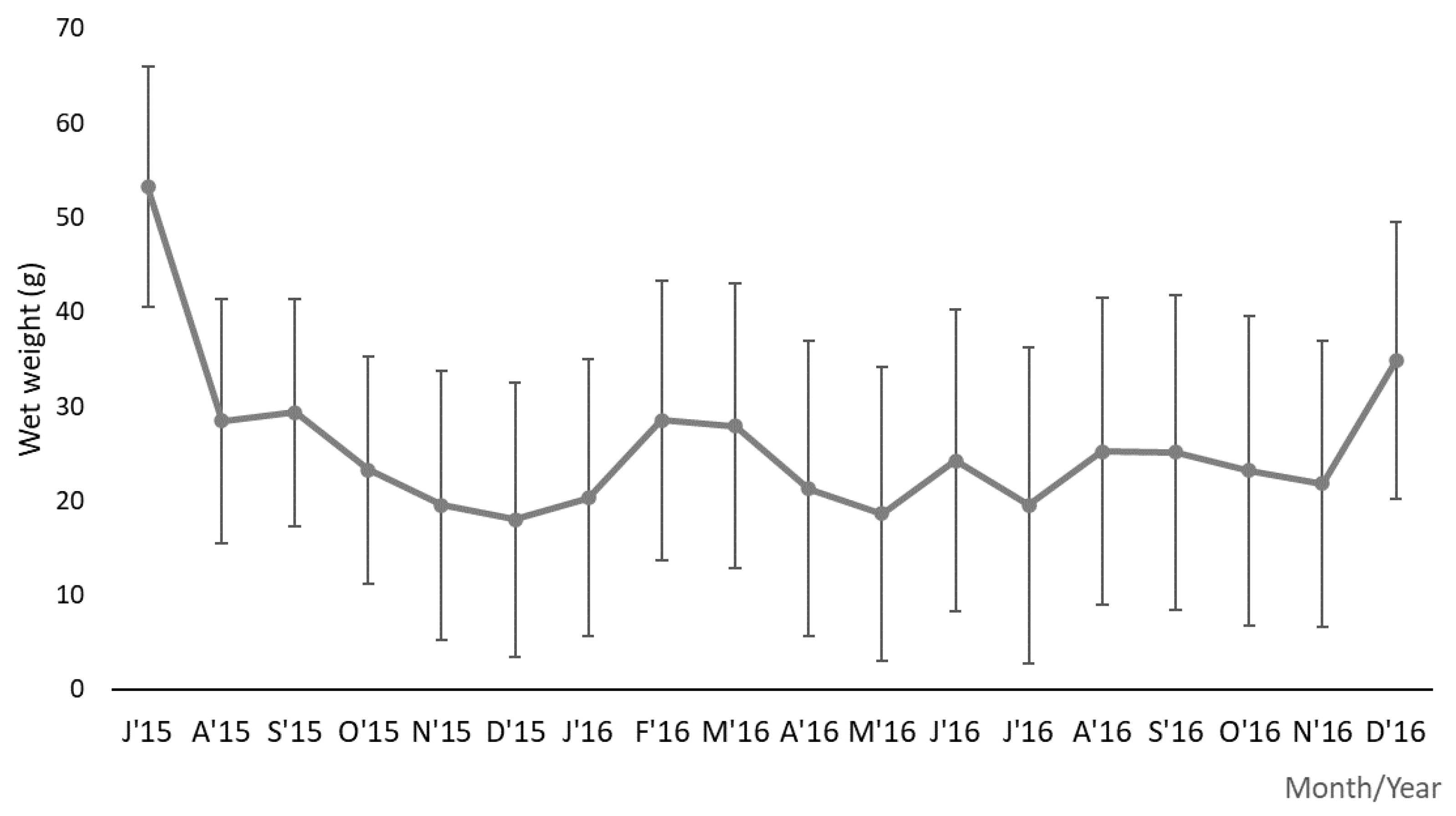
Figure 1. Monthly variation in individual wet weight (g) of sea urchins, Paracentrotus lividus, from July 2015 to December 2016 at Praia do Abalo (Peniche, Portugal).
Jmse 11 02366 g001
Figure 2. Statistical differences between the 18 months (July 2015–December 2016). (a). Total weight (TW) (b). Gonadal weight (GW) (c). Gonadosomatic index (GI) (d). Oocyte diameter (DO). Symbol indicates significant differences (p < 0.05).
Jmse 11 02366 g002
Figure 3. Monthly variation in gonadal weight (g) of sea urchins, Paracentrotus lividus, from July 2015 to December 2016 at Praia do Abalo (Peniche, Portugal).
Jmse 11 02366 g003
Figure 4. Monthly variation in the gonadosomatic index (%) of sea urchins, Paracentrotus lividus, and seawater temperature (°C) and photoperiod (hours of daylight) in Peniche (Portugal), from July 2015 to December 2016.
Jmse 11 02366 g004
Figure 5. Monthly variation in the oocyte diameter (µm) of sea urchins, Paracentrotus lividus, from July 2015 to December 2016 at Praia do Abalo (Peniche, Portugal).
Jmse 11 02366 g005
Figure 6. Monthly relative frequency (%) of male and female sea urchins, Paracentrotus lividus, from July 2015 to December 2016 at Praia do Abalo (Peniche, Portugal).
Jmse 11 02366 g006
Figure 7. Development stages of the gametogenic cycle observed in Paracentrotus lividus female gonads: (A)—Stage I: cross-section of the gonad, with visible globules (GB), derived from the lysis of non-released oocytes; noticeable nutritive phagocytes (NP) and pre-vitellogenic oocytes (PO) in the ascinal wall; (B)—stage II: growth of oocytes and ovaries; (C)—stage III: premature ovary, with oocytes in all stages of development, namely early formed vitellogenic oocytes (EV) and vitellogenic oocytes (VO) that detached from the ascinal wall, as they turned into mature oocytes (O), in which the nucleus became more evident (N). (D)—stage IV: ovary replete with mature oocytes and a very small number of nutritive phagocytes. (E)—stage V: ovary with loose ovules (LO), lack of nutritive material and emergence of empty spaces, which indicated the beginning of spawning. (F)—Partial spawning of an ovary presenting loss of the internal structure, with empty spaces (E) but with oocytes at different stages of development, but mostly vitellogenic oocytes (VO) that eventually matured and moved to the lumen; nutritious matter (NM) can be observed. (G)—Stage VI: post-spawned ovary, presenting loss of the internal structure (E), which resulted in the presence of voids; oocytes that were not released became dispersed inside the lumen, being reabsorbed later (R). (H)—Stage VII: all the non-released vitellogenic oocytes, mature oocytes and ova will be later reabsorbed; nutritive phagocytes concentrated in the lumen, forming an eosinophilic meshwork (EM), in order to enclose and lyse the oocytes (L); the ovary began to reorganise. Histological slides stained with hematoxylin and eosin (all bars = 200 μm).
Jmse 11 02366 g007
Figure 8. Development stages of the gametogenic cycle observed in Paracentrotus lividus male gonads: (A) Stage I: cross-section of an early testis, containing primary spermatocytes (PS) along the ascinal wall; a high number of nutritive phagocytes (NP) began to appear. (B) Stage II: spermatocyte columns projected to the centre (arrows). (C) Partial spawning of a testis in stage III: nutritive phagocytes (NP) in the centre and empty spaces left by released spermatozoa (V). (D) Stage III: testis filled with mature spermatozoa (S) and largely devoid of nutrient tissue. (E) Stage IV: mature testis, filled with mature spermatozoa (S) ready to be released. (F) Stage V: testis beginning to spawn, with voids (V) left by the released spermatozoa (S). (G) Stage VI: spawned testis presenting loss of the internal structure (E). (H) Stage VII: spawned testis with non-released spermatozoa that will be reabsorbed (R); nutritive phagocytes concentrated in the lumen (L), forming an eosinophilic mesh (EM) around the non-released spermatozoa. Histological slides stained with hematoxylin and eosin (A, B, D, E, F, G and H’s bars = 500 μm; C’s bar = 200 μm).
Jmse 11 02366 g008
Figure 9. Monthly relative frequency (%) of gametogenic stages during the 18 months of sampling at Praia do Abalo (Peniche, Portugal). Stage I—initial; stage II—growth; stage III—premature; stage IV—mature; stage V—spawning; stage VI—spent; stage VII—reabsorption.
Jmse 11 02366 g009
Disclaimer/Publisher’s Note: The statements, opinions and data contained in all publications are solely those of the individual author(s) and contributor(s) and not of MDPI and/or the editor(s). MDPI and/or the editor(s) disclaim responsibility for any injury to people or property resulting from any ideas, methods, instructions or products referred to in the content.