Article

Effects of the Ionic Liquid [BMIM]Cl on the Baltic Microphytobenthic Communities

by
Zuzanna Sylwestrzak
,
Aleksandra Zgrundo
* and
Filip Pniewski
Faculty of Oceanography and Geography, University of Gdańsk, Al. Piłsudskiego 46, 81-378 Gdynia, Poland
*
Author to whom correspondence should be addressed.
J. Mar. Sci. Eng. 2022, 10(9), 1223; https://doi.org/10.3390/jmse10091223
Submission received: 15 July 2022 / Revised: 26 August 2022 / Accepted: 28 August 2022 / Published: 1 September 2022

Abstract

:
Ionic liquids (IL) are regarded as the solution to the modern world’s need to create and use compounds that exhibit a range of desirable properties while having a low environmental impact. However, recent reports are shattering the image of ionic liquids as environmentally friendly substances, especially in relation to the aquatic environment, revealing their potentially toxic effects. To assess the potential environmental impact of ILs, we conducted an experiment involving 1-butyl-3-methylimidazolium chloride ([BMIM]Cl), a substance considered to be the least hazardous among the imidazolium chloride ILs, on Baltic microphytobenthic communities. Microphytobenthos collected from the environment was tested under controlled laboratory conditions, and both the cell counts and the chloroplast condition were used as endpoints. It was shown that [BMIM]Cl at concentrations of 10−3 and 10−2, considered safe based on a cumulative impact assessment, has a negative effect on the condition of the microalgal cells and causes a reduction in population size. Although, under the influence of [BMIM]Cl, only a small proportion of the species was eliminated from the communities, only two species among those important to the communities showed resistance to this compound and eventually began to dominate the communities.

1. Introduction

The concept of sustainable development was introduced at the end of the 20th century, and it is based on the idea of social and economic development, which assumes that while meeting the needs of contemporary societies it will not limit the development opportunities for future generations. Sustainable development assumes a parallel development of the economy, society, and the environment. In line with these assumptions, more and more environmentally friendly substances, such as ionic liquids (ILs), have started to be used in industry. These are substances that are gaining increasing recognition and researchers in many scientific fields are interested in them as they are characterized by a set of specific properties, such as low vapor pressure, non-flammability, thermal and electrochemical stability, good conductivity, and catalytic properties [1,2]. They are used in various chemical processes, where they represent a new alternative to traditional organic solvents [3]. ILs are often called ‘designer solvents’; the appropriate selection of anions and cations allows for the creation of a suitable chemical compound, depending on the future application [4,5]. However, with regard to ILs’ specific features (e.g., high solubility, thermal stability, and/or poor biodegradability in water), they may potentially pollute the aquatic environment [6]. Science has known of ILs as solvents since 1914 [7]. However, the first stable ILs were described in 1995 [5]. Since then, there has been a rapid increase in interest in these substances, especially in terms of their effects on human health and the environment, as in, e.g., [6,7,8,9,10,11,12,13,14,15]. A major threat from ionic liquids is their low degradation rate. For example, a 28-day experiment showed a complete lack of biodegradation of [BMIM]Cl [7]. As a result, after years of research, their “green” status has been questioned [8,9,10]. Inadequate wastewater treatment, accidental spillage, or improper storage of waste contaminated with ILs can lead to the release of these substances into the environment where they subsequently cause negative effects in the ecosystem [1]. Numerous studies have proven the deleterious effects of ILs towards microalgae [16,17,18,19,20,21]. The toxicity of ILs depends on temperature and pH. Under conditions described as moderate, i.e., room temperature and pH close to neutral, these substances are stable. During industrial processes, the physicochemical conditions can change, and it has been shown that an acidic condition (pH about 3) and high temperature (of the range between 60 °C and 100 °C) accelerate the hydrolysis of the ILs, causing an increase in their toxicity. The IL used in this study was 1-butyl-3-methylimidazolium chloride ([BMIM]Cl), which is characterized by a relatively short alkyl chain [9]. The toxicity of ILs increases with the elongation of the alkyl chain [2]; hence, the IL used here is not considered to be a highly toxic substance [22]. However, its toxicity is comparable to that of chlorinated organic substances, such as dichloromethane and chloroform; thus, it is more dangerous to the environment than common solvents of organic origin (e.g., acetone, methanol, and ethanol). Due to its properties, [BMIM]Cl is used in cellulose processing, among other areas. Although the ionic liquid itself can be recovered to a very high degree during industrial applications (such as the one mentioned above), the very process of creating the imidazole cation involves the use of large amounts of natural organic materials, energy, and solvents, causing harmful emissions to both air and water as well [7].
The photosynthetic organisms that form the microphytobenthos are extremely valuable elements of aquatic ecosystems due to their role as primary producers, combined with oxygen production and CO2 reduction. Understanding the joint response of photosynthetic organisms forming a microphytobenthic community is extremely important in order to reliably estimate the changes that may occur following the introduction of this increasingly common substance into the environment and to assess the associated risks.
Monitoring the response of organisms to potentially toxic substances introduced into the ecosystem is an important part of environmental quality control. To date, investigations into the toxicity of ionic liquids have provided information on the response of single algal strains under laboratory conditions, e.g., [21,22,23]. Previous ecotoxicological studies conducted on marine microphytobenthic communities in the Baltic Sea have tested substances such as irgarol 1051, Sea-Nine™211 (DCOIT), and TBT (trin-butyltin) [24,25]. However, most of the studies used only communities developed at salinities typical of marine waters, i.e., 32–36 PSU. Only studies conducted to determine the impact of glyphosate and copper ions on microphytobenthic communities were performed on organisms collected from environments with salinities around 8 [26,27]. Further ecotoxicological tests on other potentially toxic substances carried out on microphytobenthic communities typical of brackish waters, which are considered species-minimum waters for macrozoobenthos and macroalgae and aquatic higher plants, but which support an abundance and diversity of planktonic microorganisms [28], are an interesting contribution to the existing knowledge. In the case of ILs, the salinity aspect is extremely important because the toxicity of ILs increases in inverse proportion to the salinity [16,17,18,19,20,21]. At high salinity values, the toxicity of ILs decreases, probably due to the reduced permeability of the microalgal cell membranes which limits the migration of harmful cations [29]. Our study was designed to provide a general picture of the response of multispecies microalgal communities to an IL considered to be of relatively low toxicity, i.e., 1-butyl-3-methylimidazolium chloride—[BMIM]Cl, under brackish water conditions, and to complement the existing knowledge on the potential risks arising from the widespread use of this substance. For that purpose, observations were made at the population level, i.e., the change in species composition and the community dominance structure were determined, and at the cell level, i.e., the condition of the chloroplasts was analyzed.

2. Materials and Methods

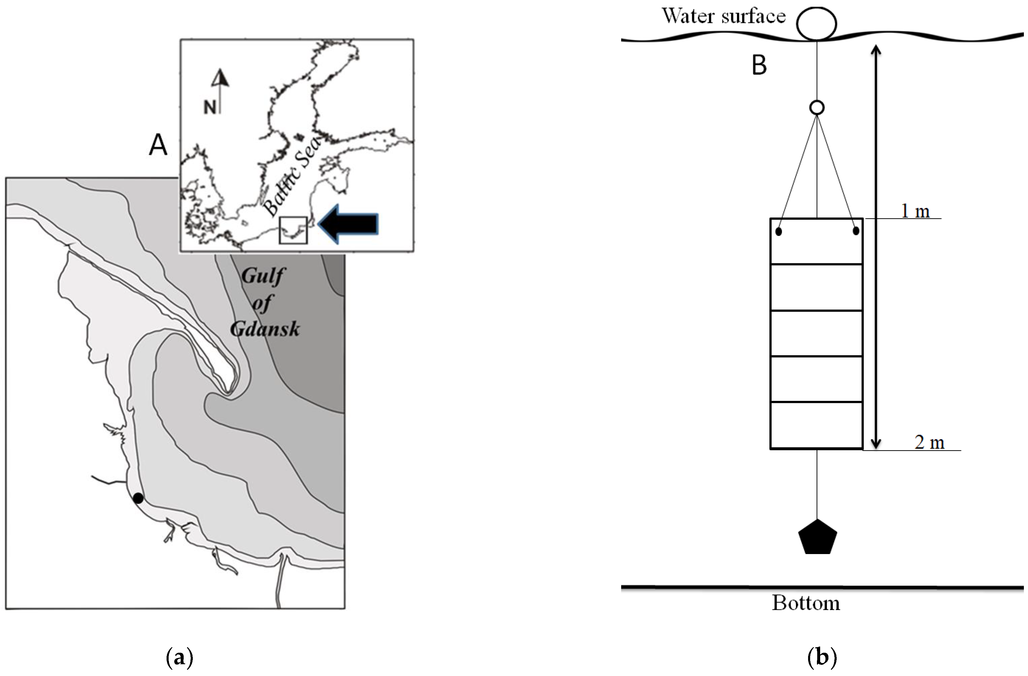
2.1. Study Area and Field Works

The experiment investigating effects of the IL [BMIM]Cl on microphytobenthic communities is one of a series of tests based on the identical methodology described in detail in [26]. The experiments were conducted in parallel on communities with identical species composition to allow for the comparison of the results. In brief, the study material was collected from glass slides mounted on a dedicated culture panel (Figure 1b) exposed in the Gulf of Gdańsk waters at a distance of 300 m from the shore (54°26′49″ N, 8°34′24″ E) (Figure 1a) for two weeks. During this time, the temperature and salinity changed within limited ranges, i.e., 17–19 °C and 7.9–8.4 PSU, respectively. The 2-week incubation period allowed for the acquisition of a relatively rich and diverse microphytobenthic community but was still devoid of organisms such as fouling macroalgae and fauna that will eventually dominate the surface of any substrate in marine waters in the long term.

2.2. Microalgal Material Preparation Procedure and Experimental Design

In the laboratory, the microphytobenthic communities were removed from the microscope slides by scraping them off with a scalpel. Subsequently, the microalgal cells were re-suspended in the seawater collected at the sampling site, which was first filtered through a glass filter (Whatman GF/C) and then autoclaved. The obtained microphytobenthos suspension was then sonicated, which allowed for the disruption and removal of cell aggregates. The sonication power was carefully chosen in order not to weaken or damage the cells [26].
The experiment was carried out in 250 mL flasks filled with 100 mL of microalgal suspension. Each microphytobenthos culture was insufflated with nitrogen for 30 s to remove heterotrophic microorganisms [30,31]. At the beginning of the experiment, the mean microalgal cell abundance was 38,800 cells/mL ± 700. Before the experiment, flasks with microalgal suspension were maintained in a thermostatic chamber for 72 h at constant light, temperature, and salinity conditions (i.e., 60 μmol photons·m−2·s−1 with a photoperiod L:D 16:8 h, 18 °C ± 1 °C, and 8 PSU, respectively) to let the communities acclimate to the experimental conditions. The natural concentrations of the nutrient compounds in the sea water were: N-NH4 9.4 mg·m−3, N-NO3 102 mg·m−3, P-PO4 36 mg·m−3, and Si-SiO4 600 mg·m−3. As the nutrient concentrations in the natural Baltic water were sufficiently high to maintain the microphytobenthos community during the experiment, any kind of culture medium was not added. This was also dictated by the fact that the high nutrient content could facilitate the growth of random species rapidly responding to the increase in nutrient concentrations.
After the acclimation phase, the [BMIM]Cl toxicity tests were performed according to the following design: control—microphytobenthic assemblages kept in filtered sea water without the addition of the tested IL and test solutions—microphytobenthic assemblages treated with two [BMIM]Cl concentrations, i.e., 1.13 × 10⁻³ g·dm−3 and 1.75 × 10⁻² g·dm−3. The lower [BMIM]Cl concentration selected for the experiment was previously proven to have significant effects on the species composition of the Baltic microphytobenthic communities [32]. The higher concentration of [BMIM]Cl was inferred from previously published values indicated as having inhibitory effects on algae [18,33]. All experimental treatments were carried out in triplicates.

2.3. Microscopic Analysis

Qualitative and quantitative changes in assemblage composition and structure, i.e., changes in taxonomic composition and taxa abundance, were the primary parameters used to assess the changes in microphytobenthos. Microscopic analysis was conducted on microalgal material preserved in Lugol solution. The observations were conducted in all cultures on the third and seventh experiment day. Fifty fields of view were checked in Utermöhl chambers (2 mL) using a Nikon Eclipse TS100 inverted light microscope (magnifications of ×200 and ×400). All cells were counted and identified as laid out in the Utermöhl method [34] and Helcom [35] guidelines (cells or threads of 100 μm length are treated as units). Species were identified using appropriate keys and floras [36,37,38,39,40,41,42,43].
The analysis of the microalgal cell condition was also performed. For this purpose, the state of chloroplasts was observed and classified in one of three classes of cells: (1) live cells with normal chloroplasts, (2) live cells with abnormal chloroplasts, and (3) dead cells. Here, the results obtained for the two first cell classes are reported (Figure 2). The microalgal cell condition was evaluated in all cells counted in 50 fields of vision under a Nikon Eclipse 80i microscope fitted with a Nikon DSU2 camera at a magnification of ×400.

2.4. Statistical Analysis

Differences between means were verified with the Student’s t-test using STATISTICA version 10 (StatSoft Polska Sp. z o.o., Kraków, Poland). Principal component analysis (PCA) was carried out with the Canoco 5 (Microcomputer Power, Ithaca, NY, USA) [44,45] and similarity percentage (SIMPER) with the PRIMER-e (PRIMER-e, Auckland, New Zealand).

3. Results

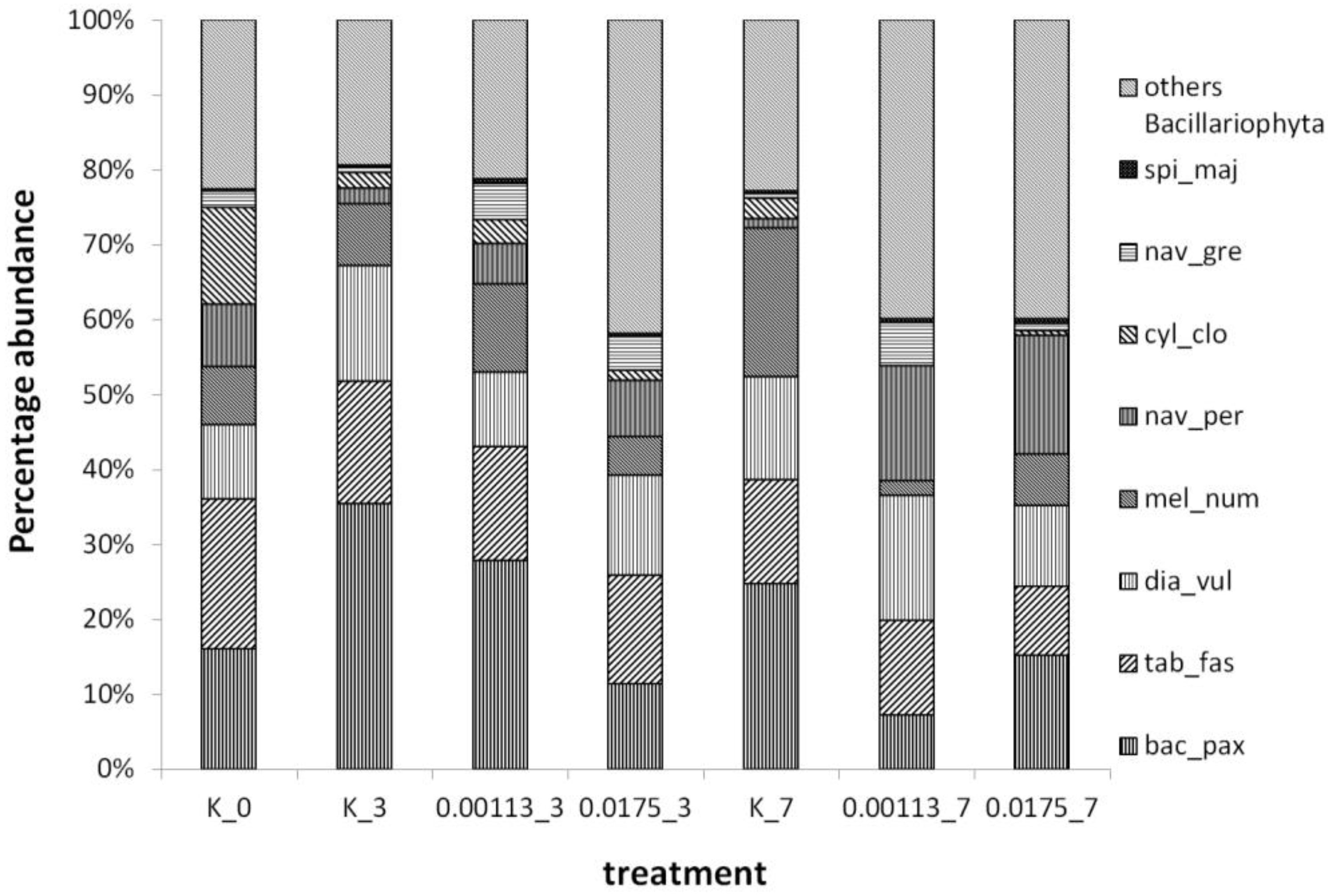
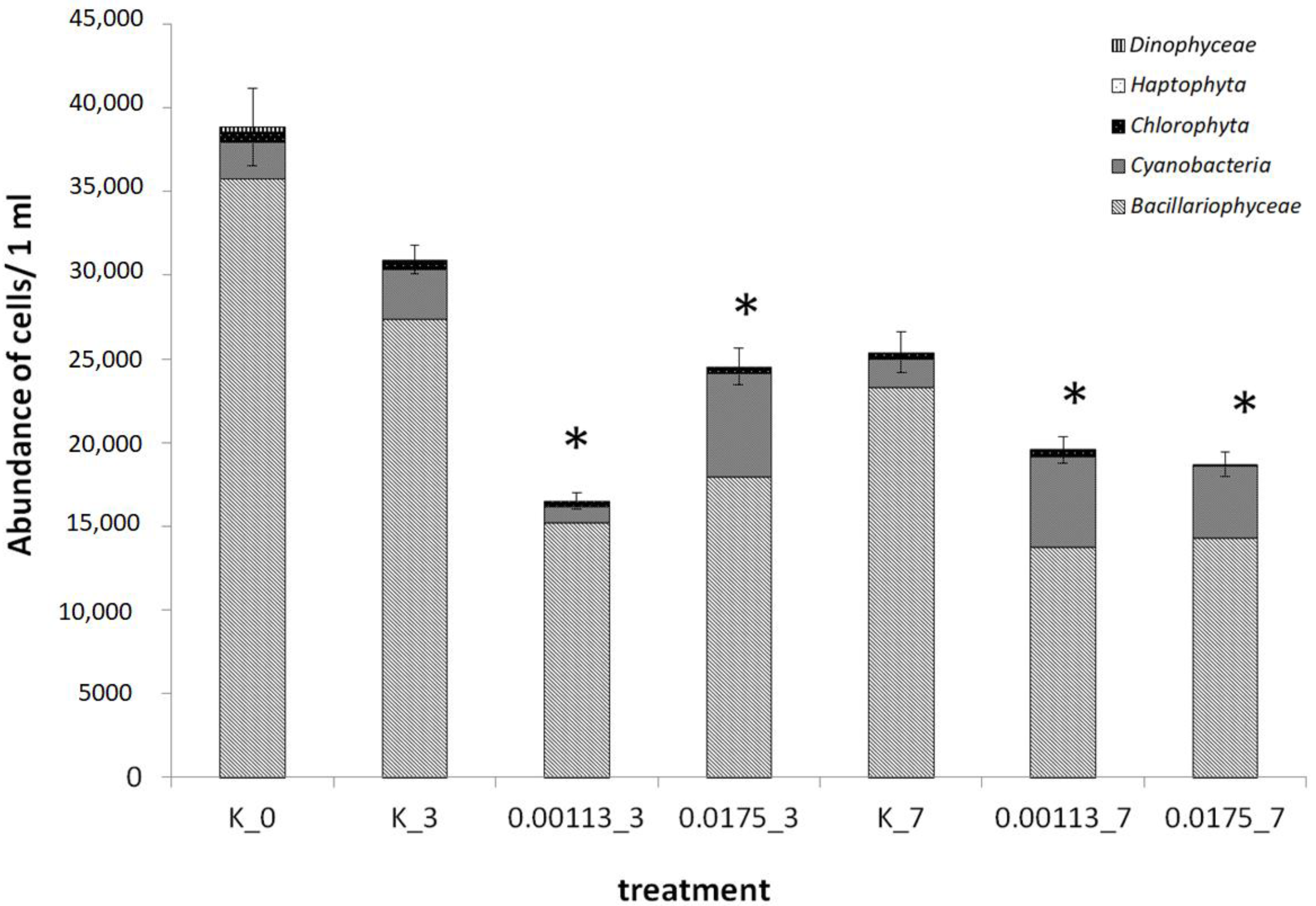
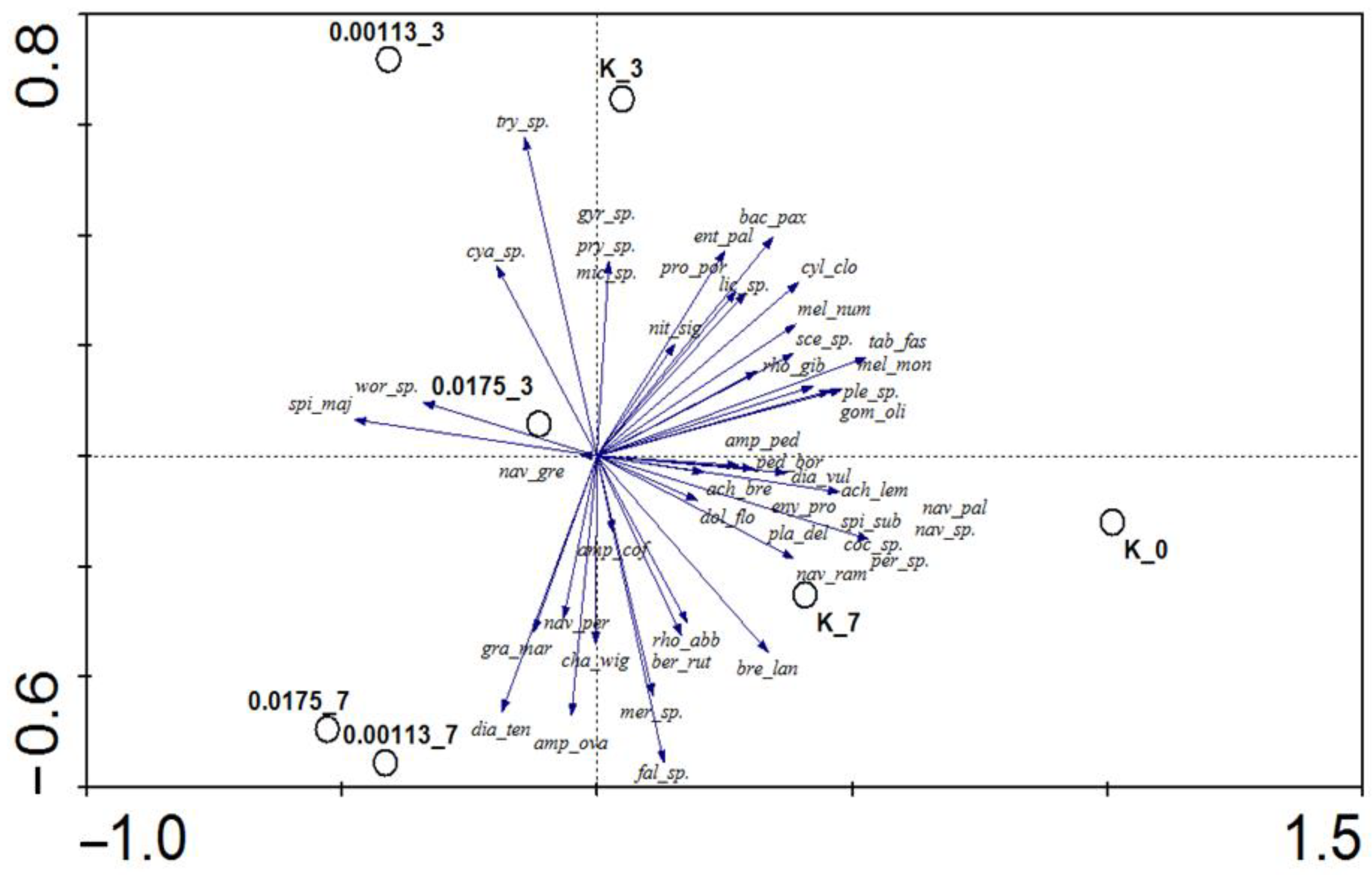
3.1. Analysis of Taxonomic Composition and Structure

A total of 46 microalgae species were identified, including 35 diatoms, 6 cyanobacteria, and 2 green algae taxa, as well as representatives of dinoflagellates (Peridinium sp.) and haptophytes (Prymnesium sp.) (the list of all identified taxa is in the Appendix A (Table A1).
At the beginning of the experiment, the highest number of cells, 38,800 ± 700 cells/mL, was found (Figure 3). On the third day, a 47% decrease in the number of microalgae cells was observed in the concentration of 1.13 × 10⁻³ g·dm−3, while in the concentration of 1.75 × 10⁻² g·dm−3 [BMIM]Cl, a drop by only 21% was observed. On the seventh day of testing, a similar abundance of microalgae was observed in both concentrations, 23–26% less than in the control solution. All differences in abundance between the concentrations tested and the control solution were statistically significant (p < 0.05).
The microphytobenthic communities were heavily dominated by diatoms, constituting from 70% to 92% of all the observed photosynthetic microorganisms. In the control cultures, the abundance of cyanobacteria did not exceed 10% (the highest number was observed on the third day of the experiment). On the seventh day, at both [BMIM]Cl concentrations, i.e., 1.13 × 10⁻³ g·dm−3 and 1.75 × 10⁻² g·dm−3, they represented 23% and 28% of the total abundance, respectively. The abundance of Prymnesium sp. (Haptophyta) did not exceed 0.3% during the whole experiment, while the abundance of Peridinium sp. (Dinophyceae) in the control solution at the start of the tests was only 0.6%.
SIMPER similarity analysis showed a high similarity in community composition and structure at the control and both ionic liquid concentrations during the experiment. The average similarity was calculated as high as 71.92%. Based on the PCA analysis performed on the quantitative data, it was found that in addition to the concentration of the ionic liquid, the duration of the experiment also influenced the transformation of communities (Figure 4). At the start of the experiment (point K_0, right part of the graph), the community was the richest (i.e., it was characterized by the highest number of species). On the third day of testing (upper left part of the graph), an increased proportion of cyanobacteria was observed (e.g., marked in graph as cya_sp., mic_sp., spi_mai, wor_sp.). The group of organisms dominating on the seventh day of testing (lower left part of the graph) included, among others, a diatom characterized by a relatively high resistance to ionic liquid—Navicula perminuta (marked as nav_per). However, based on the PCA analysis, it was not possible to delineate groups of organisms that were unequivocally sensitive or tolerant to the IL tested.
Based on SIMPER analysis, the most important species in the communities were distinguished in terms of abundance (Appendix A, Table A2). The most abundant species was Bacillaria paxilifera (up to 36% on the third day in the control solution) (Figure 5). The second most abundant species was Tabularia fasciculata (up to 23% of all cells on the initial day of the experiment). The abundance of Diatoma vulgaris ranged from 9% to 17% throughout the experiment. For Melosira nummuloides, the highest number of cells was observed on the seventh day of testing in the control solution (20% of all cells). Interesting changes were observed in the case of N. perminuta; the proportion of this species in the initial community did not exceed 8%, but on the seventh day at both [BMIM]Cl concentrations, i.e., 1.13 × 10⁻³ g·dm−3 and 1.75 × 10⁻² g·dm−3, the share of this taxon increased to 15% and 16%, respectively. The highest share of Cylindrotheca closterium in the community was observed at the start of the experiment (13%), but on subsequent days the share did not exceed 8%. In the case of Navicula gregaria, a maximum share of 6% was observed at a concentration of 1.13 × 10⁻³ g·dm−3 on the seventh day of testing. While the share of the only representative of cyanobacteria, Spirulina major, did not exceed 1% during the whole experiment.

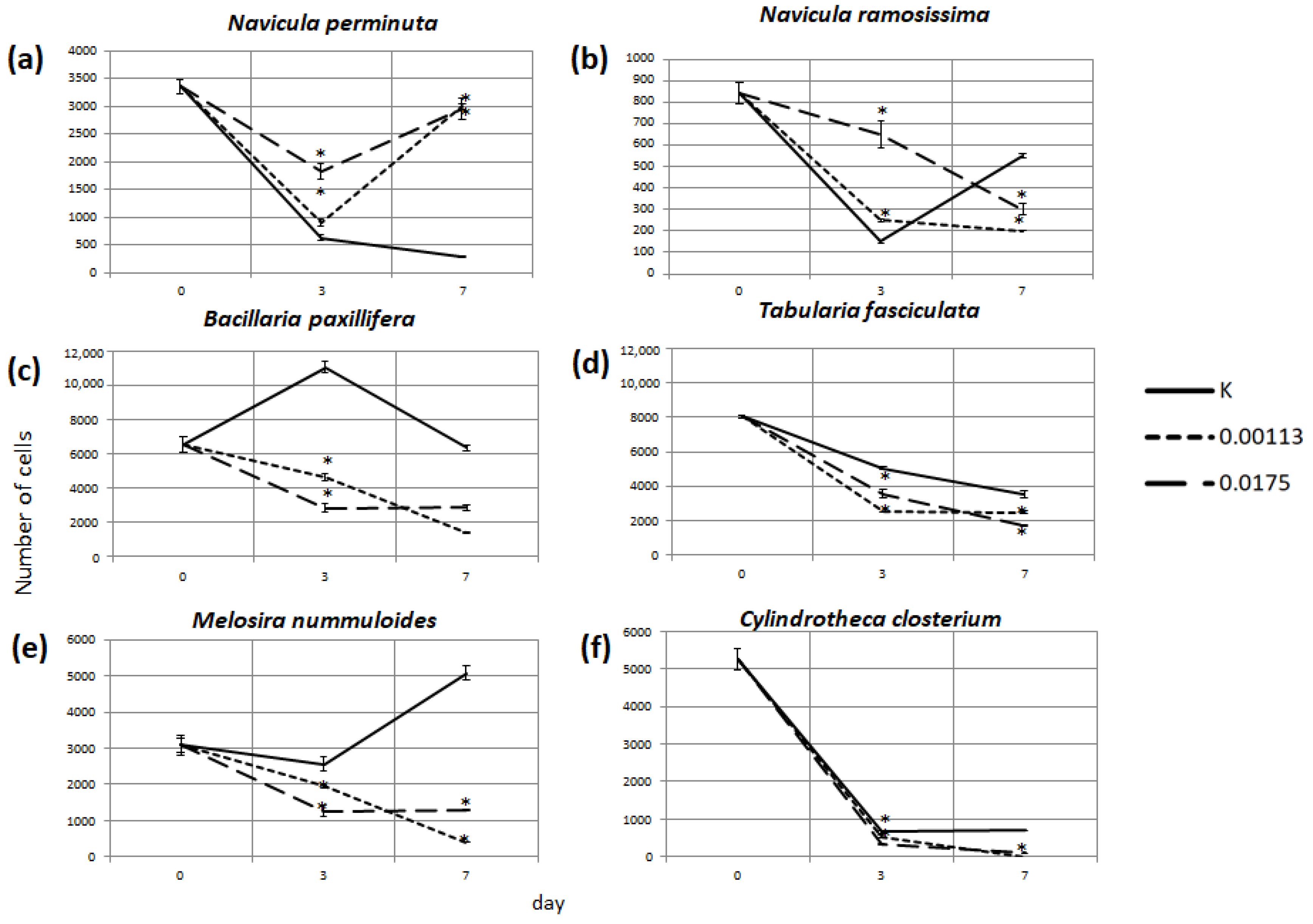
3.2. Abundance of Selected Taxa

In the presence of the IL, most of the taxa showed a statistically significant reduction in their abundance. Only for two taxa, i.e., N. perminuta and Navicula ramosissima, did the abundance in the [BMIM]Cl solutions increase compared to the control solution (Figure 6a,b). Hence, they were identified as tolerant species to this IL. The abundance of N. perminuta in the control solution on the seventh day decreased by 91% relative to the start of the experiment, while in both concentrations of the ionic liquid abundances of about 89% relative to the initial abundance were recorded. On the third day, in the lower [BMIM]Cl concentration cultures (1.13 × 10⁻³ g·dm−3), the cell abundance was 142% of the number in the control solution, while at the concentration of 1.75 × 10⁻³ g·dm−3 it was as high as 289%. On day seven, as much as a tenfold rise in the cell abundance was observed for this taxon as compared to the control solution. A similar growth stimulation was noted on day three for N. ramosissima, but on day seven, the cell number was only 45% and 64% of that in the control depending on the [BMIM]Cl concentration.
Figure 6c–f presents changes in the abundance of selected taxa considered most important in the communities in relation to the abundance based on the SIMPER analysis. The reduction in cell numbers in the [BMIM]Cl solutions tested indicated statistically significant (p < 0.05) growth inhibition. Such a response was characteristic of almost all the organisms observed in the microphytobenthic communities. However, depending on the taxon and concentration used, either a gradual reduction in abundance over time (e.g., B. paxillifera and M. nummuloides in the solution of 1.13 × 10⁻³ g·dm−3 [BMIM]Cl) or a reduction in abundance followed by an initial increase (e.g., B. paxillifera and M. nummuloides in the solution of 1.75 × 10⁻² g·dm−3 [BMIM]Cl or T. fasciculata in the solution of 1.13 × 10⁻³ g·dm−3 [BMIM]Cl) was observed. The most dramatic changes in the abundance of C. closterium were noted. In the solution of IL of 1.13 × 10⁻³ g·dm−3, no living representatives of this species were observed on day seven, and in the 1.75 × 10⁻² g·dm−3, only 14% of the abundance in the control solution was noted.

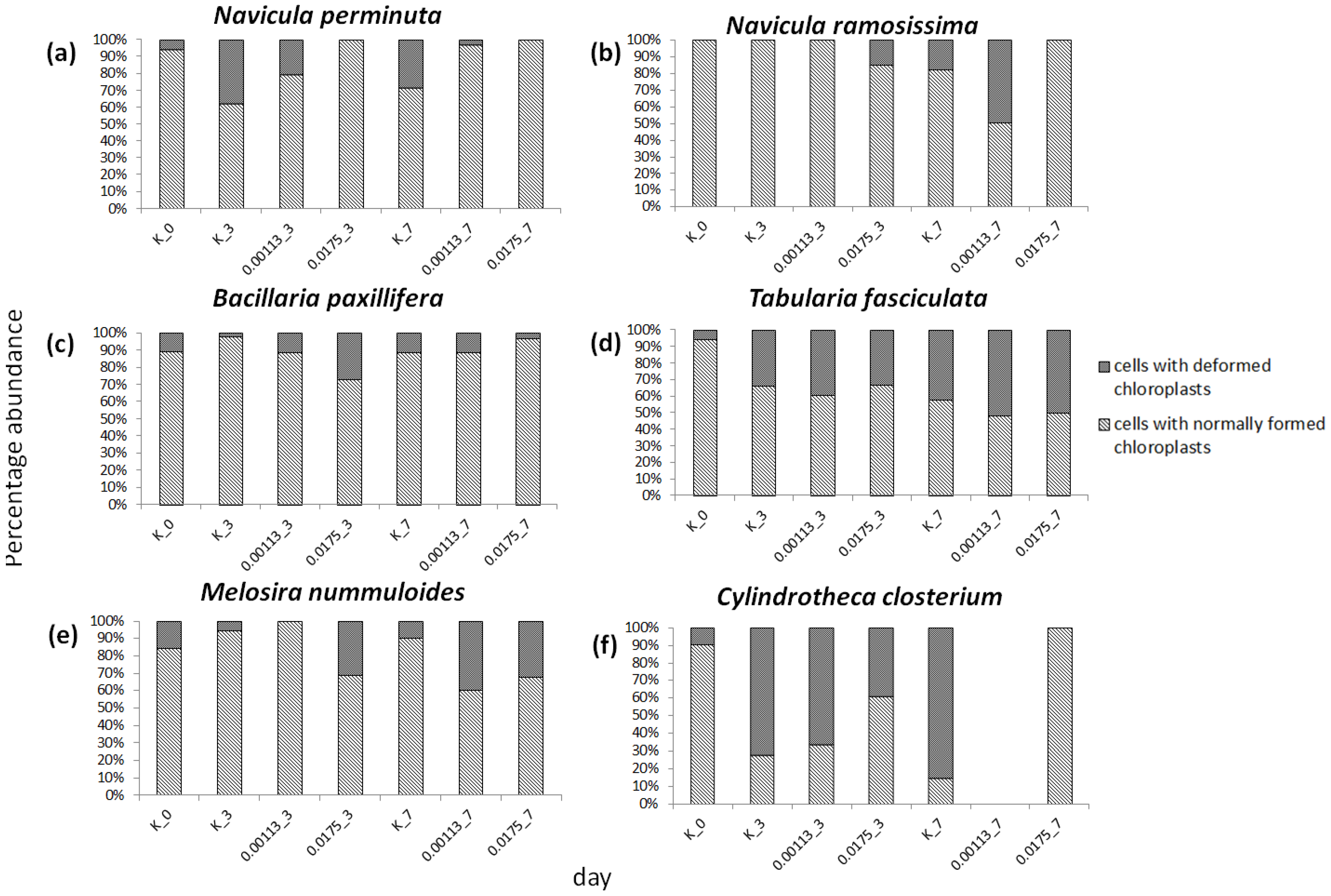
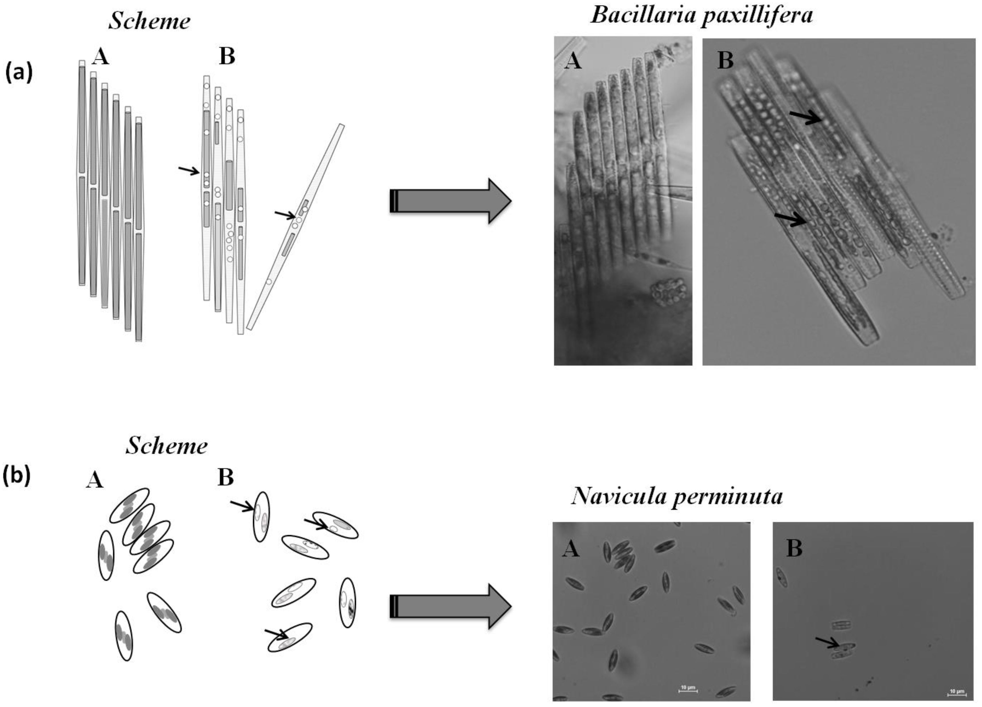
3.3. Cell Condition in Selected Taxa

Analysis of the chloroplast state in the cells provided complementary information on the differences in the response of the various taxa to [BMIM]Cl. In species considered tolerant, i.e., N. perminuta and N. ramosissima, a small number of cells with abnormally shaped chloroplasts were observed (Figure 7a,b). Interestingly, in the case of N. perminuta, cells with abnormally shaped chloroplasts were mainly observed in the control solution (e.g., up to 38% of all cells on the third day) and were not present or were only in small proportions in the IL solutions (up to 20% in the solution of 1.13 × 100−3 g·dm−3 [BMIM]Cl). Similarly, in N. ramosissima, abnormally shaped chloroplasts were not observed in the cells at the beginning of the experiment. Deformed chloroplasts were present in the cells on the third day in the solution of 1.75 × 100−2 g·dm−3 [BMIM]Cl (15% of cells) and on the seventh day in the control solution (18% of cells) and in the solution of 1.13 × 100−3 g·dm−3 (50% of cells).
For the taxa considered sensitive, cells with deformed chloroplasts were observed irrespective of the solution and day of the experiment (Figure 7c–f). For example, in B. paxillifera less than 15% of cells were characterized by deformed chloroplasts. Only in the solution of 1.75 × 100−2 g·dm−3 [BMIM]Cl on the third day was chloroplast degradation observed in 27% of the living cells. In T. fasciculata, except for at the beginning of the experiment, cells with degraded chloroplasts accounted for about 30–40% of all the cells. However, the highest number of cells with degraded chloroplasts was observed on the last day of testing in both IL solutions (about 50% of all cells). Similarly, for M. nummuloides in the control solution and at the beginning of the experiment, cells with abnormally formed chloroplasts made up a small proportion of the population (0–16%). In contrast, in the solution of 1.13 × 100−3 g·dm−3 [BMIM]Cl, up to 40% of the cells with damaged chloroplasts were observed on the seventh day and in the solution of 1.75 × 100−2 g·dm−3, 31% and 32% of cells on the third and seventh day, respectively. A significantly worse cell condition was observed in C. closterium as compared to the previously described species. In most of the solutions tested, cells with abnormally shaped chloroplasts accounted for about 40–85% of all the cells. Only at the solution of 1.75 × 100−2 g·dm−3 [BMIM]Cl did all of the cells have chloroplasts of normal shape, but the population size was low.

4. Discussion

ILs as solvents with salt structures have been known since 1914 [7]. However, the first stable ILs were described in 1995 [5]. The beginning of the 21st century brought the possibility of designing chemical compounds combining required biological properties with preferred physicochemical characteristics. Currently, these substances are being studied on a massive scale, as evidenced by the number of peer-reviewed publications e.g., [6,9,10,12,13,14]. Furthermore, the list of their potential applications as reaction media in many industrial fields is growing [9,12]. ILs have also found applications in medicine due to their antibacterial, antifungal, anticholinergic, and local anesthetic activities and agrochemistry as bactericides, fungicides, herbicides, plant growth stimulants, or wood preservatives [9,46]. Although ILs are popular in research and economics, this does not necessarily correspond to the amount of research related to the monitoring of these compounds in the environment and the subsequent risk assessment; the number of papers focusing on the presence of ILs or their compounds in the environment remains small [10,47,48,49,50]. In one of such studies, 1310 pollutants were identified in riverine waters in Germany, among which ca. 20 different compounds belonging to ILs were detected in concentrations of up to μg·dm−3 [48]. In addition, in the United States, based on analyses of sediments from lakes located within the state of Minnesota, it was shown that the concentration of the IL C4-PYR was 0.053 µg·dm−3 [51]. In this context, one of the ILs, i.e., 1-octyl-3-methyl imidazolium, is of particular significance as it was identified not only in environmental samples [52] but also in human blood [53].
The picture is completed by the fact that imadazolium-based ILs, such as [BMIM]Cl tested here, have a low rate of biodegradation and are resistant to photodegradation [54,55,56]. Previous studies have shown that ILs, after eventual emission into the environment, may behave similarly to some persistent organic pollutants [57]. An extremely important aspect is also the fact that the technology to effectively remove ILs from wastewater is still being developed [4]. Hence, it is to be expected that, due to the increasing popularity of ILs, they will be used widely and, consequently, will be uncontrollably introduced into the aquatic environment, remaining there for a long time due to the difficulties associated with water treatment and their poor biodegradability. Therefore, it is of paramount importance to investigate the ILs’ toxicity under environmental conditions and not only under controlled laboratory conditions [58]. Our tests on the effects of the IL [BMIM]Cl, considered to be relatively harmless [32,33,57], on the whole communities of microphytobenthos collected from the environment, allowed us to determine the response of a wide spectrum of microorganisms and not just single strains as in standard ecotoxicological tests. Changes in the tested communities at the population and cellular level show in a more reliable way the direction of the changes to which the microphytobenthos, an important component of the marine ecosystem, is subjected.
In a study using cumulative impact assessment, it was found that the [BMIM]Cl turned out to be the least hazardous among the imidazolium chloride ionic liquids with the Safe Environmental Concentration (SEC) as high as 750 × 100−3 mmol/L, which corresponds to the concentration of 1.31 × 100−3 g·dm−3 [59]. In this study, however, it was shown that the concentration of 1.13 × 100−3 g·dm−3 already reduced the abundance of dominant species by 20% to 60% within 3 days and up to 75% within 7 days. Only two diatom species of higher abundances showed resistance and gained a quantitative advantage in the studied communities. The number of cells of N. perminuta, for example, increased, reaching almost 290% of the abundance in the control solution on the third and 1000% on the seventh day of the experiment. As a result, the total abundance of cells comprising the communities decreased by only 25%. Although the composition of the communities remained similar, the abundance structure changed due to the strong dominance of tolerant taxa—apart from diatoms, cyanobacteria also showed some resistance. Similar observations regarding the substitution of sensitive taxa by tolerant or indifferent taxa were found during experiments on the same Baltic microalgal communities, testing the effects of copper chloride [27] and glyphosate [26]. However, effects induced by the aforementioned substances were also manifested by a drastic shift in species composition, i.e., the increased contribution of cyanobacteria to the total community abundance.
Interesting changes were observed for the second taxon selected as resistant to the presence of the IL [BMIM]Cl—N. ramosissima. On the third day of the experiment, its cell numbers increased significantly, but on the seventh day, the cell abundance again decreased in the IL concentrations tested. Such a reaction may indicate depletion of the IL molecules, the presence of which has a stimulating effect on the test organisms. For example, in toxicological tests conducted by [20] on Scenedesmus obliquus, low concentrations of ILs have been shown to stimulate cells for biological activity (e.g., by changing the activity of catalase and superoxide dismutase). However, this may also be a response related to the competitive activity of other taxa, such as the predominant N. perminuta. Similarly, [21] selected from their study several species for which the IL [BMIM]Cl was practically harmless, i.e., the cyanobacterium Anabaeana cylindrica and the green alga Chlorella pyrenoidosa, and in the case of the green alga Dunaliella salina, they concluded that it was relatively harmless.
Typically, the reactions of taxa to toxicants tested in communities are milder than in laboratory tests conducted on monocultures [26,27,60]. In our studies, conducted on communities grown in nature, B. paxillifera cell counts decreased by 55% on the seventh day of the experiment in the solution of 1.75 × 100−2 g·dm−3 [BMIM]Cl. A very similar response was observed in toxicological tests conducted on a monoculture of B. paxillifera isolated from the Baltic Sea—the concentration of 1.75 × 100−2 g·dm−3 decreased the growth of the strain by 58% on the seventh day of testing [61]. A similarly large reduction was shown for the same concentration in T. fasciculata (50% reduction on the seventh day). Moreover, [19] observed an inhibition of 50% cell growth (EC50) in the planktonic diatom Skeletonema marinoi at the concentration of 0.1 mM [BMIM]Cl (1.745 × 100−2 g·dm−3). In turn, for the green alga Chlorella pyrenoidosa, 50% inhibition of growth (IC50) was shown for the concentration of 21.4 × 100−2 g·dm−3 [21]. The phenomenon of the same species reacting the same way to a substance regardless of how it is cultured and tested (individually or in communities) may indicate that the communities as a whole, but also the individual components of the community, do not necessarily have mechanisms to protect them from the toxic effects of the IL under testing.
Exposure time was also shown to be a variable having an effect on the action of the IL because, as reported by [62], excessive accumulation of the IL in microorganisms increases its effect. In the case of M. nummuloides, which was part of the tested community in our study, a significant increase in cell number was observed in the control solution during the experiment, while on the third day at the [BMIM]Cl solution of 1.75 × 100−2 g·dm−3 50% fewer cells were observed, while on the seventh day the abundance decreased to 8% of the abundance in the control solution. A similarly rapid abundance reduction response to [BMIM]Cl was observed for the green alga Dunaliella salina [21]. This implies that there is a group of sensitive species in natural marine phytobenthic communities that may be rapidly eliminated from the environment while exposed to ILs, e.g., the aforementioned M. nummuloides or C. closterium.
The cell condition index used in our study, which consists of an assessment of the chloroplast state, confirmed the observations based on cell abundance. In the species considered sensitive, the percentage of cells with deformed chloroplasts was much higher than in the resistant species, e.g., up to a half of the observed cells of T. fasciculata on the seventh day of testing had degraded chloroplasts at both solutions of [BMIM]Cl. The negative effect on the chloroplast condition was confirmed by toxicity studies on five ILs ([Cnmim]Cl, n = 6, 8, 10, 12, 16). Ultrastructural morphology performed during the study revealed IL negative effects on various cellular structures, e.g., chloroplast grana became loose and mitochondria and their intermembranes swelled [22]. Other studies also confirmed that chloroplast damage can be considered as an indicator of microalgal degradation [27,63].
Tests carried out on the Baltic Sea microphytobenthic communities from the Gulf of Gdańsk made it possible to assess the effect of the IL [BMIM]Cl on the microorganisms comprising this formation. The study showed that 1-butyl-3-methylimidazolium chloride is a relatively harmful substance for the entire community, which contradicts the assessment carried out by reports based on cumulative impact assessment [47,59,64]. Thus, we have confirmed that even ILs considered to be of relatively low hazard have a significant impact on the aquatic environment. Hence, we are convinced that this group of compounds requires special attention in the context of testing its effects on different ecosystem components, its bioaccumulation, and its fate in the environment, as suggested recently by a growing group of authors [9,11,64].

5. Conclusions

During this study, the toxic influence of [BMIM]Cl on the marine microphytobenthic communities was demonstrated. The majority of species comprising the tested community reacted negatively to the presence of [BMIM]Cl at concentrations between 10−3 and 10−2 g·dm−3, with a reduction in cell abundance and a deterioration in cell condition. Only in the case of two dominant diatom species, N. perminuta and N. ramosissima, was a stimulation of growth observed. In conclusion, the IL [BMIM]Cl on a short time scale contributes to a reduction in the abundance of species representing diverse taxonomic groups, which translates into the decrease in the total abundance and biomass of the microphytobenthic communities. However, despite the elimination of individual taxa, it does not lead to the degradation of entire communities but to their transformation into communities strongly dominated by a few resistant taxa.

Author Contributions

Conceptualization, Z.S., A.Z. and F.P.; methodology, Z.S., A.Z. and F.P.; software, Z.S.; validation, Z.S., A.Z. and F.P.; formal analysis, Z.S., A.Z. and F.P.; investigation, Z.S.; resources, Z.S. and A.Z.; writing—original draft preparation, Z.S., A.Z. and F.P.; writing—review and editing, Z.S., A.Z. and F.P.; visualization, Z.S.; supervision, A.Z.; project administration, Z.S.; funding acquisition, Z.S. and A.Z. All authors have read and agreed to the published version of the manuscript.

Funding

This research was funded by a Research Project for Young Scientists from the Faculty of Oceanography and Geography, University of Gdansk (No. 538-G245-B209-16).

Institutional Review Board Statement

Not applicable.

Informed Consent Statement

Not applicable.

Data Availability Statement

All data are available upon request to the corresponding author.

Acknowledgments

The authors thank Adam Latała for his support in developing the idea for the experiments.

Conflicts of Interest

The authors declare no conflict of interest.

Appendix A

Table A1. List of taxa identified in the studied microphytobenthic communities with codes used in statistical analysis.
Group of OrganismsTaxon CodeTaxon NameAuthor
Bacillariophytaach_breAchnanthes adnataBory
ach_lemAchnanthes lemmermanniiHustedt
amp_ovaAmphora ovalis(Kützing) Kützing
amp_pedAmphora pediculus(Kützing) Grunow
bac_paxBacillaria paxillifera(O.F. Müller) T. Marsson
ber_rutBerkeleya rutilans(Trentepohl ex Roth) Grunow
bre_lanBrebissonia lanceolata(C. Agardh) R.K. Mahoney & Reimer
cha_wigChaetoceros wighamiiBrightwell
coc_sp.Cocconeis sp.Ehrenberg
cyl_cloCylindrotheca closterium(Ehrenberg) Reimann & J.C. Lewin
dia_tenDiatoma tenuisC. Agardh
dia_vulDiatoma vulgarisBory
eny_proEncyonema leibleinii(C. Agardh) W.J. Silva, R. Jahn, T.A.V. Ludwig, & M. Menezes
ent_palEntomoneis paludosa(W. Smith) Reimer
fal_sp.Fallacia sp.Kützing
gom_oliGomphonella olivacea(Hornemann) Rabenhorst
gram_marGrammatophora marina(Lyngbye) Kützing
gyr_sp.Gyrosigma acuminatum(Kützing) Rabenhorst
amp_cofHalamphora coffeiformis(C. Agardh) Mereschkowsky
lic_sp.Licmophora gracilis(Ehrenberg) Grunow
mel_monMelosira moniliformisC. Agardh
mel_numMelosira nummuloidesC. Agardh
nav_greNavicula gregariaDonkin
nav_palNavicula palpebralisBrébisson ex W. Smith
nav_perNavicula perminutaGrunow
nav_ramNavicula ramossisima(C. Agardh) Cleve
nav_sp.Navicula sp.Bory
nit_sigNitzschia sigma(Kützing) W. Smith
pla_delPlanothidium delicatulum(Kützing) Round & Bukhtiyarova
ple_sp.Pleurosigma sp.W. Smith
pro_porProschkinia poretzkajae(Koretkevich) D.G. Mann
rho_abbRhoicosphenia abbreviata(C. Agardh) Lange-Bertalot
rho_gibRhopalodia gibba(Ehrenberg) O. Müller
tab_fasTabularia fasciculata(C. Agardh) D.M. Williams & Round
try_sp.TryblionellaW. Smith
Cyanobacteriadol_floDolichospermum flosaquae(Brébisson ex Bornet & Flahault) P. Wacklin, L. Hoffmann & J. Komárek
cya_sp.Cyanobacteria
mer_sp.Merismopedia sp.(Turpin) Meneghini
mic_sp.Microcystis sp.Lemmermann
spi_majSpirulina majorMeyen
spi_subSpirulina subsalsaOersted ex Gomont
wor_sp.Woronichinia sp.A.A. Elenkin
Chlorophytaped_borPseudopediastrum boryanum(Turpin) E. Hegewald
sce_sp.Scenedesmus sp.Meyen
Dinophyceaeper_sp.Peridinium sp.Ehrenberg
Haptophytapry_sp.Prymnesium sp.N. Carter
Table A2. Results of the similarity analysis calculated with SIMPER. Average similarity: 71.92%.
SpeciesAv. AbundanceAv. SimilaritySim/SDContribution%Cumulative%
Bacillaria paxillifera6.066.138.848.528.52
Tabularia fasciculata5.936.069.608.4316.94
Diatoma vulgaris5.735.899.088.1925.13
Melosira nummuloides5.095.027.196.9832.12
Navicula perminuta4.944.845.106.7338.85
Merismopedia sp.5.114.501.856.2645.10
Halamphora coffeiformis3.583.566.104.9450.05
Navicula gregaria3.733.524.994.9054.94
Navicula ramossisima3.543.406.274.7259.67
Cylindrotheca closterium3.993.171.784.4064.07
Grammatophora marina3.233.167.584.4068.47
Spirulina major2.382.557.163.5572.01

References

  1. Chen, B.; Xue, C.; Amoah, P.K.; Li, D.; Gao, K.; Deng, X. Impacts of Four Ionic Liquids Exposure on a Marine Diatom Phaeodactylum Tricornutum at Physiological and Biochemical Levels. Sci. Total Environ. 2019, 665, 492–501. [Google Scholar] [CrossRef] [PubMed]
  2. Mai, N.L.; Ahn, K.; Koo, Y.-M. Methods for Recovery of Ionic Liquids—A Review. Process Biochem. 2014, 49, 872–881. [Google Scholar] [CrossRef]
  3. Liu, D.; Liu, H.; Wang, S.; Chen, J.; Xia, Y. The Toxicity of Ionic Liquid 1-Decylpyridinium Bromide to the Algae Scenedesmus Obliquus: Growth Inhibition, Phototoxicity, and Oxidative Stress. Sci. Total Environ. 2018, 622, 1572–1580. [Google Scholar] [CrossRef]
  4. Isosaari, P.; Srivastava, V.; Sillanpää, M. Ionic Liquid-Based Water Treatment Technologies for Organic Pollutants: Current Status and Future Prospects of Ionic Liquid Mediated Technologies. Sci. Total Environ. 2019, 690, 604–619. [Google Scholar] [CrossRef] [PubMed]
  5. Szepiński, E. Synteza, Właściwości i Zastosowanie Cieczy Jonowych, Pochodnych Naturalnych Związków Organicznych. Ph.D. Thesis, Department of Organic Chemistry, Faculty of Chemistry, Gdansk University of Technology, Gdansk, Poland, 2019. [Google Scholar]
  6. Cvjetko Bubalo, M.; Radošević, K.; Radojčić Redovniković, I.; Halambek, J.; Gaurina Srček, V. A Brief Overview of the Potential Environmental Hazards of Ionic Liquids. Ecotoxicol. Environ. Saf. 2014, 99, 1–12. [Google Scholar] [CrossRef] [PubMed]
  7. Flieger, J.; Flieger, M. Ionic Liquids Toxicity—Benefits and Threats. Int. J. Mol. Sci. 2020, 21, 6267. [Google Scholar] [CrossRef]
  8. Nikitenko, S.I.; Berthon, C.; Moisy, P. Instability of Actinide (IV) Hexachloro Complexes in Room-Temperature Ionic Liquid [BuMeIm] PF6 Due to Hydrolysis of the Hexafluorophosphate Anion. Comptes Rendus Chim. 2007, 10, 1122–1127. [Google Scholar] [CrossRef]
  9. Freire, M.G.; Neves, C.M.S.S.; Marrucho, I.M.; Coutinho, J.A.P.; Fernandes, A.M. Hydrolysis of Tetrafluoroborate and Hexafluorophosphate Counter Ions in Imidazolium-Based Ionic Liquids. J. Phys. Chem. A 2010, 114, 3744–3749. [Google Scholar] [CrossRef]
  10. Maculewicz, J.; Świacka, K.; Stepnowski, P.; Dołżonek, J.; Białk-Bielińska, A. Ionic Liquids as Potentially Hazardous Pollutants: Evidences of Their Presence in the Environment and Recent Analytical Developments. J. Hazard. Mater. 2022, 437, 129353. [Google Scholar] [CrossRef]
  11. Kowalska, D.; Stolte, S.; Wyrzykowski, D.; Stepnowski, P.; Dołżonek, J. Interaction of Ionic Liquids with Human Serum Albumin in the View of Bioconcentration: A Preliminary Study. Chem. Pap. 2022, 76, 2405–2417. [Google Scholar] [CrossRef]
  12. Frade, R.F.; Afonso, C.A. Impact of Ionic Liquids in Environment and Humans: An Overview. Hum. Exp. Toxicol. 2010, 29, 1038–1054. [Google Scholar] [CrossRef] [PubMed]
  13. Costa, S.P.F.; Azevedo, A.M.O.; Pinto, P.; Saraiva, L. Environmental Impact of Ionic Liquids: An Overview of Recent (Eco) Toxicological and (Bio) Degradability Literature. ChemSusChem 2017, 10, 2321–2347. [Google Scholar] [CrossRef] [PubMed]
  14. Singh, S.K.; Savoy, A.W. Ionic Liquids Synthesis and Applications: An Overview. J. Mol. Liq. 2020, 297, 112038. [Google Scholar] [CrossRef]
  15. Armitage, J.M.; Erickson, R.J.; Luckenbach, T.; Ng, C.A.; Prosser, R.S.; Arnot, J.A.; Schirmer, K.; Nichols, J.W. Assessing the Bioaccumulation Potential of Ionizable Organic Compounds: Current Knowledge and Research Priorities. Environ. Toxicol. Chem. 2017, 36, 882–897. [Google Scholar] [CrossRef] [PubMed]
  16. Latała, A.; Stepnowski, P.; Nędzi, M.; Mrozik, W. Marine Toxicity Assessment of Imidazolium Ionic Liquids: Acute Effects on the Baltic Algae Oocystis Submarina and Cyclotella Meneghiniana. Aquat. Toxicol. 2005, 73, 91–98. [Google Scholar] [CrossRef] [PubMed]
  17. Latała, A.; Nędzi, M.; Stepnowski, P. Toxicity of Imidazolium Ionic Liquids towards Algae. Influence of Salinity Variations. Green Chem. 2010, 12, 60–64. [Google Scholar] [CrossRef]
  18. Kulacki, K.J.; Lamberti, G.A. Toxicity of Imidazolium Ionic Liquids to Freshwater Algae. Green Chem. 2008, 10, 104–110. [Google Scholar] [CrossRef]
  19. Samorì, C.; Sciutto, G.; Pezzolesi, L.; Galletti, P.; Guerrini, F.; Mazzeo, R.; Pistocchi, R.; Prati, S.; Tagliavini, E. Effects of Imidazolium Ionic Liquids on Growth, Photosynthetic Efficiency, and Cellular Components of the Diatoms Skeletonema Marinoi and Phaeodactylum Tricornutum. Chem. Res. Toxicol. 2011, 24, 392–401. [Google Scholar] [CrossRef]
  20. Fan, H.; Liu, H.; Dong, Y.; Chen, C.; Wang, Z.; Guo, J.; Du, S. Growth Inhibition and Oxidative Stress Caused by Four Ionic Liquids in Scenedesmus Obliquus: Role of Cations and Anions. Sci. Total Environ. 2019, 651, 570–579. [Google Scholar] [CrossRef]
  21. Zhu, Y.; Zhong, X.; Wang, Y.; Zhao, Q.; Huang, H. Growth Performance and Antioxidative Response of Chlorella Pyrenoidesa, Dunaliella Salina, and Anabaena Cylindrica to Four Kinds of Ionic Liquids. Appl. Biochem. Biotechnol. 2021, 193, 1945–1966. [Google Scholar] [CrossRef]
  22. Liu, H.; Zhang, S.; Zhang, X.; Chen, C. Growth Inhibition and Effect on Photosystem by Three Imidazolium Chloride Ionic Liquids in Rice Seedlings. J. Hazard. Mater. 2015, 286, 440–448. [Google Scholar] [CrossRef] [PubMed]
  23. Liu, H.; Zhang, X.; Chen, C.; Du, S.; Dong, Y. Effects of Imidazolium Chloride Ionic Liquids and Their Toxicity to Scenedesmus Obliquus. Ecotoxicol. Environ. Saf. 2015, 122, 83–90. [Google Scholar] [CrossRef] [PubMed]
  24. Dahl, B.; Blanck, H. Pollution-Induced Community Tolerance (PICT) in Periphyton Communities Established under Tri-n-Butyltin (TBT) Stress in Marine Microcosms. Aquat. Toxicol. 1996, 34, 305–325. [Google Scholar] [CrossRef]
  25. Porsbring, T.; Arrhenius, Å.; Backhaus, T.; Kuylenstierna, M.; Scholze, M.; Blanck, H. The SWIFT Periphyton Test for High-Capacity Assessments of Toxicant Effects on Microalgal Community Development. J. Exp. Mar. Biol. Ecol. 2007, 349, 299–312. [Google Scholar] [CrossRef]
  26. Sylwestrzak, Z.; Zgrundo, A.; Pniewski, F. Ecotoxicological Studies on the Effect of Roundup® (Glyphosate Formulation) on Marine Benthic Microalgae. Int. J. Environ. Res. Public. Health 2021, 18, 884. [Google Scholar] [CrossRef]
  27. Sylwestrzak, Z.; Zgrundo, A.; Pniewski, F. Copper Chloride (II) Effect on the Composition and Structure of Marine Microphytobenthic Communities. Environ. Monit. Assess. 2022, 194, 1–15. [Google Scholar] [CrossRef]
  28. Telesh, I.; Schubert, H.; Skarlato, S. Life in the Salinity Gradient: Discovering Mechanisms behind a New Biodiversity Pattern. Estuar. Coast. Shelf Sci. 2013, 135, 317–327. [Google Scholar] [CrossRef]
  29. Pham, T.P.T.; Cho, C.-W.; Yun, Y.-S. Environmental Fate and Toxicity of Ionic Liquids: A Review. Water Res. 2010, 44, 352–372. [Google Scholar] [CrossRef]
  30. Rosenberg, M.; Kulkarni, G.V.; Bosy, A.; McCulloch, C.A.G. Reproducibility and Sensitivity of Oral Malodor Measurements with a Portable Sulphide Monitor. J. Dent. Res. 1991, 70, 1436–1440. [Google Scholar] [CrossRef] [PubMed]
  31. Connolly, N.M.; Crossland, M.R.; Pearson, R.G. Effect of Low Dissolved Oxygen on Survival, Emergence, and Drift of Tropical Stream Macroinvertebrates. J. N. Am. Benthol. Soc. 2004, 23, 251–270. [Google Scholar] [CrossRef]
  32. Sylwestrzak, Z.; Zgrundo, A. Preliminary Results of Laboratory Tests Using Microphytobenthic Communities in the Gulf of Gdańsk for Toxicity Testing of Ionic Liquids. In Metodologia Badań Wykorzystywana Przez Młodych Naukowców; CREATIVETIME: Kraków, Poland, 2014; pp. 74–81. (In Polish) [Google Scholar]
  33. Sylwestrzak, Z.; Zgrundo, A.; Latała, A. Effect of the Ionic Liquid [BMIM] Cl on the Baltic Diatom Navicula ramosissma (C. Agardh) Cleve in a Laboratory Experiment on Natural Microphytobenthic Communities of the Gulf of Gdansk. In Zagadnienia Aktualnie Poruszane przez Młodych Naukowców 3; CREATIVETIME: Kraków, Poland, 2015; pp. 227–231. (In Polish) [Google Scholar]
  34. Utermöhl, H. Zur Vervollkommnung der Quantitativen Phytoplankton-MethodikMitteilungen Internationale Vereinigung Theoretische und Angewandte. Limnologie 1958, 9, 1–38. [Google Scholar]
  35. HELCOM. Manual for Marine Monitoring in the COMBINE Programme of HELCOM; HELCOM: Helsinki, Finland, 2014. [Google Scholar]
  36. Snoeijs, P.; Potapova, M. Intercalibration and Distribution of Diatom Species in the Baltic Sea; Opulus Press: Uppsala, Sweden, 1993; Volume 1. [Google Scholar]
  37. Snoeijs, P.; Vilbaste, S. Intercalibration and Distribution of Diatom Species in the Baltic Sea; Opulus Press: Uppsala, Sweden, 1994; Volume 2. [Google Scholar]
  38. Snoeijs, P.; Potapova, M. Intercalibration and Distribution of Diatom Species in the Baltic Sea; Opulus Press: Uppsala, Sweden, 1995; Volume 3. [Google Scholar]
  39. Snoeijs, P.; Kasperovičiene, J. Intercalibration and Distribution of Diatom Species in the Baltic Sea; Opulus Press: Uppsala, Sweden, 1996; Volume 4. [Google Scholar]
  40. Snoeijs, P.; Balashova, N. Intercalibration and Distribution of Diatom Species in the Baltic Sea; Opulus Press: Uppsala, Sweden, 1998; Volume 5. [Google Scholar]
  41. Witkowski, A.; Lange-Bertalot, H.; Metzeltin, D. Diatom Flora of Marine Coasts; A.R.G. Gantner Verlag K.G.: Königstein, Germany, 2000; Volume I. [Google Scholar]
  42. Pliński, M.; Hindák, F. Zielenice-Chlorophyta (Green Algae):(With the English Key for the Identification to the Genus), Cz. 1, Zielenice Nienitkowate (Prasinophyceae & Chlorophyceae) = Pt. 1, Non-Filamentous Green Algae; Wydawnictwo Uniwersytetu Gdańskiego: Gdańsk, Poland, 2010. [Google Scholar]
  43. Pliński, M.; Komárek, J. Sinice – Cyanobakterie (Cyanoprokaryota):(With the English Key for the Identification to the Genus) Vol. 1; Wydawnictwo Uniwersytetu Gdańskiego: Gdańsk, Poland, 2007; p. 172. [Google Scholar]
  44. Lepš, J.; Šmilauer, P. Multivariate Analysis of Ecological Data Using CANOCO; Cambridge University Press: Cambridge, UK, 2003. [Google Scholar]
  45. Ter Braak, C.J.F.; Šmilauer, P. Program CANOCO, Version 4.52. Biometris: Quantitative Methods in the Life and Earth Sciences; Plant Research International, Wageningen University and Research Centre: Wageningen, The Netherlands, 2003. [Google Scholar]
  46. Zajac, A.; Kukawka, R.; Pawlowska-Zygarowicz, A.; Stolarska, O.; Smiglak, M. Ionic Liquids as Bioactive Chemical Tools for Use in Agriculture and the Preservation of Agricultural Products. Green Chem. 2018, 20, 4764–4789. [Google Scholar] [CrossRef]
  47. Kowalska, D.; Maculewicz, J.; Stepnowski, P.; Dołżonek, J. Ionic Liquids as Environmental Hazards–Crucial Data in View of Future PBT and PMT Assessment. J. Hazard. Mater. 2021, 403, 123896. [Google Scholar] [CrossRef] [PubMed]
  48. Ahrens, L. Polyfluoroalkyl Compounds in the Aquatic Environment: A Review of Their Occurrence and Fate. J. Environ. Monit. 2011, 13, 20–31. [Google Scholar] [CrossRef]
  49. Li, Y.; Si-Cong, L.; Shi, Y.; Yu-Ping, Q. Effects of Soil Characteristics on Sorption-Desorption of 1-Butyl-3-Methyl-Imidazolium-Based Ionic Liquids. J. Agric. Resour. Environ. 2017, 34, 30. [Google Scholar]
  50. Neuwald, I.; Muschket, M.; Zahn, D.; Berger, U.; Seiwert, B.; Meier, T.; Kuckelkorn, J.; Strobel, C.; Knepper, T.P.; Reemtsma, T. Filling the Knowledge Gap: A Suspect Screening Study for 1310 Potentially Persistent and Mobile Chemicals with SFC-and HILIC-HRMS in Two German River Systems. Water Res. 2021, 204, 117645. [Google Scholar] [CrossRef]
  51. Pati, S.G.; Arnold, W.A. Comprehensive Screening of Quaternary Ammonium Surfactants and Ionic Liquids in Wastewater Effluents and Lake Sediments. Environ. Sci. Process. Impacts 2020, 22, 430–441. [Google Scholar] [CrossRef]
  52. Probert, P.M.; Leitch, A.C.; Dunn, M.P.; Meyer, S.K.; Palmer, J.M.; Abdelghany, T.M.; Lakey, A.F.; Cooke, M.P.; Talbot, H.; Wills, C. Identification of a Xenobiotic as a Potential Environmental Trigger in Primary Biliary Cholangitis. J. Hepatol. 2018, 69, 1123–1135. [Google Scholar] [CrossRef]
  53. Leitch, A.C.; Ibrahim, I.; Abdelghany, T.M.; Charlton, A.; Roper, C.; Vidler, D.; Palmer, J.M.; Wilson, C.; Jones, D.E.; Blain, P.G. The Methylimidazolium Ionic Liquid M8OI Is Detectable in Human Sera and Is Subject to Biliary Excretion in Perfused Human Liver. Toxicology 2021, 459, 152854. [Google Scholar] [CrossRef]
  54. Gathergood, N.; Scammells, P.J. Design and Preparation of Room-Temperature Ionic Liquids Containing Biodegradable Side Chains. Aust. J. Chem. 2002, 55, 557–560. [Google Scholar] [CrossRef]
  55. Stepnowski, P.; Zaleska, A. Comparison of Different Advanced Oxidation Processes for the Degradation of Room Temperature Ionic Liquids. J. Photochem. Photobiol. Chem. 2005, 170, 45–50. [Google Scholar] [CrossRef]
  56. Coleman, D.; Gathergood, N. Biodegradation Studies of Ionic Liquids. Chem. Soc. Rev. 2010, 39, 600. [Google Scholar] [CrossRef] [PubMed]
  57. Hagiwara, R.; Lee, J.S. Ionic Liquids for Electrochemical Devices. Electrochemistry 2007, 75, 23–34. [Google Scholar] [CrossRef]
  58. Amde, M.; Liu, J.-F.; Pang, L. Environmental Application, Fate, Effects, and Concerns of Ionic Liquids: A Review. Environ. Sci. Technol. 2015, 49, 12611–12627. [Google Scholar] [CrossRef] [PubMed]
  59. Khan, M.I.; Mubashir, M.; Zaini, D.; Mahnashi, M.H.; Alyami, B.A.; Alqarni, A.O.; Show, P.L. Cumulative Impact Assessment of Hazardous Ionic Liquids towards Aquatic Species Using Risk Assessment Methods. J. Hazard. Mater. 2021, 415, 125364. [Google Scholar] [CrossRef] [PubMed]
  60. Li, B.; Zhang, X.; Deng, J.; Cheng, Y.; Chen, Z.; Qin, B.; Tefsen, B.; Wells, M. A New Perspective of Copper-Iron Effects on Bloom-Forming Algae in a Highly Impacted Environment. Water Res. 2021, 195, 116889. [Google Scholar] [CrossRef]
  61. Śliwińska-Wilczewska, S.; Sylwestrzak, Z.; Maculewicz, J.; Zgrundo, A.; Pniewski, F.; Latała, A. The Effects of Allelochemicals and Selected Anthropogenic Substances on the Diatom Bacillaria Paxillifera. Eduk. Biol. Sr. 2016, 1, 21–27. [Google Scholar]
  62. Zhang, C.; Zhu, L.; Wang, J.; Wang, J.; Zhou, T.; Xu, Y.; Cheng, C. The Acute Toxic Effects of Imidazolium-Based Ionic Liquids with Different Alkyl-Chain Lengths and Anions on Zebrafish (Danio Rerio). Ecotoxicol. Environ. Saf. 2017, 140, 235–240. [Google Scholar] [CrossRef]
  63. Kim, Y.; Ponomarev, A.V. Low-Dose Electron Beam Treatment of Red Tide Blooms Microalgae. Radiat. Phys. Chem. 2021, 179, 109201. [Google Scholar] [CrossRef]
  64. Jungnickel, C.; Mrozik, W.; Markiewicz, M.; Luczak, J. Fate of Ionic Liquids in Soils and Sediments. Curr. Org. Chem. 2011, 15, 1928–1945. [Google Scholar] [CrossRef] [Green Version]
Figure 1. (a) Sampling site. The black dot indicates the location of the culture panel during exposure in the Gulf of Gdańsk (54°26′51″ N 18°34′33″ E). (b) The design of the culture panel used in this study.
Jmse 10 01223 g001
Figure 2. Examples of cells of Bacillaria paxilifera (a) and Navicula perminuta (b) with normal (A) and abnormal chloroplasts (B). Chloroplasts treated as abnormal have a deformed shape compared to the ones correctly formed. The shape of the chloroplast itself and thus its alternations are species- and/or genus-specific.
Jmse 10 01223 g002
Figure 3. Abundance of microalgae. K indicates control cultures; the numbers 0.00113 and 0.0175 indicate cultures of lower (1.13 × 10⁻³ g·dm−3) and higher (1.75 × 10⁻² g·dm−3) tested [BMIM]Cl concentrations, respectively. The number after the underline denotes the day of the experiment. Statistically significant differences were marked with the asterisk. In each case, the value from the experimental variant was compared with the value for the control sample on the same day (p = 0.000002 for 0.00113_3 vs. K_3, p = 0.005617 for 0.0175_3 vs. K_3, p = 0.005178 for 0.00113_7 vs. K_7, p = 0.001357 for 0.0175_7 vs. K_7).
Jmse 10 01223 g003
Figure 4. PCA (principal component analysis) of microphytobenthos species composition (based on the abundance data) from control and [BMIM]Cl treatments cultures. Percentage explained variation λ1 = 26%, λ2 = 22.4% λ3 = 17.9%. K indicates control cultures, 0.00113 and 0.0175 indicate cultures of lower (1.13 × 10⁻³ g·dm−3) and higher (1.75 × 10⁻² g·dm−3) tested [BMIM]Cl concentrations, respectively. The number after the underline denotes the day of the experiment. Taxon codes are included in Table A1 in Appendix A.
Jmse 10 01223 g004
Figure 5. Percentage abundance of the 8 most important species distinguished in the tested communities based on SIMPER analysis. Labels used: spi_maj—Spirulina major, nav_gre—Navicula gregaria, cyl_clo—Cylindrotheca closterium, nav_per—Navicula perminuta, mel_num—Melosira nummuloides, dia_vul—Diatoma vulgaris, tab_fas—Tabularia fasciculata, bac_pax—Bacillaria paxilifera.
Jmse 10 01223 g005
Figure 6. Number of cells of selected microalgae during experiment: K—control solution; 0.00113—the concentration of 1.13 × 10⁻³ g·dm−3 [BMIM]Cl; 0.0175—the concentration of 1.75 × 100−2 g·dm−3 [BMIM]Cl. Statistically significant differences between the control solution and [BMIM]Cl treatments are marked with the asterisk. (a,b)—tolerant species positively affected by the ionic liquid; (cf)—sensitive species negatively affected by the ionic liquid.
Jmse 10 01223 g006
Figure 7. Condition of selected species shown as the percentage of cells with normal and abnormal chloroplasts. K indicates control cultures; 0.00113 and 0.0175 indicate cultures of lower (1.13 × 100−3 g·dm−3) and higher (1.75 × 100−2 g·dm−3) tested [BMIM]Cl concentrations, respectively. (a,b)—tolerant species positively affected by the ionic liquid; (cf)—sensitive species negatively affected by the ionic liquid.
Jmse 10 01223 g007
Publisher’s Note: MDPI stays neutral with regard to jurisdictional claims in published maps and institutional affiliations.

