Article

Increasing Light Intensity Enhances Bacillus amyloliquefaciens PMB05-Mediated Plant Immunity and Improves Biocontrol of Bacterial Wilt

1
Department of Plant Medicine, National Pingtung University of Science and Technology, Pingtung 912301, Taiwan
2
Department of Plant Industry, National Pingtung University of Science and Technology, Pingtung 912301, Taiwan
*
Author to whom correspondence should be addressed.
Agriculture 2025, 15(20), 2110; https://doi.org/10.3390/agriculture15202110
Submission received: 3 September 2025 / Revised: 4 October 2025 / Accepted: 9 October 2025 / Published: 10 October 2025
(This article belongs to the Special Issue Biocontrol Agents for Plant Pest Management)

Abstract

Bacterial wilt is a highly destructive disease affecting a wide range of crops, with no effective chemical control methods currently available. Consequently, the development of microbial strategies for disease management has become increasingly important. Among these, plant immunity-intensifying microbes have demonstrated promising efficacy in controlling bacterial wilt. However, the influence of environmental factors, particularly light intensity, on the effectiveness of these microbes remains unclear. Light intensity is a critical regulator of the photosynthetic system and plant biochemical functions, including defense responses. In this study, we specifically utilized Arabidopsis plants grown under distinct light intensities to systematically examine how light conditions affect the induction of plant immune responses and the occurrence of bacterial wilt. Our findings revealed that Arabidopsis grown under high light intensity exhibited significantly stronger immune responses and reduced disease severity, compared to plants grown under low light intensity. Further, application of Bacillus amyloliquefaciens PMB05, a plant immunity-intensifying strain, resulted in more pronounced immune signaling and disease control efficacy under high light conditions. Experiments using salicylic acid (SA)-deficient mutants demonstrated that disruption of the SA pathway abolished the enhanced suppression of bacterial wilt conferred by B. amyloliquefaciens PMB05 under high light intensity, indicating that the SA pathway is indispensable for PMB05-mediated disease resistance. Moreover, the validation experiments in tomato plants supported these results, with B. amyloliquefaciens PMB05 significantly reducing bacterial wilt development under high light intensity. Collectively, our study demonstrates that growing plants under varying light intensities provides critical insights into how environmental conditions modulate the effectiveness of plant immunity-intensifying microbes, offering a potential strategy for integrated disease management in crops.

1. Introduction

Utilizing microorganisms to enhance crop disease resistance is an important direction in agricultural science and may offer a strategy for reducing the use of chemical fungicides. Current approaches to screen microbial strains for improving plant disease resistance primarily focus on two main mechanisms: antagonistic activity and induction of disease resistance, the latter of which includes enhancement of plant immune responses [1,2,3,4]. Among these mechanisms, inducing plant disease resistance and intensifying plant immunity are intrinsic to the nature of plants. These intrinsic properties can be influenced by external environmental factors such as temperature, humidity, nutrient sources, and light conditions, which collectively alter the plant’s susceptibility or resistance to diseases. Among these factors, light-mediated photosynthesis plays a crucial role in regulating multiple plant responses such as growth and development [5].
The increase in photosynthetic efficiency can also be achieved through light intensity regulation. For instance, higher light intensity environments significantly enhance photosynthetic efficiency and promote plant growth of tomato [6]. On the other hand, there are still few studies analyzing the impact of light intensity adjustments on disease development in plants. However, studies on various plant diseases, including coffee rust, strawberry powdery mildew, gray mold, and bacterial gall, have shown that higher light intensity treatments after pathogen inoculation can indeed reduce disease occurrence [7,8,9,10]. A report demonstrates that higher light intensity can induce an increased accumulation of salicylic acid in A. thaliana. Moreover, the activation of the salicylic acid pathway is correlated with the excess energy generated from the absorption of light, which subsequently triggers cell death [11]. Our previous study shows that the electron transfer rate (ETR) in plant chlorophyll fluorescence decreases significantly when plant immunity triggers, and decreases more significantly as the light intensity increases. At this time, if treated with microorganisms that intensify plant immunity, the ETR will be further reduced [12]. However, whether the increase in light intensity can enhance plant immune signaling remains unknown.
In terms of plant immunity-intensifying microorganisms, Bacillus amyloliquefaciens PMB05 is a good example of enhancing plant disease resistance by intensifying the immune signals when PAMP-triggered immunity (PTI) occurs [1,13]. The initiation of PTI disease resistance triggers from the basic defense response activated by cell surface receptors that recognize PAMP (pathogen-associated molecular patterns) and can be regarded as the first line of defense for host plant cells in the face of pathogens. For plant pathogenic bacteria, PAMPs mainly include flagellin, peptidoglycan, lipopolysaccharide, and harpin [14]. Among them, harpin may play a role in assisting the transfer of effector proteins to host cells in the type III secretion system [15]. In addition, this protein is considered a good choice as a PAMP inducer because it can often induce stronger defense signals [16,17,18,19,20]. Harpin has been proven that its coexistence with extracellular B. amyloliquefaciens PMB05 can bring stronger plant immune signals to many plants and therefore exhibit excellent disease control ability, especially in bacterial wilt disease [1,20].
To date, no studies have systematically linked light intensity regulation with the efficacy of immunity-intensifying microorganisms. In this study, we aimed to understand whether adjusting light intensity affects PTI-induced defense responses and disease resistance. In addition, we further investigated whether the changes in light intensity can further modulate the immune signals related to B. amyloliquefaciens PMB05 and its impact on disease control. Therefore, we used bacterial wilt as a model to analyze the PopW-mediated immune responses and disease severity on Arabidopsis plants grown under different light intensities. We also analyzed whether the efficacy of B. amyloliquefaciens PMB05 in controlling bacterial wilt is modulated by light intensity. Finally, greenhouse experiments with tomato plants were conducted to assess whether combining light intensity adjustments with immunity-intensifying strains could improve bacterial wilt control.

2. Materials and Methods

2.1. Growth Conditions for Plants and Bacteria

The experiment of this study was completed between 2023–2025. The Arabidopsis thaliana plants used in this study were ecotype Columbia (Col-0), and its salicylic acid pathway-related mutants npr1-1 (CS3726), NahG (CS67159), and sid2-5 (CS16438) were used in this study. All mutant lines obtained from the Arabidopsis Biological Resource Center (ABRC, The Ohio State University, Columbus, OH, USA) are reported on their website as homozygous lines. The Solanum lycopersicum plant used in this study was commercial cultivar KnowYou 301 (Know You Seed, Kaohsiung, Taiwan). The seeds were sown in a pot containing sterilized peat moss. After germination, each individual one-week-old seedling was transplanted to a pot. The seedlings were further grown in a growth chamber under 16 h of light and 8 h of darkness (22 °C for A. thaliana and 28 °C for S. lycopersicum). The illumination was provided by a light source composed of 50% red and 50% blue wavelengths, and the photosynthetic photon flux density (PPFD) was adjusted to 50, 100, and 200 μmol m−2 s−1, respectively, by using Led Sun Light (Z series, Hipoint, Kaohsiung, Taiwan). Then, the light intensities and compositions were confirmed by a Plant Intelligence Spectrometer (HR550, Hipoint, Kaohsiung, Taiwan). The four-week-old seedlings (A. thaliana or S. lycopersicum) grown under distinct light intensities were used in further assays.
Two bacterial wilt pathogenic strains, Ralstonia solanacearum Rd15 (for A. thaliana) or Ps152 (for tomato), were purified and incubated on the Casamino acid-Peptone-Glucose (CPG) agar plate (Casein hydrolysate 1 g/L, Peptone 10 g/L, Glucose 5 g/L and Agar 20 g/L) at 28 °C for 48 h. The plant immunity-intensifying strain, Bacillus amyloliquefaciens PMB05, was cultured on nutrient broth agar plates (NA) at 28 °C for 48 h. All of the bacterial suspensions were prepared with sterilized water and adjusted to an OD600 of 0.3 (around 1 × 10⁸ CFU/mL) before assays. The PopW protein was extracted with 25 mM Tris-HCl (pH 7.0) from the collected cells of Escherichia coli BL21 (DE3). The E. coli cells were cultured in LB broth with 100 µg/mL ampicillin and 1 mM isopropylthio-β-D-galactoside at 37 °C for 8 h [1].

2.2. Disease Severity Assay

To analyze whether the light intensity is related to the occurrence of bacterial wilt disease in A. thaliana and tomato, plants grown under distinct light intensities were inoculated with the bacterial suspension of R. solanacearum (Rd15 or Ps152). The plants were transplanted into the diseased soil prepared by mixing a 1/10 v/v of bacterial suspension and peat moss. The disease scales of wilting symptoms on Arabidopsis plants and tomato were different due to the differences in growth. On Arabidopsis, the scales ranged from 0 to 6 (0, no wilting; 1, one to two leaves wilting; 2, three to four leaves wilting; 3, five to six leaves wilting; 4, seven to eight leaves wilting; 5, over nine leaves wilting; and 6, death). On tomato plant, the scales ranged from 0 to 4 (0, no wilting; 1, one leaf wilting; 2, two leaves wilting; 3, over three leaves wilting; and 4, death). Then, the disease severity was calculated using the following formula: Disease severity (%) = [Σ(i × Ni)/(MaxScale × Ntotal)] × 100, wherein i indicates the disease scale, Ni indicates the number of plants in the scale [21]. To understand the effects of light intensity and B. amyloliquefaciens PMB05 on reduction in disease development, the area under the disease progress curve (AUDPC) was calculated based on the progress of disease severity from 2 to 4 wpi in our evaluations [22]. In the treatment with B. amyloliquefaciens PMB05, each pot with a 4-week-old plant was watered with 10 mL of bacterial PMB05 suspension two days before inoculation. Each treatment was carried out with three sets as repeats, and each set had 10 plants for disease evaluation.

2.3. Rapid Reactive Oxygen Species (ROS) Generation and Callose Deposition Assay

To evaluate the intensity of immune responses activated in seedlings grown under different light conditions, we conducted rapid ROS generation and callose deposition assays, following standardized protocols [1]. Briefly, ROS accumulation was monitored in leaf tissues infiltrated with 20 μM of 2′,7′-dichlorodihydrofluorescein diacetate (Molecular Probes, Eugene, OR, USA). Fluorescence signals were detected using a Leica microscope equipped with a 465–495 nm excitation and 515–555 nm emission filter set. Callose deposition was visualized in infiltrated leaves stained with 0.01% aniline blue (Sigma-Aldrich, St. Louis, MO, USA) and observed under a fluorescence microscope with a 340–380 nm excitation and 400–425 nm emission filter. The relative intensities of ROS production and callose deposition were quantified with ImageJ version 1.54j (https://imagej.nih.gov/ij/, accessed on 1 July 2024) using consistent threshold settings across samples. For each treatment, ten individual leaf samples were analyzed. Statistical differences among treatments were assessed by analysis of variance (ANOVA), and pairwise comparisons were conducted using Tukey’s HSD test.

3. Results

3.1. Occurrence of Bacterial Wilt in Arabidopsis thaliana Grown Under Different Light Intensities

To evaluate the impact of light intensity on bacterial wilt progression, A. thaliana Col-0 seedlings were exposed to varying light intensities prior to pathogen inoculation. Disease assessment at two weeks post-inoculation revealed that plants grown under 50 and 200 PPFD exhibited a relatively slower progression of symptoms compared with those under 100 PPFD. However, by the fourth week, severe symptoms and plant death were observed across all light treatments (Figure 1A). Quantitative analysis of disease severity indicated that, at two weeks post-inoculation, plants grown under 50 PPFD and 200 PPFD exhibited lower disease severity compared with those at 100 PPFD. However, by four weeks post-inoculation, all plants succumbed to the disease, and no significant differences were detected among the light treatments (Figure 1B).

3.2. PopW-Mediated ROS Generation in Arabidopsis thaliana Grown Under Different Light Intensities

To investigate the effect of light intensity on PopW-mediated ROS generation, A. thaliana plants subjected to different light intensities were analyzed. In the Tris-treated control plants, plants could not effectively induce ROS fluorescence production, but a small amount of fluorescence signal could be observed in plants grown under higher light intensities. Upon PopW treatment, ROS fluorescence signals were enhanced in plants grown under 50, 100, and 200 PPFD, with the strongest signal observed in plants exposed to 200 PPFD (Figure 2A). Quantitative analysis further revealed that, compared with the Tris control, PopW treatment significantly increased the relative ROS fluorescence signal in leaves of plants grown under 50, 100, and 200 PPFD. Moreover, ROS generation exhibited a clear upward trend with increasing growth light intensity (Figure 2B). To further investigate the effect of Bacillus amyloliquefaciens PMB05 on PopW-mediated ROS generation in A. thaliana grown under different light intensities, PMB05 was included in the above assays. The results exhibited that treatment with PMB05 alone produced fluorescence signals comparable to those of the Tris control across all three light intensities. However, combined treatment with PMB05 and PopW markedly enhanced ROS-associated fluorescence signals compared with the control, with the most pronounced response observed in plants grown under 200 PPFD. Quantitative analysis revealed that in plants grown under 50 PPFD, combined PMB05 and PopW treatment did not result in a significant difference compared with PopW alone. In contrast, under 100 and 200 PPFD conditions, co-treatment with PMB05 and PopW significantly increased ROS fluorescence intensity compared with PopW alone, and the effect became stronger as light intensity increased. Notably, in plants grown under 200 PPFD, the combined treatment produced a 1.6-fold higher fluorescence intensity relative to PopW-alone treatment (Figure 2B).

3.3. PopW-Mediated Callose Deposition in Arabidopsis thaliana Grown Under Different Light Intensities

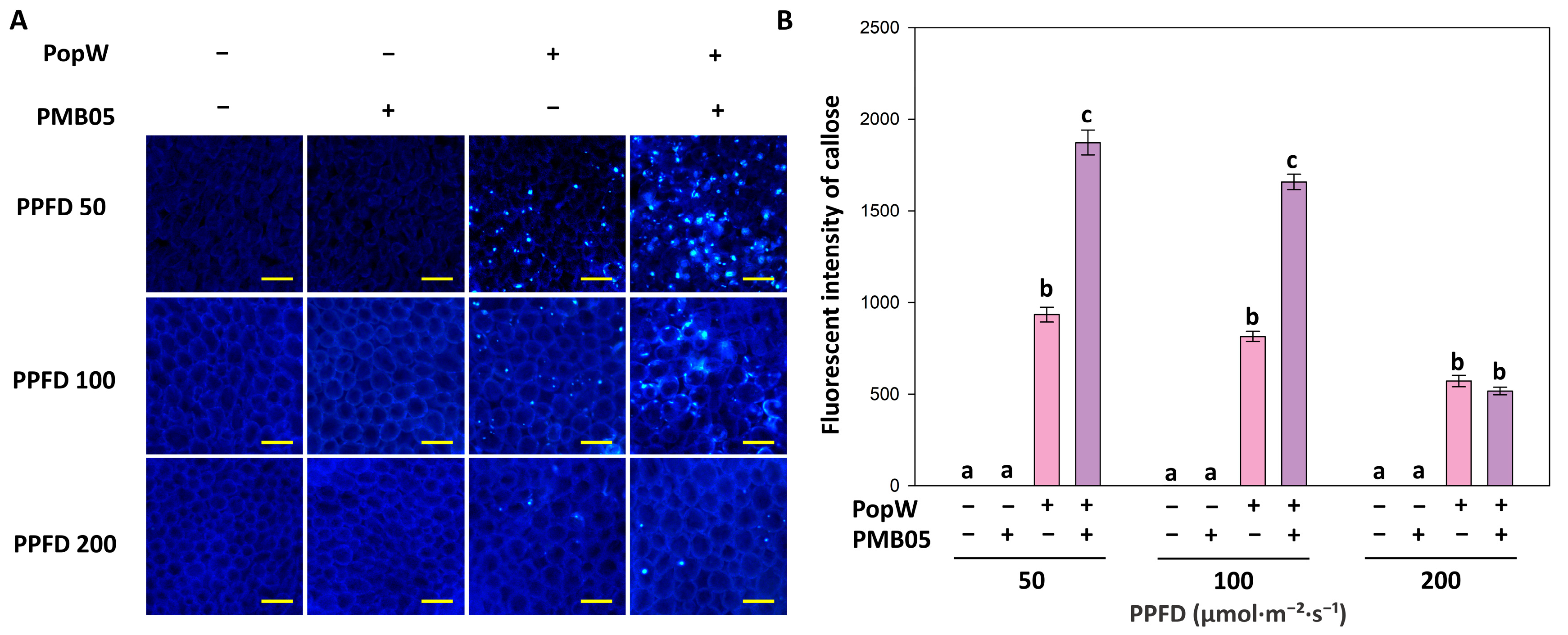
To evaluate the variation in PopW-induced callose deposition under different light intensities, A. thaliana plants exposed to various light regimes were examined. In the Tris-treated negative control plants, no callose-associated fluorescent spots were detected under any light condition. In contrast, PopW treatment induced clear fluorescent callose deposits across all light intensities, with the strongest signals observed in plants grown under 50 PPFD (Figure 3A). Quantitative analysis confirmed that PopW treatment significantly enhanced callose fluorescence compared with the Tris control in all plants; however, callose deposition decreased progressively as growth light intensity increased (Figure 3B). To further assess the influence of Bacillus amyloliquefaciens PMB05 on PopW-mediated callose deposition under different light intensities, plants grown at 50, 100, and 200 PPFD were treated with PMB05 alone or in combination with PopW. PMB05 treatment alone did not induce detectable callose deposition, and fluorescence levels were comparable to those of the Tris control. However, co-treatment with PMB05 and PopW markedly enhanced callose deposition compared with the control, although plants grown under higher light intensities displayed fewer fluorescent spots than those from lower light intensities. Among these, plants grown under 50 PPFD exhibited the strongest callose response. Quantitative analysis revealed that in 50 PPFD-grown plants, combined PMB05 and PopW treatment resulted in a 2.0-fold increase in callose fluorescence intensity compared with PopW treatment alone. By contrast, in plants grown under 200 PPFD, co-treatment with PMB05 and PopW produced only a slight increase, which was not significantly different from PopW treatment alone.

3.4. Effect of Bacillus amyloliquefaciens PMB05 on the Biocontrol of Bacterial Wilt in Arabidopsis thaliana Grown Under Different Light Intensities

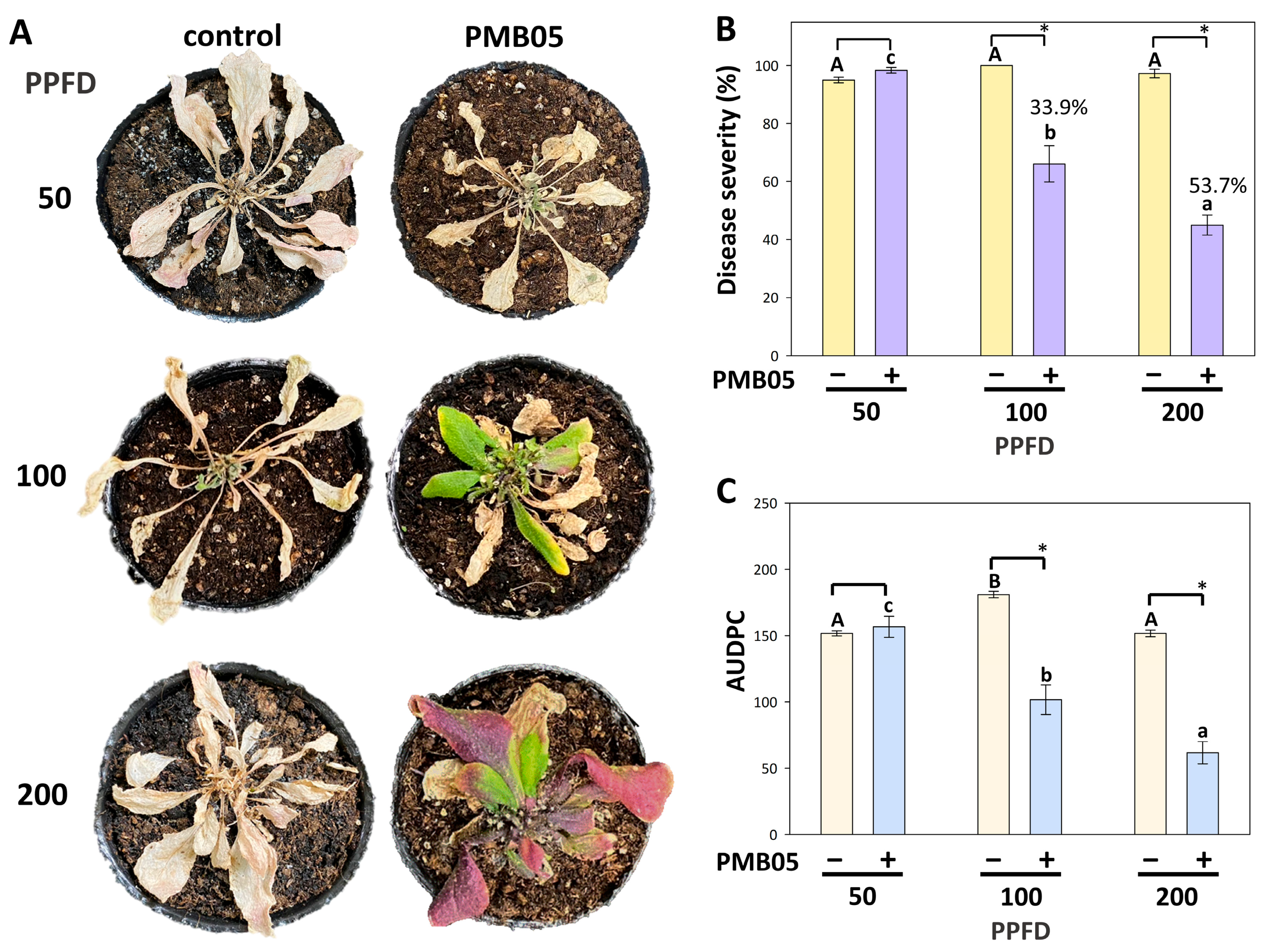
To investigate the effect of B. amyloliquefaciens PMB05 on the control of A. thaliana to bacterial wilt under different light intensities, plants were pretreated with PMB05 prior to pathogen challenge. The results showed that PMB05 application failed to effectively suppress wilt symptoms in plants grown under 50 PPFD, whereas plants cultivated under 100 and 200 PPFD exhibited a clear reduction in disease symptoms (Figure 4A). Four weeks after inoculation, the disease severity of PMB05-pretreated plants grown at 50 PPFD reached 95%, which was not significantly different from the control. In contrast, disease severity was significantly reduced to 66.1% and 45.0% in plants grown at 100 and 200 PPFD, corresponding to biocontrol efficacies of 33.9% and 53.7%, respectively (Figure 4B). Comparison of disease development among light treatments under PMB05 application further revealed that the AUDPC values were 156.6, 101.6, and 61.7 for plants grown under 50, 100, and 200 PPFD, respectively. These results indicate that PMB05-mediated biocontrol is more effective under higher light intensities, resulting in slower disease progression in plants exposed to greater light intensities (Figure 4C).

3.5. Requirement of the Salicylic Acid Pathway for Bacillus amyloliquefaciens PMB05-Mediated Biocontrol of Bacterial Wilt Under High Light Intensity

To evaluate the role of the salicylic acid pathway in B. amyloliquefaciens PMB05-mediated biocontrol of bacterial wilt, Arabidopsis thaliana transgenic lines and mutants deficient in the salicylic acid pathway (nahG, sid2, and npr1) were assessed under high light conditions. The results showed that the disease control of PMB05 observed in Col-0 was completely abolished in nahG, sid2-5, and npr1-1 plants, with no reduction in wilt symptoms detected (Figure 5A). Disease severity at four weeks post-inoculation further showed that PMB05-treated nahG, sid2-5, and npr1-1 plants exhibited diseased severity of 88.3%, 100%, and 97.2%, respectively, which were not significantly different from those of the untreated controls (90.0%, 100.0%, and 92.7%, respectively) (Figure 5B).

3.6. Effect of Bacillus amyloliquefaciens PMB05 on the Biocontrol of Bacterial Wilt in Tomato Plants Grown Under Different Light Intensities

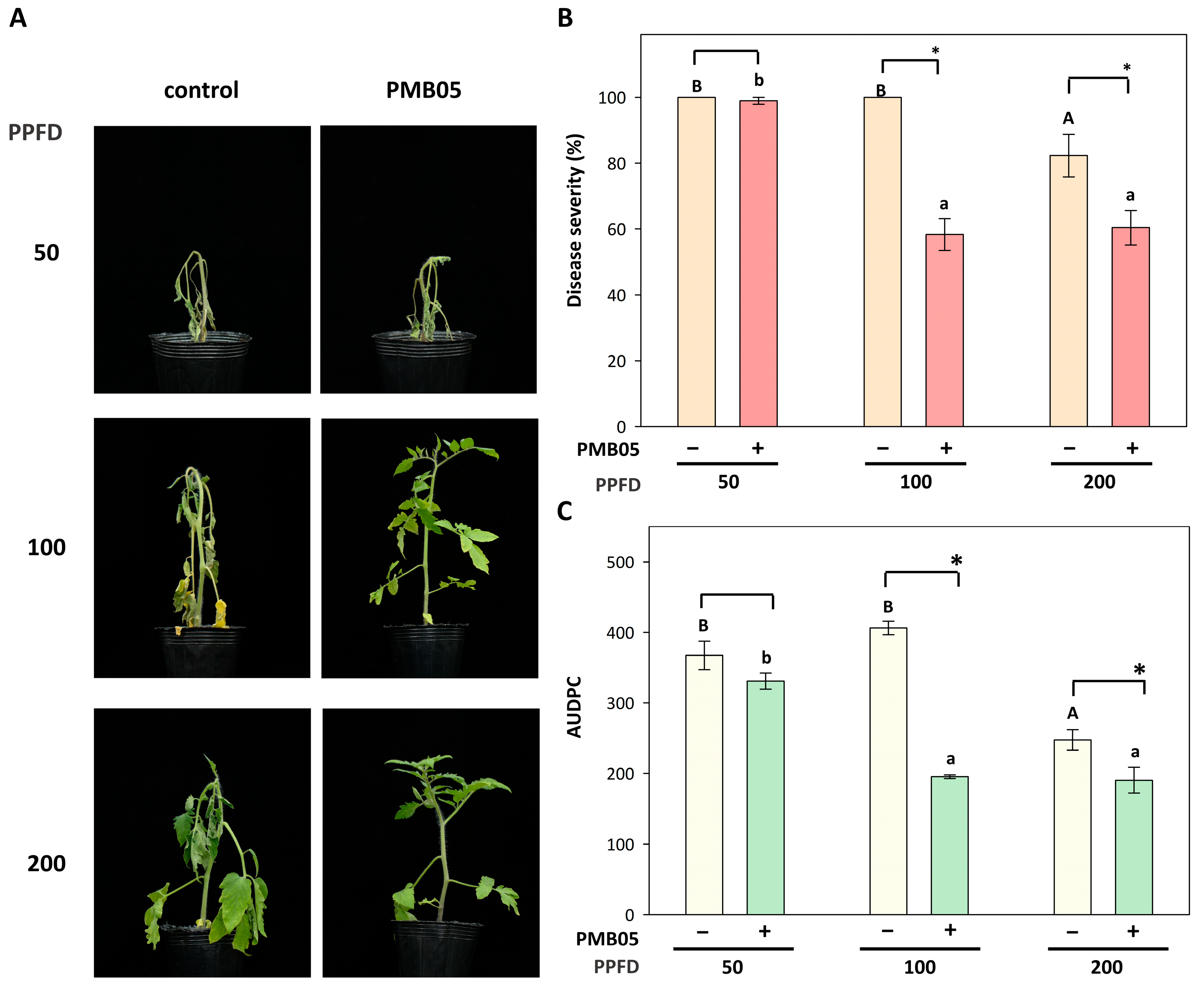
To further examine whether increased light intensity enhances the biocontrol efficacy of B. amyloliquefaciens PMB05 against bacterial wilt in tomato, plants exposed to different light regimes were pretreated with PMB05 and subjected to disease control assays. The results indicated that PMB05 treatment did not suppress disease progression in tomatoes grown under 50 PPFD; however, wilt symptoms were significantly reduced in tomatoes cultivated at 100 and 200 PPFD following PMB05 application (Figure 6A). Disease severity at 10 days post-inoculation was 98% in the PMB05-treated group at 50 PPFD, showing no significant difference compared with the untreated control. In contrast, PMB05 significantly decreased disease severity to 58.3% and 60.4% in tomatoes grown at 100 PPFD and 200 PPFD, with corresponding biocontrol efficacies of 41.7% and 26.6%, respectively (Figure 6B). Further AUDPC analysis from day 5 to day 10 after inoculation revealed values of 330.7, 195.3, and 190.1 for plants grown at 50, 100, and 200 PPFD, respectively. Only the value from 50 PPFD showed no significant difference compared to the control. Notably, in plants grown at 200 PPFD, AUDPC values under PMB05 treatment were significantly lower than those of the control, and AUDPC values in untreated plants at 200 PPFD were also markedly lower than those cultivated under lower light intensities (Figure 6C).

4. Discussion

Tomato (Lycopersicon esculentum) is the world’s second most important vegetable crop after potato [23]. However, bacterial wilt caused by Ralstonia solanacearum poses a serious threat during tomato cultivation [24]. Chemical soil disinfestation remains the primary method for bacterial wilt management, but prolonged use can cause environmental pollution and pose health risks [25,26,27]. To promote sustainable agriculture and in light of increasing understanding of plant–microbe interactions, biocontrol strategies have emerged as promising and environmentally friendly alternatives. Notably, Bacillus spp. have gained widespread application in disease management due to their spore-forming ability, resilience, and strong colonization capacity [28,29]. Recent studies have shown that Bacillus amyloliquefaciens PMB05 can effectively control bacterial wilt, a mechanism linked to its ability to enhance plant PAMP-triggered immunity (PTI) [1].
PTI is activated when plants recognize microbe-associated molecular patterns such as flg22 or harpin via surface receptors [30,31]. This process involves callose deposition, production of reactive oxygen species (ROS), and activation of downstream defense pathways [31,32,33]. As these defenses require energy, photosynthesis plays a crucial role in supplying energy for immune responses [34]. Previous research has demonstrated that chitosan application enhances disease resistance and photosynthetic rate in rose leaves [35], suggesting that improved disease resistance may rely on energy derived from photosynthesis. Infection by pathogens often leads to suppressed photosynthetic electron transport and increased non-photochemical and photochemical quenching in leaves [36,37,38]. Additionally, flg22 treatment reduces chlorophyll non-photochemical quenching (NPQ) fluorescence, indicating that PTI induction compromises photoprotection mechanisms and limits the ability of plants to dissipate excess light energy as heat during immune activation [39]. These results imply that the regulation of light use efficiency is tightly linked to the enhanced generation of ROS for strengthening defensive responses. In this study, since B. amyloliquefaciens PMB05 effectively reduces bacterial wilt by enhancing PAMP-triggered immunity (PTI) responses, we aimed to investigate whether variations in light intensity during plant growth influence its biocontrol efficacy. First, PopW from R. solanacearum was used as a PAMP to analyze PTI-related responses in A. thaliana plants cultivated under 50, 100, and 200 PPFD. The results demonstrated that PopW-induced ROS generation significantly increased with higher growth light intensities. Furthermore, treatment with B. amyloliquefaciens PMB05 further accelerated ROS generation. These findings indicate that increasing light intensity in the plant growth environment effectively enhances immune signaling strength in plants. Conversely, PopW-induced callose deposition was suppressed under higher light conditions, possibly due to elevation of abscisic acid (ABA) synthesis, which is known to inhibit callose deposition [40,41]. Disease progression analysis showed that both low (50 PPFD) and high (200 PPFD) light conditions slow the development of bacterial wilt. At low light, diminished root development (Supplementary Figure S1) may limit entry points for pathogen invasion. This restriction may contribute to slower disease progression. In contrast, under 200 PPFD, plants develop more vigorous root systems. This increased root growth correlates with stronger immune signaling (as shown in Figure 2), which also results in delayed disease development. Importantly, recent studies have demonstrated that the biocontrol efficacy of PMB05 in strawberry anthracnose differs between leaves and fruit, with immunity enhancement effective in leaves but requiring direct microbial antagonism for fruit disease control [42]. Such findings suggest that PMB05-mediated resistance enhancement relies on photosynthetic energy, with higher light intensities optimizing biocontrol activity without negatively affecting plant growth. Consistent with our previous observations, increased light intensity during PMB05-mediated PTI induction further suppressed ETR, supporting a link between light conditions and immune signaling strength [12]. In our results, callose deposition was strongly inhibited during the induction of PTI under high light intensity. There is currently no direct evidence that increased light intensity enhances callose deposition during the PTI process. A report indicates that abscisic acid (ABA) can inhibit callose deposition under low light conditions and subsequently suppress salicylic acid signaling [41]. Additionally, studies have shown that high light intensity induces ABA biosynthesis and activates its signaling pathways in Arabidopsis [40]. Therefore, whether the improved salicylic acid signaling observed during PTI under high light intensity is mediated by ABA pathway activation remains to be further elucidated. While callose deposition can inhibit fungal pathogens such as powdery mildew and gray mold [43,44], the enhanced resistance to bacterial wilt observed under high light conditions appears to be independent of callose deposition, indicating the need for further investigation into the relationship between light intensity and resistance signaling against fungal diseases. In addition, light treatments prior to wheat rust inoculation have been shown to promote infection efficiency, and field surveys also highlight a positive correlation between light intensity and rust incidence [45]. Thus, these findings suggest that increasing the light intensity in the plant growth environment may enhance the effectiveness of B. amyloliquefaciens PMB05 in reducing bacterial disease.
Our recent research further reveals that PMB05 can enhance PopW recognition and activate MPK3/6 and subsequent salicylic acid signaling (PR-1 expression) in Arabidopsis [13]. This suggests that higher light intensity may promote MAPK pathway activation, facilitating enhanced ROS generation and immune signaling by PMB05. Since MAPK modules (MEKK1/MKK4-MKK5/MPK6) positively regulate the salicylic acid pathway and defense gene expression such as PR1, PR2, and PR5 [46,47], this study demonstrates that under a light intensity of 200 PPFD, B. amyloliquefaciens PMB05 is unable to effectively suppress bacterial wilt development in Arabidopsis plants when salicylic acid biosynthesis is disrupted or its downstream transcription factors are nonfunctional. These results suggest that the salicylic acid defense pathway remains essential for PMB05-mediated bacterial wilt control, even under enhanced light conditions.
Extending these observations to tomato, a similar trend was observed: bacterial wilt incidence was significantly reduced at 200 PPFD, consistent with reduced disease progression noted in Pseudomonas syringae pv. tomato infection trials at 300 PPFD [48]. PMB05 showed stronger biocontrol under higher light conditions, though further increases beyond 100 PPFD did not yield additional benefits. Based on the results from Arabidopsis and tomato, it is inferred that different plant species require varying light intensities to activate disease resistance, warranting further investigation in future studies. With advances in LED technology, the effects of light quantity and spectrum on disease susceptibility have gained attention. Enhanced light intensity reduces cucumber mosaic virus resistance in tobacco, while light spectrum adjustments can alter leaf thickness and photosynthetic efficiency, influencing disease response [6,49]. The impact of spectral variation on PMB05 biocontrol efficacy is thus worthwhile for future research.

5. Conclusions

Taken together, our findings indicate that increased light intensity during plant growth enhances PMB05-mediated PAMP-triggered immunity (PTI) signaling, thereby improving suppression of bacterial wilt. Activation of the salicylic acid pathway is essential for this biocontrol effect. These mechanisms are also applicable to crops such as tomato, suggesting that optimizing growth light conditions could maximize the biocontrol potential of B. amyloliquefaciens PMB05 in agriculture. Future research can explore how different light spectra combined with high light intensity affect the effectiveness of plant immunity-enhancing bacteria like B. amyloliquefaciens PMB05. Moreover, the application of short high-light treatments during the night could be tested under field conditions to further boost plant immune responses and achieve more efficient disease reduction. These approaches may provide valuable strategies for sustainable disease management.

Supplementary Materials

The following supporting information can be downloaded at https://www.mdpi.com/article/10.3390/agriculture15202110/s1. Figure S1: Root development of Arabidopsis thaliana grown under different light intensities.

Author Contributions

Y.-H.L.: Conceptualization, methodology, data curation, writing—review and editing, supervision, funding acquisition; S.-H.L.: methodology, software, formal analysis, investigation, writing—original draft preparation; A.-T.L.: methodology and investigation; M.-Q.S., Y.-X.L. and L.-Y.H.: validation, formal analysis; H.-Y.C.: review and supervision. All authors have read and agreed to the published version of the manuscript.

Funding

This study was supported by the National Science and Technology Council (NSTC), Taiwan, through grants (NSTC-113-2313-B-020-011) to Yi-Hsien Lin.

Institutional Review Board Statement

Not applicable.

Data Availability Statement

The original contributions presented in the study are included in the article; further inquiries can be directed to the corresponding author.

Conflicts of Interest

The authors declare no conflicts of interest.

References

  1. Ho, T.-H.; Chung, C.-Y.; Zheng, J.-L.; Chen, H.-H.; Liang, Y.-S.; Huang, T.-P.; Lin, Y.-H. Bacillus amyloliquefaciens Strain PMB05 Intensifies Plant Immune Responses to Confer Resistance Against Bacterial Wilt of Tomato. Phytopathology 2020, 110, 1877–1885. [Google Scholar] [CrossRef]
  2. Morales, P.; González, M.; Salvatierra-Martínez, R.; Araya, M.; Ostria-Gallardo, E.; Stoll, A. New Insights into Bacillus-Primed Plant Responses to a Necrotrophic Pathogen Derived from the Tomato-Botrytis Pathosystem. Microorganisms 2022, 10, 1547. [Google Scholar] [CrossRef] [PubMed]
  3. Zhang, N.; Wang, Z.; Shao, J.; Xu, Z.; Liu, Y.; Xun, W.; Miao, Y.; Shen, Q.; Zhang, R. Biocontrol mechanisms of Bacillus: Improving the efficiency of green agriculture. Microb. Biotechnol. 2023, 16, 2250–2263. [Google Scholar] [CrossRef]
  4. Zhou, A.; Wang, F.; Yin, J.; Peng, R.; Deng, J.; Shen, D.; Wu, J.; Liu, X.; Ma, H. Antifungal action and induction of resistance by Bacillus sp. strain YYC 155 against Colletotrichum fructicola for control of anthracnose disease in Camellia oleifera. Front. Microbiol. 2022, 13, 956642. [Google Scholar] [CrossRef]
  5. de Wit, M.; Galvão, V.C.; Fankhauser, C. Light-Mediated Hormonal Regulation of Plant Growth and Development. Annu. Rev. Plant Biol. 2016, 67, 513–537. [Google Scholar] [CrossRef] [PubMed]
  6. Fan, X.-X.; Xu, Z.-G.; Liu, X.-Y.; Tang, C.-M.; Wang, L.-W.; Han, X.-L. Effects of light intensity on the growth and leaf development of young tomato plants grown under a combination of red and blue light. Sci. Hort. 2013, 153, 50–55. [Google Scholar] [CrossRef]
  7. Amsalem, L.; Freeman, S.; Rav-David, D.; Nitzani, Y.; Sztejnberg, A.; Pertot, I.; Elad, Y. Effect of climatic factors on powdery mildew caused by Sphaerotheca macularis f. sp. fragariae on strawberry. Eur. J. Plant Pathol. 2006, 114, 283–292. [Google Scholar] [CrossRef]
  8. Eskes, A.B. The effect of light intensity on incomplete resistance of coffee to Hemileia vastatrix. Netherlands J. Plant Pathol. 1982, 88, 191–202. [Google Scholar] [CrossRef]
  9. Shafia, A.; Sutton, J.C.; Yu, H.; Fletcher, R.A. Influence of preinoculation light intensity on development and interactions of Botrytis cinerea and Clonostachys rosea in tomato leaves. Can. J. Plant Pathol. 2010, 23, 346–357. [Google Scholar] [CrossRef]
  10. Ikeda, T.; Okuda, M.; Ishihara, M.; Kon-No, Y. Effect of different light conditions on anatomical and histological features of galls in bacterial gall disease of Cerasus × yedoensis. Phytopathology 2024, 114, 2196–2206. [Google Scholar] [CrossRef] [PubMed]
  11. Karpinski, S.; Szechynska-Hebda, M.; Wituszynska, W.; Burdiak, P. Light acclimation, retrograde signalling, cell death and immune defences in plants. Plant Cell Environ 2013, 36, 736–744. [Google Scholar] [CrossRef]
  12. Wang, Y.-H.; Lai, I.-L.; Zheng, J.-L.; Lin, Y.-H. Using dynamic changes of chlorophyll fluorescence in Arabidopsis thaliana to evaluate plant immunity-intensifying Bacillus spp. strains. Phytopathology 2019, 109, 1566–1576. [Google Scholar] [CrossRef]
  13. Chuang, C.-Y.; Lin, S.-T.; Li, A.-T.; Li, S.-H.; Hsiao, C.-Y.; Lin, Y.-H. Bacillus amyloliquefaciens PMB05 increases resistance to bacterial wilt by activating MAPK and ROS pathway crosstalk in Arabidopsis thaliana. Phytopathology 2022, 6, 10.1094. [Google Scholar] [CrossRef]
  14. Postel, S.; Kemmerling, B. Plant systems for recognition of pathogen-associated molecular patterns. Semin. Cell Dev. Biol. 2009, 20, 1025–1031. [Google Scholar] [CrossRef]
  15. Oh, C.-S.; Beer, S.V. Molecular genetics of Erwinia amylovora involved in the development of fire blight. FEMS Microbiol. Lett. 2005, 253, 185–192. [Google Scholar] [CrossRef]
  16. Bauer, D.W.; Wei, Z.M.; Beer, S.V.; Collmer, A. Erwinia chrysanthemi harpinEch: An elicitor of the hypersensitive response that contributes to soft-rot pathogenesis. Mol. Plant-Microbe Interact. 1995, 8, 484–491. [Google Scholar] [CrossRef]
  17. Desikan, R.; Clarke, A.; Atherfold, P.; Hancock, J.T.; Neill, S.J. Harpin induces mitogen-activated protein kinase activity during defence response in Arabidopsis thaliana suspension cultures. Planta 1999, 210, 97–103. [Google Scholar] [CrossRef] [PubMed]
  18. Liu, H.; Wang, Y.; Zhou, X.; Wang, C.; Wang, C.; Fu, J.; Wei, T. Overexpression of a harpin-encoding gene popW from Ralstonia solanacearum primed antioxidant defenses with enhanced drought tolerance in tobacco plants. Plant Cell Rep. 2016, 35, 1333–1344. [Google Scholar] [CrossRef] [PubMed]
  19. Wei, Z.M.; Laby, R.J.; Zumoff, C.H.; Bauer, D.W.; He, S.Y.; Collmer, A.; Beer, S.V. Harpin, elicitor of the hypersensitive response produced by the plant pathogen Erwinia amylovora. Science 1992, 257, 85–88. [Google Scholar] [CrossRef]
  20. Zheng, J.-L.; Li, J.-R.; Li, A.-T.; Li, S.-H.; Blanco, S.D.; Chen, S.-Y.; Lai, Y.-R.; Shi, M.-Q.; Lin, T.-C.; Su, J.-F.; et al. A Rapid Method for Screening Pathogen-Associated Molecular Pattern-Triggered Immunity-Intensifying Microbes. Plants 2024, 13, 2185. [Google Scholar] [CrossRef] [PubMed]
  21. Lin, Y.-H.; Huang, H.-E.; Chen, Y.-R.; Liao, P.-L.; Chen, C.-L.; Feng, T.-Y. C-terminal region of plant ferredoxin-like protein is required to enhance resistance to bacterial disease in Arabidopsis thaliana. Phytopathology 2011, 101, 741–749. [Google Scholar] [CrossRef] [PubMed]
  22. Simko, I.; Piepho, H.-P. The Area Under the Disease Progress Stairs: Calculation, Advantage, and Application. Phytopathology 2011, 102, 381–389. [Google Scholar] [CrossRef] [PubMed]
  23. Quinet, M.; Angosto, T.; Yuste-Lisbona, F.J.; Blanchard-Gros, R.; Bigot, S.; Martinez, J.-P.; Lutts, S. Tomato fruit development and metabolism. Front. Plant Sci. 2019, 10, 1554. [Google Scholar] [CrossRef]
  24. Kim, B.-S.; French, E.; Caldwell, D.; Harrington, E.J.; Iyer-Pascuzzi, A.S. Bacterial wilt disease: Host resistance and pathogen virulence mechanisms. Physiol. Mol. Plant Pathol. 2016, 95, 37–43. [Google Scholar] [CrossRef]
  25. Li, J.; Huang, B.; Wang, Q.; Li, Y.; Fang, W.; Yan, D.; Guo, M.; Cao, A. Effect of fumigation with chloropicrin on soil bacterial communities and genes encoding key enzymes involved in nitrogen cycling. Environ. Pollut. 2017, 227, 534–542. [Google Scholar] [CrossRef]
  26. Mao, L.; Wang, Q.; Yan, D.; Ma, T.; Liu, P.; Shen, J.; Li, Y.; Ouyang, C.; Guo, M.; Cao, A. Evaluation of Chloropicrin as a Soil Fumigant against Ralstonia solanacarum in Ginger (Zingiber officinale Rosc.) Production in China. PLoS ONE 2014, 9, e91767. [Google Scholar] [CrossRef]
  27. Pesonen, M.; Vähäkangas, K. Chloropicrin-induced toxicity in the respiratory system. Toxicol. Lett. 2020, 323, 10–18. [Google Scholar] [CrossRef]
  28. Bonaterra, A.; Badosa, E.; Daranas, N.; Francés, J.; Roselló, G.; Montesinos, E. Bacteria as Biological Control Agents of Plant Diseases. Microorganisms 2022, 10, 1759. [Google Scholar] [CrossRef] [PubMed]
  29. Soni, R.; Keharia, H. Phytostimulation and biocontrol potential of Gram-positive endospore-forming Bacilli. Planta 2021, 254, 49. [Google Scholar] [CrossRef] [PubMed]
  30. Chinchilla, D.; Bauer, Z.; Regenass, M.; Boller, T.; Felix, G. The Arabidopsis receptor kinase FLS2 binds flg22 and determines the specificity of flagellin perception. Plant Cell 2006, 18, 465–476. [Google Scholar] [CrossRef]
  31. Zipfel, C.; Kunze, G.; Chinchilla, D.; Caniard, A.; Jones, J.D.; Boller, T.; Felix, G. Perception of the bacterial PAMP EF-Tu by the receptor EFR restricts Agrobacterium-mediated transformation. Cell 2006, 125, 749–760. [Google Scholar] [CrossRef]
  32. Ellinger, D.; Voigt, C.A. Callose biosynthesis in Arabidopsis with a focus on pathogen response: What we have learned within the last decade. Ann. Bot. 2014, 114, 1349–1358. [Google Scholar] [CrossRef]
  33. Kadota, Y.; Sklenar, J.; Derbyshire, P.; Stransfeld, L.; Asai, S.; Ntoukakis, V.; Jones, J.D.; Shirasu, K.; Menke, F.; Jones, A.; et al. Direct regulation of the NADPH oxidase RBOHD by the PRR-associated kinase BIK1 during plant immunity. Mol. Cell 2014, 54, 43–55. [Google Scholar] [CrossRef]
  34. Cheaib, A.; Killiny, N. Photosynthesis Responses to the Infection with Plant Pathogens. Mol. Plant-Microbe Interact. 2025, 38, 9–29. [Google Scholar] [CrossRef]
  35. Li, J.; Li, R.; Zhang, C.; Guo, Z.; Wu, X.; An, H. Co-Application of Allicin and Chitosan Increases Resistance of Rosa roxburghii against Powdery Mildew and Enhances Its Yield and Quality. Antibiotics 2021, 10, 1449. [Google Scholar] [CrossRef] [PubMed]
  36. Bechtold, U.; Karpinski, S.; Mullineaux, P.M. The influence of the light environment and photosynthesis on oxidative signalling responses in plant-biotrophic pathogen interactions. Plant Cell Environ. 2005, 28, 1046–1055. [Google Scholar] [CrossRef]
  37. Suárez, J.C.; Vanegas, J.I.; Contreras, A.T.; Anzola, J.A.; Urban, M.O.; Beebe, S.E.; Rao, I.M. Chlorophyll Fluorescence Imaging as a Tool for Evaluating Disease Resistance of Common Bean Lines in the Western Amazon Region of Colombia. Plants 2022, 11, 1371. [Google Scholar] [CrossRef]
  38. Yang, H.; Luo, P. Changes in Photosynthesis Could Provide Important Insight into the Interaction between Wheat and Fungal Pathogens. Int. J. Mol. Sci. 2021, 22, 8865. [Google Scholar] [CrossRef] [PubMed]
  39. Göhre, V.; Jones, A.M.; Sklenář, J.; Robatzek, S.; Weber, A.P. Molecular crosstalk between PAMP-triggered immunity and photosynthesis. Mol. Plant Microbe Interact. 2012, 25, 1083–1092. [Google Scholar] [CrossRef]
  40. Galvez-Valdivieso, G.; Fryer, M.J.; Lawson, T.; Slattery, K.; Truman, W.; Smirnoff, N.; Asami, T.; Davies, W.J.; Jones, A.M.; Baker, N.R.; et al. The high light response in Arabidopsis involves ABA signaling between vascular and bundle sheath cells. Plant Cell 2009, 21, 2143–2162. [Google Scholar] [CrossRef]
  41. Wang, Y.; Li, X.; Fan, B.; Zhu, C.; Chen, Z. Regulation and Function of Defense-Related Callose Deposition in Plants. Int. J. Mol. Sci. 2021, 22, 2393. [Google Scholar] [CrossRef]
  42. Wu, Y.-M.; Chen, X.; Wang, F.; Hsiao, C.-Y.; Yang, C.-Y.; Lin, S.-T.; Wu, L.-H.; Chen, Y.-K.; Liang, Y.-S.; Lin, Y.-H. Bacillus amyloliquefaciens strains control strawberry anthracnose through antagonistic activity and plant immune response intensification. Biol. Control 2021, 157, 104592. [Google Scholar] [CrossRef]
  43. Bayles, C.J.; Ghemawat, M.S.; Aist, J.R. Inhibition by 2-deoxy-D-glucose of callose formation, papilla deposition, and resistance to powdery mildew in an ml-o barley mutant. Physiol. Mol. Plant Pathol. 1990, 36, 63–72. [Google Scholar] [CrossRef]
  44. Sanmartín, N.; Pastor, V.; Pastor-Fernández, J.; Flors, V.; Pozo, M.J.; Sánchez-Bel, P. Role and mechanisms of callose priming in mycorrhiza-induced resistance. J. Exp. Bot. 2020, 71, 2769–2781. [Google Scholar] [CrossRef]
  45. de Vallavieille-Pope, C.; Huber, L.; Leconte, M.; Bethenod, O. Preinoculation Effects of Light Quantity on Infection Efficiency of Puccinia striiformis and P. triticina on Wheat Seedlings. Phytopathology 2002, 92, 1308–1314. [Google Scholar] [CrossRef] [PubMed]
  46. Feri dit Frey, N.; Garcia, A.V.; Bigeard, J.; Zaag, R.; Bueso, E.; Garmier, M.; Pateyron, S.; de Tauzia-Moreau, M.-L.; Brunaud, V.; Balzergue, S.; et al. Functional analysis of Arabidopsis immune-related MAPKs uncover a role for MPK3 as negative regulator of inducible defences. Genome Biol. 2014, 15, R87. [Google Scholar]
  47. Kumar, D. Salicylic acid signaling in disease resistance. Plant Sci. 2014, 228, 127–134. [Google Scholar] [CrossRef] [PubMed]
  48. Luo, Q.; Wang, J.; Wang, P.; Liang, X.; Li, J.; Wu, C.; Fang, W.; Ding, S.; Shao, S.; Shi, K. Transcriptomic and genetic approaches reveal that low-light-induced disease susceptibility is related to cellular oxidative stress in tomato. Hortic. Res. 2023, 10, uhad173. [Google Scholar] [CrossRef] [PubMed]
  49. Chen, L.-J.; Zhao, F.-F.; Zhang, M.; Lin, H.-H.; Xi, D.-H. Effects of Light Quality on the Interaction between Cucumber Mosaic Virus and Nicotiana tabacum. J. Phytopathol. 2015, 163, 1002–1013. [Google Scholar] [CrossRef]
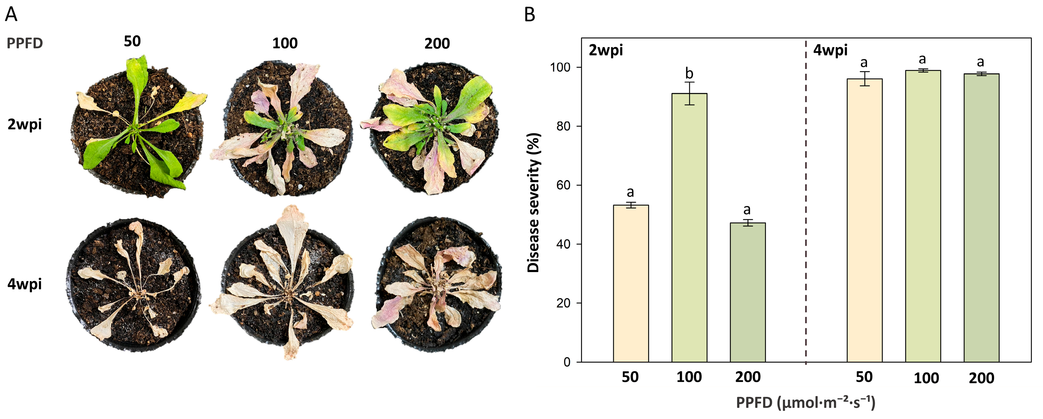
Figure 1. The occurrence of bacterial wilt in Arabidopsis thaliana grown under different light intensities. To assay the disease occurrence, four-week-old (A) thaliana plants grown under 50, 100, or 200 PPFD of light intensity were transplanted into diseased soil containing Ralstonia solanacearum Rd15. Panel indicates the symptoms of bacterial wilt at 2- and 4-week post-inoculation (wpi). Panel (B) indicates the disease severity of bacterial wilt. Different letters above the columns indicate significant differences between plants from distinct light intensities based on Tukey’s HSD test (p < 0.05).
Agriculture 15 02110 g001
Figure 2. Effect of PopW-mediated rapid reactive oxygen species (ROS) generation in Arabidopsis thaliana plants grown under different light intensities. Four-week-old A. thaliana Col-0 plants that had been cultivated under 50, 100, or 200 PPFD were used in this assay. ROS generation was induced with 0.5 μM PopW in the presence or absence of Bacillus amyloliquefaciens PMB05. Panel (A) shows representative images of ROS generation (scale bar = 20 μm). Panel (B) presents the quantification of ROS fluorescence intensity. The symbols “−” and “+” indicate whether or not the treatment contained PopW or PMB05, respectively. Different letters indicate significant differences among plants grown under different light intensities, as determined by Tukey’s HSD test (p < 0.05).
Agriculture 15 02110 g002
Figure 3. Effect of PopW-mediated callose deposition in Arabidopsis thaliana plants grown under different light intensities. Four-week-old A. thaliana Col-0 plants that had been cultivated under 50, 100, or 200 PPFD were used in this assay. Callose deposition was induced with 0.5 μM PopW in the presence or absence of Bacillus amyloliquefaciens PMB05. Panel (A) shows representative images of callose deposition (scale bar = 20 μm). Panel (B) presents the quantification of callose deposition intensity. The symbols “−” and “+” indicate whether or not the treatment contained PopW or PMB05, respectively. Different letters indicate significant differences among plants grown under different light intensities, as determined by Tukey’s HSD test (p < 0.05).
Agriculture 15 02110 g003
Figure 4. Effects of Bacillus amyloliquefaciens PMB05 on the control of bacterial wilt in Arabidopsis thaliana grown under different light intensities. Four-week-old A. thaliana Col-0 plants cultivated under 50, 100, or 200 PPFD were pretreated with PMB05 and, two days later, transplanted into soil infested with Ralstonia solanacearum Rd15. Panel (A) shows bacterial wilt symptoms at 4 weeks post-inoculation (wpi) with or without PMB05 treatment. Panel (B) presents disease severity at 4 wpi. Panel (C) shows the area under the disease progress curve (AUDPC) calculated from disease severity between 2 and 4 wpi. The symbols “−” and “+” denote absence or presence of PMB05 treatment, respectively. Asterisks (*) above bars indicate significant differences from the untreated control by t-test (p < 0.05). Different lowercase and uppercase letters represent significant differences among plants grown under different light intensities based on Tukey’s HSD tests.
Agriculture 15 02110 g004
Figure 5. Effects of Bacillus amyloliquefaciens PMB05 on the control of bacterial wilt in salicylic acid pathway mutants of Arabidopsis thaliana grown under high light intensity. Four-week-old A. thaliana Col-0 and its SA pathway mutants nahG, sid2-5, and npr1-1 were cultivated at 200 PPFD, pretreated with PMB05 bacterial suspensions, and transplanted two days later into soil infested with Ralstonia solanacearum Rd15. Panel (A) shows bacterial wilt symptoms at 4 weeks post-inoculation (wpi) with or without PMB05 treatment. Panel (B) presents disease severity at 4 wpi. The symbols “−” and “+” denote absence or presence of PMB05 treatment, respectively. Asterisks (*) indicate significant differences compared to the untreated control by t-test (p < 0.05).
Agriculture 15 02110 g005
Figure 6. Evaluation of Bacillus amyloliquefaciens PMB05 on the biocontrol of bacterial wilt in tomato plants grown under different light intensities. Four-week-old tomato plants cultivated under 50, 100, or 200 PPFD were pretreated with PMB05 and, two days later, transplanted into soil containing Ralstonia solanacearum Ps152. Panel (A) shows bacterial wilt symptoms at 10 days post-inoculation (dpi) with or without PMB05 treatment. Panel (B) presents disease severity at 10 dpi. Panel (C) displays the area under the disease progress curve (AUDPC), calculated from disease severity between 5 and 10 dpi. The symbols “−” and “+” denote absence or presence of PMB05 treatment, respectively. Asterisks (*) above bars indicate significant differences from untreated controls by t-test (p < 0.05). Different lowercase and uppercase letters indicate significant differences among plants grown under different light intensities within each treatment based on Tukey’s HSD tests.
Agriculture 15 02110 g006
Disclaimer/Publisher’s Note: The statements, opinions and data contained in all publications are solely those of the individual author(s) and contributor(s) and not of MDPI and/or the editor(s). MDPI and/or the editor(s) disclaim responsibility for any injury to people or property resulting from any ideas, methods, instructions or products referred to in the content.