Abstract
Chrysanthemum (Chrysanthemum × morifolium (Ramat.) Hemsl.) holds a prominent position in the market of ornamental plants. To further advance chrysanthemum breeding efforts, the development of haploids may be useful. Therefore, the effect of various chemical and thermal treatments on regeneration efficiency and ploidy level in chrysanthemum was studied. Ovaries and ovules of three chrysanthemum cultivars, i.e., ‘Brasil,’ ‘Capitola,’ and ‘Jewel Time Yellow,’ were cultured either on a medium with 1 mg·L−1 2,4-dichlorophenoxyacetic acid (2,4-D) and different concentrations (0.5–1.5 mg·L−1) of thidiazuron (TDZ) or subjected to thermal shock (pretreatment temperature of 4 °C or 32 °C) and cultured on a medium with 1 mg·L−1 2,4-D and 1 mg·L−1 6-benzylaminopurine (BAP). It was found that ovaries had a greater organogenic potential (both in terms of callogenesis and shoot formation) than ovules. Microscopic analyses revealed that shoots mainly developed via indirect somatic embryogenesis from a callus developed from the ovary wall. The highest number of shoots was produced in cooled (at 4 °C) ovaries of chrysanthemum ‘Brasil’ and in ‘Jewel Time Yellow’ ovaries cultured on a medium with 1.0–1.5 mg·L−1 TDZ. The latter cultivar also had the highest potential to produce plants with an altered ploidy level (doubled and halved the number of chromosomes). This study demonstrates that manipulating factors such as temperature and thidiazuron concentration can enhance regeneration efficiency and induce altered ploidy levels in selected cultivars, offering valuable insights for chrysanthemum breeding programs.
1. Introduction
Chrysanthemum (Chrysanthemum × morifolium) holds a prominent position in the market of ornamental plants due to its vibrant and diverse floral cultivars, making them highly sought after for decorative purposes in gardens and floral arrangements. Beyond the visual allure, chrysanthemum has become a valuable model organism in plant biology, offering unique opportunities for exploring various aspects of genetics, physiology, and development [1,2].
The creation of new chrysanthemum cultivars with enhanced traits such as flower shape, color, and disease resistance has been a primary focus of breeders. Therefore, the species has garnered substantial attention in breeding programs using crossing, somatic hybridization, mutagenesis, and genetic transformation. These breeding methods, however, often involve time-consuming and labor-intensive practices. Moreover, the high ploidy level, heterozygosity, and self-incompatible state of the chrysanthemum complicate the entire process [3,4].
The Chrysanthemum genus comprises plants with a basic chromosome count equal to nine, but the range of ploidy levels in particular species varies from diploid 2n = 2x = 18 in Ch. nankingense to octaploid 2n = 8x = 72 in Ch. arcticum [5]. Ch. morifolium is considered an allopolyploid, i.e., hexapolyploid with the basic chromosome number 2n = 6x = 54, showing mixed polysomic and disomic inheritance [6]. To further advance chrysanthemum breeding efforts, the development of haploids, i.e., plants having only a single set of each homologous chromosome in somatic cells, is essential. Haploids, with their simplified genetic makeup, provide a valuable resource for breeders to expedite the selection of desirable traits, ultimately enhancing the quality and variety of chrysanthemum cultivars available to consumers [7]. This approach has gained attention for its potential to accelerate the breeding process [8].
Gynogenesis involves the development of embryos from the female sporophyte, bypassing the need for paternal contribution. The application of gynogenesis has opened new possibilities for the rapid generation of homozygous lines in ornamental plants [9]. It is currently one of the most popular methods of inducing haploidy in plants. Moreover, gynogenic embryos often exhibit increased genetic stability, making them valuable resources for breeding programs seeking to maintain cultivar purity [10].
There are many factors that affect the process of gynogenesis in vitro [11]. Thidiazuron (TDZ) is a plant growth regulator that has been widely used in plant tissue culture [12]. According to the literature, TDZ is more stable than BAP, yet it effectively stimulates morphogenesis processes, including regeneration from meristematic tissues, even at low concentrations [13]. TDZ is particularly effective in the induction of haploids through its ability to stimulate the formation of adventitious shoots or calli from haploid explants [14]. This compound plays a role in facilitating chromosome reduction, which is crucial for producing haploid plants. TDZ is often used to induce the formation of embryos from the microspores during androgenesis in ornamental plants [15]. Nonetheless, the exact mechanism of action of TDZ in haploid induction is not fully understood, but it is believed to promote cell division and growth.
Temperature is another vital factor in inducing haploidy in plants, and its impact varies depending on the specific plant species and the methods employed for haploid induction. It was found that heat stress promotes haploid formation during centromere-mediated genome elimination in Arabidopsis [16,17]. On the other hand, cold treatment or vernalization is commonly used to induce haploids in cereals such as rice, oats, and barley [18,19].
To date, very limited progress in producing chrysanthemum haploids has been obtained. In a study by Wang et al. [20], a total of 2579 non-fertilized chrysanthemum ovules pollinated by Argyranthemum frutescens were cultured in vitro to initiate gynogenesis. A very low shoot regeneration efficiency was reported. Chromosome counts and microsatellite fingerprinting showed that only one of the regenerants was a haploid. An attempt was made by Miler and Muszczyk [21] to regenerate haploids (2n = 3x) from ovary and ovule cultures, but despite abundant calli and hexaploid shoot regeneration, no plants with reduced ploidy were found. Therefore, more research in this regard is needed.
The study aimed to investigate the influence of temperature and chemical stimuli on the in vitro organogenesis and regeneration of ovaries and ovules in three cultivars of chrysanthemum. The impact of these treatments on the stability of the produced plant material was investigated using flow cytometry. Moreover, the origin of regenerated plants was indicated by microscopic observations of cultured ovaries.
2. Materials and Methods
2.1. Plant Material and In Vitro Culture Conditions
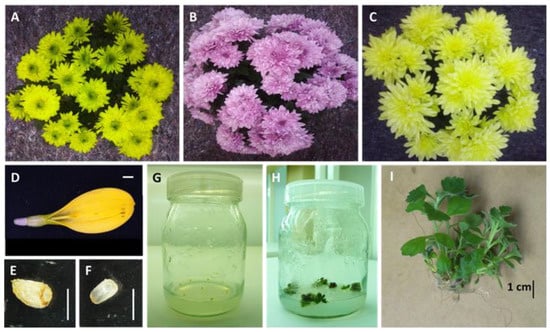
Three cultivars of Chrysanthemum × morifolium (Ramat.) Hemsl., i.e., ‘Brasil,’ ‘Capitola,’ and ‘Jewel Time Yellow,’ were used in this experiment (Figure 1). These commercial pot cultivars are characterized by a medium growth force and full, flat green, purple, or yellow inflorescences, respectively. The mother plants were obtained from Maciej Raszka’s gardening farm in Łabiszyn, Poland.

Figure 1.
Three chrysanthemum cultivars used in the study: ‘Brasil’ (A), ‘Capitola’ (B) and ‘Jewel Time Yellow’ (C); ligulate flower of ‘Jewel Time Yellow’ cultivar (D); cut off ovary (E); isolated ovule (F); ovaries inoculated on the induction medium (G); regenerating explants in the growth room (H); regenerated plantlet (I); bar = 1 mm (D–F) or 1 cm (I).
Two types of explants were used in this experiment, i.e., ovaries and ovules from ligulate flowers. Ligulate flowers were collected from the third and fourth outer whorls of the inflorescence of in vivo-grown plants in November when the inner whorls were not yet open. The mother plants were protected from cross-pollination, and since they did not possess disc flowers, self-pollination was not possible.
Approximately 50 flowers were collected from one inflorescence. Then, the flowers were disinfected as follows: washing under running tap water, followed by treatment with 0.5% (v/v) detergent (5 min), 70% ethanol (5 s), 0.5% (v/v) sodium hypochlorite (5 min), and finally, rinsing with sterile distilled water (3 × 5 min). Ovaries and ovules were isolated using the MS-Z TRI stereoscopic microscope (Precoptic, Warsaw, Poland) in a laminar airflow cabinet.
Ovaries and ovules of each cultivar were horizontally cultured in 350 mL glass jars filled with 40 mL MS [22] medium supplemented with different plant growth regulators (PGRs) (Sigma-Aldrich, St. Louis, MO, USA) as described in detail below. The media were supplemented with 30 g·L−1 of sucrose and solidified with 8.0 g·L−1 agar (Biocorp, Warsaw, Poland). After adding all the components, the pH was adjusted to 5.8 before the medium was autoclaved at 121 °C and 105 kPa for 20 min.
The cultures were grown at 24 ± 1 °C and exposed to a 16 h photoperiod. Standard cool daylight was provided by TLD54/36W fluorescent lamps (Koninklijke Philips Electronics N.V., Eindhoven, The Netherlands) with a photosynthetic photon flux density of approximately 14 µmol·m2·s−1 for the first six weeks of induction stage and 35 µmol·m2·s−1 after the explants transfer onto the regeneration medium; the light color temperature was 6200 K.
2.2. Regeneration of In Vitro Culture Depending on TDZ Application
In the first experiment, ovaries and ovules were cultured on the MS medium supplemented with 1.0 mg·L−1 2,4-dichlorophenoxyacetic acid (2,4-D) and thidiazuron (TDZ) ranging from 0.5 to 1.5 mg·L−1.
According to the two-step protocol [21], after six weeks in the induction stage, the explants that started regenerating calli were transferred to the modified MS medium supplemented with 2 mg·L−1 kinetin (KIN), 1 mg·L−1 indole-3-acetic acid (IAA), and an additional 2 mg·L−1 glycine for another 10 weeks. The regenerated shoots were transferred to the MS medium devoid of PGRs.
2.3. Regeneration of In Vitro Culture Depending on the Pretreatment Temperature
In the second experiment, the inflorescences in full bloom, after cutting, were stored for four days either in a C2N340 refrigerator (Polar, Łódź, Poland) at 4 °C or in a laboratory oven at 32 °C (SML Zalmel, Poland). After incubation, ligulate flowers were disinfected, and the explants (ovaries and ovules) were isolated and inoculated on the MS medium with 1.0 mg·L−1 2,4-D and 1.0 mg·L−1 BAP, according to the previously mentioned protocol [21].
After six weeks, as in the first experiment, the explants that started regenerating calli were transferred to the modified MS medium supplemented with 2 mg·L−1 KIN, 1 mg·L−1 IAA, and an additional 2 mg·L−1 glycine for another 10 weeks. Next, the regenerated shoots were transferred to the PGRs-free MS medium.
The control consisted of ovaries and ovules isolated from inflorescences not subjected to thermal shock. Control plants were grown in a greenhouse at 14 °C.
2.4. Flow Cytometry (FCM) Analysis
Fresh leaves of each 16-week-old shoot were sampled to measure the ploidy level using flow cytometry. Samples for FCM were prepared according to Galbraith et al. [23], using nuclei-isolation buffer (0.1 M Tris; 2.5 mM MgCl2∙6H2O; 85 mM NaCl; 0.1% v/v Triton X-100; pH 7.0) supplemented with 4′,6-diamidino-2-phenylindole (DAPI; 2 μg·mL−1). For each sample, at least 5000 nuclei were analyzed using a CCA flow cytometer (Partec GmbH, Münster, Germany). Analyses were performed using linear amplification. Histograms were evaluated using DPAC v.2.2. software (Partec GmbH, Münster, Germany). The ploidy of plants was determined by comparing the position of the G0/G1 peak in an external standard with the position of the G0/G1 peak in a tested sample.
2.5. Evaluation of Regeneration Pattern with Microscopic Observations
For the identification of the origin of regenerated shoots, ligulate flowers of ‘Capitola’ chrysanthemum were disinfected as described above. Ovaries were cut off the flowers and cultured for six weeks on the induction medium (MS supplemented with 1.0 mg·L−1 2,4-D and 1.0 mg·L−1 BAP) in the growth room conditions as described above. Weekly, 18 explants from three randomly selected culture vessels were thoroughly screened using a stereoscopic microscope (Nikon SMZ 1500, Tokyo, Japan) to assess the shape and fitness of the ovule, observe callus formation, and indicate the origin of shoots formed from ovaries.
2.6. Experimental Design and Data Analysis
The experiment was set in a completely randomized design (CRD). For each cultivar, 10 ovaries or 10 ovules were cultured in each culture jar. Two jars were considered a single repetition. The experiment was repeated six times. A total of 120 explants were tested in each experimental combination. The disinfection efficiency was evaluated after 14 days. After 16 weeks of culture, the share of explants regenerating callus, shoots, and the number of shoots produced per explant were counted. The results were evaluated using the analysis of variance (ANOVA) and Fisher’s test at p = 0.05 using Statistica 12.0 (Statsoft, Cracow, Poland).
3. Results
3.1. Effect of Medium Supplementation with TDZ on the Regeneration of In Vitro Culture
It was found that ‘Capitola’ explants had the highest disinfection efficiency (91.7–100%). On the other hand, 41.7–83.3% of ovaries and 58.3–66.7% of ovules of chrysanthemum ‘Brasil’ were aseptic after the disinfection step (Table 1). In general, ovaries regenerated callus more often (90.1% explants) than ovules (32.6%), especially in the chrysanthemum ‘Jewel Time Yellow’ (Table 1). The highest share of explants forming shoots was found in the ovaries of the ‘Jewel Time Yellow’ cultivar (12.0–22.2%). In this experiment, a positive correlation between the concentration of TDZ and the share of regenerating explants was found (Table 1). Likewise, the highest mean number of shoots per explant (0.42–0.53) was counted in the ovaries of ‘Jewel Time Yellow’ cultured on the medium with 1.0 and 1.5 mg·L−1 TDZ (Table 1).

Table 1.
Effect of cultivar, explant type, and thidiazuron (TDZ) concentration on the disinfection efficiency, the share of explants forming callus, shoots, and the mean number of shoots regenerated per one explant.
3.2. Effect of Pretreatment Temperature on the Regeneration of In Vitro Culture
As for the effect of temperature, the highest disinfection efficiency (92–100%) was reported in ovules of chrysanthemum ‘Brasil’ treated with thermal shock (both 4 °C and 32 °C), as well as ovaries heated at 32 °C (Table 2). The efficiency of callus regeneration reached from 16.6% (in the ovaries of chrysanthemum ‘Capitola’ stored at 32 °C) to 98.8% (control ovaries of ‘Capitola’). The highest share of explants regenerating shoots was found in control ovaries of ‘Jewel Time Yellow’ (33%) and ovaries of ‘Capitola’ stored at 4 °C (24%). On the other hand, the highest number of shoots (0.55–0.68 per explant) was regenerated by the ovaries of ‘Capitola’ and ‘Brasil’ treated at 32 °C and 4 °C, respectively, although it was not significantly different from the control (0.44–0.45 shoot per explant) (Table 2).

Table 2.
Effect of cultivar, explant type, and pretreatment temperature on the disinfection efficiency, share of explants forming callus, shoots, and the mean number of shoots regenerated per one explant.
3.3. Ploidy Level in In Vitro-Derived Plantlets
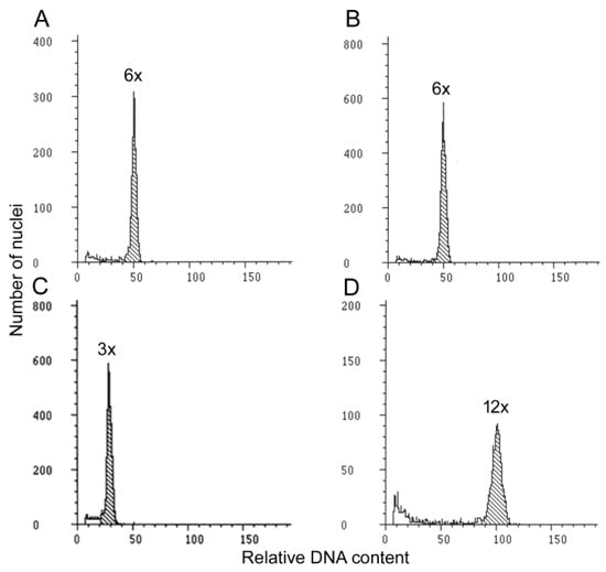
Most of the studied plants remained hexaploids (2n = 6x). Only one sample (regenerated from the ovary of the ‘Jewel Time Yellow’ cultivar) was a haploid with a halved set of chromosomes in the somatic cells (2n = 3x). This plant was regenerated on the medium with 1.0 mg·L−1 TDZ. Another plant, also obtained from the ‘Jewel Time Yellow’ ovary but heated at 32 °C, had a doubled level of ploidy (2n = 12x) compared with the control (Figure 2).

Figure 2.
Histograms obtained in the FCM analysis in chrysanthemum ‘Jewel Time Yellow’: in vivo-grown mother plant (A); plant regenerated in vitro from an ovary with a ploidy level consistent with the control (2n = 6x) (B); haploid plant regenerated in vitro from an ovary on the medium with 1.0 mg·L−1 TDZ (2n = 3x) (C); plant regenerated in vitro from an ovary treated with high temperature with a doubled ploidy level (2n = 12x) (D).
3.4. Indication of the Origin of Shoots Regenerated from Ovaries
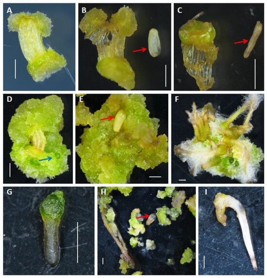
Throughout the experiment, all the ovules remained independent structures and could be easily isolated from the ovary wall (at the beginning of the experiment) and from the callus that overgrown the ovary wall in the later weeks (Figure 3). Intact ovules were whitish, shiny, and swollen, while damaged ones were shrunk, desiccated, and brownish. After the first week of culture, all ovules remained intact–whitish and brilliant. Starting from the second week, damaged ovules were observed, but simultaneously, even in the sixth week of the experiment, intact ovules were still present in 33.3% of the explants, though they were hidden in the abundance of callus and other regenerating structures. Nonetheless, no growth, development, or regeneration was observed from the ovules during the six week period of observations: ovules either stayed intact and unchanged or damaged.

Figure 3.
Callogenesis and somatic embryogenesis in ovaries of chrysanthemum ‘Capitola’ cultured in vitro on the MS medium supplemented with 1.0 mg·L−1 2.4 D and 1.0 mg·L−1 BAP: week 1–callus regeneration on the proximal and distal end of an ovary (A), week 1–intact ovule isolated from an ovary (B); week 2–shrink, damaged ovule dissected from an ovary (C); week 3–ovary overgrown with vivid greenish callus, blue arrow indicates SE at globular stage (D); week 4–intact ovule surrounded by callus regenerated from an ovary wall (E); week 5–the ovary covered with callus from which numerous somatic embryos with hairy roots develop (F); week 5–somatic embryo isolated from callus (G); week 6–intact ovule dissected from callus originating from an ovary wall (H); week 6–mature somatic embryo dissected from callus originating from an ovary wall (I). Bar = 1 mm; red arrows indicate ovules.
Callus started to regenerate in the first week of culture at both the proximal and distal ends of the ovaries (Figure 3). It was greenish at the beginning but turned yellowish and brownish in the later weeks, although its structure remained loose and spongy up to the sixth week of the experiment. In the last week of culture, the callus overgrew the ovary wall and increased its volume remarkably compared with the first week.
Starting from the third week of culture, globular somatic embryos (SE) were found on the surface of the callus in 44.4% of explants. In the fourth week, embryo roots with well-developed hairy roots were present, indicating the further development of SE. In the fifth and sixth weeks, the torpedo stage and mature SE were found together with newly formed globular SE in 66.6% of explants (Figure 3). The highest mean number of SE (4.5 per explant) was observed in the fifth week of culture.
4. Discussion
Chrysanthemum stands as an emblematic and cherished member of the Asteraceae family, captivating the world with its ornate and diverse array of floral forms and colors [24]. In recent years, the utilization of in vitro culture techniques and haploids in plant breeding has emerged as a promising strategy to expedite the development of improved cultivars [25,26]. Ovaries cultured in vitro were reported as a valuable explant source for the development of phenotypic variants with variegated, marble-like, and lighter-green leaves, and changes in the morphology of inflorescences and ligulate florets, as well as changes in the shape of corymb [27]. Our research sheds new insights into the application of haploidization in chrysanthemum.
In the present study, significant variability in disinfection efficiency among the chrysanthemum cultivars tested was found. Chrysanthemum ‘Capitola’ exhibited the highest disinfection efficiency, with an impressive range of 91.7% to 100% aseptic explants. In contrast, chrysanthemum ‘Brasil’ demonstrated a lower disinfection efficiency, with 41.7% to 83.3% of ovules and ovaries remaining aseptic. This difference underscores the genetic diversity among chrysanthemum cultivars, reported also by Din et al. [28], leading to differential responses during the disinfection process. It is possible that the ‘Capitola’ cultivar contains a higher content of metabolites with antimicrobial properties, which could explain the obtained results and shed new light on the utilization of this cultivar in further breeding. On the other hand, the effectiveness of disinfection may also be related to the shape and size of ligulate flowers, which affects the penetration of disinfectants and their action.
Interestingly, the type of explant (ovules versus ovaries) played a crucial role in the regeneration potential of chrysanthemum [21]. In our study, ovaries, in general, exhibited a higher capability for callus formation (90.1% of explants) compared with ovules (32.6%), particularly in the case of chrysanthemum ‘Jewel Time Yellow.’ This divergence may be attributed to variations in the physiological state of these explant types and their competence for tissue culture responses. Ovaries are more complex structures than ovules. In chrysanthemums, they are female reproductive organs comprising a single anatropous ovule wrapped with several layers of parenchyma cells and epidermis forming an ovary wall [29]. Our microscopic observations showed that the callus originates from the ovary wall tissues. Ovaries, as floral organs, are formed late in a plant’s ontogenic development, and early-stage tissues tend to have a higher capacity for callus formation. A similar tendency was found in Beta vulgaris [30] and Allium cepa cultures [31]. The authors justify this phenomenon by the fact that ovules, which are delicate by nature, are less exposed to damage when whole ovaries are cultured. Moreover, in ovule culture, the stage of ovule sac development is more important than in ovary cultures. Direct exposure to light may also negatively affect the ability of ovules to regenerate. When they are not in direct contact with the environment—as it happens in ovary cultures—fewer harmful phenolic substances are produced.
Furthermore, the influence of TDZ concentration on explant regeneration was evident, especially in the chrysanthemum ‘Jewel Time Yellow.’ A positive correlation was observed between the TDZ concentration and the percentage of explants capable of regeneration. This suggests that TDZ, as a cytokinin-like growth regulator, effectively promotes the initiation of shoot formation in chrysanthemum ovaries, particularly in the ‘Jewel Time Yellow’ cultivar [32]. This hypothesis is supported by the fact that the highest mean number of shoots per explant was regenerated in ovaries cultured on the media containing high (1.0 and 1.5 mg·L−1) concentrations of TDZ. TDZ stimulated the quick regeneration of multiple shoots from the nodal explant of Vitex trifolia [33]. In chrysanthemums, the combinations of BAP and TDZ stimulated the regeneration of shoots from leaf explant [34]. Perhaps using this or other PGRs combination would also enhance caulogenesis in ovaries and ovules. The capacity for shoot regeneration in chrysanthemum depends strongly on the composition of PGRs, genotype, and explant type. The same growth regulators can show a different effect even in cultivars and clones of the same species [35].
The results obtained in this experiment demonstrate a complex interplay between temperature treatments and the efficiency of chrysanthemum ovary and ovule regeneration in vitro. Regarding disinfection efficiency, these data reveal that both low and high-temperature treatments can significantly improve the effectiveness of disinfection in chrysanthemum explants. Explants subjected to thermal shock at both 4 °C and 32 °C exhibited the highest disinfection efficiency, ranging from an impressive 92% to a remarkable 100%. This finding confirms that temperature fluctuations, either cold or heat shock, can disrupt the integrity of microbial contaminants or pathogens in the ovules and ovaries, resulting in a highly effective disinfection process, as suggested by Gélinas et al. [36]. This high level of disinfection is crucial for subsequent plant regeneration, as it reduces the risk of contamination and increases the chances of obtaining high-quality, pathogen-free haploid cultures.
The efficiency of callus regeneration, however, exhibited a wider range of variation across different temperature treatments. Callus regeneration ranged from 16.6% (in the ovaries of chrysanthemum ‘Capitola’ stored at 32 °C) to 98.8% (in control ovaries of ‘Capitola’). This indicates that some genotypes are more sensitive to temperature changes than others when it comes to callus regeneration. Likewise, Benderradji et al. [37] and Lee and Seo [38] reported a genotype- and temperature-dependent callus formation in wheat and Arabidopsis, respectively. On the other hand, Mostafiz et al. [39] reported that all three wetland Malaysian rice cultivars (MR232, MR220, and MR220-CL2) and upland rice (Bario) exhibited the highest callus induction percentage from 45 °C pre-heated seeds. Our data on shoot regeneration further emphasize the intricate relationship between temperature treatments and plant response. Surprisingly, neither cold- nor heat-shock treatment increased the number of regenerated shoots compared with the control. Usually, stress increases the regeneration efficiency in plant tissue culture. For example, warm temperature promotes shoot regeneration in A. thaliana through the enhanced expression of several regeneration-associated genes, such as CUP-SHAPED COTYLEDON 1 (CUC1) encoding a transcription factor involved in shoot meristem formation and YUCCAs (YUCs) encoding auxin biosynthesis enzymes [40,41]. In summary, the effects of temperature on plant tissue culture can be complex and multifaceted. While temperature shock may improve disinfection and promote callus formation by increasing metabolic activity, it can disrupt the hormonal balance necessary for shoot formation. Additionally, the specific responses can vary between plant cultivars, highlighting the importance of optimizing culture conditions for each unique case. Our results underscore the importance of considering factors beyond temperature alone, such as different concentrations of sucrose, culturing on liquid or solid medium, and the complex interaction of various culture conditions in determining the success of chrysanthemum gynogenesis [42].
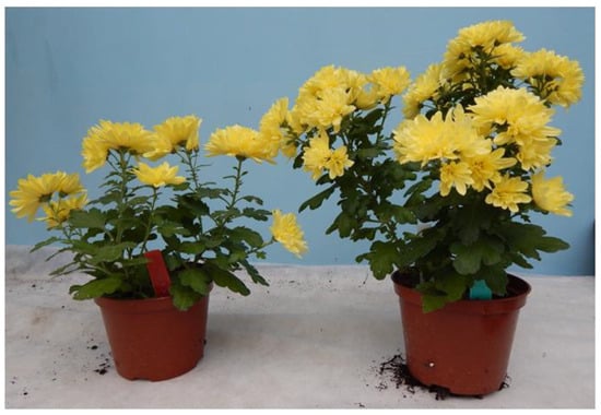
Stress is known to be a successful factor in inducing chromosome elimination in plants [43]. For example, reactive oxygen species burst caused haploid induction in maize [44]. Remarkably, in the present study, the majority of the regenerated plants did not differ in terms of ploidy from the control. Only one individual, originating from the ovary of the ‘Jewel Time Yellow’ cultivar, displayed haploidy with a halved set of chromosomes (2n = 3x). This plant may have originated from the unfertilized egg cell within the ovule. As for the remaining plants, it is possible that regeneration took place from haploid cells, but spontaneous diploidization occurred. However, it is more likely that the shoots regenerated from the somatic tissues of explants and had the same ploidy level as the mother plant. The latter statement is supported by our findings concerning the microscopic observations of embryogenesis in ovaries; many somatic embryos developed from callus, which overgrown the ovary wall, while ovules did not develop during the six weeks of observations. Thus, the regenerated plantlets are more likely to originate as vegetative progeny from somatic tissue of the ovary wall than from generative cells of the embryo sac. Furthermore, another plant obtained from the ‘Jewel Time Yellow’ ovary, subjected to a heat shock of 32 °C, exhibited a doubled level of ploidy (2n = 12x) compared with the control. The phenomenon of chromosome doubling has been previously reported during plant regeneration in vitro [45]. The chromosome doubling observed in the ‘Jewel Time Yellow’ plant may have been induced by the elevated temperature, triggering changes in cell division and chromosome replication [46]. This dodecaploid plant could originate from callus cell(s), which duplicated its genome without cytokinesis. The phenomenon of polyploidization often occurs in tissue-cultured callus, leading to the development of plants with multiplied chromosome sets [47]. Polyploidy plays a major role in driving species formation and facilitating the evolution of novel gene functions [48]. The dodecaploids show a higher number of genes with upregulated differentially expressed genes (DEGs) compared with their hexaploid counterparts, which may contribute to the enhanced environmental adaptability of dodecaploid plants [49]. The 2n = 12x plant regenerated in this study from the heat-shock treatment was taller than the original cultivar. No changes in the shape or color of inflorescences were observed (Figure 4). The dodecaploid chrysanthemum can be employed in genetic and inheritance studies. Moreover, it can display some beneficial traits resulting from polyploidization, such as higher pest or stress resistance, which will be evaluated in following research studies.

Figure 4.
Chrysanthemum ‘Jewel Time Yellow’: true-to-type, hexaploid (2n = 6x) reference plant (left) and dodecaploid (2n = 12x) plant regenerated from heat-treated ovary (right).
Nonetheless, considering the relatively low share of plants with an altered chromosome number, further research into genotype-specific responses and optimization of culture conditions is essential to harness the full potential of temperature-based treatments in haploid production and chrysanthemum breeding programs.
5. Conclusions
In this study, the effect of genotype, TDZ, and pretreatment temperature on the regeneration of chrysanthemum ovaries and ovules was studied. It was found that ovaries had a greater organogenic potential (both in terms of callogenesis and shoot formation) than ovules. The highest number of shoots was produced in cooled (at 4 °C) ovaries of chrysanthemum ‘Brasil’ and in ‘Jewel Time Yellow’ explants cultured on a medium with 1.0–1.5 mg·L−1 TDZ. The latter cultivar also had the highest potential to produce plants with an altered ploidy level (doubled and halved chromosome number). This research study underscores the promising potential of ovules and ovaries as explant sources not only for the reproduction of chrysanthemum in vitro but also for the production of plants with altered ploidy levels. The practical implications of our findings for horticultural practices are significant, as they open doors for the development of haploids and dodecaploids, desirable in breeding.
Author Contributions
Conceptualization, N.M. and A.T.; methodology, N.M., A.T. and M.R.; software, N.M., A.T., D.K. and M.R.; validation, N.M., A.T. and D.K.; formal analysis, N.M. and A.T.; investigation, N.M., A.T. and M.R.; resources, N.M., A.T. and M.R.; data curation, N.M. and A.T.; writing—original draft preparation, D.K.; writing—review and editing, N.M., A.T. and D.K.; visualization, N.M.; supervision, N.M., A.T. and D.K.; project administration, N.M. and A.T. All authors have read and agreed to the published version of the manuscript.
Funding
This research received no external funding.
Institutional Review Board Statement
Not applicable.
Data Availability Statement
Data are available by email on reasonable request.
Acknowledgments
The authors would like to thank Elżbieta Szydłowska and Agnieszka Skrzeszewska for their technical support in this research study.
Conflicts of Interest
The authors declare no conflict of interest.
References
- Schroeter-Zakrzewska, A.; Pradita, F.A. Effect of colour of light on rooting cuttings and subsequent growth of chrysanthemum (Chrysanthemum × grandiflorum /Ramat./Kitam.). Agriculture 2021, 11, 671. [Google Scholar] [CrossRef]
- Zhang, Y.; Guo, C.; Hu, J.; Liu, F.; Fu, S.; Guo, X.; Chen, Q.; Zhang, L.; Zhu, L.; Hou, X. Effects of 6-benzylaminopurine combined with prohexadione-Ca on yield and quality of Chrysanthemum morifolium Ramat cv. Hangbaiju. Agriculture 2023, 13, 444. [Google Scholar] [CrossRef]
- Mekapogu, M.; Kwon, O.-K.; Song, H.-Y.; Jung, J.-A. Towards the improvement of ornamental attributes in chrysanthemum: Recent progress in biotechnological advances. Int. J. Mol. Sci. 2022, 23, 12284. [Google Scholar] [CrossRef] [PubMed]
- Sasaki, K.; Tanaka, T. Overcoming difficulties in molecular biological analysis through a combination of genetic engineering, genome editing, and genome analysis in hexaploid Chrysanthemum morifolium. Plants 2023, 12, 2566. [Google Scholar] [CrossRef] [PubMed]
- Anderson, N.O. Chrysanthemum. In Flower Breeding and Genetics; Anderson, N.O., Ed.; Springer: Dordrecht, The Netherlands, 2007; pp. 389–437. [Google Scholar] [CrossRef]
- Klie, M.; Schie, S.; Linde, M.; Debener, T. The type of ploidy of chrysanthemum is not black or white: A comparison of a molecular approach to published cytological methods. Front. Plant Sci. 2014, 5, 479. [Google Scholar] [CrossRef] [PubMed]
- Karjee, S.; Mahapatra, S.; Singh, D.; Saha, K.; Viswakarma, P.K. Production of double haploids in ornamental crops. J. Pharmacogn. Phytochem. 2020, 9, 555–565. [Google Scholar]
- Zargar, M.; Zavarykina, T.; Voronov, S.; Pronina, I.; Bayat, M. The recent development in technologies for attaining doubled haploid plants in vivo. Agriculture 2022, 12, 1595. [Google Scholar] [CrossRef]
- Jacquier, N.M.A.; Gilles, L.M.; Pyott, D.E.; Martinant, J.P.; Rogowsky, P.M.; Widiez, T. Puzzling out plant reproduction by haploid induction for innovations in plant breeding. Nat. Plants 2020, 6, 610–619. [Google Scholar] [CrossRef]
- Portemer, V.; Renne, C.; Guillebaux, A.; Mercier, R. Large genetic screens for gynogenesis and androgenesis haploid inducers in Arabidopsis thaliana failed to identify mutants. Front. Plant Sci. 2015, 6, 147. [Google Scholar] [CrossRef]
- Ashapkin, V.V.; Kutueva, L.I.; Aleksandrushkina, N.I.; Vanyushin, B.F. Epigenetic regulation of plant gametophyte development. Int. J. Mol. Sci. 2019, 20, 3051. [Google Scholar] [CrossRef]
- Ashokhan, S.; Othman, R.; Abd Rahim, M.H.; Karsani, S.A.; Yaacob, J.S. Effect of plant growth regulators on coloured callus formation and accumulation of azadirachtin, an essential biopesticide in Azadirachta indica. Plants 2020, 9, 352. [Google Scholar] [CrossRef]
- Zayachkovskaya, T.; Domblides, E.; Zayachkovsky, V.; Kan, L.; Domblides, A.; Soldatenko, A. Production of gynogenic plants of red beet (Beta vulgaris L.) in unpollinated ovule culture in vitro. Plants 2021, 10, 2703. [Google Scholar] [CrossRef]
- Zou, J.; Zou, X.; Gong, Z.; Song, G.; Ren, J.; Feng, H. Thidiazuron promoted microspore embryogenesis and plant regeneration in curly kale (Brassica oleracea L. convar. acephala var. sabellica). Horticulturae 2023, 9, 327. [Google Scholar] [CrossRef]
- Kumari, P.T.; Aswath, C. Haploid and double haploids in ornamentals—A review. Int. J. Curr. Microbiol. App. Sci. 2018, 7, 1322–1336. [Google Scholar] [CrossRef]
- Ahmadli, U.; Kalidass, M.; Khaitova, L.C.; Fuchs, J.; Cuacos, M.; Demidov, D.; Zuo, S.; Pecinkova, J.; Mascher, M.; Ingouff, M.; et al. High temperature increases centromere-mediated genome elimination frequency and enhances haploid induction in Arabidopsis. Plant Commun. 2023, 4, 100507. [Google Scholar] [CrossRef]
- Jin, C.; Sun, L.; Trinh, H.K.; Danny, G. Heat stress promotes haploid formation during CENH3-mediated genome elimination in Arabidopsis. Plant Reprod. 2023, 36, 147–155. [Google Scholar] [CrossRef]
- Kiviharju, E.; Pehu, E. The effect of cold and heat pretreatments on anther culture response of Avena sativa and A. sterilis. Plant Cell Tissue Organ Cult. 1998, 54, 97–104. [Google Scholar] [CrossRef]
- Mayakaduwa, R.; Silva, T. Haploid induction in indica rice: Exploring new opportunities. Plants 2023, 12, 3118. [Google Scholar] [CrossRef]
- Wang, H.; Dong, B.; Jiang, J.; Fang, W.; Guan, Z.; Liao, Y.; Chen, S.; Chen, F. Characterization of in vitro haploid and doubled haploid Chrysanthemum morifolium plants via unfertilized ovule culture for phenotypical traits and DNA methylation pattern. Front. Plant Sci. 2014, 5, 738. [Google Scholar] [CrossRef]
- Miler, N.; Muszczyk, P. Regeneration of callus and shoots from the ovules and ovaries of chrysanthemum in vitro. Acta Hortic. 2015, 1083, 103–106. [Google Scholar] [CrossRef]
- Murashige, T.; Skoog, F. A revised medium for rapid growth and bioassays with tobacco tissue culture. Plant Physiol. 1962, 15, 473–497. [Google Scholar] [CrossRef]
- Galbraith, D.W.; Harkins, K.R.; Maddox, J.R.; Ayres, N.M.; Sharma, D.P.; Firoozabady, E. Rapid flow cytometric analysis of the cell cycle in intact plant tissues. Science 1983, 220, 1049–1051. [Google Scholar] [CrossRef]
- Liu, J.-Z.; Du, L.-D.; Chen, S.-M.; Cao, J.-R.; Ding, X.-Q.; Zheng, C.-S.; Sun, C.-H. Comparative analysis of the effects of internal factors on the floral color of four chrysanthemum cultivars of different colors. Agriculture 2022, 12, 635. [Google Scholar] [CrossRef]
- Liu, C.; Song, G.; Zhao, Y.; Fang, B.; Liu, Z.; Ren, J.; Feng, H. Trichostatin A induced microspore embryogenesis and promoted plantlet regeneration in ornamental kale (Brassica oleracea var. acephala). Horticulturae 2022, 8, 790. [Google Scholar] [CrossRef]
- Starosta, E.; Szwarc, J.; Niemann, J.; Szewczyk, K.; Weigt, D. Brassica napus haploid and double haploid production and its latest applications. Curr. Issues Mol. Biol. 2023, 45, 4431–4450. [Google Scholar] [CrossRef]
- Miler, N.; Jedrzejczyk, I. Chrysanthemum plants regenerated from ovaries: A study on genetic and phenotypic variation. Turk. J. Bot. 2018, 42, 5. [Google Scholar] [CrossRef]
- Din, A.; Qadri, Z.A.; Rather, Z.A.; Mir, M.S.; Murtaza, I.; Khan, F.A.; Neelofar; Wani, M.A. In vitro sterilization of different explants of Chrysanthemum (Dendranthemum morifolium L.) cvs. “Candid” and “Flirt”. Cur. J. Appl. Sci. Technol. 2018, 31, 1–14. [Google Scholar] [CrossRef]
- Sun, C.Q.; Chen, F.D.; Teng, N.J.; Liu, Z.L.; Fang, W.M.; Hou, X.L. Factors affecting seed set in the crosses between Dendranthema grandiflorum (Ramat.) Kitamura and its wild species. Euphytica 2010, 171, 181–192. [Google Scholar] [CrossRef]
- Van Geyt, J.; Speckmann, G.J.; D’Halluin, K.; Jacobs, M. In vitro induction of haploid plants from unpollinated ovules and ovaries of the sugarbeet (Beta vulgaris L.). Theoret. Appl. Genetics 1987, 73, 920–925. [Google Scholar] [CrossRef]
- Ihan, A.; Javornik, B. Studies of gynogenesis in onion (Allium cepa L.): Induction procedures and genetic analysis of regenerants. Plant Sci. 1995, 104, 215–224. [Google Scholar] [CrossRef]
- Ali, H.M.; Khan, T.; Khan, M.A.; Ullah, N. The multipotent thidiazuron: A mechanistic overview of its roles in callogenesis and other plant cultures in vitro. Biotechnol. Appl. Biochem. 2022, 69, 2624–2640. [Google Scholar] [CrossRef]
- Ahmed, M.R.; Anis, M. Role of TDZ in the quick regeneration of multiple shoots from nodal explant of Vitex trifolia L.—An important medicinal plant. Appl. Biochem. Biotechnol. 2012, 168, 957–966. [Google Scholar] [CrossRef]
- Sjahril, R.; Haring, F.; Riadi, M.; Rahim, M.D.; Khan, R.S.; Arjunayanti, A.; Trisnawaty, A.R. Performance of NAA, 2iP, BAP and TDZ on callus multiplication, shoots initiation and growth for efficient plant regeneration system in Chrysanthemum (Chrysanthemum morifolium Ramat.). IJAS 2016, 4, 52–61. [Google Scholar]
- Tymoszuk, A.; Zalewska, M. In vitro adventitious shoots regeneration from ligulate florets in the aspect of application in chrysanthemum breeding. Acta Sci. Pol. Hort. Cult. 2014, 13, 45–58. [Google Scholar]
- Gélinas, P.; Goulet, J.; Tastayre, G.M.; Picard, G.A. Effect of temperature and contact time on the activity of eight disinfectants—A classification. J. Food Prot. 1984, 47, 841–847. [Google Scholar] [CrossRef]
- Benderradji, L.; Brini, F.; Kellou, K.; Ykhlef, N.; Djekoun, A.; Masmoudi, K.; Bouzerzour, H. Callus induction, proliferation, and plantlets regeneration of two bread wheat (Triticum aestivum L.) genotypes under saline and heat stress conditions. ISRN Agronomy 2012, 2012, 367851. [Google Scholar] [CrossRef]
- Lee, K.; Seo, P.J. High-temperature promotion of callus formation requires the BIN2-ARF-LBD axis in Arabidopsis. Planta 2017, 246, 797–802. [Google Scholar] [CrossRef]
- Mostafiz, S.B.; Wagiran, A.; Johan, N.S.; Zulkifli, N.S.A.; Ming, N.J. The effects of temperature on callus induction and regeneration in selected Malaysian rice cultivar Indica. Sains Malays. 2018, 47, 2647–2655. [Google Scholar] [CrossRef]
- Haque, M.; Islam, S.M.S.; Subramaniam, S. Effects of salt and heat pre-treatment factors on efficient regeneration in barley (Hordeum vulgare L.). 3 Biotech. 2017, 7, 63. [Google Scholar] [CrossRef]
- Lambolez, A.; Kawamura, A.; Takahashi, T.; Rymen, B.; Iwase, A.; Favero, D.S.; Ikeuchi, M.; Suzuki, T.; Cortijo, S.; Jaeger, K.E.; et al. Warm temperature promotes shoot regeneration in Arabidopsis thaliana. Plant Cell Physiol. 2022, 63, 618–634. [Google Scholar] [CrossRef]
- Zayachkovskaya, T.; Alyokhina, K.; Mineykina, A.; Romanova, O.; Vjurtts, T.; Tukuser, Y.; Zayachkovsky, V.; Ermolaev, A.; Kan, L.; Fomicheva, M.; et al. Optimizing different medium component concentration and temperature stress pretreatment for gynogenesis induction in unpollinated ovule culture of sugar beet (Beta vulgaris L.). Horticulturae 2023, 9, 900. [Google Scholar] [CrossRef]
- Zheng, M.; Fournier, A.; Weng, Y. Differential effects of cold and heat shock on embryogenic induction and green plant regeneration from wheat (Triticum aestivum L.) microspores. Am. J. Plant Sci. 2023, 14, 308–322. [Google Scholar] [CrossRef]
- Jiang, C.; Sun, J.; Li, R.; Yan, S.; Chen, W.; Guo, L.; Qin, G.; Wang, P.; Luo, C.; Huang, W.; et al. A reactive oxygen species burst causes haploid induction in maize. Mol. Plant. 2022, 15, 943–955. [Google Scholar] [CrossRef] [PubMed]
- Kasha, K.J.; Shim, Y.S.; Simion, E.; Letarte, J. Haploid production and chromosome doubling. Acta Hortic. 2006, 725, 817–828. [Google Scholar] [CrossRef]
- Bertin, N. Analysis of the tomato fruit growth response to temperature and plant fruit load in relation to cell division, cell expansion and DNA endoreduplication. Ann. Bot. 2005, 95, 439–447. [Google Scholar] [CrossRef]
- Betekhtin, A.; Rojek, M.; Jaskowiak, J.; Milewska-Hendel, A.; Kwasniewska, J.; Kostyukova, Y.; Kurczynska, E.; Rumyantseva, N.; Hasterok, R. Nuclear genome stability in long-term cultivated callus lines of Fagopyrum tataricum (L.) Gaertn. PLoS ONE 2017, 12, e0173537. [Google Scholar] [CrossRef] [PubMed]
- Aversano, R.; Ercolano, M.R.; Caruso, I.; Fasano, C.; Rosellini, D.; Carputo, D. Molecular tools for exploring polyploid genomes in plants. Int. J. Mol. Sci. 2012, 13, 10316–10335. [Google Scholar] [CrossRef] [PubMed]
- Bolaños-Villegas, P.; Chen, F.-C. Advances and perspectives for polyploidy breeding in orchids. Plants 2022, 11, 1421. [Google Scholar] [CrossRef]
Disclaimer/Publisher’s Note: The statements, opinions and data contained in all publications are solely those of the individual author(s) and contributor(s) and not of MDPI and/or the editor(s). MDPI and/or the editor(s) disclaim responsibility for any injury to people or property resulting from any ideas, methods, instructions or products referred to in the content. |
© 2023 by the authors. Licensee MDPI, Basel, Switzerland. This article is an open access article distributed under the terms and conditions of the Creative Commons Attribution (CC BY) license (https://creativecommons.org/licenses/by/4.0/).