Article

In Vitro Antimicrobial Activity of Plant Species against the Phytopathogens Ralstonia solanacearum, Phytophthora infestans, and Neopestalotiopsis javaensis

1
Grupo de Investigación Productos Naturales Bioactivos, Escuela de Ciencias Agrícolas y Ambientales, Pontificia Universidad Católica del Ecuador-Ibarra, Av. Jorge Guzmán Rueda y Av. Padre Aurelio Espinosa Polit, Ibarra 100112, Ecuador
2
Escuela de Ciencias Agrícolas y Ambientales, Pontificia Universidad Católica del Ecuador-Ibarra, Av. Jorge Guzmán Rueda y Av. Padre Aurelio Espinosa Polit, Ibarra 100112, Ecuador
3
Grupo de Investigación Aplicada en Materiales y Procesos (GIAMP), School of Chemical Sciences and Engineering, Yachay Tech University, Hda. San José s/n y Proyecto Yachay, Urcuquí 100119, Ecuador
*
Authors to whom correspondence should be addressed.
Agriculture 2023, 13(10), 2029; https://doi.org/10.3390/agriculture13102029
Submission received: 21 August 2023 / Revised: 20 September 2023 / Accepted: 25 September 2023 / Published: 20 October 2023
(This article belongs to the Special Issue Advances in Biological Control of Plant Diseases)

Abstract

:
Plants are prone to be attacked by various pathogenic microorganisms, leading to significant crop yield losses. Pesticides are used to control agricultural pathogens; however, frequently, they are toxic synthetic products. This work evaluated the antibacterial and antifungal activity of Pernettya prostrata, and Rubus roseus plant extracts against three phytopathogens Ralstonia solanacearum, Phytophthora infestans, and Neopestalotiopsis javaensis responsible for causing banana bacterial wilt, late blight y scab diseases, respectively. The extracts were obtained in methanol. The phytopathogens were isolated from diseased plants grown in Ecuador, R. solanacearum was isolated from samples of Musa paradiasiaca, P. infestans from samples of Solanum lycopersicum and N. javaensis from diseased samples of Persea americana—morphological and molecular methods identified the isolated pathogens. The antibacterial activity was determined by the microtiter broth dilution method; six serial concentrations of the extracts were tested. The antifungal activity was determined based on the effects of the plant extracts on the inhibition of radial growth of fungi; five serial concentrations of the extracts were tested. The extracts of P. prostrata and R. roseus showed activity against R. solanacearum and P. infestans with a MIC of 22.5 and 31.25 mg/mL, respectively.

1. Introduction

Agriculture has a direct influence on the world economy [1]. However, plants exposed to environmental conditions are prone to be attacked by pathogenic microorganisms such as bacteria, viruses, nematodes, and fungi, which cause significant crop yield losses.
Pesticides are frequently toxic synthetic products used to carry out chemical controls on pathogenic organisms; their deliberate use has constituted a risk factor for human health and the ecosystem [2]. In recent decades, the environmental contamination caused by these chemical compounds has been increasing remarkably [3]. Therefore, there is interest in searching for alternative compounds to be used as natural control.
Organic farming suggests applying natural products in the search to reduce the use of synthetic products. One of the most effective options for this reduction is using organic formulas from plant extracts to achieve ecological pesticides that control or eliminate diseases caused by transmissible vectors and reduce the impacts caused [3].
The geographic location of Ecuador and its geological features make this region one of the richest in the world in biodiversity. Ecuador is among the 17 megadiverse countries, accounting for about 10% of the world’s plant species [4,5]. This fact makes Ecuador an invaluable source of bioactive extracts and molecules that could be used by themselves or as raw materials for developing products focused on the pharmaceutical, nutraceutical, or agricultural industry [6]. Plants produce a wide range of secondary metabolites; they exert their biological effect on other organisms [7] and can act against microbial pathogens based on their toxic nature. Secondary metabolites in higher plants play a crucial role in various ways in developing natural products for the agricultural industry. Firstly, crude extracts can act as natural pesticides in the unmodified state. In addition, they supply the chemical compounds necessary for synthesizing more complex compounds. Finally, new modes of pesticide action allow the complete synthesis of new products, which in turn help to counteract the resistance currently shown by synthetic products used to combat phytopathogens [8].
Ericaceae and Rosaceae families are plants rich in alkaloids, terpenoids, flavonoid glycosides, tannins, and terpenes, active compounds against microorganisms that affect crops [9]. This work focused on studying two plant species belonging to the previously mentioned families: Pernettya prostrata and Rubus roseus.
P. prostrata (Cav.) DC. belongs to the family Ericaceae. It is a shrub up to 30 cm tall, distributed from 1500 to more than 4000 m above sea level; it produces a few purple fleshy fruits, and inside, it has many seeds smaller than one millimeter [10]. In Ecuador, it is commonly known as “yuruñiwi” (kichwa language), “mortiño,” “manzana,” or “moridera”. Its fruits are edible in small quantities since they have narcotic, hallucinogenic, and intoxicating properties [11], and poisonings have been reported in animals after their administration [12]. Studies suggest that the toxicity may be due to andrometoxin or grayanotoxins [9]. In Ecuador, the infusion of P. prostrata is used therapeutically by the Saraguros and Shuar indigenous communities to treat headaches [13]. The genus Pernettya includes 20 species of plants distributed in America and Oceania [14]. In America, they are distributed from Mexico to Argentina [15]. Plant extracts and isolated compounds from this genus have shown pharmacological properties such as antioxidant, anti-inflammatory, insecticide, antifungal [9,16,17,18,19,20], and allelopathic properties [9].
R. roseus Poir belongs to the Rosaceae family. It is a shrub distributed from 1900 to 4000 m altitude [21]. It is commonly known as “mora” or “mora silvestre”. The fruit is edible and used to prepare juices, sweets, and jams; it also serves birds [5]. Phenolic compounds are the most active metabolites of the genus Rubus [22] and are widely used in ethnomedicinal applications [23]. Extracts of species of this genus present bacteriostatic activity [24], antibacterial, and antifungal activity [25].
No previous studies were found about the effect of P. prostrata or R. roseus on phytopathogens used in this research. This work aims to isolate and characterize the pathogens responsible for causing bacterial wilt, late blight, and scab in the crops of Musa paradisiaca, Solanum lycopersicum, Persea american, respectively, and evaluate the antimicrobial effect of the extracts obtained from P. prostrata, and R. roseus on the isolated phytopathogens.

2. Materials and Methods

2.1. Plant Material and Extraction

The aerial parts of R. roseus (17N 813854 38744, 3456 m.a.s.l.) and P. prostrata (17N 802976 15112, 3525 m.a.s.l.) were collected in Mojanda Lake, in Imbabura province of Ecuador. Voucher specimens for P. prostrata (N° 006453) and R. roseus (N° 006454) were deposited at the Herbarium of the Pontificia Universidad Católica del Ecuador Ibarra, Ecuador. The samples were collected under governmental permission (006-2019-IC-FAU-FLO-DPAI/MAE). Dust, insect contamination, and other plant species contamination were removed from each sample.
Plant material was dried in a plant drier using heat (50 °C) and an airflow source for 24 h. Dry plant materials were ground into a fine powder and extracted with methanol (3 times), using a ratio of 1 g of plant material: 10 mL of MeOH, at room temperature overnight with constant agitation (250 rpm). Filtered extracts were combined and concentrated under reduced pressure until dryness.

2.2. Isolation and Characterization of Agricultural Microorganisms

2.2.1. Ralstonia solanacearum

Isolation of Ralstonia solanacearum: Samples of the rachis of Musa paradiasiaca were collected in El Carmen, in Manabí province of Ecuador (17M 669899 9967526, 245 m.a.s.l.). A transversal cut of the rachis was made, evidencing reddish dots; the fruit presented brown internal necrosis, early maturation in a non-uniform way, and a dark, necrotic, and cracked peel (see Figure 1A). The plant sample was subjected to a disinfection process with sterile distilled water for 30 min and with 70% alcohol for 1 min in the laminar flow chamber. To obtain the pure colonies of R. solanacearum, a rachis that presented bacterial exudate was used with a previously sterilized wire loop; a small amount of the exudate was taken and placed in a petri dish with nutrient agar, incubated for 24 to 48 h at 28 ± 1 °C. The Petri dishes that showed colonies with irregular borders, fluid, and mucoid, the typical form of R. solanacearum, were placed in new dishes of nutrient agar until a pure colony was observed [26].
Characterization of Ralstonia solanacearum: this pathogen was identified through phenotypic, biochemical tests, and molecular characterization.
Gram staining: Aqueous crystal violet solution (0.5%) was added to the smear for 30 s and washed with tap water for one minute. Then, it was placed with iodine for one minute, rinsed with running water and decolorized with 95% ethanol. The sample was counterstained with safranin for 10 s, washed with water, dried, and observed microscopically at 100× with oil immersion [27].
Potassium hydroxide test: The bacterium was aseptically removed from the Petri dish with an inoculation wire loop, placed on the glass slide in a drop of 3% KOH solution, and shaken for 20 s to observe the formation of threads [27].
Catalase test: Bacterial cultures 24 h old were contacted with 3% hydrogen peroxide (H2O2) on a glass slide, and the production of gas bubbles was observed with the naked eye and at 25× magnification [27].
Kovacs oxidase test: One drop of oxidase reagent (1% tetra-methyl-p-phenyl diamine dihydrochloride) was added to a piece of filter paper placed inside a glass Petri dish. Colonies (24 h old) were rubbed onto the filter paper with an oxidase reagent solution. Wait for 60 s to see if the bacteria develop a purple color [28].
TSI Agar test (Triple Sugar Iron Agar): The test was performed to determine the fermentation of the three sugars (glucose, lactose, and sucrose). For this, a colony 24 h old was taken and inoculated into the TSI agar tube at medium temperature. Depth, making a striation in the tip of the tube, the sample was incubated at 28 °C for 24 h to show results; after the time elapsed, the sample should turn yellow, which means that the bacteria ferment the three sugars and negative if it does not present changes in its coloration [29].
Simmons Citrate Test: This test is used to determine if the microorganism under study is capable of using citrate as a carbon source. A colony 24 h old was taken and inoculated into the agar tube at medium temperature. A striatum at the top of the tube, the sample was incubated at 28 °C for 24 h, after which time the sample should change its color from green to blue to assume a positive result, while if it remains green, it is a negative result [30].
The molecular identification of R. solanacearum was confirmed by 16s rRNA gene sequence comparison with GenBank of NCBI. The 16s rRNA gene sequencing was completed at Zurita & Zurita Laboratories.

2.2.2. Phytophthora infestans

Isolation of Phytophthora infestans: Samples of the leaves of Solanum lycopersicum were collected in the locality of La Esperanza, in Imbabura province of Ecuador (0.297646N −78.11780, 2350 m.a.s.l.); the samples presented damage, including wilting of the foliar system due to root rot (see Figure 1B). The leaves were subjected to a disinfection process with a soapy solution for 5 min, then placed in distilled water for 3 min, and immersed into a 3% sodium hypochlorite solution for 5 min, rinsed three times with distilled water. For 3 min, the samples were dried with sterilized absorbent towels. The tissue was sown in potato-dextrose-agar (PDA) culture medium; the plates were incubated at 17 ± 1 °C for five days. When the mycelia were formed, they were replicated to obtain pure cultures in rye-agar culture medium; the plates were kept incubated for six days at 17 ± 1 °C, and then antifungal analyses and molecular characterization of the fungus were carried out [31].
Morphological and Molecular Characterization of Phytophthora infestans: The structure of the fungus was analyzed with an optical microscope (MicrosAustria, Veit an der Glan, Austria) in a 40× lens, for which the shape and color of the sporangia were analyzed, and they were compared with those reported by Fernández Maura et al. [32]. The identification of fungal strain was confirmed by 18S rRNA gene sequence comparison with GenBank of NCBI. The 18S rRNA gene sequencing was completed at Laboratorio de Fitopatología y Control Biológico in Ecuador. The sequence alignment was completed at a BLAST server.

2.2.3. Neopestalotiopsis javaensis

Isolation of Neopestalotiopsis javaensis: Samples of damaged fruits of Persea americana were collected in the locality of San Vicente de Pusir, in El Carchi province of Ecuador (17 N 829001.34 m E 54818.96 m N, 2.550 m.a.s.l) (see Figure 1C). The samples were washed with sterilized water and allowed to dry. Scab lesions of 3 mm in diameter were taken. The tissue was disinfected for 90 s in a 2% hypochlorite solution, followed by a 30-s wash with sterile water. The tissue was immersed in 70% alcohol for 60 s and washed with sterile water for 30 s. It was dried with sterilized filter paper. Petri dishes were seeded with potato-dextrose-agar (PDA) culture medium containing 100 mg of streptomycin sulfate. The plates were incubated at 25 ± 1 °C in the dark and checked at 24 h intervals until mycelium growth was observed. When the mycelium was formed, it was transferred to Petri dishes containing PDA to obtain pure cultures. Incubate at 25 ± 1 °C in the dark for seven days [33].
Morphological and molecular identification of Neopestalotiopsis javaensis: The macroscopic features of the fungi, such as colony color, colony diameter, and the exudates of the isolate, were recognized. Microscopic characteristics such as acervuli were examined under an optical microscope 40× and compared with the features in identification keys and species descriptions [33]. The molecular identification of N. javaensis was confirmed by 18S rRNA gene sequence comparison with GenBank of NCBI. The 18S rRNA gene sequencing was completed at Identification Molecular Services in Ecuador, and the sequence alignment was completed at a BLAST serve [34].

2.3. Antimicrobial Activity of Plant Extracts

2.3.1. Bacteria and Culture Conditions

Ralstonia solanacearum was grown in Nutritive Broth for 24 h at 28 °C. A 0.5 McFarland Standard [35] was used to create inoculum densities of 1.5 × 10⁸ cfs/mL in Nutritive Broth using the direct suspension method for MIC assays.

2.3.2. Determination of Minimum Inhibitory Concentrations (MICs)

MIC was determined by the microtiter broth dilution method in sterile flat-bottom 96-well polystyrene plates. Six serial dilution techniques were used to determine the MIC of two extracts at concentrations of 45, 22.5, 11.25, 5.62, 2.81, and 1.41 mg/mL after 18 h growth. Negative controls (cells + Nutritive Broth), positive controls (cells + Nutritive Broth + antibiotics − gentamicin), vehicle controls (cells + Nutritive Broth + methanol), and media controls (Nutritive Broth) were included. Positive controls for antibiotics were prepared at 7 g/mL. All tests were performed in triplicate. Optical density readings were taken using a microplate reader (BioTek Instruments, Inc., Winooski, VT, USA) at 650 nm at 0 and 18 h post-inoculation. Results are reported as the MIC for growth at 18 h post-inoculation. The lowest concentration of extract at which growth of microorganisms was not demonstrated after incubation at 37 °C for 18 h is considered the MIC value. The percent inhibition was calculated using Equation (1) [24].
%   I n h i b i t i o n = 1 O D t 18   O D t 0 O D v c 18 O D v c 0   ×   100
ODt18 = optical density (650 nm) of the test well at 18 h post-inoculation; ODt0 = optical density (650 nm) of the test well at 0 h post-inoculation; ODvc18 = optical density (650 nm) of the vehicle control well at 18 h post-inoculation; ODvc0 = optical density (650 nm) of the vehicle control well at 0 h post-inoculation. The Minimum Inhibitory concentration (MIC) was recorded as the lowest concentration of the extract that did not permit any bacteria growth on the appropriate agar plate after incubation.

2.3.3. Antifungal Assays

The antifungal activity of P. prostratra and R. roseus against P. infestans was determined using the agar dilution method according to EUCAST standard antifungal susceptibility testing procedure [36]. 10 mL agar-rye was poured into the Petri dishes and allowed to solidify. Plugs of mycelium (∅ = 5 mm), from the margin of 1 week old cultivated, were transferred to the center of the plates; discs of 6 mm in diameter soaked with the total extracts were incorporated at five serial concentrations: 500, 250, 125, 62.50, 31.50 mg/mL for each treatment (3 plates per treatment/concentration, with three replicates). The plates were incubated at 17 ± 1 °C in the dark for 18 h. Fungal growth was measured after six days, and the time the phytopathogenic agent took to fully colonize the culture medium of the treatment was determined as a control.
The antifungal activity of P. prostratra and R. roseus against N. javaensis was determined using the Kirby-Bauer method. N. javaensis mycelium was taken with a sterile swab and placed on Petri dishes with potato dextrose agar (PDA) and kept at a temperature of 25 ± 1 °C for 7 days in the dark, discs the 6 mm diameter were placed previously submerged in the concentration of the extract to be evaluated: 500, 250, 125, 62.50, 31.50 mg/mL (3 plates per treatment/concentration, with three repetitions), incubated at a temperature of 25 ± 1 °C for seven days. Fungal growth was measured after seven days, the time the phytopathogenic agent took to fully colonize the culture medium of the treatment determined as a control [37].
The mycelial growth inhibition percentage for the two fungi under study was calculated using Equation (2):
Inhibition   percentage   of   mycelial   growth = d c d t d c   ×   100
dc = average diameters of the fungal colony on the control, dt = average diameters of the treated fungal colony [38].
Negative controls (fungal colony + media), positive controls (fungal colony + media + antibiotics − copper oxychloride), vehicle controls (fungal colony + media + methanol), and media controls (rye-agar for P. infestans and PDA for N. javaensis without any treatment) were included. All tests were performed in triplicate. The MIC was recorded as the lowest extract concentration that did not permit any visible fungal colony growth on the appropriate agar plate after the incubation period.

2.3.4. Statistical Analysis

Results are expressed as mean values. All procedures were repeated three times. Microsoft Excel was extracted to collect the data; for data analysis, the R project was used [39].

3. Results and Discussion

3.1. Characterization of the Phytopathogens

3.1.1. Characterization of Ralstonia solanacearum

Samples of the rachis of Musa paradiasiaca (banana) where symptoms of the disease were evident, including reddish dots, fruit with brown internal necrosis, non-uniform early ripening, and dark necrotic and cracked skin, were collected. The phenotypic characterization was completed using the Gram staining, showing a reddish pink color of the colonies, indicating a Gram-negative bacterium. This result was confirmed when we performed five biochemical tests. In the potassium hydroxide test, the isolated bacteria showed the formation of mucilaginous threads; in the catalase test, the isolated bacterium produced gas bubbles during the test on the glass slide; the Kovacs oxidase test showed a purple color after 60 s of exposure on the filter paper impregnated with oxidase reagent. When we performed the TSI Agar test (Triple Sugar Iron Agar), the samples turned yellow, which means that the bacteria fermented three sugars (glucose, lactose, and sucrose). In the Simmons Citrate test, after 24 h of reaction, a positive culture for citrate was obtained since the samples changed from green to blue. The molecular characterization of the isolated bacteria identified it as R. solanacearum. This was achieved by the sequencing of its 16S rRNA gene (See Supplementary Materials).
R. solanacearum is a β-proteobacteria pathogenic for more than 200 species of plants belonging to more than 50 different botanical families [40]. The phytopathogen affects crops of tomato, potato, tobacco, banana, and eggplant, as well as some ornamental plants [41]. It is the cause of bacterial wilt, whose most frequent symptoms in most plants are yellowing, wilting, flattening of the foliage, and darkening in the vascular bundles when making a cross-section in the stem a greyish viscous substance can be observed in severe cases [41].

3.1.2. Characterization of Phytophthora infestans

P. infestans fungus was isolated from the leaves of Solanum lycopersicum (tomato) and presented wilting of the foliar system due to root rot. The morphological characterization revealed ovoid-shaped colonies produced in succession and colorless or slightly yellowish sporangia [32]. The identification was confirmed by its 18S rRNA gene sequence, which gave a 98.05% sequence similarity with those accessible in the BLAST of P. infestans with access number ON705717.1. (See Supplementary Materials).
P. infestans is one of the primary pathogens causing late blight disease and yield losses in economically important crops such as potatoes and tomatoes [42,43]. This pathogen has become devastating to control, even 180 years after its identification, due to its tremendous ability to overcome host resistance [43]. P. infestans is a member of the Peronosporaceae family of the phylum Oomycota. The significant symptoms of the disease include the formation of small blackish/brown lesions on leaves, fruit, and stems that quickly progress to necrosis in the entire plant [44]. The control of aggressiveness of this devastating pathogen is challenged by its remarkable speed of adaptation to control strategies such as genetically resistant crops. Another factor they consider is the large amounts of fungicides reported to protect these crops [45]; for example, in the US, in 2001 alone, more than 2000 tons of fungicides were applied to potatoes to suppress this disease [46].

3.1.3. Characterization of Neopestalotiopsis javaensis

N. javaensis fungus was isolated from the fruits of Persea americana (avocado). The morphological characteristics of the fungus were observed in the PDA medium after seven days of growth at 25 ± 1 °C. Raised circular colonies were observed, whitish and cottony in texture, with a yellow-brown color on the reverse. Acervuli light brown to dark tones. Its characterization was based on the sequence of its 18S rRNA gene, which gave 100% sequence similarity with those accessible in the BLAST of N. javaensis with accession number MH855207.1. (See Supplementary Materials).
N. javaensis is a pathogen that harms avocado fruit; its incidence can be observed in the epidermis of the fruit. The avocado scab was first exposed in Florida in 1918, and today, this disease covers all avocado production regions worldwide [47]. This disease prevents the fruits from reaching the desired size and makes their appearance uncommercial.

3.2. Antimicrobial Activity of Extract against Phytopathogens

In this study, methanolic extracts of P. prostrata and R. roseus were obtained with a yield of 33.4% and 24.5%, respectively. Its antibacterial activity was tested at concentrations of 45, 22.5, 11.25, 5.62, 2.81, and 1.4 mg/mL, and the antifungal activity was evaluated at concentrations of 500, 250, 125, 62.5, and 31.25 mg/mL.

3.2.1. Antibacterial Activity of Extracts against Ralstonia solanacearum

Plant extracts of P. prostrata and R. roseus showed inhibitory activity against the bacteria R. solanacearum. The methanolic extract of P. prostrata showed a 69.25% growth inhibition at 45.00 mg/mL, followed by a 30.08% growth inhibition at 22.50 mg/mL. The R. ruseus extract exerted a percentage of inhibition of 66.24% at 45 mg/mL and 51.44% at 22.5 mg/mL against R. solanacearum (see Table 1). As a consequence, the highest percentage of inhibition corresponds to 45 mg/mL.
Both extracts did not show activity at the lower tested concentrations (11.25, 5.62, 2.81, and 1.4 mg/mL). The positive control gentamicin, at a concentration of 7 mg/mL, inhibited the growth of the bacteria; the vehicle control (MeOH), negative control, and medium control showed no inhibitory activity against R. solanacearum.
The minimum inhibitory concentration (MIC) value was 22.50 mg/mL for the extract of P. prostrate and R. roseus against R. solanacearum. Previous research reports the activity of plant extracts against R. Solanacearum, including the aqueous ginger extract at a MIC of 3.91 mg/mL [48] and Aloe vera rind methanolic extract at a MIC of 25 mg/mL [49].

3.2.2. Antifungal Activity against Phytophthora infestans and Neopestalotiopsis javaensis

The extracts of P. prostrata and R. roseus showed activity against P. infestans at the five tested concentrations (500, 250, 125, 62.5, and 31.25 mg/mL). P. prostrata give a similar response at concentrations of 500, 250, and 125 mg/mL, whereas the extract of R. roseus turned out to be the most active against P. infestans, with mycelial growth inhibition of 34.89% at the highest tested concentration, 500 mg/mL (see Table 2).
The activity of other plant extracts has been evaluated against P. infestans. For example, the aqueous extracts of Allium sativum, at a concentration of 15%, inhibited the radial growth by 58.4% [50], and the extract of Magnolia officinalis reported inhibition of 52% at a concentration of 1 mg/mL [51].
The plant extracts of P. prostrata and R. roseus were inactive against N. javaensis at the tested concentrations (500, 250, 125, 62.5, and 31.25 mg/mL); these results could be interpreted as the development of resistance of the pathogen. However, it must be taken into account that the activity of the extracts is variable between plant species; it also varies depending on the solvent used and type of extraction, among others [52].
The positive control, copper oxychloride at a concentration of 8 mg/mL, inhibited the growth of the pathogen, the vehicle control (MeOH), the negative control, and the medium control did not show inhibitory activity against either P. infestans or N. javaensis.
Plant extracts contain secondary metabolites that can be rich sources to prepare agricultural products. Some examples of medicinal plants with antimicrobial properties that are used in the agricultural industry are Artemisia persica, Zingiber officinale, Thymus vulgaris, Lavandula angustifolia, Pimpinella anisum, Nigella sativa, and Juniper galbulid [53].
Molecular studies have revealed various mechanisms of action of plant extracts on microorganisms. Plant extracts substantially influence the cell membranes of Gram-positive and Gram-negative bacteria, which is attributed to a decrease in pH levels and the induction of hyperpolarization within the cell membrane [54]. While the precise mechanism underlying their action against fungi remains elusive, it is widely hypothesized to be linked to their capacity to dissolve or disrupt the structural integrity of fungal cell walls and membranes [8].
Using plant extracts or pure chemical compounds isolated from plants to control phytopathogens seems a viable alternative, given the number of plants with activity against the bacteria and fungi already found. Plant extracts or derivatives can be used as less hazardous natural antimicrobials as they tend to be less toxic, and their residues are rapidly biodegraded. Although bio-products have some challenges, including field validation of experimental formulations, these do not prevent the use of natural products for plant disease management [55,56,57].
P. prostrata and R. roseus are widely distributed in Ecuador, and both plant extracts warrant further investigation to identify the secondary metabolites present in these species. Additionally, given that the microorganisms responsible for crop diseases have been isolated and characterized in this work, the research can continue by evaluating extracts from other Ecuadorian plants against these phytopathogens.

4. Conclusions and Future Perspectives

In this study, we report for the first time the antimicrobial activity of P. prostate and R. roseus against the phytopathogens R. solanacearum and P. infestans isolated from Ecuadorian crops. The extracts of P. prostrata and R. roseus showed activity against R. solanacearum and P. infestans with a MIC of 22.5 and 31.25 mg/mL, respectively. These extracts could be considered potential bio-controllers to treat diseases known as banana bacterial wilt and tomato late blight, diseases that have caused low yield losses worldwide and are considered devastating pathogens to control because of their ability to overcome resistance. This research also contributes to scientific evidence of the therapeutic efficacy of Ecuadorian medicinal plants.
Future directions based on this research include bioactivity-guided fractionation and in vivo assays in the field.

Supplementary Materials

The following supporting information can be downloaded at: https://www.mdpi.com/article/10.3390/agriculture13102029/s1.

Author Contributions

Conceptualization, Y.F.O. and P.E.O.; writing—original draft preparation, Y.F.O. and P.E.O.; investigation, Y.F.O., J.R. and P.A.; state-of-the-art Y.F.O., P.E.O. and L.B.; writing—review and editing Y.F.O. and P.E.O.; collection and botanical identification of plant species, P.C.G. All authors have read and agreed to the published version of the manuscript.

Funding

This research received no external funding.

Institutional Review Board Statement

Not applicable.

Data Availability Statement

Not applicable.

Acknowledgments

Financial support from the Pontificia Universidad Católica del Ecuador Ibarra (PUCE-I) is acknowledged. We thank Dario Sanguano for his technical assistance.

Conflicts of Interest

The authors declare no conflict of interest.

References

  1. Rohne Till, E. The Role of Agriculture in Economic Development. In Agriculture for Economic Development in Africa; Springer International Publishing: Cham, Switzerland, 2022; pp. 9–17. [Google Scholar]
  2. Karam, M.Á.; Ramírez, G.; Montes, L.P.B.; Galván, J.M. Plaguicidas y Salud de La Población. Cienc. Ergo-Sum Rev. Cient. Multidiscip. Prospect. 2004, 11, 246–254. [Google Scholar]
  3. Del Puerto Rodríguez, A.M.; Suárez Tamayo, S.; Palacio Estrada, D.E. Efectos de Los Plaguicidas Sobre El Ambiente y La Salud. Rev. Cubana Hig. Epidemiol. 2014, 52, 372–387. [Google Scholar]
  4. Worldatlas. 17 Most Ecologically Diverse Countries on Earth. Available online: http://www.worldatlas.com/articles/ecologically-megadiverse-countries-of-the-world.html (accessed on 21 December 2022).
  5. De la Torre, L.; Navarrete, H.; Muriel, P.; Macías, M.J.; Balslev, H. Enciclopedia de Las Plantas Útiles Del Ecuador; Herbario QCA de La Escuela de Ciencias Biológicas de La Pontificia Universidad Católica Del Ecuador y Herbario AAU Del Departamento de Ciencias Biológicas de La Universidad de Aarhus: Aarhus, Denmark, 2008; Volume 55, Available online: https://www.researchgate.net/profile/Hugo-Navarrete-4/publication/310828407_Enciclopedia_de_las_Plantas_Utiles_del_Ecuador/links/583897f608ae3a74b49d1ca5/Enciclopedia-de-las-Plantas-Utiles-del-Ecuador.pdf (accessed on 15 September 2023).
  6. Panda, S.; Mohanta, Y.; Padhi, L.; Park, Y.-H.; Mohanta, T.; Bae, H. Large Scale Screening of Ethnomedicinal Plants for Identification of Potential Antibacterial Compounds. Molecules 2016, 21, 293. [Google Scholar] [CrossRef]
  7. Haas, B.J.; Kamoun, S.; Zody, M.C.; Jiang, R.H.Y.; Handsaker, R.E.; Cano, L.M.; Grabherr, M.; Kodira, C.D.; Raffaele, S.; Torto-Alalibo, T.; et al. Genome Sequence and Analysis of the Irish Potato Famine Pathogen Phytophthora infestans. Nature 2009, 461, 393–398. [Google Scholar] [CrossRef]
  8. Zaker, M. Natural Plant Products as Eco-Friendly Fungicides for Plant Diseases Control-A Review. Agriculturists 2016, 14, 134–141. [Google Scholar] [CrossRef]
  9. Teillier, S.; Amarilla, L.D.; Anton, A.M. Contribución A La Flora Vascular De La República Argentina: Familia Ericaceae. Darwiniana Nueva Ser. 2019, 7, 68–92. [Google Scholar] [CrossRef]
  10. Romero-Saritama, J.M.; Cueva-Ojeda, D.N. Tamaño de Semillas y Germinación de Pernettya prostrata (Ericaceae): Una Especie de Páramo Andino. Caldasia 2020, 42, 326–329. [Google Scholar] [CrossRef]
  11. Romoleroux, K.; Cárate-Tandalla, D.; Erler, R.; Navarrete, H. Pernettya prostrata En: Plantas Vasculares de Los Bosques de Polylepis En Los Páramos de Oyacachi. Version 2019.0. 2019. Available online: https://Bioweb.Bio/Floraweb/Polylepis/FichaEspecie/Pernettya%20prostrata (accessed on 15 September 2023).
  12. Mezey, K. Pernettya prostrata: Var. Pentlandii. Rev. la Fac. Med. Vet. y Zootec. 1943, 12, 40–58. [Google Scholar]
  13. Tene, V.; Malagón, O.; Finzi, P.V.; Vidari, G.; Armijos, C.; Zaragoza, T. An Ethnobotanical Survey of Medicinal Plants Used in Loja and Zamora-Chinchipe, Ecuador. J. Ethnopharmacol. 2007, 111, 63–81. [Google Scholar] [CrossRef]
  14. Chaves, Ó.M. Retraso Del Enverdecimiento En Las Hojas Nuevas de Pernettya prostrata (Ericaceae): Posibles Funciones Adaptativas. Pensam. Actual 2007, 7, 96–104. [Google Scholar]
  15. The New York Botanical Garden. 2005. Available online: https://www.nybg.org/bsci/res/lut2/pernettya_prostrata.html (accessed on 11 December 2022).
  16. Li, J.; Li, F.; Lu, Y.-Y.; Su, X.-J.; Huang, C.-P.; Lu, X.-W. A New Dilactone from the Seeds of Gaultheria Yunnanensis. Fitoterapia 2010, 81, 35–37. [Google Scholar] [CrossRef]
  17. Wang, L.-Q.; Ding, B.-Y.; Qin, G.-W.; Lin, G.; Cheng, C.-F. Grayanoids from Pieris formosa. Phytochemistry 1998, 49, 2045–2048. [Google Scholar] [CrossRef]
  18. Wang, L.-Q.; Qin, G.-W.; Chen, S.-N.; Li, C.-J. Three Diterpene Glucosides and a Diphenylamine Derivative from Pieris Formosa. Fitoterapia 2001, 72, 779–787. [Google Scholar] [CrossRef]
  19. Szulc, Z.M.; Mayroo, N.; Bai, A.; Bielawski, J.; Liu, X.; Norris, J.S.; Hannun, Y.A.; Bielawska, A. Novel Analogs of D-e-MAPP and B13. Part 1: Synthesis and Evaluation as Potential Anticancer Agents. Bioorg. Med. Chem. 2008, 16, 1015–1031. [Google Scholar] [CrossRef]
  20. Rincón Aguilar, C.M.; Patiño Ladino, O.J.; Plazas González, E.A.; Bulla Nieto, M.E.; Torres, G.R.; Puyana Hegedu, M. Estudio Químico Preliminar y Evaluación de La Actividad Antioxidante, Antialimentaria y Tóxica, de La Especie Pernettya prostrata (Ericaceae) Preliminary Chemical Study and Evaluation of Antioxidant, Antifeedant and Toxic Activity of the Species Pernettya. Rev. Cuba. Plantas Med. 2014, 19, 138–150. [Google Scholar]
  21. Romoleroux, K.; Bastidas-León, E.; Espinel-Ortiz, D. Guía de Mpras Del Ecuador; Pontificia Universidad Católica del Ecuador: Quito, Ecuador, 2018; ISBN 978-9978-77-360-4. [Google Scholar]
  22. Hummer, K.E. Rubus Pharmacology: Antiquity to the Present. HortScience 2010, 45, 1587–1591. [Google Scholar] [CrossRef]
  23. Määttä-Riihinen, K.R.; Kamal-Eldin, A.; Törrönen, A.R. Identification and Quantification of Phenolic Compounds in Berries of Fragaria and Rubus Species (Family Rosaceae). J. Agric. Food Chem. 2004, 52, 6178–6187. [Google Scholar] [CrossRef]
  24. Quave, C.L.; Plano, L.R.W.; Pantuso, T.; Bennett, B.C. Effects of Extracts from Italian Medicinal Plants on Planktonic Growth, Biofilm Formation and Adherence of Methicillin-Resistant Staphylococcus Aureus. J. Ethnopharmacol. 2008, 118, 418–428. [Google Scholar] [CrossRef]
  25. Muhammad, R.; Mansoor, A.; Najmur, R. Antimicrobial Screening Of Fruit, Leaves, Root and Stem of Rubus Fruticosus. J. Med. Plants Res. 2011, 5, 5920–5924. [Google Scholar]
  26. Zubeda, C.H.R. Isolation and characterization of Ralstonia solanacearum from infected tomato plants of soan skesar valley of punjab. Pak. J. Bot. 2011, 6, 2979–2985. [Google Scholar]
  27. Schaad, N.W.; Jones, J.B.; Chun, W. Laboratory Guide for Identification of Plant Pathogenic Bacteria, 3rd ed.; APS Press: St. Paul, MN, USA, 2001; ISBN 0-89054-263-5. [Google Scholar]
  28. Shields, P.; Cathcart, L. Oxidase Test Protocol. Am. Soc. Microbiol. 2016, 1–9. Available online: https://asm.org/getattachment/00ce8639-8e76-4acb-8591-0f7b22a347c6/oxidase-test-protocol-3229.pdf?fbclid=IwAR3Y0UGSj7mlg-G20q4ZNqFmNItCvZSrXtauhRVQ0q69_uMa7B7HIeco43M (accessed on 15 September 2023).
  29. Lehman, D. Triple Sugar Iron Agar Protocols. Am. Soc. Microbiol. 2005, 1–7. Available online: https://asm.org/ASM/media/Protocol-Images/Triple-Sugar-Iron-Agar-Protocols.pdf?ext=.pdf (accessed on 15 September 2023).
  30. MacWilliams, M.P. Citrate Test Protocol; American Society for Microbiology: Washington, DC, USA, 2009. [Google Scholar]
  31. Shimelash, D.; Dessie, B. Novel Characteristics of Phytophthora infestans Causing Late Blight on Potato in Ethiopia. Curr. Plant Biol. 2020, 24, 100172. [Google Scholar] [CrossRef]
  32. Fernández Maura, Y.; Lachenaud, P.; Decock, C.; Rodríguez, A.D.; Romero, N.A. Characterization of Phytophthora, the Etiological Agent of Black Pod Rots of Cocoa Cob in Cuba and French Guiana. Rev. Cent. Agrícola 2018, 45, 17–26. [Google Scholar]
  33. Maharachchikumbura, S.S.N.; Hyde, K.D.; Groenewald, J.Z.; Xu, J.; Crous, P.W. Pestalotiopsis Revisited. Stud. Mycol. 2014, 79, 121–186. [Google Scholar] [CrossRef]
  34. Darapanit, A.; Boonyuen, N.; Leesutthiphonchai, W.; Nuankaew, S.; Piasai, O. Identification, Pathogenicity and Effects of Plant Extracts on Neopestalotiopsis and Pseudopestalotiopsis Causing Fruit Diseases. Sci. Rep. 2021, 11, 22606. [Google Scholar] [CrossRef]
  35. Arivudainambi, U.S.E.; Anand, T.D.; Shanmugaiah, V.; Karunakaran, C.; Rajendran, A. Novel Bioactive Metabolites Producing Endophytic Fungus Colletotrichum Gloeosporioides against Multidrug-Resistant Staphylococcus Aureus. FEMS Immunol. Med. Microbiol. 2011, 61, 340–345. [Google Scholar] [CrossRef]
  36. Arendrup, M.C.; Cuenca-Estrella, M.; Lass-Flörl, C.; Hope, W. EUCAST Technical Note on the EUCAST Definitive Document EDef 7.2: Method for the Determination of Broth Dilution Minimum Inhibitory Concentrations of Antifungal Agents for Yeasts EDef 7.2 (EUCAST-AFST). Clin. Microbiol. Infect. 2012, 18, E246–E247. [Google Scholar] [CrossRef]
  37. Bernal, M.; Guzman, M. El Antibiograma de Discos. Normalización de La Técnica de Kirby-Bauer. Biomedica 1984, 4, 112–121. [Google Scholar] [CrossRef]
  38. Langa-Lomba, N.; Buzón-Durán, L.; Sánchez-Hernández, E.; Martín-Ramos, P.; Casanova-Gascón, J.; Martín-Gil, J.; González-García, V. Antifungal Activity against Botryosphaeriaceae Fungi of the Hydro-Methanolic Extract of Silybum Marianum Capitula Conjugated with Stevioside. Plants 2021, 10, 1363. [Google Scholar] [CrossRef]
  39. R Core Team. R: A Language and Environment for Statistical Computing. R Foundation for Statistical Computing, Vienna, Austria. 2022. Available online: https://www.r-project.org/ (accessed on 5 April 2023).
  40. Genin, S.; Denny, T.P. Pathogenomics of the Ralstonia solanacearum Species Complex. Annu. Rev. Phytopathol. 2012, 50, 67–89. [Google Scholar] [CrossRef] [PubMed]
  41. González, I.; Arias, Y.; Peteira, B. Interacción Planta-Bacterias Fitopatógenas: Caso de Estudio Ralstonia solanacearum-Plantas Hospedantes. Rev. protección Veg. 2009, 24, 69–80. [Google Scholar]
  42. Abd-El-Khair, H.; Haggag, W.M. Application of Some Egyptian Medicinal Plant Extracts against Potato Late and Early Blights. Res. J. Agric. Biol. Sci 2007, 3, 166–175. [Google Scholar]
  43. Mazumdar, P.; Singh, P.; Kethiravan, D.; Ramathani, I.; Ramakrishnan, N. Late Blight in Tomato: Insights into the Pathogenesis of the Aggressive Pathogen Phytophthora infestans and Future Research Priorities. Planta 2021, 253, 119. [Google Scholar] [CrossRef] [PubMed]
  44. Pauwelyn, E.; Huang, C.-J.; Ongena, M.; Leclère, V.; Jacques, P.; Bleyaert, P.; Budzikiewicz, H.; Schäfer, M.; Höfte, M. New Linear Lipopeptides Produced by Pseudomonas Cichorii SF1-54 Are Involved in Virulence, Swarming Motility, and Biofilm Formation. Mol. Plant-Microbe Interact. 2013, 26, 585–598. [Google Scholar] [CrossRef]
  45. Fry, W.E.; Birch, P.R.J.; Judelson, H.S.; Grünwald, N.J.; Danies, G.; Everts, K.L.; Gevens, A.J.; Gugino, B.K.; Johnson, D.A.; Johnson, S.B.; et al. Five Reasons to Consider Phytophthora infestans a Reemerging Pathogen. Phytopathology 2015, 105, 966–981. [Google Scholar] [CrossRef]
  46. Haverkort, A.J.; Boonekamp, P.M.; Hutten, R.; Jacobsen, E.; Lotz, L.A.P.; Kessel, G.J.T.; Visser, R.G.F.; van der Vossen, E.A.G. Societal Costs of Late Blight in Potato and Prospects of Durable Resistance Through Cisgenic Modification. Potato Res. 2008, 51, 47–57. [Google Scholar] [CrossRef]
  47. Everett, K.R.; Rees-George, J.; Pushparajah, I.P.S.; Manning, M.A.; Fullerton, R.A. Molecular Identification of Sphaceloma Perseae (Avocado Scab) and Its Absence in New Zealand. J. Phytopathol. 2011, 159, 106–113. [Google Scholar] [CrossRef]
  48. Zhang, L.; Qin, M.; Yin, J.; Liu, X.; Zhou, J.; Zhu, Y.; Liu, Y. Antibacterial Activity and Mechanism of Ginger Extract against Ralstonia solanacearum. J. Appl. Microbiol. 2022, 133, 2642–2654. [Google Scholar] [CrossRef]
  49. Kankamol, C.; Srikamb, W. Antimicrobial Activities of Aloe Vera Rind Extracts against Plant Pathogenic Bacteria and Fungi. Agric. Nat. Resour. 2021, 55, 715–723. [Google Scholar]
  50. Mehmood, B.; Azad, A.; Rahim, N.; Arif, S.; Khan, M.R.; Hussain, A.; Tariq-Khan, M.; Younis, M.T.; Bashir, A.; Ahmed, S.; et al. Management of Late Blight of Potato Caused by Phytophthora infestans through Botanical Aqueous Extracts. Int. J. Phytopathol. 2022, 11, 35–42. [Google Scholar] [CrossRef]
  51. Thuerig, B.; Ramseyer, J.; Hamburger, M.; Ludwig, M.; Oberhänsli, T.; Potterat, O.; Schärer, H.-J.; Tamm, L. Efficacy of a Magnolia Officinalis Bark Extract against Grapevine Downy Mildew and Apple Scab under Controlled and Field Conditions. Crop Prot. 2018, 114, 97–105. [Google Scholar] [CrossRef]
  52. Soto-García, M.; Rosales-Castro, M. Efecto Del Solvente y de La Relación Masa/Solvente, Sobre La Extracción de Compuestos Fenólicos y La Capacidad Antioxidante de Extractos de Corteza de Pinus Durangensis y Quercus Sideroxyla. Maderas. Cienc. y Tecnol. 2016, 18, 701–714. [Google Scholar] [CrossRef]
  53. Bolouri, P.; Salami, R.; Kouhi, S.; Kordi, M.; Asgari Lajayer, B.; Hadian, J.; Astatkie, T. Applications of Essential Oils and Plant Extracts in Different Industries. Molecules 2022, 27, 8999. [Google Scholar] [CrossRef] [PubMed]
  54. Gonelimali, F.D.; Lin, J.; Miao, W.; Xuan, J.; Charles, F.; Chen, M.; Hatab, S.R. Antimicrobial Properties and Mechanism of Action of Some Plant Extracts against Food Pathogens and Spoilage Microorganisms. Front. Microbiol. 2018, 9, 1639. [Google Scholar] [CrossRef]
  55. Agwunobi, D.O.; Pei, T.; Wang, K.; Yu, Z.; Liu, J. Effects of the Essential Oil from Cymbopogon Citratus on Mortality and Morphology of the Tick Haemaphysalis Longicornis (Acari: Ixodidae). Exp. Appl. Acarol. 2020, 81, 37–50. [Google Scholar] [CrossRef]
  56. Abbey, J.A.; Percival, D.; Abbey, L.; Asiedu, S.K.; Prithiviraj, B.; Schilder, A. Biofungicides as Alternative to Synthetic Fungicide Control of Grey Mould (Botrytis Cinerea)—Prospects and Challenges. Biocontrol Sci. Technol. 2019, 29, 207–228. [Google Scholar] [CrossRef]
  57. Qi, P.-Y.; Zhang, T.-H.; Wang, N.; Feng, Y.-M.; Zeng, D.; Shao, W.-B.; Meng, J.; Liu, L.-W.; Jin, L.-H.; Zhang, H. Natural Products-Based Botanical Bactericides Discovery: Novel Abietic Acid Derivatives as Anti-Virulence Agents for Plant Disease Management. J. Agric. Food Chem. 2023, 71, 5463–5475. [Google Scholar] [CrossRef]
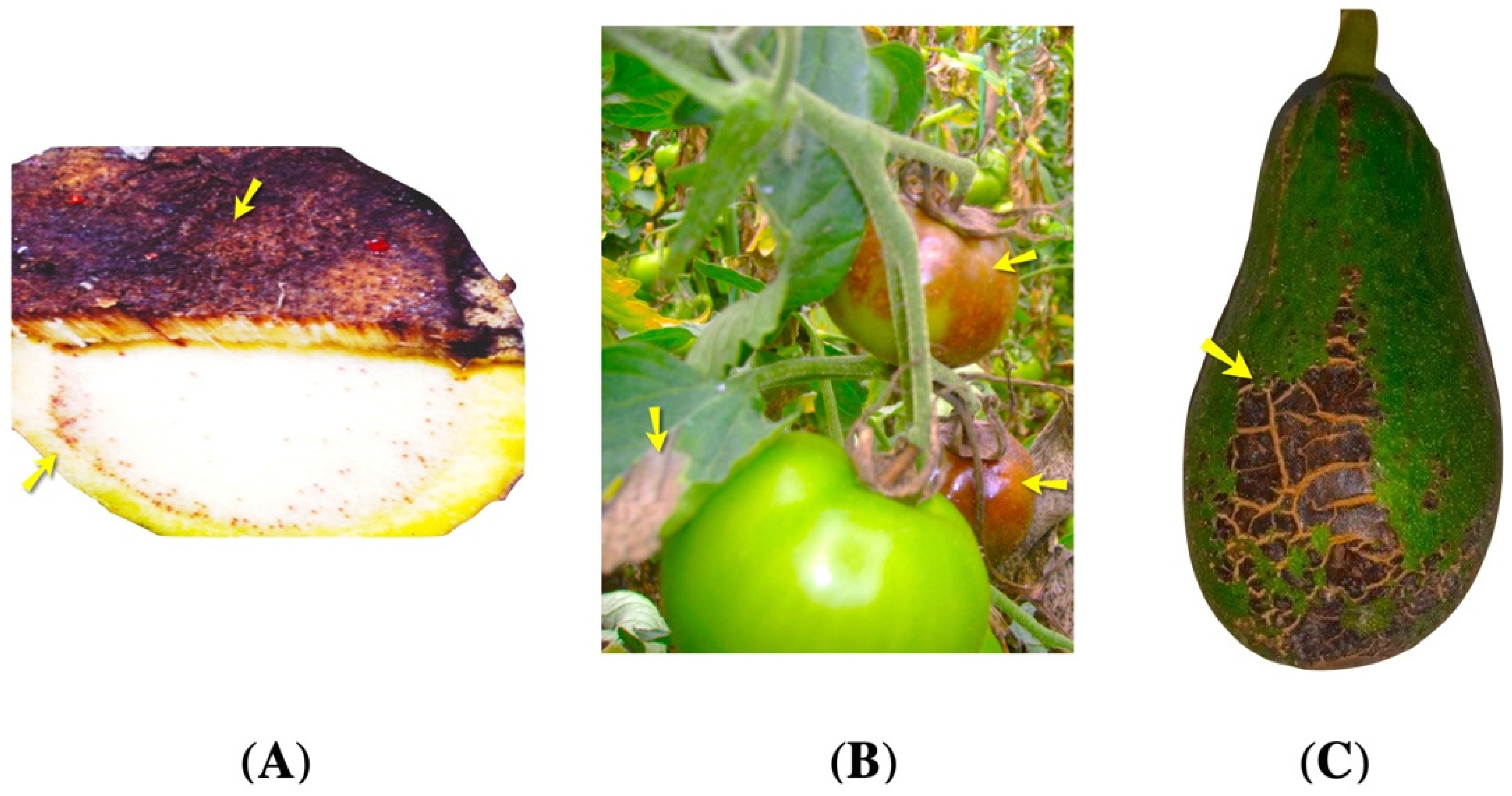
Figure 1. Infected plants. (A) Rachis of Musa paradiasiaca, sample with banana bacterial wilt symptoms (B) Leaves of Solanum lycopersicum, sample with late blight symptoms (C) Fruit of Persea americana, sample with scab lesions. The yellow arrows point out the regions infected regions.
Agriculture 13 02029 g001
Table 1. Antibacterial activity of Pernettya prostrata and Rubus roseus extracts against Ralstonia solanacearum.
Concentration/Control% Inhibition
Pernettya prostrata ExtractRubus roseus Extract
45.00 mg/mL69.25 ± 2.83 a66.24 ± 3.16 a
22.50 mg/mL30.08 ± 1.89 b51.44 ± 1.35 b
Control (+) gentamicin 7 mg/mL92.22 ± 1.992.22 ± 1.9
Control (−)++
Vehicle control++
Media control++
Data are expressed as mean ± SD (n = 3). P. prostrata and R. roseus extract test: ANOVA (p < 0.01) followed by a Tukey test (p = 0.05). Highly significant differences were found. a, b values indicate statistical significance.
Table 2. Mycelial growth inhibition of Phytoptora infestans treated with Pernettya prostrata and Rubus roseus extracts.
Concentration/Control% Mycelial Growth Inhibition
Pernettya prostrata ExtractRubus roseus Extract
500.00 mg/mL28.65 ± 0.96 a34.89 ± 0.74 a
250.00 mg/mL27.54 ± 1.74 a29.86 ± 1.22 b
125.00 mg/mL26.13 ± 1.15 ab17.28 ± 1.31 c
62.50 mg/mL22.44 ± 1.43 bc15.39 ± 1.20 c
31.25 mg/mL22.15 ± 1.88 c14.34 ± 1.05 c
Control (+) Oxicloruro de cobre 8 mg/mL--
Control (−)++
Vehicle control++
Media control++
Data are expressed as mean ± SD (n = 3). P. prostrata test: ANOVA (p < 0.001) followed by a Tukey test (p = 0.05), significant differences were found. R. roseus test: ANOVA (p < 0.001) followed by a Tukey test (p = 0.05), significant differences were found. a, b, c values indicate statistical significance.
Disclaimer/Publisher’s Note: The statements, opinions and data contained in all publications are solely those of the individual author(s) and contributor(s) and not of MDPI and/or the editor(s). MDPI and/or the editor(s) disclaim responsibility for any injury to people or property resulting from any ideas, methods, instructions or products referred to in the content.