Wound Healing: In Vitro and In Vivo Evaluation of a Bio-Functionalized Scaffold Based on Hyaluronic Acid and Platelet-Rich Plasma in Chronic Ulcers

 , , ,
, , , 
Abstract
1. Introduction
2. Methods
2.1. Patient Population and Study Protocol
2.2. Patient Selection
2.3. In Vivo Application of Bio-Functionalized Scaffold PRP + HA
- Evaluation of the number of platelets (in all patients the PRP had a platelet concentration equal to 1 × 10⁶/µL + 20%)
- Blood volume withdrawn (within 55 cc for each patient);
- Volume of PRP obtained (variable according to the type of use and the size of the ulcer);
- Labeling of each sample of PRP for each patient for laboratory control;
- Adverse reaction signaling (did not occur).
2.4. Follow-Up
2.5. Timing Control
2.6. In Vitro Procedures
2.7. Statistical Analysis
3. Results
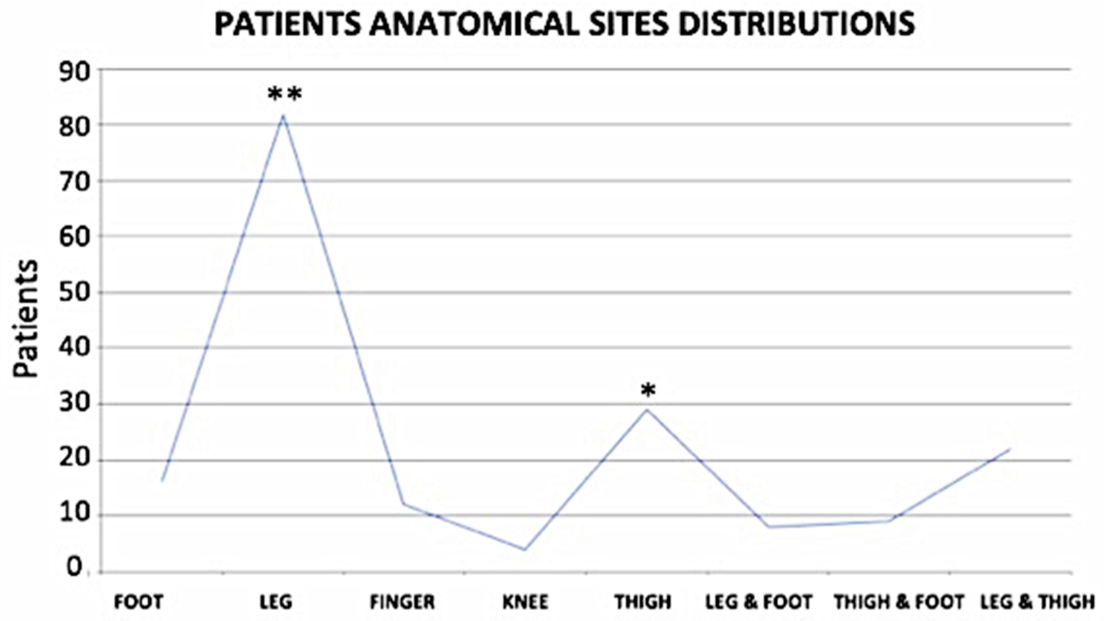
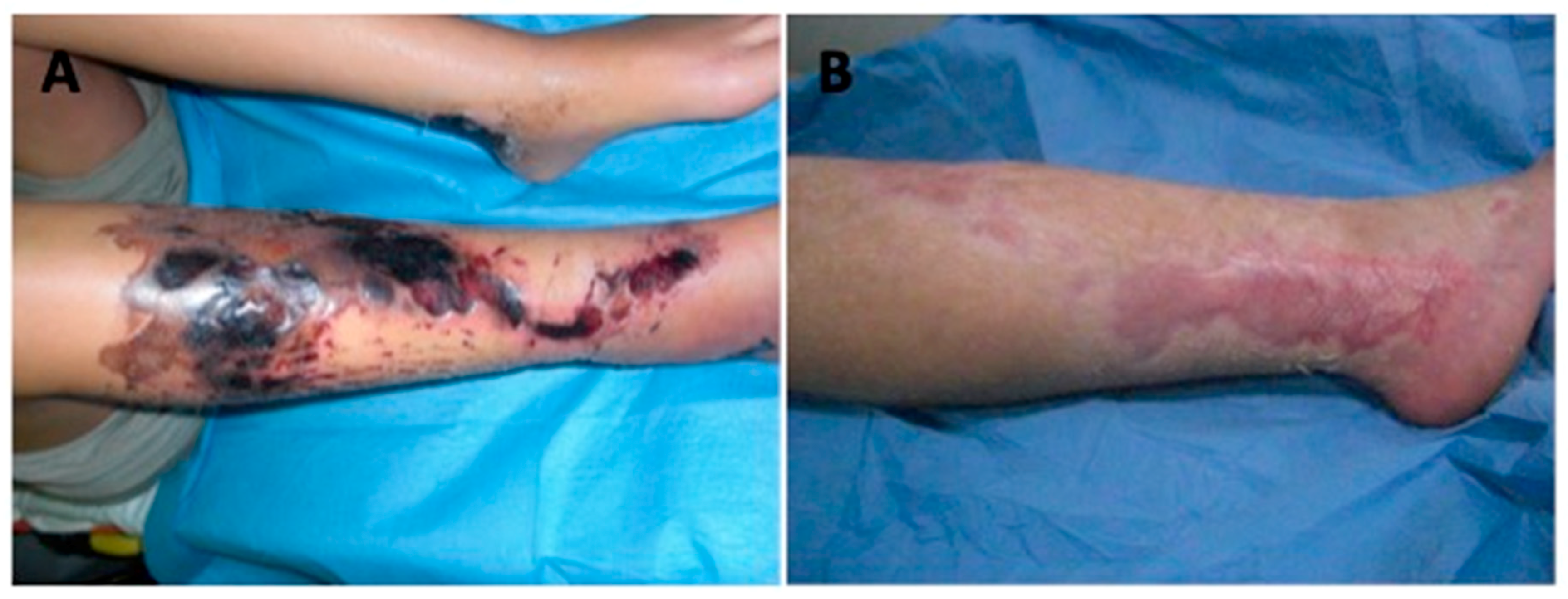
3.1. Wound Features
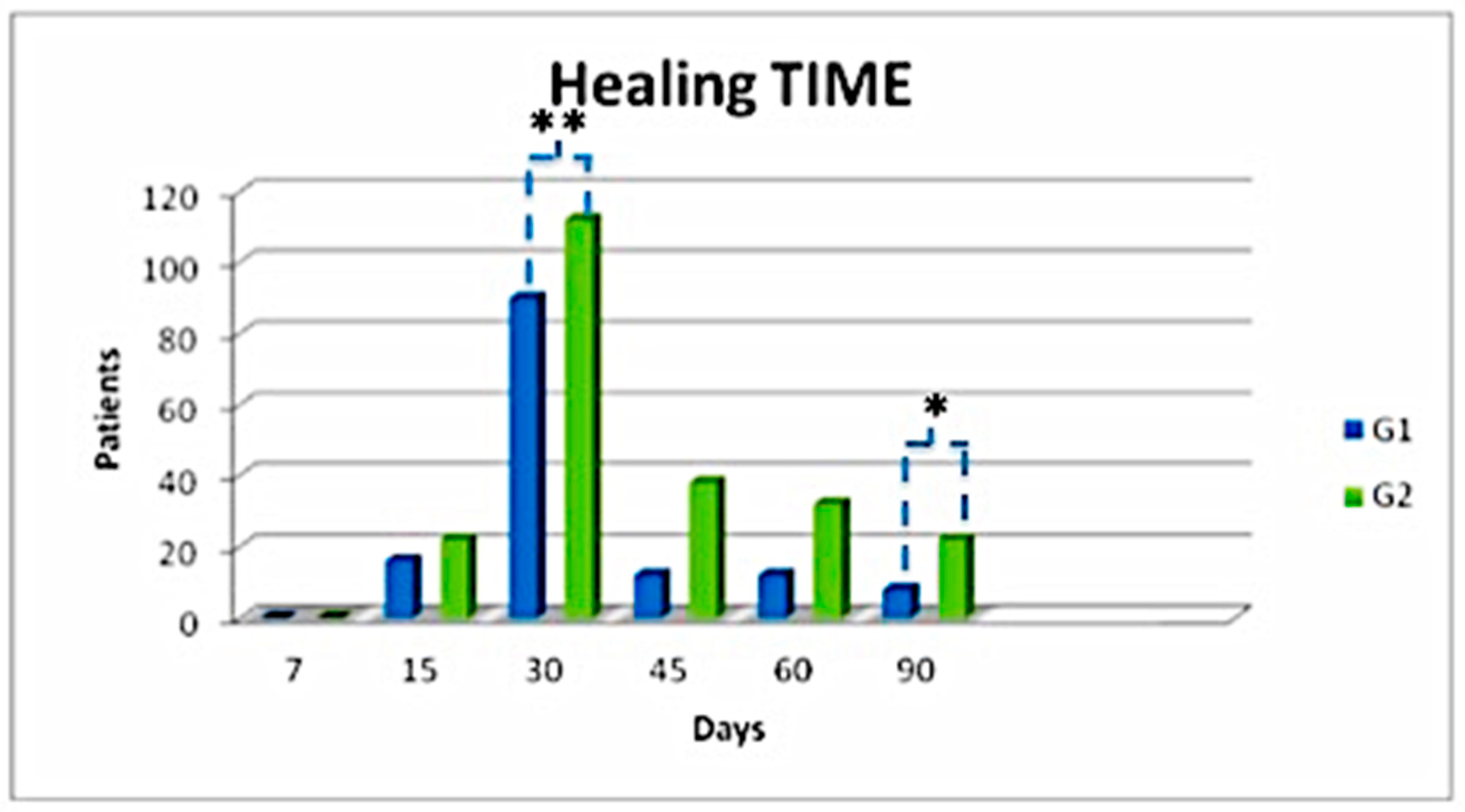
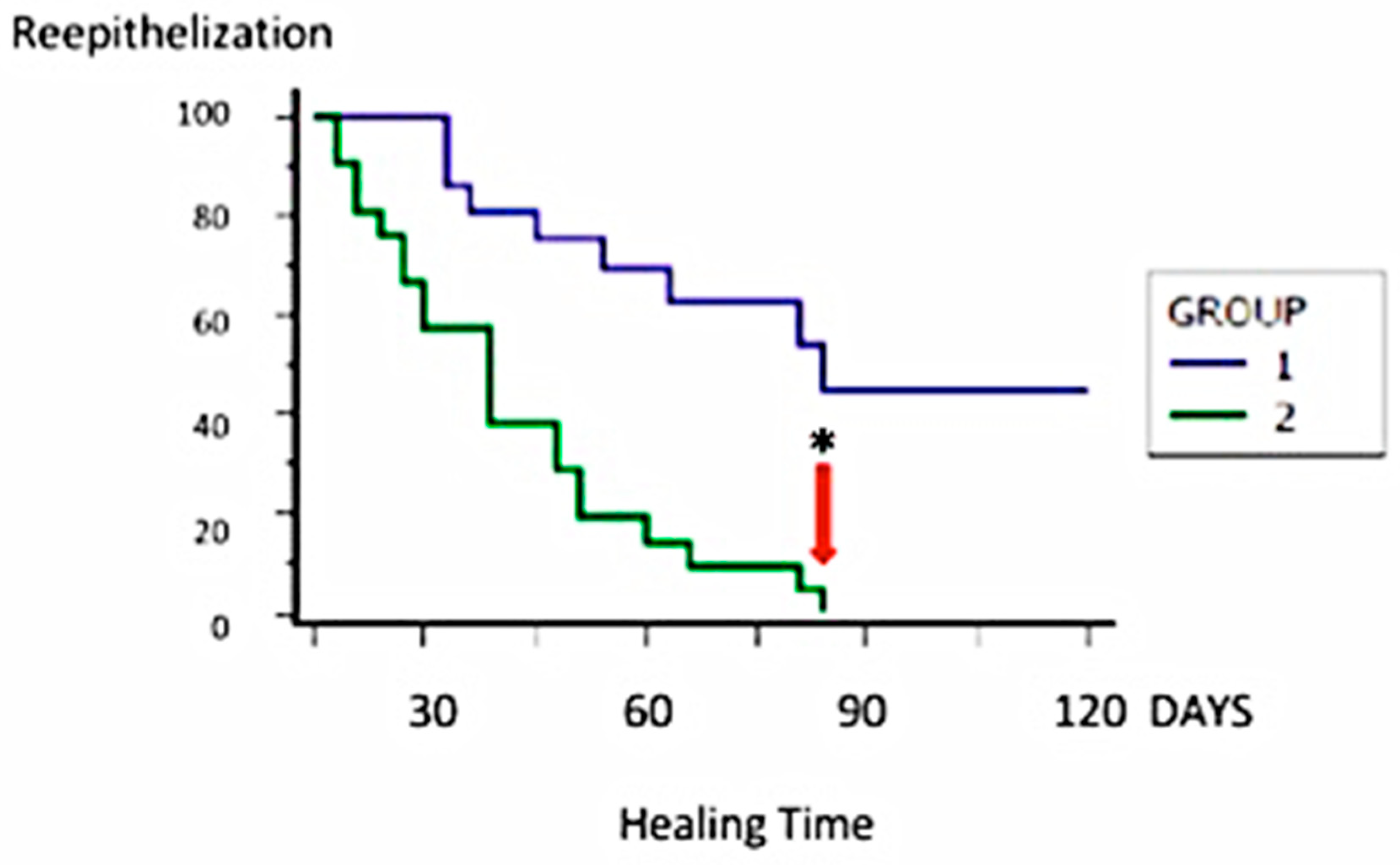
3.2. In Vivo Evaluation
3.3. In Vitro Evaluation
4. Discussion
5. Conclusions
Supplementary Materials
Author Contributions
Acknowledgments
Conflicts of Interest
References
- Prichard, H.L.; Reichert, W.M.; Klitzman, B. Adult adipose-derived stem cell attachment to biomaterial. Biomaterials 2007, 28, 936–946. [Google Scholar] [CrossRef] [PubMed][Green Version]
- Pinto, J. Application of platelet-rich plasma in the treatment of chronic skin ulcer—Case report. Anais Bras. Dermatol. 2014, 89, 638–640. [Google Scholar] [CrossRef] [PubMed]
- Dowsett, C. Wound care bed preparation: Time in practice. Clin. Pract. Dev. 2005, 1, 58–70. [Google Scholar]
- Chiaretti, M. Gel piastrinico nel piede diabetico: Case report. Med Estet. 2014. [Google Scholar]
- Crane, D.; Everts, P. Platelet rich plasma (PRP) matrix grafts. Pract. Pain Manag. 2008, 8, 11–26. [Google Scholar]
- Gentile, P.; Cole, J.P.; Cole, M.A.; Garcovich, S.; Bielli, A.; Scioli, M.G.; Orlandi, A.; Insalaco, C.; Cervelli, V. Evaluation of not-activated and activated PRP in hair loss treatment: Role of growth factor and cytokine concentrations obtained by different collection systems. Int. J. Mol. Sci. 2017, 18, 408. [Google Scholar] [CrossRef] [PubMed]
- Margolis, D.J.; Cohen, J.H. Management of chronic venous leg ulcers: A literature-guided approach. Clin. Dermatol. 1994, 12, 19–26. [Google Scholar] [CrossRef]
- Cervelli, V.; De Angelis, B.; Lucarini, L.; Spallone, D.; Balzani, A.; Palla, L.; Gentile, P.; Cerulli, P. Tissue regeneration in loss of substance on the lower limbs through use of platelet riche plasma, steem cells from adipose tissue and hyaluronic acid. Adv. Skin Wound Care 2010, 23, 262–272. [Google Scholar] [CrossRef]
- Costagliola, M.; Agrosi, M. Second-degree burns: A comparative, multicenter, randomized trial of hyaluronic acid plus silver sulfadiazine vs silver sulfadiazine alone. Curr. Med. Res. Opin. 2005, 21, 1235–1240. [Google Scholar] [CrossRef]
- Cervelli, V.; Lucarini, L.; Spallone, D.; Brinci, L.; De Angelis, B. Use of platelet rich plasma and hyaluronic acid on exposed tendons of the foot and ankle. J. Wound Care 2010, 19, 5. [Google Scholar] [CrossRef]
- Goto, T.; Naito, A.; Tamai, N.; Nakagami, G.; Mo, M.; Sanada, H. Objective evaluation for venous leg ulcer-related nociceptive pain using thermography. Chronic Wound Care Manag. Res. 2014, 1, 23–30. [Google Scholar] [CrossRef][Green Version]
- Gentile, P.; De Angelis, B.; Agovino, A.; Orlandi, F.; Migner, A.; Di Pasquali, C.; Cervelli, V. Use of platelet rich plasma and hyaluronic acid in the treatment of complications of achilles tendon reconstruction. World J. Plast. Surg. 2016, 5, 124–132. [Google Scholar] [PubMed]
- Whitlock, P.W.; Smith, T.L.; Poehling, G.G.; Shilt, J.S.; Van Dyke, M. A naturally derived, cytocompatible, and architecturally optimized scaffold for tendon and ligament regeneration. Biomaterials 2007, 28, 4321–4329. [Google Scholar] [CrossRef] [PubMed]
- Rossi, S.; Mori, M.; Vigani, B.; Bonferoni, M.C.; Sandri, G.; Riva, F.; Caramella, C.; Ferrari, F. A novel dressing for the combined delivery of platelet lysate and vancomycin hydrochloride to chronic skin ulcers: Hyaluronic acid particles in alginate matrices. Eur. J. Pharm. Sci. 2018, 118, 87–95. [Google Scholar] [CrossRef] [PubMed]
- Cervelli, V.; Gentile, P.; De Angelis, B.; Calabrese, C.; Di Stefani, A.; Scioli, M.G.; Curcio, B.C.; Felici, M.; Orlandi, A. Application of enhanced stromal vascular fraction and fat grafting mixed with PRP in post-traumatic lower extremity ulcers. Stem Cell Res. 2011, 6, 103–111. [Google Scholar] [CrossRef] [PubMed]
- Russo, F.; D’Este, M.; Vadalà, G.; Cattani, C.; Papalia, R.; Alini, M.; Denaro, V. Platelet rich plasma and hyaluronic acid blend for the treatment of osteoarthritis: Rheological and biological evaluation. PLoS ONE 2016, 16, e0157048. [Google Scholar] [CrossRef] [PubMed]
- Iio, K.; Furukawa, K.; Tsuda, E.; Yamamoto, Y.; Maeda, S.; Naraoka, T.; Kimura, Y.; Ishibashi, Y. Hyaluronic acid induces the release of growth factors from platelet-rich plasma. Asia Pac. J. Sports Med. Arthrosc. Rehabil. Technol. 2016, 28, 27–32. [Google Scholar] [CrossRef] [PubMed]
- Cervelli, V.; Lucarini, L.; Spallone, D.; Palla, L.; Colicchia, G.M.; Gentile, P.; De Angelis, B. Use of platelet-rich plasma and hyaluronic acid in the loss of substance with bone exposure. Adv. Skin Wound Care 2011, 24, 176–181. [Google Scholar] [CrossRef] [PubMed]
- Schuklenk, U.; Ashcroft, R. International research ethics. Bioethics 2000, 14, 158–172. [Google Scholar] [CrossRef] [PubMed]
- Von Elm, E.; Altman, D.G.; Egger, M.; Pocock, S.J.; Gøtzsche, P.C.; Vandenbroucke, J.P. The Strengthening the Reporting of Observational Studies in Epidemiology (STROBE) statement: Guidelines for reporting observational studies. J. Clin. Epidemiol. 2008, 61, 344–349. [Google Scholar] [CrossRef]
- De Angelis, B.; Gentile, P.; Orlandi, F.; Bocchini, I.; Di Pasquali, C.; Agovino, A.; Gizzi, C.; Patrizi, F.; Scioli, M.G.; Orlandi, A.; et al. Limb rescue: A new autologous-peripheral blood mononuclear cells technology in critical limb ischemia and chronic ulcers. Tissue Eng. Part C Methods 2015, 21, 423–435. [Google Scholar] [CrossRef] [PubMed]
- Myers, S.R.; Partha, V.N.; Soranzo, C.; Price, R.D.; Navsaria, H.A. Hyalomatrix: A temporary epidermal barrier, hyaluronan delivery, and neodermis induction system for keratinocyte stem cell therapy. Tissue Eng. 2007, 13, 2733–2741. [Google Scholar] [CrossRef] [PubMed]
- Gentile, P.; Colicchia, G.M.; Nicoli, F.; Cervelli, G.; Curcio, C.B.; Brinci, L.; Cervelli, V. Complex abdominal wall repair using a porcine dermal matrix. Surg. Innov. 2013, 20, 12–15. [Google Scholar] [CrossRef] [PubMed]
- Everts, P.A.; Overdevest, E.P.; Jakimowicz, J.J.; Oosterbos, C.J.; Schönberger, J.P.; Knape, J.T.; van Zundert, A. The use of autologous platelet-leukocyte gels to enhance the healing process in surgery, a review. Surg. Endosc. 2007, 21, 2063–2068. [Google Scholar] [CrossRef] [PubMed]
- Persoon, A.; Heinen, M.M.; van der Vleuten, C.J.; de Rooij, M.J.; van de Kerkhof, P.C.; van Achterberg, T. Leg ulcers: A review of their impact on daily life. J. Clin. Nurs. 2004, 13, 341–354. [Google Scholar] [CrossRef] [PubMed]
- Schaible, H.G. Peripheral and central mechanisms of pain generation. In Analgesia; Springer: Berlin, Germany, 2006; pp. 3–28. [Google Scholar]
- Franks, P.J.; McCullagh, L.; Moffatt, C.J. Assessing quality of life in patients with chronic leg ulceration using the Medical Outcomes Short Form-36 questionnaire. Ostomy Wound Manag. 2003, 49, 26–37. [Google Scholar]
- Reed, G.L. Platelet secretory mechanisms. In Seminars in Thrombosis and Hemostasis; Thieme Medical Publishers, Inc.: New York, NY, USA, 2004; Volume 30, pp. 441–450. [Google Scholar]
- Mazzucco, L.; Balbo, V.; Cattana, E.; Guaschino, R.; Borzini, P. Not every PRP gel is born equal. Evaluation of growth factor availability for tissues through four PRP-gel preparations: Fibrinet, RegenPRP-Kit, Plateltex and one manual procedure. Vox Sang. 2009, 97, 110–118. [Google Scholar] [CrossRef]
- Anitua, E.; Sánchez, M.; Zalduendo, M.M.; de la Fuente, M.; Prado, R.; Orive, G.; Andía, I. Fibroblastic response to treatment with different preparations rich in growth factors. Cell. Prolif. 2009, 42, 162–170. [Google Scholar] [CrossRef]
- Pizzicannella, J.; Gugliandolo, A.; Orsini, T.; Fontana, A.; Ventrella, A.; Mazzon, E.; Bramanti, P.; Diomede, F.; Trubiani, O. Engineered extracellular vesicles from human periodontal-ligament stem cells increase VEGF/VEGFR2 expression during bone regeneration. Front. Physiol. 2019, 30, 512. [Google Scholar] [CrossRef]
- Manusukiatti, W.; Maibach, H.I. Hyaluronic acid and skin: Wound healing and aging. Int. J. Dermatol. 1996, 35, 539–544. [Google Scholar] [CrossRef]
- Barrientos, S.; Stojadinovic, O.; Golinko, M.S.; Brem, H.; Tomic-Canic, M. Growth factors and cytokines in wound healing. Wound Repair Regen. 2008, 16, 585–601. [Google Scholar] [CrossRef] [PubMed]
- Stark, H.J.; Willhauck, M.J.; Mirancea, N.; Boehnke, K.; Nord, I.; Breitkreutz, D.; Pavesio, A.; Boukamp, P.; Fusenig, N.E. Authentic fibroblast matrix in dermal equivalents normalises epidermal histogenesis and dermoepidermal junction in organotypic coculture. Eur. J. Cell. Biol. 2004, 83, 631–645. [Google Scholar] [CrossRef] [PubMed]
- Scioli, M.G.; Bielli, A.; Gentile, P.; Cervelli, V.; Orlandi, A. Combined treatment with platelet-rich plasma and insulin favours chondrogenic and osteogenic differentiation of human adipose-derived stem cells in three-dimensional collagen scaffolds. J. Tissue Eng. Regen. Med. 2017, 11, 2398–2410. [Google Scholar] [CrossRef] [PubMed]
- Kim, D.H.; Je, Y.J.; Kim, C.D.; Lee, Y.H.; Seo, Y.J.; Lee, J.H.; Lee, Y. Can platelet-rich plasma be used for skin rejuvenation? Evaluation of effects of platelet-rich plasma on human dermal fibroblast. Ann. Dermatol. 2011, 23, 424–431. [Google Scholar] [CrossRef] [PubMed]
- Falanga, V. Classifications for wound bed preparation and stimulation of chronic wounds. Wound Repair Regen. 2000, 8, 347–352. [Google Scholar] [PubMed]
- Halim, A.S.; Khoo, T.L.; Saad, A.Z. Wound bed preparation from a clinical perspective. Indian J. Plast. Surg. 2012, 45, 193–202. [Google Scholar] [CrossRef] [PubMed]
- Mehrabani, D.; Manafi, N. Role of cultured skin fibroblasts in aesthetic and plastic surgery. World J. Plast. Surg. 2013, 2, 2–5. [Google Scholar] [PubMed]
- Kakudo, N.; Minakata, T.; Mitsui, T.; Kushida, S.; Notodihardjo, F.Z.; Kusumoto, K. Proliferation promoting effect of platelet-rich plasma on human adipose derived stem cells and human dermal fibroblasts. Plast. Reconstr. Surg. 2008, 122, 1352–1360. [Google Scholar] [CrossRef]
- Cervelli, V.; Bocchini, I.; Di Pasquali, C.; De Angelis, B.; Cervelli, G.; Curcio, C.B.; Orlandi, A.; Scioli, M.G.; Tati, E.; Delogu, P.; et al. P.R.L. platelet rich lipotransfert: Our experience and current state of art in the combined use of fat and PRP. Biomed. Res. Int. 2013, 2013, 434191. [Google Scholar] [CrossRef]
- Ramos-Torrecillas, J.; García-Martínez, O.; De Luna-Bertos, E.; Ocaña-Peinado, F.M.; Ruiz, C. Effectiveness of platelet-rich plasma and hyaluronic acid for the treatment and care of pressure ulcers. Biol. Res. Nurs. 2015, 17, 152–158. [Google Scholar] [CrossRef] [PubMed]
- Martin, V.; Maibach, H.I. The Principles of Wound Care Bed Preparation. Available online: https://www.woundsource.com/ (accessed on 17 September 2019).
- Mammana, S.; Gugliandolo, A.; Cavalli, E.; Diomede, F.; Iori, R.; Zappacosta, R.; Bramanti, P.; Conti, P.; Fontana, A.; Pizzicannella, J.; et al. Human gingival mesenchymal stem cells pretreated with vesicular moringin nanostructures as a new therapeutic approach in a mouse model of spinal cord injury. J. Tissue Eng. Regen. Med. 2019. [Google Scholar] [CrossRef] [PubMed]
- Cervelli, V.; Bottini, D.J.; Arpino, A.; Grimaldi, M.; Rogliani, M.; Gentile, P. Bone-anchored implant in cosmetic finger reconstruction. Ann. Chir. Plast. Esthet. 2008, 53, 365–367. [Google Scholar] [CrossRef] [PubMed]
- Gentile, P.; Bottini, D.J.; Gravante, G.; Nicoli, F.; Caruso, R.; Cervelli, V. The use of bone-anchored implants for absent ear. J. Craniofac. Surg. 2008, 19, 744–747. [Google Scholar] [CrossRef] [PubMed]
- Rozman, P.; Bolta, Z. Use of platelet growth factors in treating wounds and soft-tissue injuries. Acta Dermatovenerol. Alp. Panonica Adriat. 2007, 16, 156–165. [Google Scholar]
- Salzano, A.; De Rosa, A.; Rossi, E.; Brunese, L.; Carbone, M.; Nocera, V.; Tuccillo, M.; Scialpi, M. The topicality and use of the radiological exam in gunshot wounds of the limbs an assessment of 132 cases. Radiol. Med. 1999, 98, 468–471. [Google Scholar] [PubMed]
- Strem, B.M.; Hedrick, M.H. The growing importance of fat in regenerative medicine. Trends Biotechnol. 2005, 23, 64–66. [Google Scholar] [CrossRef] [PubMed]
- American Society of Anesthesiologists Task Force on Acute Pain Management. Practice guidelines for acute pain management in the perioperative setting an updated report by the American society of anesthesiologists task force on acute pain management. Anesthesiology 2012, 116, 248–273. [Google Scholar] [CrossRef] [PubMed]
- Sarvajnamurthy, S.; Suryanarayan, S.; Budamakuntala, L.; Suresh, D.H. Autologous platelet rich plasma in chronic venous ulcers: Study of 17 cases. J. Cutan. Aesth. Surg. 2013, 6, 97–99. [Google Scholar]
- Nicoli, F.; Balzani, A.; Lazzeri, D.; Gentile, P.; Chilgar, R.M.; Di Pasquali, C.; Nicoli, M.; Bocchini, I.; Agovino, A.; Cervelli, V. Severe hidradenitis suppurativa treatment using platelet-rich plasma gel and Hyalomatrix. Int. Wound J. 2015, 12, 338–343. [Google Scholar] [CrossRef] [PubMed]
- Gentile, P.; Scioli, M.G.; Orlandi, A.; Cervelli, V. Breast reconstruction with enhanced stromal vascular fraction fat grafting: What is the best method? Plast. Reconstr. Surg. Glob. Open 2015, 8, e406. [Google Scholar] [CrossRef] [PubMed]


© 2019 by the authors. Licensee MDPI, Basel, Switzerland. This article is an open access article distributed under the terms and conditions of the Creative Commons Attribution (CC BY) license (http://creativecommons.org/licenses/by/4.0/).
Share and Cite
De Angelis, B.; D’Autilio, M.F.L.M.; Orlandi, F.; Pepe, G.; Garcovich, S.; Scioli, M.G.; Orlandi, A.; Cervelli, V.; Gentile, P. Wound Healing: In Vitro and In Vivo Evaluation of a Bio-Functionalized Scaffold Based on Hyaluronic Acid and Platelet-Rich Plasma in Chronic Ulcers. J. Clin. Med. 2019, 8, 1486. https://doi.org/10.3390/jcm8091486
De Angelis B, D’Autilio MFLM, Orlandi F, Pepe G, Garcovich S, Scioli MG, Orlandi A, Cervelli V, Gentile P. Wound Healing: In Vitro and In Vivo Evaluation of a Bio-Functionalized Scaffold Based on Hyaluronic Acid and Platelet-Rich Plasma in Chronic Ulcers. Journal of Clinical Medicine. 2019; 8(9):1486. https://doi.org/10.3390/jcm8091486
Chicago/Turabian StyleDe Angelis, Barbara, Margarida Fernandes Lopes Morais D’Autilio, Fabrizio Orlandi, Giampiero Pepe, Simone Garcovich, Maria Giovanna Scioli, Augusto Orlandi, Valerio Cervelli, and Pietro Gentile. 2019. "Wound Healing: In Vitro and In Vivo Evaluation of a Bio-Functionalized Scaffold Based on Hyaluronic Acid and Platelet-Rich Plasma in Chronic Ulcers" Journal of Clinical Medicine 8, no. 9: 1486. https://doi.org/10.3390/jcm8091486
APA StyleDe Angelis, B., D’Autilio, M. F. L. M., Orlandi, F., Pepe, G., Garcovich, S., Scioli, M. G., Orlandi, A., Cervelli, V., & Gentile, P. (2019). Wound Healing: In Vitro and In Vivo Evaluation of a Bio-Functionalized Scaffold Based on Hyaluronic Acid and Platelet-Rich Plasma in Chronic Ulcers. Journal of Clinical Medicine, 8(9), 1486. https://doi.org/10.3390/jcm8091486