Article

Metastatic Tumors of the Sinonasal Cavity: A 15-Year Review of 17 Cases

1
Department of Otorhinolaryngology-Head and Neck Surgery, Taipei Veterans General Hospital, Taipei 11217, Taiwan
2
Department of Pathology, Taipei Veterans General Hospital, Taipei 11217, Taiwan
3
School of Medicine, National Yang-Ming University, Taipei 11221, Taiwan
4
Department of Otolaryngology, Cheng Hsin Hospital, Taipei 11220, Taiwan
5
Department of Otolaryngology-Head and Neck Surgery, University of California, Irvine, Orange, CA 92868, USA
*
Author to whom correspondence should be addressed.
J. Clin. Med. 2019, 8(4), 539; https://doi.org/10.3390/jcm8040539
Submission received: 1 March 2019 / Revised: 22 March 2019 / Accepted: 16 April 2019 / Published: 19 April 2019
(This article belongs to the Section Otolaryngology)

Abstract

:
Extranasal cancers that metastasize to the sinonasal cavity are very rare. To date, there are only limited reports regarding this rare condition within the literature. Therefore, we retrospectively reviewed all patients diagnosed with metastatic cancer of the sinonasal tract from 2003 to 2018 at a tertiary academic medical center. Patient demographic data, clinical presentation, treatment modalities, and outcomes were investigated. There were a total of 17 patients (9 males and 8 females) included in the analysis. The mean age was 56.8 years (range 27–80). The most common primary malignancies were hepatocellular carcinoma (n = 3) and gastrointestinal tract adenocarcinoma (n = 3). The most common site of metastasis was the nasal cavity (n = 8). Five patients received radical tumor resection and the others underwent radiotherapy, chemotherapy, or combined chemoradiotherapy. The 2-year survival was 28%. In summary, metastasis to the sinonasal cavity remains extremely rare. A high degree of suspicion regarding the possibility of metastatic spread to the sinonasal region is necessary for patients with a previous history of malignancy who present with new sinonasal symptoms. The treatment strategy of sinonasal metastatic cancer is usually palliative therapy and the prognosis remains poor. However, early detection and diagnosis, coupled with aggressive treatment, may improve patient quality of life.

1. Introduction

Sinonasal malignancies are extremely rare and generally present as primary malignancies. They account for approximately 3% of upper respiratory tract malignancies [1,2,3,4]. The incidence of metastatic tumors in the sinonasal cavity is even more unlikely [5,6,7,8]. The most common origins of metastatic sinonasal malignancy are the kidneys, lungs, urogenital ridge, breasts, gastrointestinal tract, and thyroid [5,9]. In the literature, renal cell carcinoma (RCC) is the most common metastatic cancer of the sinonasal tract, accounting for almost half of all cases. Other common metastatic cancers are bronchogenic carcinoma and urogenital ridge malignancies, such as prostate cancer and female urogenital cancer [10,11].
Due to its limited occurrence, information regarding sinonasal metastases remains to be investigated. In the current study, we present tumor clinicopathologic characteristics, treatment modalities, and prognosis of 17 patients diagnosed with sinonasal metastases.

2. Materials and Methods

2.1. Patients

This was a retrospective review of patients diagnosed with sinonasal metastases and treated at Taipei General Veterans Hospital in Taiwan between 1 January 2003 and 31 December 2018. Institutional Review Board approval was obtained. Data regarding patient demographics, presenting signs and symptoms, tumor pathology, primary tumor site, metastatic sites, laboratory studies, relevant imaging scans, treatment modalities, complications, and outcomes were collected.

2.2. Statistical analysis

Quantitative data were summarized as the mean ± standard deviation (SD) and categorical variables as percentages. IBM SPSS 20.0 software (IBM Corp., Armonk, NY, USA) was used for the statistic analysis. The Kaplan–Meier method was used to calculate survival trends.

3. Results

3.1. Patients

Demographic data are summarized in Table 1. There were 17 patients identified during the study period, including 9 males and 8 females. Age at diagnosis ranged from 27 to 80 years (mean 56.8 ± 14.2 years). In three cases, the primary and metastatic lesions were found synchronously, and the other 14 cases experienced metastasis after the primary tumors were treated. Time from treatment to the development of metastases ranged from 2 months to 18 years and 7 months.
In our study, there were eight unilateral and seven bilateral sinonasal metastases. As for metastatic sites, eight cases metastasized to the nasal cavity, six cases to the skull base, five cases to the nasal septum, four cases to the maxillary sinus, four cases to the ethmoid sinus, four cases to the sphenoid sinus, and one case to the frontal sinus. Among them, nine cases manifested as a single lesion, while seven cases were multifocal lesions. Common symptoms included epistaxis (n = 7), headache (n = 3), nasal obstruction (n = 2), diplopia (n = 1), extraocular movement limitation (n = 1), and facial swelling (n = 1) (Table 1). For cases with metastatic lesions involving the nasal cavity, endoscopy often revealed hypervascularity, friable, or polypoid lesions, which bled easily after touch (Figure 1). CT or MRI were commonly utilized to evaluate the extent of tumor invasion (Figure 2).
Pathology of the primary tumors were diverse, including hepatocellular carcinoma (HCC) (n = 3), gastrointestinal tract adenocarcinoma (n = 3), invasive ductal carcinoma of the breast (n = 2), retroperitoneal leiomyosarcoma (n = 2), poorly differentiated thyroid carcinoma (n = 1), thyroid papillary carcinoma (n = 1), melanoma of the foot (n = 1), osteosarcoma of the tibia (n = 1), laryngeal mucoepidermoid carcinoma (n = 1), retrorectal neuroendocrine carcinoma (n = 1), and lung adenocarcinoma (n = 1) (Table 2) (Figure 3). There were no cases of renal cell carcinoma.

3.2. Treatment Modality
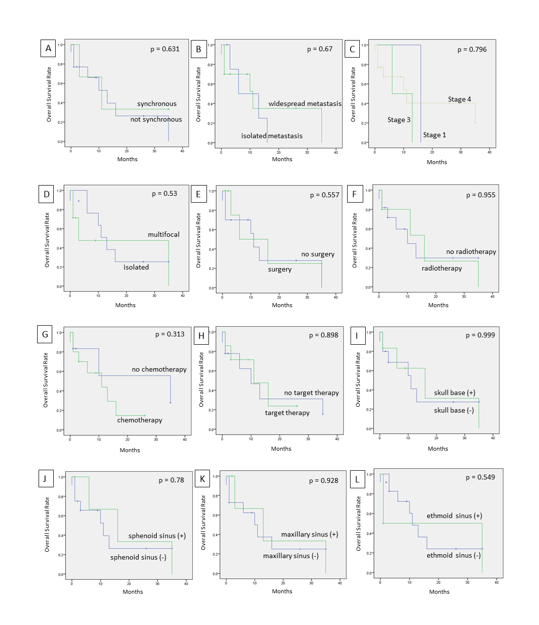
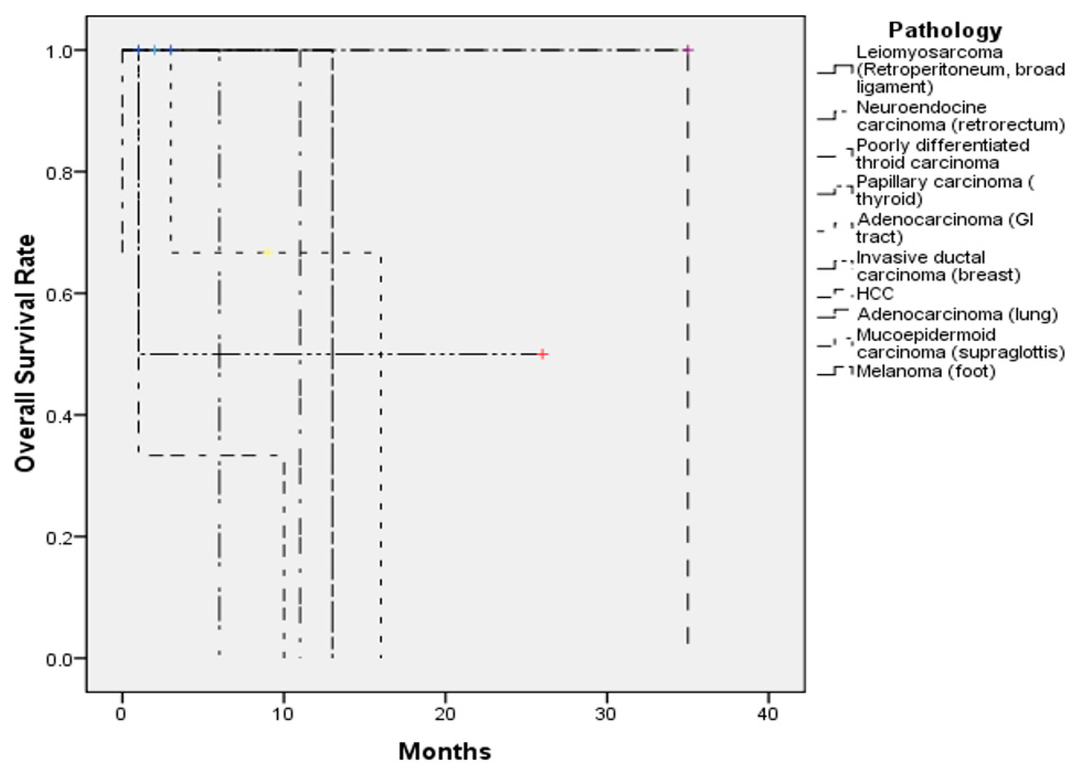
There were five patients who underwent radical tumor excision, and one who underwent tumor debulking. Five patients received radiotherapy, and 10 patients underwent palliative chemotherapy (Table 3).

3.3. Survival

The follow-up duration ranged from 1 month to 35 months. One patient was lost to follow-up (case No. 1). At the last follow-up, three patients were free from disease (cases 2, 5, and 16), four patients were still receiving regular follow-ups or treatment for residual and/or recurrent disease (cases 4, 8, 11, and 12), and nine patients died from disease (cases 3, 6, 7, 9, 10, 13, 14, 15, and 17) (Table 3). The 1-year survival was 47%, and the 2-year survival was 28% (Figure 4).

4. Discussion

Sinonasal metastases from distant extranasal sites is exceedingly rare. In the literature, nearly half of sinonasal metastases originated from the kidneys, followed by the lungs, breasts, colon, prostate, urogenital ridge, gastrointestinal tract, thyroid, and pancreas (Table 4) [1,5,9,12,13,14,15,16,17,18,19]. In our study, there were four sinonasal metastatic cases originating from the stomach and three cases from the liver, making gastrointestinal adenocarcinoma and hepatocellular carcinoma the top two common primary tumors. Previous cases have been largely reported in the Western literature [5,6,20,21,22,23,24,25]. However, the incidence and distribution of malignant tumors might be diverse between different geographic areas due to distinct genetic or environmental factors [26]. Among sinonasal metastases, the incidence of prostate cancer and breast cancer ranked first and second highest in North America and Europe, respectively. In North America, the incidence of kidney cancer ranked after colon cancer, melanoma, and thyroid cancer. However, in Europe, the incidence of RCC is higher than that of melanoma. In East Asia, the incidence of cancers of the respiratory tract have been reported to be the highest, followed by stomach, liver, breast, and colorectal cancer; in contrast, the incidence of kidney cancer was relevantly low [27]. According to the Taiwan Food and Drug Administration (TFDA) statistics and a study investigating cancers between 2002 and 2012, the most common malignancy overall in Taiwan was also breast cancer, followed by colorectal, respiratory tract, and liver cancers [28]. Huang et al. proposed that the differences in primary cancers leading to metastatic sinonasal cancers compared to that in Western countries might be due to a unique cancer epidemiology exclusive to Taiwan [26].
It is well-established that chronic hepatitis B virus (HBV) and hepatitis C virus (HCV) infections are important risk factors for the development of HCC [29,30]. Incidence of HBV and HCV infection was high in Taiwan before the launch of the HBV vaccine and other means of hepatitis virus control, which explains the relatively higher incidence of HCC in Taiwan [13,31,32]. Therefore, HCC was identified as one of the most common sinonasal metastases in our study, which is different from Western studies.
Prior reports have identified the maxillary sinus as the most common site of metastasis, followed by the sphenoid, ethmoid, frontal sinuses, and, finally, the nasal cavity [5,9,11,18,33,34,35]. However, in our study, the nasal cavity was the most common site for metastasis. This is different from previous studies and may suggest different biological behavior for distant metastatic spread based on different primaries.
The symptoms for such tumors are usually non-specific, similar to that of primary sinonasal tumors [36], and are related to the characteristics of the primary tumor and the metastatic sites [37,38]. Due to the fact that RCC is often highly vascular [39], metastasis from RCC to the sinonasal tract is usually accompanied by epistaxis. A tumor affecting the nasal cavity may present with epistaxis, increased nasal secretions, and nasal obstruction. Maxillary sinus tumors may present with symptoms of facial swelling, facial numbness, or eye symptoms. Ethmoid and sphenoid sinus tumors may result in headache, proptosis, diplopia, or vision changes [5,20,33]. Horner’s syndrome has been reported to result from lateral sphenoid sinus erosion or infratemporal fossa lesion [12]. If the tumor involves the cavernous sinus or the skull base, patients may present with various cranial nerve palsies, such as diplopia, ptosis, or visual impairment [13,35,40]. In our study, the most common symptom was epistaxis, followed by headache and nasal obstruction. For those with frequent epistaxis and severe nasal obstruction, surgery helps alleviate the symptoms, which might improve quality of life.
Time from treatment to the development of metastases ranged from 2 months to 18 years and 7 months. The wide variation may be related to the initial M (the presence of metastasis) stage of the patient when diagnosed with the cancer. For cases with initial presentation of other metastatic sites, which suggests widespread disease, the duration of the development of sinonasal metastasis should be shorter. However, in our study, the mean metastatic duration for an initial M0 stage was 22 months (4 cases), which was shorter compared to patients with an initial M1 stage (25 months, 8 cases), with no significant difference between the two groups (p > 0.05). Tumor biology may play a major role in metastatic duration, though the small sample size of this study limits more in-depth analysis.
Hematogenous [5,11,41,42,43,44] and lymphatic spread [13,37,45] are two different pathways of metastasis. For hematogenous travel, tumor cells have to undergo intravasation, which is the process of passing through endothelial cells into the blood vessels. Tumor cells circulate via the valveless vertebral venous (Batson’s) plexus and can enter the pterygoid plexus and cavernous sinus, as well as the nasal and paranasal sinuses [9,41,46,47]. As for lymphatic spread, the tumor emboli flow into regional lymphatic channels and drain to the thoracic duct, and then flow in retrograde through the intercostal, mediastinal, or supraclavicular lymph vessels to the head and neck region [9,13,37]. Whether tumor cells metastasize via hematogenous or lymphogenous pathways depends on multiple factors, including the density of vascularization around the tumor or the secondary site, the biologic behavior of the primary tumor, and the presence or absence of lymph node involvement [48].
Though rare, it is critical to keep sinonasal metastases within the differential diagnoses for sinonasal masses. Identification of the origin of the metastatic sinonasal cancer differs from case to case. In general, histopathologic analysis will be required to make the diagnosis. It would be prudent to obtain imaging (CT and/or MRI) ahead of time to rule out intracranial extension, as well as to determine the vascularity of the lesion, and also for pre-operative surgical evaluation. For extranasal primary cancers, expression of specific biomarkers, such as carcinoembryonic antigen (CEA) and prostate-specific antigen (PSA), may be considered. For instance, if the patient had a history of breast cancer, the expression of human epidermal growth factor receptor 2 (HER2) and glandular morphology in malignant cells should be checked.
Primary sinonasal malignancies and sinonasal metastases may overlap in clinical behavior and histopathologic appearance. Three of our cases were metastatic adenocarcinomas of gastrointestinal or colorectal origins. This is to be distinguished from primary sinonasal intestinal-type adenocarcinoma, which may share similar morphology and immunophenotyping, such as immunoreactivity for CK20 and cdx2. In our study, there were also two metastatic breast carcinomas, of which the morphology may be variable and may lack specificity. Therefore, the lesion may not be easily separated from a high-grade non-intestinal type adenocarcinoma. One of our patients was a metastatic rectal neuroendocrine carcinoma, and its pathologic findings were identical to that of a primary sinonasal neuroendocrine carcinoma. Therefore, the precise diagnosis of sinonasal metastases relies on the combination of clinical, radiological, and pathologic information.
Metastases to the sinonasal cavity represent advanced distant disease and are naturally associated with poor prognosis, and, in the vast majority of cases, may not be treated with a curative intent. Mickel and Zimmerman reviewed the survival in 26 cases of malignancies that metastasized to the sphenoid sinus. Only nine cases lived for more than six months and four cases survived for at least two years [12]. Multifocal metastases in the sinonasal region have been noted to be much more common than a single metastasis [21]. In our study, there were seven cases with multifocal metastatic tumor in the sinonasal region, and nine cases with a single lesion. The location and number of metastatic lesions mainly determine whether the lesion may be operable or not. Multiple metastatic lesions and proximity to vital structures increase the challenges of surgical resection for palliation, which would be the only possible goal in these cases [37]. In many cases, resections may be approached endoscopically. For lesions that cannot always be excised radically, other multimodality treatments, such as radiotherapy, chemotherapy, hormone therapy for urogenital cancer, or iodine ablation therapy for thyroid cancer, may be considered, and should be chosen based on the characteristics of the primary tumor. One consideration for palliative surgical candidates is preoperative embolization of feeding vessels to reduce intraoperative bleeding [9]. Most sinonasal tumors receive blood supply from external carotid artery branches, which would be amenable to embolization [7,9,12]. In our opinion, whether or not patients receive surgery depends on several factors, including the general health condition of the patient, the preferences of the surgeon, and tumor biology. We would recommend surgery if the tumor caused frequent nasal bleeding or severe nasal obstruction (to improve quality of life), and if the patient could tolerate general anesthesia. The location and size of the metastatic tumor should be readily accessible. Classically, surgery is not routinely considered for metastatic disease. However, surgery may provide some chance for these patients to live longer (decreasing tumor burden for later chemotherapy control), and to also achieve a better quality of life (alleviating symptoms of nasal bleeding and nasal obstruction).
In our study, 10 (58.8%) of the 17 patients died in the follow-up period. The shortest survival time was 2 months and the longest was 35 months. There were three patients alive without obvious residual sinonasal metastatic lesions, and three patients alive with either residual sinonasal metastases or other distant metastases. The 1-year survival was 47%, and the 2-year survival was 28.1%. There are studies that showed that differences in prognosis are apparent based on the presence of isolated or widespread disseminated disease [7], with longer survival noted for an isolated sinonasal metastasis [22]. In addition, slightly higher survival has been noted in patients that receive surgery than those who receive radiotherapy alone [7]. However, in our study, there was no significant difference in survival whether patients received surgery, whether the sinonasal lesions were multifocal, or whether there was isolated or widespread disseminated disease (Supplementary Figure S1). The insignificant findings may be related to the small number of cases, which is harder for meaningful statistical analysis.
Indeed, pathology may play a major role in patient survival. All patients with HCC and melanoma died of the disease, which is characteristic of the known poor survival when compared to other tumor types. One case with thyroid papillary carcinoma, a malignancy with a good prognosis, received surgical excision of a nasal septal metastatic lesion and was still alive without obvious recurrence. In regard to targeted therapy for metastatic breast cancer, one patient receiving target therapy was still alive, while another who did not receive targeted therapy died of the disease. Four out of seven patients receiving target therapy died regardless of the cancer type, including one case of HCC, one case of lung adenocarcinoma, one case of gastric adenocarcinoma, and one case of cecal adenocarcinoma. Since this was a retrospective study, there may be some bias to stratify the patients based on receiving targeted therapy, due to its recent development. Cancer cases in more recent years would be more likely to receive the novel targeted therapies compared to those from prior years. In our study, 11 patients had widespread metastatic disease when sinonasal metastasis (e.g., lung, bone, liver, brain) was diagnosed, while 5 cases presented with isolated sinonasal metastasis.
In our study, gastrointestinal adenocarcinoma and hepatocellular carcinoma were the top two common primary tumors that metastasized to the sinonasal tract, which is quite different from reports on Western countries. The nasal cavity was the most common site for metastasis, which is also quite different from previous reports where the maxillary sinus was the most common site. The similar findings included the mean age, the sex distribution, and the poor prognosis (Table 4). There are several limitations to our study. First, it is a retrospective study, which makes complete data collection and verification difficult. Second, the small number of cases for such a rare condition, may prevent meaningful statistical analysis. Finally, treatment plans for each patient may be impacted by physician and/or patient preference, where electing to be more aggressive with treatment may affect survival.

5. Conclusions

Sinonasal metastases from extranasal primary malignancies are rare, and patients usually suffer from non-specific symptoms. Treatment modalities differ based on the origin and characteristics of the primary tumor. The prognosis remains poor, though early detection and aggressive treatment may help to improve quality of life.

Supplementary Materials

The following are available online at https://www.mdpi.com/2077-0383/8/4/539/s1, Figure S1: Survival curves for patients with sinonasal metastasis analyzed based on several parameters.

Author Contributions

Data curation, M.-H.C., Y.-J.K. and C.-Y.H.; Formal analysis, M.-Y.L.; Investigation, M.-H.C. and Y.-J.K.; Methodology, Y.-J.K. and E.C.K.; Project administration, M.-Y.L.; Supervision, M.-Y.L.; Writing–original draft, M.-H.C.; Writing–review & editing, C.-Y.H., E.C.K. and M.-Y.L. All authors reviewed the manuscript and approved the final version.

Funding

This research was supported by a grant from the Ministry of Science and Technology, Taiwan (MOST 106-2314-B-075-035-MY3).

Conflicts of Interest

The authors declare no conflict of interest.

References

  1. Frazell, E.L.; Lewis, J.S. Cancer of the nasal cavity and accessory sinuses. A report of the management of 416 patients. Cancer 1963, 16, 1293–1301. [Google Scholar] [CrossRef]
  2. Carrau, R.L.; Myers, E.N. Neoplasms of the nose and paranasal sinuses. In Head and Neck Surgery-Otolaryngology, 3rd ed.; Bailey, B.J., Calhoun, K.H., Healy, G.B., Johnson, J.T., Jackler, R.K., Pillsbury, H.C., III, Tardy, M.E., Eds.; Lippincott Williams & Wilkins: Philadelphia, PA, USA, 2001; pp. 1247–1264. [Google Scholar]
  3. Austin, J.R.; Kershiznek, M.M.; McGill, D.; Austin, S.G. Breast carcinoma metastatic to paranasal sinuses. Head Neck 1995, 17, 161–165. [Google Scholar] [CrossRef] [PubMed]
  4. Silverberg, E.; Grant, R.N. Cancer statistics, 1970. CA Cancer J. Clin. 1970, 20, 11–23. [Google Scholar] [CrossRef]
  5. Bernstein, J.M.; Montgomery, W.W.; Balogh, K., Jr. Metastatic tumors to the maxilla, nose, and paranasal sinuses. Laryngoscope 1966, 76, 621–650. [Google Scholar] [CrossRef]
  6. Azarpira, N.; Ashraf, M.J.; Khademi, B.; Asadi, N. Distant metastases to nasal cavities and paranasal sinuses case series. Indian J. Otolaryngol. Head Neck Surg. 2011, 63, 349–352. [Google Scholar] [CrossRef] [PubMed]
  7. Kent, S.E.; Majumdar, B. Metastatic tumours in the maxillary sinus. A report of two cases and a review of the literature. J. Laryngol. Otol. 1985, 99, 459–462. [Google Scholar] [CrossRef]
  8. Barnes, L. Metastases to the head and neck: An overview. Head Neck Pathol. 2009, 3, 217–224. [Google Scholar] [CrossRef]
  9. Lopez, F.; Devaney, K.O.; Hanna, E.Y.; Rinaldo, A.; Ferlito, A. Metastases to nasal cavity and paranasal sinuses. Head Neck 2016, 38, 1847–1854. [Google Scholar] [CrossRef] [PubMed]
  10. Miyamoto, R.; Helmus, C. Hypernephroma metastatic to the head and neck. Laryngoscope 1973, 83, 898–905. [Google Scholar] [CrossRef] [PubMed]
  11. Frigy, A.F. Metastatic hepatocellular carcinoma of the nasal cavity. Arch. Otolaryngol. 1984, 110, 624–627. [Google Scholar]
  12. Mickel, R.A.; Zimmerman, M.C. The sphenoid sinus—A site for metastasis. Otolaryngol. Head Neck Surg. 1990, 102, 709–716. [Google Scholar] [CrossRef]
  13. Huang, H.H.; Chang, P.H.; Fang, T.J. Sinonasal metastatic hepatocellular carcinoma. Am. J. Otolaryngol. 2007, 28, 238–241. [Google Scholar] [CrossRef]
  14. Pourseirafi, S.; Shishehgar, M.; Ashraf, M.J.; Faramarzi, M. Papillary Carcinoma of Thyroid with Nasal Cavity Metastases: A Case Report. Iran. J. Med. Sci. 2018, 43, 90–93. [Google Scholar]
  15. Pittoni, P.; Di Lascio, S.; Conti-Beltraminelli, M.; Valli, M.C.; Espeli, V.; Bongiovanni, M.; Richetti, A.; Pagani, O. Paranasal sinus metastasis of breast cancer. BMJ Case Rep. 2014, 2014. [Google Scholar] [CrossRef] [Green Version]
  16. Terada, T. Renal cell carcinoma metastatic to the nasal cavity. Int. J. Clin. Exp. Pathol. 2012, 5, 588–591. [Google Scholar]
  17. Liu, C.Y.; Chang, L.C.; Yang, S.W. Metastatic hepatocellular carcinoma to the nasal cavity: A case report and review of the literature. J. Cancer Sci. Ther. 2011, 3, 81–83. [Google Scholar] [CrossRef]
  18. Barbosa, E.B.; Ferreira, E.; Mariano, F.V.; Altemani, A.; Sakuma, E.T.I.; Sakano, E. Metastasis to Paranasal Sinuses from Carcinoma of Prostate: Report of a Case and Review of the Literature. Case Rep. Otolaryngol. 2018, 2018, 5428975. [Google Scholar] [CrossRef]
  19. Bin Sabir Husin Athar, P.P.; bte Ahmad Norhan, N.; bin Saim, L.; bin Md Rose, I.; bte Ramli, R. Metastasis to the sinonasal tract from sigmoid colon adenocarcinoma. Ann. Acad. Med. Sing. 2008, 37, 788–790. [Google Scholar]
  20. Friedmann, I.; Osborn, D.A. Metastatic tumours in the ear, nose and throat region. J. Laryngol. Otol. 1965, 79, 576–591. [Google Scholar] [CrossRef]
  21. Choong, C.V.; Tang, T.; Chay, W.Y.; Goh, C.; Tay, M.H.; Zam, N.A.; Tan, P.H.; Tan, M.H. Nasal metastases from renal cell carcinoma are associated with Memorial Sloan-Kettering Cancer Center poor-prognosis classification. Chin. J. Cancer 2011, 30, 144–148. [Google Scholar] [CrossRef] [Green Version]
  22. Simo, R.; Sykes, A.J.; Hargreaves, S.P.; Axon, P.R.; Birzgalis, A.R.; Slevin, N.J.; Farrington, W.T. Metastatic renal cell carcinoma to the nose and paranasal sinuses. Head Neck 2000, 22, 722–727. [Google Scholar] [CrossRef]
  23. Kaminski, B.; Kobiorska-Nowak, J.; Bien, S. [Distant metastases to nasal cavities and paranasal sinuses, from the organs outside the head and neck]. Otolaryngol. Polska = Pol. Otolaryngol. 2008, 62, 422–425. [Google Scholar] [CrossRef]
  24. Parida, P.K. Renal cell carcinoma metastatic to the sinonasal region: Three case reports with a review of the literature. Ear Nose Throat J. 2012, 91, E11–E16. [Google Scholar] [CrossRef] [PubMed]
  25. Ravnik, J.; Smigoc, T.; Bunc, G.; Lanisnik, B.; Ksela, U.; Ravnik, M.; Velnar, T. Hypophyseal metastases: A report of three cases and literature review. Neurologia i Neurochirurgia Polska 2016, 50, 511–516. [Google Scholar] [CrossRef] [PubMed]
  26. Huang, H.H.; Fang, T.J.; Chang, P.H.; Lee, T.J. Sinonasal metastatic tumors in Taiwan. Chang Gung Med. J. 2008, 31, 457–462. [Google Scholar] [PubMed]
  27. Fitzmaurice, C.; Allen, C.; Barber, R.M.; Barregard, L.; Bhutta, Z.A.; Brenner, H.; Dicker, D.J.; Chimed-Orchir, O.; Dandona, R.; Dandona, L.; et al. Global, Regional, and National Cancer Incidence, Mortality, Years of Life Lost, Years Lived with Disability, and Disability-Adjusted Life-years for 32 Cancer Groups, 1990 to 2015: A Systematic Analysis for the Global Burden of Disease Study. JAMA Oncol. 2017, 3, 524–548. [Google Scholar] [CrossRef]
  28. Chiang, C.J.; Lo, W.C.; Yang, Y.W.; You, S.L.; Chen, C.J.; Lai, M.S. Incidence and survival of adult cancer patients in Taiwan, 2002–2012. J. Formos. Med. Assoc. = Taiwan Yi Zhi 2016, 115, 1076–1088. [Google Scholar] [CrossRef] [Green Version]
  29. Ringelhan, M.; McKeating, J.A.; Protzer, U. Viral hepatitis and liver cancer. Philos. Trans. R. Soc. Lond. Ser. B Biol. Sci. 2017, 372. [Google Scholar] [CrossRef]
  30. Trepo, C.; Chan, H.L.; Lok, A. Hepatitis B virus infection. Lancet 2014, 384, 2053–2063. [Google Scholar] [CrossRef]
  31. Parkin, D.M.; Pisani, P.; Ferlay, J. Estimates of the worldwide incidence of 25 major cancers in 1990. Int. J. Cancer 1999, 80, 827–841. [Google Scholar] [CrossRef] [Green Version]
  32. Chen, D.S. Hepatocellular carcinoma in Taiwan. Hepatol. Res. 2007, 37 (Suppl. 2), S101–S105. [Google Scholar] [CrossRef] [PubMed]
  33. Prescher, A.; Brors, D. Metastases to the paranasal sinuses: Case report and review of the literature. Laryngo Rhino Otologie 2001, 80, 583–594. [Google Scholar] [CrossRef]
  34. Dogan, S.; Can, I.H.; Sayn, M.; Ozer, E.; Bayz, U.; Yazici, G.; Samim, E.E. The nasal septum: An unusual presentation of metastatic renal cell carcinoma. J. Craniofac. Surg. 2009, 20, 1204–1206. [Google Scholar] [CrossRef]
  35. Sountoulides, P.; Metaxa, L.; Cindolo, L. Atypical presentations and rare metastatic sites of renal cell carcinoma: A review of case reports. J. Med. Case Rep. 2011, 5, 429. [Google Scholar] [CrossRef]
  36. Weber, A.L.; Stanton, A.C. Malignant tumors of the paranasal sinuses: Radiologic, clinical, and histopathologic evaluation of 200 cases. Head Neck Surg. 1984, 6, 761–776. [Google Scholar] [CrossRef]
  37. Gottlieb, M.D.; Roland, J.T., Jr. Paradoxical spread of renal cell carcinoma to the head and neck. Laryngoscope 1998, 108, 1301–1305. [Google Scholar] [CrossRef]
  38. Fukuda, M.; Miyata, M.; Okabe, K.; Sakashita, H. A case series of 9 tumors metastatic to the oral and maxillofacial region. J. Oral Maxillofac. Surg. 2002, 60, 942–944. [Google Scholar] [CrossRef]
  39. Aziz, S.A.; Sznol, J.; Adeniran, A.; Colberg, J.W.; Camp, R.L.; Kluger, H.M. Vascularity of primary and metastatic renal cell carcinoma specimens. J. Transl. Med. 2013, 11, 15. [Google Scholar] [CrossRef]
  40. Evgeniou, E.; Menon, K.R.; Jones, G.L.; Whittet, H.; Williams, W. Renal cell carcinoma metastasis to the paranasal sinuses and orbit. BMJ Case Rep. 2012, 2012. [Google Scholar] [CrossRef] [Green Version]
  41. Nahum, A.M.; Bailey, B.J. malignant tumors metastatic to the paranasal sinuses: case report and review of the literature. Laryngoscope 1963, 73, 942–953. [Google Scholar] [CrossRef]
  42. Sim, R.S.; Tan, H.K. A case of metastatic hepatocellular carcinoma of the sphenoid sinus. J. Laryngol. Otol. 1994, 108, 503–504. [Google Scholar] [CrossRef] [PubMed]
  43. Lin, C.D.; Cheng, K.S.; Tsai, C.H.; Chen, C.L.; Tsai, M.H. Metastatic hepatocellular carcinoma in the nasal septum: Report of a case. J. Formos. Med. Assoc. = Taiwan Yi Zhi 2002, 101, 715–718. [Google Scholar] [PubMed]
  44. Matsuda, H.; Tanigaki, Y.; Yoshida, T.; Matsuda, R.; Tsukuda, M. A case of metastatic hepatocellular carcinoma in the nasal cavity. Eur. Arch. Oto-Rhino-Laryngol. 2006, 263, 305–307. [Google Scholar] [CrossRef] [PubMed]
  45. Stacker, S.A.; Baldwin, M.E.; Achen, M.G. The role of tumor lymphangiogenesis in metastatic spread. FASEB J. 2002, 16, 922–934. [Google Scholar] [CrossRef] [PubMed] [Green Version]
  46. Batson, O.V. The function of the vertebral veins and their role in the spread of metastases. Ann. Surg. 1940, 112, 138–149. [Google Scholar] [CrossRef] [PubMed]
  47. Wiltse, L.L.; Fonseca, A.S.; Amster, J.; Dimartino, P.; Ravessoud, F.A. Relationship of the dura, Hofmann’s ligaments, Batson’s plexus, and a fibrovascular membrane lying on the posterior surface of the vertebral bodies and attaching to the deep layer of the posterior longitudinal ligament. An anatomical, radiologic, and clinical study. Spine 1993, 18, 1030–1043. [Google Scholar] [PubMed]
  48. Mehlen, P.; Puisieux, A. Metastasis: A question of life or death. Nat. Rev. Cancer 2006, 6, 449–458. [Google Scholar] [CrossRef] [PubMed]
  49. Garrett, M.J. Metastatic tumours of the paranasal sinuses simulating primary growths. J. Fac. Radiol. 1959, 10, 151–155. [Google Scholar] [CrossRef]
  50. Som, P.M.; Norton, K.I.; Shugar, J.M.; Reede, D.L.; Norton, L.; Biller, H.F.; Som, M.L. Metastatic hypernephroma to the head and neck. AJNR Am. J. Neuroradiol. 1987, 8, 1103–1106. [Google Scholar] [PubMed]
Figure 1. Endoscopic views of various sinonasal metastatic cancers. Endoscopic examinations showed various metastatic sinonasal malignancies, which usually appeared as reddish, fragile, and hemorrhagic masses in the nasal cavity or sinuses: (A) retroperitoneum leiomyosarcoma, (B) thyroid poorly differentiated carcinoma, (C) breast invasive ductal carcinoma, (D) rectal adenocarcinoma, (E) hepatic cell carcinoma, and (F) lung adenocarcinoma.
Jcm 08 00539 g001
Figure 2. CT and MRI images of sinonasal metastatic cancers: (A) A 37-year-old female diagnosed with metastatic retroperitoneum leiomyosarcoma. The CT scan revealed that the tumor involved the nasal chamber, bilateral sphenoid sinus, left side of the posterior ethmoid sinus, left aspect of the sellar floor, and the clivus. (B) A 45-year-old female diagnosed with retrorectal neuroendocrine carcinoma. The MRI scans revealed that the tumor involved the sphenoid sinus, sella, suprasella, left cavernous sinus, and pituitary gland.
Jcm 08 00539 g002
Figure 3. Pathologic images of various sinonasal metastatic cancers: (A) Metastatic neuroendocrine carcinoma; (B) Metastatic colorectal adenocarcinoma of the colon; (C) Metastatic hepatocellular carcinoma; (D) Metastatic papillary thyroid carcinoma; (E) Metastatic breast carcinoma; and (F) Metastatic pulmonary adenocarcinoma.
Jcm 08 00539 g003
Figure 4. The overall survival of the 17 patients with sinonasal metastases in our study.
Jcm 08 00539 g004
Table 1. The demographic and clinical characteristics of the patients and the tumors.
Case Number (n = 17)
Age27–80 (mean 56.8 ± 14.2)
SexM:F = 9:8
Metastatic time2 months–18 years and 7 months
Symptoms
Epistaxis7
Headache 3
Nasal obstruction2
Diplopia1
Extraocular movement limitation1
Facial swelling1
Side
 Unilateral (L/R)8 (3/5)
 Bilateral7
Sinonasal metastases
 Single metastases9
 Multifocal metastases7
Widespread metastasis11
Sinonasal metastatic sites
Nasal cavity8
Skull base6
Nasal septum5
 Maxillary sinus4
 Ethmoid sinus4
 Sphenoid sinus4
 Frontal sinus1
Table 2. Primary tumor origins and pathologies.
Number (n = 17)
Primary tumor site
-GI tract4
-Liver3
-Breast2
-Thyroid2
-Retroperitoneum1
-Broad ligament1
-Supraglottis1
-Lung1
-Tibia1
-Foot1
Pathology
-Adenocarcinoma (GI tract)3
-Hepatocellular carcinoma (HCC)3
-Invasive ductal carcinoma (breast)2
-Leiomyosarcoma (retroperitoneum, broad ligament)2
-Poorly differentiated carcinoma (thyroid)1
-Papillary carcinoma (thyroid)1
-Adenocarcinoma (lung)1
-Neuroendocrine carcinoma (retrorectum)1
-Mucoepidermoid carcinoma (supraglottis)1
-Osteosarcoma (tibia)1
-Melanoma (foot)1
Table 3. Summary of the cases of sinonasal metastatic cancer in our study.
No.Age/SexSymptomsPrimary TumorClinical Stage before Metastasis to Sinonasal RegionSinonasal Metastatic SiteTime Before MetastasisExtrasinonasal Metastasis (Initial) Extrasinonasal Metastasis (Before Sinonasal Metastasis)Treatment ModalityFollow-up DurationDisease Status
127MHeadache PtosisTibiaosteosarcomaStage III (M1)NA4y5mNLungCT (Ifosfamide + Etoposide)NANA
237FEpistaxisRetroperitoneum leiomyosarcomaStage IV (M1)Sphenoid, ethmoid sinuses, clivus3y4mNLiverTumor resection + RT1mAlive, no obvious residual tumor
345FHeadacheRetrorectalneuroendocrine carcinomaStage IIIa (T2N1M0)Sphenoid sinus, pituitary gland4mNNDebulking surgery + CT (cisplatin+ etoposide)6mDead
447MEpistaxisThyroidpoorly differentiated carcinomaStage IVb (pT3N1bM1)Maxillary, ethmoid, sphenoid sinuses, nasal cavity, orbit, brain6mLeft iliacLeft iliacTumor biopsy + RT + iodine ablation therapy3y11mDead
548FNasal massThyroid papillary carcinomaStage IVc (M1)Nasal septumSynchronousNNTumor excision2y11mAlive, no obvious residual tumor
649MDiplopiaGastricadenocarcinomaStage Ib (T1N1M0)Sphenoid sinus, cavernous sinus6y3mNNTumor excision + CT (Capecitabine+ oxaliplatin+ Paclitaxel)+ Target therapy (Cetuximab+ Uracil-Tegafur)1y4mDead
751FEOM limitation, hearing impairmentBreast invasive ductal carcinomaStage IV (pT3N3M1)Ethmoid sinus, nasal cavity, nasal septum11yBone, pleural, lungBone, pleural, lungTumor biopsy + CT1m22dDead
853FFacial pain, headache, ptosisRectal adenocarcinomaStage IVb (M1)Nasal cavity, skull base,1y1mLung, liver, adrenal, bone Lung, liver, adrenal, boneTumor biopsy + CT (Irinotecan + Fluorouracil + Leucovorin) + Target therapy (Bevacizumab + Cetuximab)9mAlive with residual tumor
956MEpistaxis, nasal obstructionHepatocellular carcinomaStageIVb (M1)Frontal, ethmoid sinuses, nasal septum, nasal cavity, orbit10yNLung, brainTumor biopsy + RT+ Target therapy (Sorafenib)2mDead
1057FEpistaxisLungadenocarcinomaStage IVa (cT3N2M1b)Nasal cavitySynchronousLung, boneLung, bone Tumor biopsy + RT + CT (Alimta + Cisplatin + Docetaxel) + Target therapy (Erlotinib + Pembrolizumab)11mDead
1166FNasal mass EpistaxisBreast invasive ductal carcinomaStage IV (pT4cN0M1)Nasal vestibule4y3mLungLungTumor biopsy + Hormone therapy (Tamoxifen)2y2mAlive with residual tumor
1266FNoBroad ligamentleiomyosarcomaStage IV (M1) ITF18y7mNLiver, abdomen, muscle, boneTumor biopsy + CT (Gemcitabine + Taxotere + Cisplatin + Everolimus + Adriamycin) + Target therapy (Pazopanib)3mAlive with residual tumor
1367MEpistaxisRight plantar foot melanomaStage IIIb (T4aN2bM0)Maxillary sinus6 mNNTumor biopsy + CT (Dacarbazine + Cisplatin + Vinblastine + Proleukin + Cyclophosphamide + paclitaxel) + interferon-A + immunotherapy (IL-2)1y1mDead
1469MNasal massHepatocellular carcinomaStageIVb (M1)Nasal cavity2mLungLungTumor biopsy + CT (Etoposide + Doxorubicin + Cisplatin +5-FU +Leucovorin)3dDead
1570MEpistaxisCecaladenocarcinomaStage IVc (M1)MaxillaSynchronousNNTumor resection + CT (fluorouracil + oxaliplatin + calcium folinate + irinotecan) + Target therapy (bevacizumab)3mDead
1677MNasal obstructionSupraglottic mucoepidermoid carcinomaStage IVa (pT4N2cM0)Maxillary sinus, nasal cavity, nasal septum6mNNTumor resection + RT2mAlive, no obvious residual tumor
1780MNAHepatocellular carcinomaStage IVb (M1) nasal septum5y9mNLung, muscle, boneTumor biopsy10mDead
Abbreviations: F: female; M: male; EOM: extraocular movement; NA: not available; y: year; m: months; d: days; PPF: pterygoid palatine fossa; ITF: infratemporal fossa; RT: radiotherapy; CT: chemotherapy; CCRT: concurrent chemoradiotherapy.
Table 4. Review of major case series (cases ≥ 3) of sinonasal metastatic cancers in the English language literature.
Years of PublicationAuthorsTotal PatientsAge (mean, y/o)SexMetastatic SitePrimary Tumor SitesFollow-up (months)Alive with Residual Tumor (grossly)Death
1959Garrett [49]658.85M1F5M, 1E1B, 1G, 1L, 1K, 2T0.3–1614
1966Bernstein et al. [5]1054.23M7F5M, 1E, 2F, 2N1B, 2G, 5K, 1L, 1U1–4845
1987Som et al. [50]654NA3M, 2S, 1 E,1F6KNA03
1990Mickel et al. [12]749.45M2F7S3L, 1U, 2O, 1 humerus0.25–7NANA
2000Simo et al. [22]667.83M3FNA6K6–4824
2008Huang et al. [26]1750.89M8F7M, 7N, 3E, 1S, 1NP5G, 3H, 3K, 3B, 2T, 1LNA07
2008Kaminski et al. [23]4611M3F2M,1S,1N1B, 2G, 1K2–2303
2011Azarpira et al. [6]3582M1F1E, 1M, 1S1B, 1K, 1U6–1103
2011Choong et al. [21]455.83M1F4N4K8–2413
2012Parida et al. [24]354.31M2F2F, 1N3K4–610
2016Ravnik et al. [25]3571M2F3P1B, 1K, 1 lymphoma8–4820
2019Chang et al. (our study)1756.89M8F8N, 6SB, 5 septum, 4M, 4E, 4S, 1F,2B, 4G, 3H, 1L, 2T, 1 supraglottis, 2 leg, 1 retroperitoneum, 1 broad ligament2–22879
Total 8655.542M38F27M, 11E, 16S, 4F, 23N, 1 NP, 6 SB, 5 septum10B, 14G, 6H, 7L, 31K, 6T, 3U, 1 supraglottis, 3 extremities, 1 retroperitoneum, 1 broad ligament0.3–2281841
NA: not available; M: maxillary; S: sphenoid; E: ethmoid; F: frontal; N: nasal cavity; P: pituitary gland; NP: nasopharynx; SB: skull base; B: breast; G: gastrointestinal tract; H: hepatic region; K: kidney; L: lung; T: thyroid; U: urogenital region; O: oral.

