Review

MicroRNAs in the Cholangiopathies: Pathogenesis, Diagnosis, and Treatment

by
Maria Jose Lorenzo Pisarello
1,
Lorena Loarca
1,
Tommy Ivanics
2,
Leslie Morton
1 and
Nicholas LaRusso
1,*
1
Division of Gastroenterology and Hepatology, and the Mayo Clinic Center for Signaling in Gastroenterology, Mayo Clinic, Rochester, MN 55905, USA
2
Department of Surgery, Mayo Clinic Rochester, MN 55905, USA
*
Author to whom correspondence should be addressed.
J. Clin. Med. 2015, 4(9), 1688-1712; https://doi.org/10.3390/jcm4091688
Submission received: 7 August 2015 / Revised: 7 August 2015 / Accepted: 11 August 2015 / Published: 26 August 2015
(This article belongs to the Special Issue MicroRNAs: Novel Biomarkers for Liver Diseases)

Abstract

:
The cholangiopathies are a group of liver diseases resulting from different etiologies but with the cholangiocyte as the primary target. As a group, the cholangiopathies result in significant morbidity and mortality and represent one of the main indications for liver transplant in both children and adults. Contributing to this situation is the absence of a thorough understanding of their pathogenesis and a lack of adequate diagnostic and prognostic biomarkers. MicroRNAs are small non-coding RNAs that modify gene expression post-transcriptionally. They have been implicated in the pathogenesis of many diseases, including the cholangiopathies. Thus, in this review we provide an overview of the literature on miRNAs in the cholangiopathies and discuss future research directions.

1. Introduction

The biliary tree is a complex network of conduits that start at the canals of Hering and progressively merge into a system of intralobular, septal, and major intrahepatic ducts ultimately coalescing to form the extrahepatic bile ducts, which deliver bile to the gallbladder and the intestine. Area (zonal) and segmental ducts are considered large intrahepatic bile ducts, whereas septal ducts represent the transition point between the large and the interlobular biliary system. This classification is based on the diameter of the ducts: bile ductules (15 μm), interlobular ducts (15–100 μm), septal ducts (100–300 μm), area (zonal) ducts (300–400 μm), segmental ducts (400–800 μm), and hepatic ducts (800 μm) [1,2,3]. Small bile ducts are lined by 4 to 5 cuboidal cholangiocytes while larger bile ducts are lined by 10–12 cholangiocytes. The lining by the cholangiocytes of the latter is larger and more columnar in shape than the former [3,4,5].
Cholangiocytes possess an apical and basolateral plasma membrane. Extending from the apical membrane is a single primary cilium. Cilia are microtubule-based organelles that function as mechano-, chemo-, and osmosensors [6,7,8,9,10].
The major physiologic function of cholangiocytes is ductal bile formation which occurs through modification of primary bile secreted by hepatocytes through a series of secretory and reabsorptive processes. In addition, cholangiocytes are involved in regenerative/reparative processes.
The cholangiopathies are diseases originating from the biliary tree, with biliary epithelial cells currently considered the primary cellular targets in their pathogenesis. These diseases include neonatal biliary atresia (BA), the main indication for liver transplantation in children. Indeed, about a third of all liver transplants in adults involve the cholangiopathies, primarily primary biliary cirrhosis (PBC) and primary sclerosing cholangitis (PSC) [11]. The cholangiopathies can be classified into several broad categories: immune-mediated, drug- or toxin-induced, infectious, genetic, ischemic, and idiopathic. Many of them evolve from chronic inflammation of the bile ducts, which subsequently leads to the development of cholestasis, bile duct proliferation and/or ductopenia (Table 1). This process can progress to biliary fibrosis and malignant transformation [12,13]. The pathogenic mechanisms involved in the cholangiopathies remain unknown; however, there is increasing evidence suggesting a role for miRNAs in the etiopathogenesis of at least some of the cholangiopathies. As a result, miRNAs have been postulated as potential targets for diagnosis and therapy. First described in 1993, miRNAs are a type of highly conserved, small non-coding RNAs with an average length of 22 nucleotides [14]. The biogenesis of miRNAs is shown in Figure 1.
Table 1. Classification of Cholangiopathies.
Genetic
  Alagille’s syndrome
  Cystic fibrosis
  Fibropolycystic diseases (i.e., Caroli’s syndrome, congenital hepatic fibrosis, ADPKD, ARPKD, ADPLD)
Immune-mediated
  Primary biliary cirrhosis
  Primary sclerosing cholangitis
  Hepatic allograft rejection
  Graft vs. host disease involving the liver
  Autoimmune cholangitis
Infectious
  Bacterial cholangitis
  Parasitic cholangitis
  Fungal cholangitis
  Viral cholangitis (i.e., AIDS cholangiopathy)
  Drug-induced (i.e., Floxuridine-induced cholangiopathy)
  Vascular/Ischemic (i.e., postliver transplantation hepatic artery stenosis)
Idiopathic
  Biliary atresia
  Sarcoidosis Idiopathic childhood/adulthood ductopenia
Malignant
  Cholangiocarcinoma (i.e., bile duct adenocarcinoma)
ADPKD, Adult dominant polycystic kidney disease; ARPKD, Adult recessive polycystic kidney disease; ADPLD, Adult dominant polycystic liver disease; Adapted from Lazaridis et al., 2004 [13].
Many key cellular processes involve regulation by miRNAs including cellular development, proliferation, apoptosis, metabolism, and morphogenesis [15,16,17]. Recently, aberrant miRNA expression has also been linked to disease. As a consequence, differentially expressed miRNAs in diseased tissues and circulating miRNAs in blood or other biological fluids may have a potential role in diagnosis and prognosis [18,19,20,21,22,23,24,25,26,27,28].
We previously published a review article highlighting the role of miRNAs in benign biliary tract diseases [29]. This review focuses on recent discoveries regarding miRNAs as biomarkers with respect to the cholangiopathies.
Figure 1. microRNA biogenesis. MicroRNA (miRNA) genes are transcribed by RNA polymerase II (Pol II) to generate the primary transcripts (pri-miRNAs). The initial processing of the primary transcript is mediated by the Drosha-DiGeorge syndrome critical region gene 8 (DGCR8; Pasha in Drosophila melanogaster and Caenorhabditis elegans) complex (also known as the Microprocessor complex) that generates ~70 nucleotide (nt) pre-miRNAs. Pre-miRNA has a short stem plus a ~2-nt 3′ overhang, which is recognized by the nuclear export factor exportin 5. Once exported from the nucleus, the cytoplasmic RNase III Dicer catalyses the production of miRNA duplexes. Dicer, TRBP (TAR RNA-binding protein; also known as TARBP2), and Argonaute (AGO) 1–4 mediate the processing of pre-miRNA and the assembly of the RISC (RNA-induced silencing complex). Within this complex, one strand of the miRNA duplex is removed resulting in a single stranded miRNA, partially complementary to target mRNA, which remains in the complex. .The “seed” sequence (positions 2–7 from the 5′ end of miRNAs) is complementary to the 3′ UTR of mRNA targets. microRNA complex interaction with mRNA induces posttranscriptional silencing through as both mRNA destabilization and translational repression (author’s figure) [14,15,16,17,18,19].
Jcm 04 01688 g001

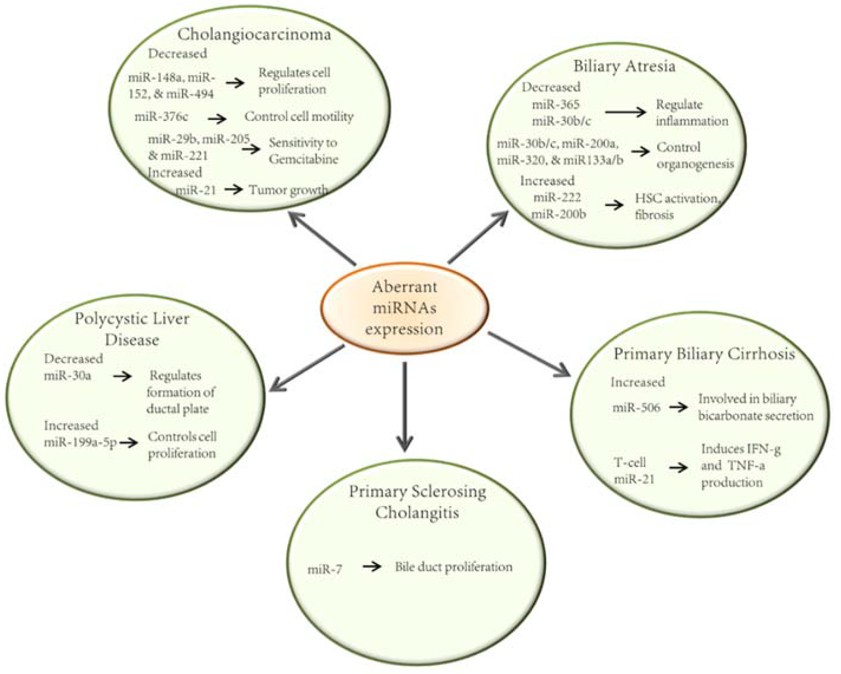
2. MicroRNAs in the Pathobiology of the Cholangiopathies

2.1. Biliary Atresia

Biliary atresia (BA) is a liver disease that occurs in infancy. It is characterized by inflammation, fibrosis, and obstruction of the extra-hepatic bile ducts (EHBD) [30].
A number of miRNAs have been described to have altered expression in BA (Figure 2). These findings have been confirmed in both animals and humans. Bessho and colleagues have studied the biliary transcriptome in the Rhesus Rotavirus type-A (RRV) mouse model of BA. This model consists of three stages that occur within 14 days of RRV injection. Initially, biliary epithelial cells are injured (3 days), triggering an inflammatory response. This is followed by a blockade of bile duct lumens (7 days) and ultimately atresia of the bile duct (14 days). The group then investigated the presence of candidate miRNAs involved in bile duct obliteration during the development of BA. Their results demonstrated a 25.8-fold decrease in the expression of miR-let-7e in the RRV group compared to controls in the early stage of BA development (3 days following RRV challenge). Between 7 and 14 days of RRV exposure, the underexpression of at least 7 additional miRNAs was identified as potential candidates in the pathogenesis of BA. The levels of miR-30b/c, miR-133a/b, miR-200a, miR-320, and miR-365 were decreased by at least 2.36-fold compared to controls [31] Studies performed in embryonic mouse liver have demonstrated that miR-30b and miR-30c are induced at day 18.5 of embryogenesis and that miR-30c is necessary for ductal plate formation [31]. Furthermore, in situ hybridization studies of liver samples have shown miR-30c to be present in bile ducts of normal subjects. Moreover, miR-30c has been found to be upregulated in the ductal reaction that occurs in BA [32]. Importantly, miR-195, miR-365 and miR-30b/c regulate the expression of genes implicated in inflammation and miR-30b/c, miR-200a, miR-320 and miR-133a/b control organogenesis [31].
Aberrant miRNA levels have been suggested to play a crucial role in the development of liver fibrosis and hepatocellular carcinoma (HCC). In the RRV-mouse model of BA, miR-222 has been found to be increased in the EHBD. This increase has been shown to induce activation of hepatic stellate cells (HSC), which are important mediators of the fibrogenic response in BA [33]. The same group showed that miR-222 expression was considerably increased in liver tissue from BA patients compared to controls. A miR-222 binding site within the 3′UTR of the PPP2R2A gene was also demonstrated [33]. This gene is known to control cell proliferation through the Akt pathway. Additionally, abrogation of miR-222 in the human immortalized HSC cell line, LX-2, resulted in decreased cell proliferation via impairment of Akt signaling [34]. Almost in parallel, another group found that miR-200b promotes cell motility and replication in studies using the same cell line [35]. The mechanism involved activation of the PI3K/Akt pathway. Importantly, miR-200b was found to be overexpressed in liver specimens from children with BA-induced hepatic fibrosis. Furthermore, the expression of miR-200b increased with liver fibrosis progression [35]. Additionally, miR-21 has been implicated in the fibrogenesis of kidney, lung, and heart; and has been reported to be elevated in patients with BA [33]. Other miRNAs have been described to be associated with liver fibrosis in patients with BA. For instance, Roderburg and colleagues uncovered a link between miR-29 and the development of liver fibrosis [36]. In RRV-challenged mice, miR-29 is upregulated in hepatocytes and cholangiocytes. These changes in miR-29 expression result in decreased levels of insulin-like growth factor 1 (Igf1) and interleukin-1 receptor accessory protein (Il-1 RAP), two genes that have been implicated in the pathogenesis of BA [30].
Figure 2. The roles of miRNAs on the pathogenesis of cholangiopathies. Schematic representation of the most relevant miRNAs whose expression is altered in cholangiophaties and their involvement in the pathogenesis of these diseases [31,33,34,35,37,38,39,40,41,42,43,44,45].
Jcm 04 01688 g002

2.2. Primary Biliary Cirrhosis

Primary biliary cirrhosis is a chronic autoimmune liver disease that primarily affects women. It is characterized by the presence of auto-reactive antibodies and T-cells, causing injury to the intrahepatic bile ducts and subsequently an uncontrolled fibrogenic response. This cascade of events can eventually lead to the development of liver cirrhosis [37].
Altered expression of miRNAs has also been associated with autoimmune disorders of the liver. Padgett and colleagues have reported the downregulation of miR-122a and miR-26a and the upregulation of miR-328 and miR-299-5p in liver tissue from patients with end-stage PBC. Importantly miR-122a represents 70% of all the miRNAs in the liver and is also involved in both NASH and HCC pathogenesis [46]. Another study identified elevated levels of miR-155 and miR-146a in peripheral blood mononuclear cells (PBMCs) in PBC patients compared to control patients. MiR-299-5p, also elevated in PBMCs of PBC patients, deemed resistant to ursodeoxycholic acid compared to control and treatment-responders [47].
A role of impaired biliary bicarbonate secretion has been suggested in the pathogenesis of PBC. Indeed, mice deficient in the Cl/HCO3 anion exchanger (AE2), a regulator of biliary bicarbonate flux, develop an autoimmune disease that resembles human PBC. Moreover, AE2 is decreased in PBMCs and in liver samples of patients with PBC compared to controls. MiR-506, was found to be upregulated in cholangiocytes in patients with PBC. It was also found that miR-506 binds to the 3′UTR of AE2 mRNA, blocking its translation. Interestingly, AE2 protein expression and function were restored when an anti-miR-506 was transfected into PBC cholangiocytes [37].
To further study PBC, an animal model was generated to express a dominant form of the TGF-β receptor type II (dnTGF-β RII). TGF-β signaling in this mouse is partially abrogated which leads to T-cell-induced autoimmunity, consequently, resulting in a cholangitic response similar to human PBC. Mice expressing dnTGF-β RII have increased levels of IL-17A, TNF-α and IL-6 in serum, colon, and liver vs. controls [38]. Furthermore, the T-cell miR-21 expression in dnTGF-β RII mice is also increased compared to control mice. The TGF-β pathway regulates an array of miRNAs. For instance, decreased TGF-β signaling in T-cells induces their miR-21 expression, typically known to amplify the production of IFN-γ and IL-17A via T-cells. Interestingly, transfection of miR-21 in wild type B6 T-cells induced increased production of IFN-γ and TNF-α, modulating the T-cell phenotype seen in dnTGF-β RII mice [38].

2.3. Primary Sclerosing Cholangitis

Primary sclerosing cholangitis is a liver disease of unknown etiology characterized by chronic biliary epithelial injury that leads to destruction of the extra- and intrahepatic bile ducts and biliary fibrosis. PSC is a strong risk factor for the development of cholangiocarcinoma (CCA) [48].
Currently there are no studies that have investigated the impact of miRNAs in the pathogenesis of PSC, or whether a potential role exists for miRNAs as diagnostic markers for this disease.
Li and colleagues investigated the potential use of miRNAs as diagnostic tools for the detection of CCA. As a result, they were one of the first groups to report the miRNA content of extracellular vesicles from bile. The bile samples were obtained from patients with CCA, benign biliary tree obstructions, benign bile leaks, and PSC. Using a panel of miRNAs, they were able to distinguish between patients with CCA and PSC with a sensitivity and specificity of 67% and 96%, respectively [49].
Another study investigated the role of miR-7 in cholangiocyte proliferation in the context of bile duct injury in the 3,5-diethoxycarbonyl-1,4-dihydrocoline (DDC) mouse model of sclerosing cholangitis. In this model, wild type mice fed with DDC developed a disease similar to human sclerosing cholangitis. Neurogenin-3 (Ngn-3), which is required for bile duct formation during embryogenesis, is upregulated in DDC mice. Marzioni and colleagues demonstrated that in DDC mice, Ngn-3 controls cholangiocyte replication and collagen secretion in a miR-7 dependent mechanism. Importantly, they also showed that Ngn-3 is overexpressed in bile ducts of patients with PSC [39].

2.4. Polycystic Liver Disease

Polycystic liver disease (PLD) is a group of uncommon human disorders that result from structural changes in the biliary tree development. This disorder can occur in isolation, i.e., Autosomal Dominant Polycystic Liver Disease—ADPLD, or more commonly, with an associated renal cystic component, i.e., Autosomal Dominant Polycystic Kidney Disease (ADPKD). Autosomal recessive polycystic kidney disease (ARPKD) is characterized by the formation of multiple cysts in the kidney, liver, and pancreas, and by various vascular abnormalities. Its incidence is 1:20,000 to 1:40,000 [50]. ADPKD occurs in 1:400 patients and results in a slow, gradual, and massive enlargement of the kidneys and ultimately kidney failure. This occurs in the majority of patients by the fifth or sixth decade [51,52]. More than 85% of ADPKD cases are caused by mutations in the PKD1 gene, with primarily all remaining cases being associated with PKD2 gene mutations. These genes encode membrane glycoproteins polycistin-1 and 2 (PC1 and PC2). ARPKD is caused by mutations of the polycystic kidney and hepatic disease 1 gene (PKHD1), which encodes the protein fibrocystin/polyductin [53]. ADPLD is a less common disease with the exclusion of renal involvement but otherwise sharing many clinical features of ADPKD, resulting from mutations in either SEC63 or PRKCSH [39]. The majority of proteins encoded by those genes are located in the primary cilia of cholangiocytes and other epithelia [50].
Although the mechanisms involved in the pathogenesis of liver cysts in PLDs are unclear, they are believed to be a result of the dysfunction of several pathways including those of cell proliferation, apoptosis, cell polarity, and fluid secretion. In addition, several studies have demonstrated changes in miRNA patterns present in both cystic cholangiocytes and renal epithelial cells suggesting a novel regulatory mechanism of cystic progression [40,54,55,56].
Previously, our group demonstrated that Cdc25A protein expression, which plays an essential role in cell-cycle progression, is overexpressed in the PCK rats, an animal model of ARPKD, as well as in patients with cystic liver diseases despite no difference in Cdc25A mRNA level. Similarly, we found that miR-15a is down-regulated in cystic cholangiocytes compared to normal cholangiocytes. The fact that miR-15a can bind to Cdc25A mRNA suggests the presence of a posttranslational mechanisms for the regulation of protein expression [55].
Patel et al. [57] demonstrated that the miR-17~92 cluster acts as a regulator for kidney cyst growth by increasing proliferation and inhibition of PKD genes. PKD1 and PKD2 contain conserved binding sites for members of the miR-17 family. One member of the miR-200 family, miR-200b/c/429 represses the expression of Pkd1 through interaction with these binding sites [58]. Another group, Dweep et al. identified, using microarrays, changes in the miRNA expression patterns in PKD/mhm (cy/+), a model of ADPKD [59]. Those miRNAs may have a role in specific pathways underlying PKD [56,60].
Using in silico analysis, we examined miRNA signatures in normal and PCK rats. From this, we demonstrated that miR-1, -17, -20, -23, -31, -106, -130, -150, -194, -218, and -342 are predicted to target the mRNAs of the genes mutated in PLD. All of these miRNAs are aberrantly expressed in cystic cholangiocytes and may function in concert with each other affecting critical cellular functions and thus influencing liver cyst growth; 29% of miRNAs target genes related to the cAMP signaling pathway; 5% to the intracellular calcium signaling, 29% to the cell cycle progression; and 12% to fluid secretion [40,56].
Cyst development is driven by somatic second hit mutations, which affects wild type alleles of biliary type cells during early hepatic organogenesis. In PCLD it is thought that subsets of cells behave abnormally during maturation of the ductal plate [61]. MiR-30a plays an important role in ductal plate formation which may promote excess of EGFR (the epidermal growth factor receptor). EGFR is expressed in the ductal plate and downregulated in mature cholangiocytes [32]. In human cystic cholangiocytes, the level of miR-30a is decreased [40].
Recently, Sun et al. have reported that miR-199a-5p is up-regulated both in vivo and in vitro and may promote cell proliferation through suppression of CDKN1C, which plays an important role in cell cycle regulation [41].

2.5. Cholangiocarcinoma

Cholangiocarcinoma is a primary malignancy of the bile ducts [62], and has been increasing in both incidence and prevalence worldwide in recent decades [63]. CCA has poor sensitivity to traditional chemo- and radiation therapy, permitting surgical resection as the only treatment option with curative potential. Some miRNAs can contribute to CCA tumorigenesis by acting as oncogenes or through inhibition of tumor suppressor genes. In addition, deregulated expression of miRNA in malignant cells may contribute to various biological processes [64,65].
Modulation of cell proliferation is an established mechanism through which miRNAs can contribute to tumor growth. Several studies have reported that miR-let-7a, miR-21, -26a, -34a, -421, and -494 are involved in these processes [66,67,68]. Olaru et al. found that miR-494 was down-regulated in human CCA samples and its up-regulation induced a decrease in cancer cell growth through multiples targets involved in G1-S (gap 1 to synthesis) transition [42]. Likewise, Yamanaka et al. reported the G2/M (gap 2 to mitosis) arrest is induced by miR-494. In particular, the protein levels of six genes are modulated by miR-494: Polo-like Kinase 1 (PLK1), pituitary tumor-transforming gene 1 (PTTG1), Cyclin B1 (CCNB1), cell-division cycle 2 (CDC2), cell-division cycle 20 (CDC20) and topoisomerase II alpha (TOP2A) [69]. Moreover, miR-21, which is overexpressed in CCA, induces posttranscriptional inhibition of both PDCD4 (Programmed Cell Death 4) and 15-PGDH (15-hydroxyprostrangaldin dehydrogenase) and consequently increases the rate of tumor growth [43,44]. Another miRNA overexpressed in CCA is miR-31 which inhibits the protein expression of RAS p21 GTPase-activating protein 1 (RASA1). This promotes proliferation and prevents apoptosis of the cell [70]. Increased levels of miR-25 have shown anti-apoptotic properties by repressing the Death Receptor-4 (DR4) protein expression which protects cells from TRAIL-induced death [71]. miR-144 is down-regulated in CCA both in vivo and in vitro, and its overexpression is able to attenuate cell proliferation, migration, and invasion. This occurs through the inhibition of AKT and its direct target: LIS1 (platelet-activating factor acetylhydrolase 1b) [72]. Additionally, down-regulation of miR-138 enhances the proliferation, migration, and invasion of cholangiocarcinoma cells. The mRNA of the Ras homolog gene family, member C (RhoC), was identified as a direct target of miR-138.
Based on an established association between chronic inflammation and malignancy, there have been studies investigating the role of miRNAs in IL-6-mediated tumor survival. One group that has studied the relationship between IL-6 and miRNA is Meng et al. They assessed the effects of IL-6 on miRNA expression and the subsequent downstream effects of these miRNAs. They demonstrated that IL-6 is able to alter the expression of miRNAs such as miR-let-7a. This particular miRNA contributes to the increased phosphorylation of STAT-3 leading to cell survival [73]. Additionally, the expression of DNA methyltransferase 1 (DNMT-1) can be increased by IL-6. The miRNAs miR-148a, miR-152, and miR-301 have a sequence which is complementary to the 3′-UTR of DNMT-1. The authors found that DNMT-1 was a target of miR-148a and miR-152 using luciferase reporter constructs. The expression of these miRNAs is decreased in CCA cells. Moreover, they noted that precursors of these miRNAs decreased DNMT-1 protein expression, and increased Rassf1a and p16INK4a expression which resulted in a reduction of cell proliferation [45].
An additional process which may be affected by miRNAs is migration which is a phenotypic characteristic of cells required for metastasis. An example is the down-regulation of miR-376c in HuCCT1 cells. This can subsequently regulate EGF-dependent cell migration through repression of GRB2 translation [74].
The regulation of angiogenesis may be yet another mechanism involved in the modulation of tumor cell proliferation. Indeed, it has been demonstrated that miR-101 inhibits CCA angiogenesis by targeting the 3′UTR of VEGF (vascular endothelial growth factor) mRNA. This can occur either directly or indirectly via inhibition of COX2-derived PGE2 signaling [75].
CCA is very resistant to common chemotherapies, like Gemcitabine (Gem). Okamoto et al. analyzed miRNA expression in HuH28, a CCA Gem-resistant cell line compared to a sensitive cell line (HuCCT1). They found that miR-29b, miR-205, and miR-221 were down-regulated, and that ectopic overexpression of any one of these miRNAs could restore Gem sensitivity to these cells. Using in silico analysis, PIK and MMP-2 were identified as potential targets of these miRNAs [76]. MiR-200b, a highly overexpressed miRNA in malignant cholangiocytes, is involved in cell growth, cell differentiation, and oncogenic transformation by targeting the protein tyrosine phosphatase non-receptor type 12. Furthermore, miR-200b is believed to contribute to CCA chemoresistance by modulating the chemotherapy-induced apoptosis [77].

3. MiRNAs as Potential Biomarkers and Therapeutic Targets in the Cholangiopathies

3.1. Circulating MicroRNAs

As described above, miRNAs have been identified to be important in the pathophysiology of the cholangiopathies by affecting cell proliferation, apoptosis, migration, inflammation, and chemoresistance of cholangiocytes. In addition, several aspects of miRNAs make them useful for clinical diagnostics. In particular, circulating miRNAs are emerging as potential biomarkers in disease, including most recently, the cholangiopathies.
Exosomes, nano-sized (0.040–0.100 µm) membrane-enclosed vesicles, are a subpopulation of extracellular vesicles (EVs) [78] and have been identified to be potential miRNA shuttles to mediate intercellular communication between cells [79]. EVs are generated within the cytoplasm via inward budding of early endosomal membranes producing intraluminal vesicles (ILVs) [80]. Upon generation of ILVs within the endosome, a multivesicular body (MVB) is formed [80]. Exosomes are released from the cell via endosomal trafficking of multivesicular bodies (MVBs) subsequently followed by MVB fusion to the plasma membrane and exocytic release of the ILVs, now notably referred to as exosomes [80]. Recent studies describe multiple pathways, e.g., ESCRT (endosomal sorting complex required for transport)-dependent [81] and ESCRT-independent [82] (tetraspanins, e.g., CD63) mechanisms, for exosome generation and cellular secretion. Combined interest in exosomes and miRNA targets for diagnostic biomarkers and therapeutic targets has led to several studies analyzing specific miRNA molecules encapsulated within these lipid-enclosed vesicles. More specifically, research has shown specific upregulation and down-regulation of exosomal miRNA and their specific impact on the pathology of certain diseases following target cell uptake [83]. Although, miRNAs have been extensively studied from peripheral blood and serum-free cell culture samples, these miRNAs can often be indiscriminate between diseased and normal patients and susceptible to RNase degradation [83], further supporting increased efforts to isolate enriched miRNA populations packaged as exosomal cargo.
Exosome-induced target cell effects have peaked interest in the significance of exosomal cargo. Functional exosomal miRNA extracted and purified from isolated exosomes via serum and cell culture systems, have been profiled from a variety of cells to study their potential impact on target cells [84,85,86]. Exosomal membrane encapsulation of these small non-coding RNAs is protected and evades degradation from RNases present in circulation, facilitating effective intercellular transfer [87]. The mechanisms of miRNA packaging within exosomes is an area of active investigation; current studies suggest that this packaging may be dependent on the endosomal uptake of the RNA-induced silencing complex [88,89].
Recent data support the concept that exosomal miRNA from a donor cell may induce target cell effects by modifying gene expression of target cells in normal and diseased states. One example discusses miRNA uptake via exosomes derived from embryonic stem cells that can induce alterations in gene expression in target embryonic fibroblasts [90]. In addition, further studies show how uninfected target cells became infected via exosomal miRNA following internalization of exosomes derived from Epstein-Barr virus cells [91]. Other data specifically show how active miRNA packaged within glioblastoma-derived exosomes induced gene modifications in target endothelial cells [86]. Further data show exosomes derived from human colorectal cell lines contain miRNAs known to be involved with colorectal cancer (CRC) metastasis [92], e.g., miR-21, miR-192, and miR-221, further confirmed by RT-PCR [93]. More studies have also reported functional exosomal miRNA following target cell interaction, suggesting their significant role in intercellular communication and disease pathology.
Exosomal miRNA transfer into target cells has also been explored in liver diseases. In HCC, which is the third leading cause of cancer-related deaths in the world [94], miR-21 was detected from exosomes in patient serum [95]. Due to enriched miRNA levels present in exosomes in contrast to whole serum, elevated levels of exosomal miR-21 were observed in patients with HCC compared to patients diagnosed with chronic hepatitis B (CHB) or healthy volunteers. Interestingly, functional clinical tests assessed how elevated levels of miR-21 present in serum of HCC patients directly correlated to cirrhosis and tumorigenesis [95]. More specifically, a positive correlation was demonstrated between high exosomal miR-21 expression and clinical-pathological elements monitored in HCC patients, e.g., liver cirrhosis and tumor stage. Current studies have also observed the specific transfer of exosomal miRNA into a human liver carcinoma cell line, HepG2. These observations were generated through confocal microscopy via tracking exosomal tetraspanin markers, e.g., CD63, CD9, and CD81 [93]. To the best of our knowledge, a cholangiocyte-specific miRNA has not yet been identified from cholangiocyte-derived exosomes; however, these studies are currently being pursued. Taken together, these data further suggest the intercellular communication directed by exosomal miRNAs between cells to not only influence target cellular behavior but also liver disease pathogenesis.
Intercellular transfer of circulating miRNAs via exosome shuttles can be conceptualized to occur in several ways [87]. Exosomes may bind to receptors exposed on the surface of target cells, inducing a signaling cascade and further altering changes in cellular behavior [96,97]. In contrast, exosomes may directly fuse to the plasma membrane via membrane protein complexes followed by cytoplasmic internalization [98]. Further studies also describe endocytic uptake of exosomes mediated by a diverse range of mechanisms, e.g., clathrin-dependent, caveolin-dependent, and micropinocytosis [99]. Specific to the latter targeting mechanism described above, exosomal content, including miRNA, is released into the cell where it can alter gene expression and cellular function; thus, intercellular communication via circulating miRNAs packaged within exosomes may have a distinct role in liver disease pathogenesis.

3.2. MiRNAs as Biomarkers for the Cholangiopathies

3.2.1. Cholangiocarcinoma

Cholangiocarcinoma is the only cholangiopathy for which miRNAs in exosomes have already shown potential value as a diagnostic marker (Table 2). Soluble miRNAs extracted from whole bile are rapidly degraded. This has prompted efforts for isolation of human bile EVs to enhance miRNA stability in biliary EVs. In an attempt to develop a novel miRNA panel of specific markers for CCA, miRNAs were extracted from human biliary EVs [49]. Li et al. focused on identifying and analyzing an expansive pool of CCA-based miRNAs present in human bile [49]. Assessing the propensity of highly expressed miRNAs to predict CCA diagnosis, a mathematical algorithm was utilized and chosen based on sensitivity and specificity. These studies are critical in showing the potential roles and impact of EV-packaged miRNA and their impact on improving CCA diagnosis.
Shigehara et al. have identified 10 miRNAs (miR-9, -145, -105, -147b, -302c, -199a-3p, -222, -942, -let-7f-2, and let7i) in human bile whose levels differ between malignant and benign conditions. After several analyses, they concluded that miR-9 has the potential to be used as a diagnostic indicator for biliary tract cancers [100]. Levels of miR-192 were also reported to be significantly higher in the serum of CCA patients than that of healthy subjects [101]. In addition, the correlation of serum miR-192 with clinicopathological features of CCA showed an association between tumor metastasis and poor survival. When evaluated as a prognostic indicator for CCA, this miRNA demonstrated a sensitivity of 74% and a specificity of 72%. Karakatsanis et al. analyzed tissue samples from 60 patients with primary HCC, 21 patients with primary intrahepatic cholangiocarcinoma (ICC) and 98 healthy controls. They found that miR-21, -31, and -223 levels were increased in patients with ICC while miR-122, -145, -200c, -221 and -222 were down-regulated in patients with ICC compared to normal tissues. However, no correlation with clinicopathological features was found [102]. In a retrospective study, McNally et al. found 17 upregulated and 26 down-regulated miRNAs in CCA tissue compared to normal bile duct epithelium. The overexpression of miR-151-3p and the down-regulation of miR-126 were identified as potential prognostic markers for CCA progression. The best overall correlation with survival was demonstrated when they were concomitantly dysregulated (58.7 months vs. 15.1 months) [103]. Similarly, the overexpression of miR-21 showed a potential role in cancer prognosis. MiR-21 expression was higher in tumors with (1) lower tumor differentiation degree; (2) lymph node metastasis and (3) perineural invasion. All of which are associated with poor survival of CCA patients [104]. The prognostic value of these miRNAs therefore may be important for the stratification of patients for clinical trials as well as identifying those that might benefit from adjuvant therapies.
Table 2. miRNAs and their potential as biomarkers of cholangiopathies.
CholangiopathySourcemiRNAClinical CorrelationHumanReference
CCA
1. Bile samples from patients who underwent prognostic and/or therapeutic bile drainageSensitivity level of 88.9%
miR-9
miR-302c
miR-199a-3p
miR-222
Largest Area under ROC-curve:
miR-9 and miR-145
Most promising miR:
miR-9
DiagnosisBile samples from patients with CCA (cholangiocarcinoma n = 7, gall bladder cancer n = 2)
Bile samples from patients with choledocholithiasis without malignancy or inflammatory condition (age-matched) (n = 9)
[100]
2. Surgical specimens of ICC (chemo and radiotherapy naïve)Overexpressed in ICC compared to control:
miR-21
miR-31
miR-223
Down-regulated in ICC compared to control:
miR-122
miR-145
miR-146a
miR-200c
miR-221
miR-222
No correlation found with clinicopathological features but encouraged further prospective studies to explore the significance of findingsSurgical specimens of ICC (n = 21)
Liver specimens of healthy controls (n = 98)
[102]
3. Surgical specimens of CCA (chemo and radiotherapy naïve)Higher expression correlated with worse prognosis:
miR-21
PrognosisSurgical specimens of CCA (n = 41)[104]
4. Surgical specimens of CCAConcordant dysregulation:
miR-151-3p
miR-126
Prognosis and potential therapeutic targetsSurgical specimens of CCA with adjacent uninvolved bile duct epithelium (n = 32)[103]
5. Bile samples obtained during ERCP or PTCmiR-species panel:
miR-191
miR-486-3p
miR-1274b
miR-16
miR-484
DiagnosisBile samples obtained during ERCP from CCA patients (n = 46)
Controls (n = 50)
[49]
6. Tumor tissue from liver fluke (Opisthorchis Vierrini) (Ov)—associated cholangiocarcinoma and serummiR significantly higher in the serum of CCA patients compared to healthy subjects:
miR-192
Prognosis and diagnosisTumor with adjacent non-tumor tissues (n = 30)
Sera from patients with ICC (n=51)
Sera from healthy subjects negative for Ov (n = 32)
Sera from subjects infected with Ov (n = 10)
Subjects with periductal fibrosis (n = 20)
[101]
7. Plasma from patients with Ov induced ICCDetected in ICC but not control:
miR-483-5p
miR-505-3p
miR-874
miR-885-5p
miR-320b
miR-92b-3p
miR-1275
miR-1307-3p
DiagnosisPlasma samples of Ov-induced ICC from patients:
Well differentiated ICC (n = 4)
Moderately differentiated ICC (n = 2)
Papillary ICC (n = 6)
Plasma controls (n = 5)
[105]
PLD
1. Urine specimens of ADPKD patients Higher in urine cells from ADPKD compared to other chronic kidney disease (CKD) patients:
miR-143(2)
Lower in urine cells from APKD compared to other CKD patients:
miR-133b(2)
miR-1(4)
Increased abundance in ADPKD urine cells:
miR-223(1)
miR-199a(3)
miR-199b(1)
Less abundant in ADPKD urine microvesicles compared to other CKD patients
miR-1(2)
miR-133a(2)
Monitoring disease progression and treatment responseUrine specimens of ADPKD patients (n = 20)
Urine specimens of patients with CKD of other etiologies (n = 20)
[106]
PBC
1. Serum from patients with PBCDownregulated in PBC:
hsa-miR-505-3p
miR-197-3p
DiagnosisSera of patients with PBC (n = 10) (treatment naïve), sera of patients with CH-B (n = 5), sera of patients with CH-C (n = 5), and sera of healthy controls (n = 5).[107]
2. Peripheral blood mononuclear cells (PBMCs) from PBC patientsUpregulated in PBC:
miR-15a-5p
miR-20a-5p
miR-140-3p
miR-106b-5p
Down-regulated in PBC:
miR-3654
miR-181a-5p
Diagnosis or treatmentPBC patients (n = 9) and healthy controls (n = 9) matched by gender and age[108]
3. Serum from patients with PBCUpregulated in PBC:
hsa-miR-122-5p
hsa-miR-34a-5p
hsa-miR-141-3p
hsa-miR-27b-3p
Downregulated in PBC:
hsa-miR-26b-5p
DiagnosisSera of patients with PBC (n = 207)
Sera of healthy controls (n = 173)
[109]
BA
1. SerumUpregulated in BA:
miR-200b/429
Diagnosis [110]
ICC: Intrahepatic Cholangiocarcinoma; ERCP: Endoscopic Retrograde Cholangiopancreatography; PTC: Percutaneous Transhepatic Cholangiography; OV: Opisthorchis Vierrini; PLD: Polycystic Liver Disease; ADPKD: Autosomal dominant polycystic kidney disease; PBC: Primary biliary cirrhosis; CH-B: chronic hepatitis B; CH-C: chronic hepatitis C; BA: Biliary atresia.
Recently, Plieskatt et al. analyzed tumor tissue samples and matched plasma samples from patients with Ov-induced ICC and controls. They found that adjacent-non-tumor liver tissue obtained from ICC-patients had miRNA expression profiles more similar to the tumor than to control normal liver. In addition, they identified 8 miRNAs which were present in plasma from ICC but not from controls, regardless of histology. Hence, they proposed that such a miRNA profile may have a role as a diagnostic biomarker for this select group of patients and encouraged further studies to explore this potential [105].

3.2.2. MiRNAs as Biomarkers in Other Cholangiopathies

Recently, the utility of miRNAs as a primary diagnostic tool for PBC, PSC, PLD, and other cholangiopathies has begun to be explored. Zahm et al. analyzed serum from patients with BA and cholestatic controls matched on age and gender [110]. They identified the presence of increased levels of the miR-200b/429 cluster in BA serum. The same group validated this finding using 24 additional samples from each group. Using this miRNA cluster, they were able to correctly identify 85% of BA patients from the rest of the cholestatic cohort with a sensitivity and specificity ranging between 71%–92%. Using their mouse model of BA, Zahm et al. identified a 29-fold increase of miR-200b/429 in EHBDs compared to levels in liver tissue from BA mice. These findings would suggest that EHBDs are the source of miR-200b/429 overexpression. In contrast to other neonatal cholestatic diseases, EHBD injury is a unique feature of BA. As a result, the miR-200b/429 cluster may have a role as a potential biomarker in the diagnosis of BA [110].

4. Conclusions

In this review, we have summarized information that is important for understanding the role of miRNAs in the cholangiopathies. We reviewed the mechanisms of potential genetic alterations regulated by miRNAs and the feasibility of utilizing miRNAs as early diagnostic and prognostic biomarkers as well as potential therapeutic targets in the cholangiopathies. MiRNAs not only play a significant role in regulating various processes in normal cells, such as cell proliferation, differentiation, and apoptosis, but also in functioning as oncogenes and tumor suppressors, which can influence chemoresistance. Indeed, the data summarized here justifies continued investigation, including replication of previous findings in larger cohorts of patients and the standardization of miRNA isolation, purification, and amplification protocols.
While the value of miRNAs as diagnostic tools for PBC, PSC, and polycystic liver disease is limitless, there may also be a role for miRNAs in clarifying the pathogenesis of these conditions and in forecasting their progression. Furthermore, miRNAs have great potential value in understanding CCA development and progression as well as serving as potential biomarkers for early detection.
Indeed, the isolation and characterization of miRNAs in biological fluids (e.g., serum, bile, urine, etc.) has the potential to change the landscape of patient care, providing a noninvasive and potentially accurate method to identify molecular signatures for early diagnosis and prognosis.
As there have been recent studies suggesting a role for microRNAs in the pathogenesis of the cholangiopathies, particularly in the activation of HSCs, it is imperative to elucidate the specific contribution that cholangiocyte miRNAs have in the development of biliary fibrosis. The involvement of miRNAs in cholangiocyte injury and, consequently, the induction of cholestasis has not yet been revealed.
Furthermore, extensive literature assessed in this review indicates that aberrant expression of various microRNAs is closely associated with chemoresistance of CCA cell lines. It would be interesting to investigate key mediators that regulate the miRNA expression in this setting and in the pathogenesis of other cholangiopathies described herein.

Acknowledgments

This work was supported by National Institutes of Health Grants AI089713 (to S.P.O.), DK57993 (to N.L.F.) the Mayo Foundation, PSC Partners Seeking a Cure, and the Mayo Clinic Center for Cell Signaling in Gastroeterology (P30DK084567).

Conflicts of Interest

The authors declare no conflict of interest.

References

  1. Han, Y.; Glaser, S.; Meng, F.; Francis, H.; Marzioni, M.; McDaniel, K.; Alvaro, D.; Venter, J.; Carpino, G.; Onori, P.; et al. Recent advances in the morphological and functional heterogeneity of the biliary epithelium. Exp. Biol. Med. 2013, 238, 549–565. [Google Scholar] [CrossRef] [PubMed]
  2. Masyuk, A.I.; M.T.; LaRusso, N.F. Physiology of Cholangiocytes. In Physiology of the Gastrointestinal Tract, 5th ed.; Johnson, L., Ghishan, F., Kaunitz, J., Merchant, J., Said, H., Wood, J., Eds.; Elsevier: Amsterdam, The Netherlands, 2011; pp. 1431–1459. [Google Scholar]
  3. Schaffner, F.; Popper, H. Electron. Microscopic Studies of Normal and Proliferated Bile Ductules. Am. J. Pathol. 1961, 38, 393–410. [Google Scholar] [PubMed]
  4. Sasaki, H.; Schaffner, F.; Popper, H. Bile ductules in cholestasis: Morphologic evidence for secretion and absorption in man. Lab. Investig. 1967, 16, 84–95. [Google Scholar] [PubMed]
  5. Alpini, G.P.J.; LaRusso, N.F. The biology of biliary epithelia. In The Liver: Biology and Pathobiology; Arias, J.B.I., Fausto, N., Jakoby, W., Schachter, D., Shafritz, D., Eds.; Raven Press: New York, NY, USA, 1994; pp. 623–653. [Google Scholar]
  6. Masyuk, A.I.; Masyuk, T.V.; Splinter, P.L.; Huang, B.Q.; Stroope, A.J.; LaRusso, N.F. Cholangiocyte cilia detect changes in luminal fluid flow and transmit them into intracellular Ca2+ and cAMP signaling. Gastroenterology 2006, 131, 911–920. [Google Scholar] [CrossRef] [PubMed]
  7. Gradilone, S.A.; Masyuk, A.I.; Splinter, P.L.; Banales, J.M.; Huang, B.Q.; Tietz, P.S.; Masyuk, T.V.; Larusso, N.F. Cholangiocyte cilia express TRPV4 and detect changes in luminal tonicity inducing bicarbonate secretion. Proc. Natl. Acad. Sci. USA 2007, 104, 19138–19143. [Google Scholar] [CrossRef] [PubMed]
  8. Masyuk, A.I.; Gradilone, S.A.; Banales, J.M.; Huang, B.Q.; Masyuk, T.V.; Lee, S.O.; Splinter, P.L.; Stroope, A.J.; Larusso, N.F. Cholangiocyte primary cilia are chemosensory organelles that detect biliary nucleotides via P2Y12 purinergic receptors. Am. J. Physiol. Gastrointest. Liver Physiol. 2008, 295, G725–G734. [Google Scholar] [CrossRef] [PubMed]
  9. Masyuk, A.I.; Huang, B.Q.; Ward, C.J.; Gradilone, S.A.; Banales, J.M.; Masyuk, T.V.; Radtke, B.; Splinter, P.L.; LaRusso, N.F. Biliary exosomes influence cholangiocyte regulatory mechanisms and proliferation through interaction with primary cilia. Am. J. Physiol. Gastrointest. Liver Physiol. 2010, 299, G990–G999. [Google Scholar] [CrossRef] [PubMed]
  10. Masyuk, A.I.; Masyuk, T.V.; LaRusso, N.F. Cholangiocyte primary cilia in liver health and disease. Dev. Dyn. 2008, 237, 2007–2012. [Google Scholar] [CrossRef] [PubMed]
  11. Bogert, P.T.; LaRusso, N.F. Cholangiocyte biology. Curr. Opin. Gastroenterol. 2007, 23, 299–305. [Google Scholar] [CrossRef] [PubMed]
  12. Lazaridis, K.N.; Strazzabosco, M.; Larusso, N.F. The cholangiopathies: Disorders of biliary epithelia. Gastroenterology 2004, 127, 1565–1577. [Google Scholar] [CrossRef] [PubMed]
  13. O’Hara, S.P.; Tabibian, J.H.; Splinter, P.L.; LaRusso, N.F. The dynamic biliary epithelia: Molecules, pathways, and disease. J. Hepatol. 2013, 58, 575–582. [Google Scholar] [CrossRef] [PubMed]
  14. Lee, R.C.; Feinbaum, R.L.; Ambros, V. The C. elegans heterochronic gene lin-4 encodes small RNAs with antisense complementarity to lin-14. Cell 1993, 75, 843–854. [Google Scholar] [CrossRef]
  15. Carthew, R.W.; Sontheimer, E.J. Origins and Mechanisms of miRNAs and siRNAs. Cell 2009, 136, 642–655. [Google Scholar] [CrossRef] [PubMed]
  16. Iorio, M.V.; Croce, C.M. microRNA involvement in human cancer. Carcinogenesis 2012, 33, 1126–1133. [Google Scholar] [CrossRef] [PubMed]
  17. Ambros, V. MicroRNA pathways in flies and worms: Growth, death, fat, stress, and timing. Cell 2003, 113, 673–676. [Google Scholar] [CrossRef]
  18. Wang, N.; Bu, R.; Duan, Z.; Zhang, X.; Chen, P.; Li, Z.; Wu, J.; Cai, G.; Chen, X. Profiling and initial validation of urinary microRNAs as biomarkers in IgA nephropathy. Peer J. 2015, 3, e990. [Google Scholar] [CrossRef] [PubMed]
  19. Cappelletti, V.; Appierto, V.; Tiberio, P.; Fina, E.; Callari, M.; Daidone, M.G. Circulating Biomarkers for Prediction of Treatment Response. J. Natl. Cancer Inst. Monogr. 2015, 2015, 60–63. [Google Scholar] [CrossRef] [PubMed]
  20. Aguado-Fraile, E.; Ramos, E.; Conde, E.; Rodríguez, M.1.; Martín-Gómez, L.; Lietor, A.; Candela, Á.; Ponte, B.; Liaño, F.; García-Bermejo, M.L. A Pilot Study Identifying a Set of microRNAs As Precise Diagnostic Biomarkers of Acute Kidney Injury. PLoS ONE 2015, 10, e0127175. [Google Scholar] [CrossRef] [PubMed]
  21. Yun, S.J.; Jeong, P.; Kang, H.W.; Kim, Y.H.; Kim, E.A.; Yan, C.; Choi, Y.K.; Kim, D.; Kim, J.M.; Kim, S.K.; et al. Urinary MicroRNAs of Prostate Cancer: Virus-Encoded hsv1-miRH18 and hsv2-miR-H9–5p Could Be Valuable Diagnostic Markers. Int. Neurourol. J. 2015, 19, 74–84. [Google Scholar] [CrossRef] [PubMed]
  22. Nouraee, N.; Mowla, S.J. miRNA therapeutics in cardiovascular diseases: Promises and problems. Front. Genet. 2015, 6, 232. [Google Scholar] [CrossRef] [PubMed]
  23. Turchinovich, A.; Tonevitsky, A.G.; Cho, W.C.; Burwinkel, B. Check and mate to exosomal extracellular miRNA: New lesson from a new approach. Front. Mol. Biosci. 2015, 2, 11. [Google Scholar] [CrossRef] [PubMed]
  24. Tan, Y.; Lin, B.; Ye, Y.; Wen, D.; Chen, L.; Zhou, X. Differential expression of serum microRNAs in cirrhosis that evolve into hepatocellular carcinoma related to hepatitis B virus. Oncol. Rep. 2015, 33, 2863–2870. [Google Scholar] [PubMed]
  25. Li, G.; Shen, Q.; Li, C.; Li, D.; Chen, J.; He, M. Identification of circulating MicroRNAs as novel potential biomarkers for hepatocellular carcinoma detection: A systematic review and meta-analysis. Clin. Transl. Oncol. 2015. [Google Scholar] [CrossRef] [PubMed]
  26. Vlasov, V.V.; Rykova, E.; Ponomareva, A.A.; Zaporozhchenko, I.A.; Morozkin, E.S.; Cherdyntseva, N.V.; Laktionov, P.P. Circulating microRNAs in lung cancer: Prospects for diagnostics, prognosis and prediction of antitumor treatment efficiency. Mol. Biol. 2015, 49, 55–66. [Google Scholar] [CrossRef]
  27. Lan, H.; Lu, H.; Wang, X.; Jin, H. MicroRNAs as potential biomarkers in cancer: Opportunities and challenges. Biomed. Res. Int. 2015, 2015, 125094. [Google Scholar] [CrossRef] [PubMed]
  28. Schena, F.P.; Sallustio, F.; Serino, G. microRNAs in glomerular diseases from pathophysiology to potential treatment target. Clin. Sci. 2015, 128, 775–788. [Google Scholar] [CrossRef] [PubMed]
  29. Gradilone, S.A.; O’Hara, S.P.; Masyuk, T.V.; Pisarello, M.J.; LaRusso, N.F. MicroRNAs and benign biliary tract diseases. Semin. Liver Dis. 2015, 35, 26–35. [Google Scholar] [PubMed]
  30. Hand, N.J.; Horner, A.M.; Zankhana, R.M.; Boateng, L.A.; LeGuen, C.; Uvaydova, M.; Friedman, J.R. MicroRNA profiling identifies miR-29 as a regulator of disease-associated pathways in experimental biliary atresia. J. Pediatr. Gastroenterol. Nutr. 2012, 54, 186–192. [Google Scholar] [CrossRef] [PubMed]
  31. Bessho, K.; Shanmukhappa, K.; Sheridan, R.; Shivakumar, P.; Mourya, R.; Walters, S.; Kaimal, V.; Dilbone, E.; Jegga, A.G.; Bezerra, J.A. Integrative genomics identifies candidate microRNAs for pathogenesis of experimental biliary atresia. BMC Syst. Biol. 2013, 7, 104–113. [Google Scholar] [CrossRef] [PubMed]
  32. Hand, N.J.; Master, Z.R.; Eauclaire, S.F.; Weinblatt, D.E.; Matthews, R.P.; Friedman, J.R. The microRNA-30 family is required for vertebrate hepatobiliary development. Gastroenterology 2009, 136, 1081–1090. [Google Scholar] [CrossRef] [PubMed]
  33. Shen, W.; Chen, G.; Dong, R.; Zhao, R.; Zheng, S. MicroRNA-21/PTEN/Akt axis in the fibrogenesis of biliary atresia. J. Pediatr. Surg. 2014, 49, 1738–1741. [Google Scholar] [CrossRef] [PubMed]
  34. Dong, R.; Zheng, Y.; Chen, G.; Zhao, R.; Zhou, Z.; Zheng, S. miR-222 Overexpression May Contribute to Liver Fibrosis in Biliary Atresia by Targeting PPP2R2A. Hepatology 2015, 60, 84–90. [Google Scholar] [CrossRef] [PubMed]
  35. Xiao, Y.; Wang, J.; Chen, Y.; Zhou, K.; Wen, J.; Wang, Y.; Zhou, Y.; Pan, W.; Cai, W. Up-regulation of miR-200b in biliary atresia patients accelerates proliferation and migration of hepatic stellate cells by activating PI3K/Akt signaling. Cell. Signal. 2014, 26, 925–932. [Google Scholar] [CrossRef] [PubMed]
  36. Roderburg, C.U.; Urban, G.W.; Bettermann, K.; Vucur, M.; Zimmermann, H.; Schmidt, S.; Janssen, J.; Koppe, C.; Knolle, P.; Castoldi, M.; et al. Micro-RNA Profiling Reveals a Role for miR-29 in Human and Murine Liver Fibrosis. Hepatology 2011, 53, 209–218. [Google Scholar] [CrossRef] [PubMed]
  37. Banales, J.M.; Saez, E.; Uriz, M.; Sarvide, S.; Urribari, A.D.; Splinter, P.; Tietz Bogert, P.S.; Bujanda, L.; Prieto, J.; Medina, J.F.; et al. Upregulation of mir-506 Leads to Decreased AE2 Expression in Biiary Epithelium of Patients with Primary Biliary Cirrhosis. Hepatology 2012, 56, 687–697. [Google Scholar] [CrossRef] [PubMed]
  38. Ando, Y.; Yang, G.X.; Penny, T.P.; Kawata, K.; Zhang, W.; Huang, W.; Leung, P.S.C.; Lian, Z.X.; Okazaki, K.; Ansari, A.A.; et al. Overexpression of microRNA-21 is associated with elevated pro-inflammatory cytokines in dominant-negative TGF-β receptor type II mouse. J. Autoimmun. 2013, 41, 111–119. [Google Scholar] [CrossRef] [PubMed]
  39. Perugorria, M.J.; Masyuk, T.V.; Marin, J.J.; Marzioni, M.; Bujanda, L.; LaRusso, N.F.; Banales, J.M. Polycystic liver diseases: Advanced insights into the molecular mechanisms. Nat. Rev. Gastroenterol. Hepatol. 2014, 11, 750–761. [Google Scholar] [CrossRef] [PubMed]
  40. Masyuk, T.; Masyuk, A.; LaRusso, N. MicroRNAs in cholangiociliopathies. Cell Cycle 2009, 8, 1324–1328. [Google Scholar] [CrossRef] [PubMed]
  41. Sun, L.; Zhu, J.; Wu, M.; Sun, H.; Zhou, C.; Fu, L.; Xu, C.; Mei, C. Inhibition of MiR-199a-5p reduced cell proliferation in autosomal dominant polycystic kidney disease through targeting CDKN1C. Med. Sci. Monit. 2015, 21, 195–200. [Google Scholar] [PubMed]
  42. Olaru, A.V.; Ghiaur, G.; Yamanaka, S.; Luvsanjav, D.; An, F.; Popescu, I.; Alexandrescu, S.; Allen, S.; Pawlik, T.M.; Torbenson, M.; et al. MicroRNA down-regulated in human cholangiocarcinoma control cell cycle through multiple targets involved in the G1/S checkpoint. Hepatology 2011, 54, 2089–2098. [Google Scholar] [CrossRef] [PubMed]
  43. Selaru, F.M.; Olaru, A.V.; Kan, T.; David, S.; Cheng, Y.; Mori, Y.; Yang, J.; Paun, B.; Jin, Z.; Agarwal, R.; et al. MicroRNA-21 is overexpressed in human cholangiocarcinoma and regulates programmed cell death 4 and tissue inhibitor of metalloproteinase 3. Hepatology 2009, 49, 1595–1601. [Google Scholar] [CrossRef] [PubMed]
  44. Lu, L.; Byrnes, K.; Han, C.; Wang, Y.; Wu, T. miR-21 targets 15-PGDH and promotes cholangiocarcinoma growth. Mol. Cancer Res. 2014, 12, 890–900. [Google Scholar] [CrossRef] [PubMed]
  45. Braconi, C.; Huang, N.; Patel, T. MicroRNA-dependent regulation of DNA methyltransferase-1 and tumor suppressor gene expression by interleukin-6 in human malignant cholangiocytes. Hepatology 2010, 51, 881–890. [Google Scholar] [CrossRef] [PubMed]
  46. Padgett, K.A.; Lan, R.Y.; Leung, P.C.; Lleo, A.; Dawson, K.; Pfeiff, J.; Mao, T.K.; Coppel, R.L.; Ansari, A.A.; Gershwin, M.E. Primary biliary cirrhosis is associated with altered hepatic microRNA expression. J. Autoimmun. 2009, 32, 246–253. [Google Scholar] [CrossRef] [PubMed]
  47. Katsushima, F.; Takahashi, A.; Sakamoto, N.; Kanno, Y.; Abe, K.; Ohira, H. Expression of micro-RNAs in peripheral blood mononuclear cells from primary biliary cirrhosis patients. Hepatol. Res. 2014, 44, E189–E197. [Google Scholar] [PubMed]
  48. Tabibian, J.H.; O’Hara, S.P.; Splinter, P.L.; Trussoni, C.E.; LaRusso, N.F. Cholangiocyte Senescence by Way of N-Ras Activation Is a Characteristic of Primary Sclerosing Cholangitis. Hepatology 2014, 59, 2263–2275. [Google Scholar] [CrossRef] [PubMed]
  49. Li, L.; Masica, D.; Ishida, M.; Tomuleasa, C.; Umegaki, S.; Kalloo, A.N.; Georgiades, C.; Singh, V.K.; Khashab, M.; Amateu, S.; et al. Human Bile Contains MicroRNA-Laden Extracellular Vesicles That Can. Be Used for Cholangiocarcinoma Diagnosis. Hepatology 2014, 60, 896–907. [Google Scholar] [CrossRef] [PubMed]
  50. Masyuk, T.; Masyuk, A.; LaRusso, N. MicroRNAs in cholangiociliopathies. Cell Cycle 2009, 8, 1324–1328. [Google Scholar] [CrossRef] [PubMed]
  51. Gabow, P.A. Autosomal dominant polycystic kidney disease. N. Engl. J. Med. 1993, 329, 332–342. [Google Scholar] [CrossRef] [PubMed]
  52. Grantham, J.J.; Chapman, A.B.; Torres, V.E. Volume progression in autosomal dominant polycystic kidney disease: The major factor determining clinical outcomes. Clin. J. Am. Soc. Nephrol. 2006, 1, 148–157. [Google Scholar] [CrossRef] [PubMed]
  53. Onori, P.; Franchitto, A.; Mancinelli, R.; Carpino, G.; Alvaro, D.; Francis, H.; Alpini, G.; Gaudio, E. Polycystic liver diseases. Dig. Liver Dis. 2010, 42, 261–271. [Google Scholar] [CrossRef] [PubMed]
  54. Bhatt, K.; Mi, Q.S.; Dong, Z. microRNAs in kidneys: Biogenesis, regulation, and pathophysiological roles. Am. J. Physiol. Renal Physiol. 2011, 300, F602–F610. [Google Scholar] [CrossRef] [PubMed]
  55. Lee, S.O.; Masyuk, T.; Splinter, P.; Banales, J.M.; Masyuk, A.; Stroope, A.; Larusso, N. MicroRNA15a modulates expression of the cell-cycle regulator Cdc25A and affects hepatic cystogenesis in a rat model of polycystic kidney disease. J. Clin. Investig. 2008, 118, 3714–3724. [Google Scholar] [CrossRef] [PubMed]
  56. Pandey, P.; Brors, B.; Srivastava, P.K.; Bott, A.; Boehn, S.N.; Groene, H.J.; Gretz, N. Microarray-based approach identifies microRNAs and their target functional patterns in polycystic kidney disease. BMC Genom. 2008, 9, 624. [Google Scholar] [CrossRef] [PubMed] [Green Version]
  57. Patel, V.; Williams, D.; Hajarnis, S.; Hunter, R.; Pontoglio, M.; Somlo, S.; Igarashi, P. miR-17~92 miRNA cluster promotes kidney cyst growth in polycystic kidney disease. Proc. Natl. Acad. Sci. USA 2013, 110, 10765–10770. [Google Scholar] [CrossRef] [PubMed]
  58. Patel, V.; Hajarnis, S.; Williams, D.; Hunter, R.; Huynh, D.; Igarashi, P. MicroRNAs regulate renal tubule maturation through modulation of Pkd1. J. Am. Soc. Nephrol. 2012, 23, 1941–1948. [Google Scholar] [CrossRef] [PubMed]
  59. Dweep, H.; Sticht, C.; Kharkar, A.; Pandey, P.; Gretz, N. Parallel analysis of mRNA and microRNA microarray profiles to explore functional regulatory patterns in polycystic kidney disease: Using PKD/Mhm rat model. PLoS ONE 2013, 8, e53780. [Google Scholar] [CrossRef] [PubMed]
  60. Pandey, P.; Qin, S.; Ho, J.; Zhou, J.; Kreidberg, J.A. Systems biology approach to identify transcriptome reprogramming and candidate microRNA targets during the progression of polycystic kidney disease. BMC Syst. Biol. 2011, 5, 56. [Google Scholar] [CrossRef] [PubMed]
  61. Wills, E.S.; Roepman, R.; Drenth, J.P. Polycystic liver disease: Ductal plate malformation and the primary cilium. Trends Mol. Med. 2014, 20, 261–270. [Google Scholar] [CrossRef] [PubMed]
  62. De Groen, P.C.; Gores, G.J.; LaRusso, N.F.; Gunderson, L.L.; Nagorney, D.M. Biliary tract cancers. N. Engl. J. Med. 1999, 341, 1368–1378. [Google Scholar] [CrossRef] [PubMed]
  63. Razumilava, N.; Gores, G.J. Cholangiocarcinoma. Lancet 2014, 383, 2168–2179. [Google Scholar] [CrossRef]
  64. Nikitina, E.G.; Urazova, L.N.; Stegny, V.N. MicroRNAs and human cancer. Exp. Oncol. 2012, 34, 2–8. [Google Scholar] [PubMed]
  65. Mendell, J.T.; Olson, E.N. MicroRNAs in stress signaling and human disease. Cell 2012, 148, 1172–1187. [Google Scholar] [CrossRef] [PubMed]
  66. Munoz-Garrido, P.; García-Fernández de Barrena, M.; Hijona, E.; Carracedo, M.; Marín, J.J.; Bujanda, L.; Banales, J.M. MicroRNAs in biliary diseases. World J. Gastroenterol. 2012, 18, 6189–6196. [Google Scholar] [CrossRef] [PubMed]
  67. Haga, H.; Yan, I.; Takahashi, K.; Wood, J.; Patel, T. Emerging insights into the role of microRNAs in the pathogenesis of cholangiocarcinoma. Gene Expr. 2014, 16, 93–99. [Google Scholar] [CrossRef] [PubMed]
  68. Chen, L.; Yan, H.X.; Yang, W.; Hu, L.; Yu, L.X.; Liu, Q.; Li, L.; Huang, D.D.; Ding, J.; Shen, F.; et al. The role of microRNA expression pattern in human intrahepatic cholangiocarcinoma. J. Hepatol. 2009, 50, 358–369. [Google Scholar] [CrossRef] [PubMed]
  69. Yamanaka, S.; Campbell, N.R.; An, F.; Kuo, S.C.; Potter, J.J.; Mezey, E.; Maitra, A.; Selaru, F.M. Coordinated effects of microRNA-494 induce G(2)/M arrest in human cholangiocarcinoma. Cell Cycle 2012, 11, 2729–2738. [Google Scholar] [CrossRef] [PubMed]
  70. Hu, C.; Huang, F.; Deng, G.; Nie, W.; Huang, W.; Zeng, X. miR-31 promotes oncogenesis in intrahepatic cholangiocarcinoma cells via the direct suppression of RASA1. Exp. Ther. Med. 2013, 6, 1265–1270. [Google Scholar] [PubMed]
  71. Razumilava, N.; Bronk, S.F.; Smoot, R.L.; Fingas, C.D.; Werneburg, N.W.; Roberts, L.R.; Mott, J.L. miR-25 targets TNF-related apoptosis inducing ligand (TRAIL) death receptor-4 and promotes apoptosis resistance in cholangiocarcinoma. Hepatology 2012, 55, 465–475. [Google Scholar] [CrossRef] [PubMed]
  72. Yang, R.; Chen, Y.; Tang, C.; Li, H.; Wang, B.; Yan, Q.; Hu, J.; Zou, S. MicroRNA-144 suppresses cholangiocarcinoma cell proliferation and invasion through targeting platelet activating factor acetylhydrolase isoform 1b. BMC Cancer 2014, 14, 917. [Google Scholar] [CrossRef] [PubMed]
  73. Meng, F.; Henson, R.; Wehbe-Janek, H.; Smith, H.; Ueno, Y.; Patel, T. The MicroRNA let-7a modulates interleukin-6-dependent STAT-3 survival signaling in malignant human cholangiocytes. J. Biol. Chem. 2007, 282, 8256–8264. [Google Scholar] [CrossRef] [PubMed]
  74. Iwaki, J.; Kikuchi, K.; Mizuguchi, Y.; Kawahigashi, Y.; Yoshida, H.; Uchida, E.; Takizawa, T. MiR-376c down-regulation accelerates EGF-dependent migration by targeting GRB2 in the HuCCT1 human intrahepatic cholangiocarcinoma cell line. PLoS ONE 2013, 8, e69496. [Google Scholar] [CrossRef] [PubMed]
  75. Zhang, J.; Han, C.; Zhu, H.; Song, K.; Wu, T. miR-101 inhibits cholangiocarcinoma angiogenesis through targeting vascular endothelial growth factor (VEGF). Am. J. Pathol. 2013, 182, 1629–1639. [Google Scholar] [CrossRef] [PubMed]
  76. Okamoto, K.; Miyoshi, K.; Murawaki, Y. miR-29b, miR-205 and miR-221 enhance chemosensitivity to gemcitabine in HuH28 human cholangiocarcinoma cells. PLoS ONE 2013, 8, e77623. [Google Scholar] [CrossRef] [PubMed]
  77. Meng, F.; Henson, R.; Lang, M.; Wehbe, H.; Maheshwari, S.; Mendell, J.T.; Jiang, J.; Schmittgen, T.D.; Patel, T. Involvement of human micro-RNA in growth and response to chemotherapy in human cholangiocarcinoma cell lines. Gastroenterology 2006, 130, 2113–2129. [Google Scholar] [CrossRef] [PubMed]
  78. Ji, H.; Greening, D.W.; Barnes, T.W.; Lim, J.W.; Tauro, B.J.; Rai, A.; Xu, R.; Adda, C.; Mathivanan, S.; Zhao, W.; et al. Proteome profiling of exosomes derived from human primary and metastatic colorectal cancer cells reveal differential expression of key metastatic factors and signal transduction components. Proteomics 2013, 13, 1672–1686. [Google Scholar] [CrossRef] [PubMed]
  79. Chiam, K.; Wang, T.; Watson, D.I.; Mayne, G.C.; Irvine, T.S.; Bright, T.; Smith, L.; White, I.A.; Bowen, J.M.; Keefe, D.; et al. Circulating Serum Exosomal miRNAs As Potential Biomarkers for Esophageal Adenocarcinoma. J. Gastrointest. Surg. 2015, 19, 1208–1215. [Google Scholar] [CrossRef] [PubMed]
  80. Urbanelli, L.; Magini, A.; Buratta, S.; Brozzi, A.; Sagini, K.; Polchi, A.; Brunella, T.; Emiliani, C. Signaling pathways in exosomes biogenesis, secretion and fate. Genes 2013, 4, 152–170. [Google Scholar] [CrossRef] [PubMed]
  81. Hurley, J.H.; Odorizzi, G. Get on the exosome bus with ALIX. Nat. Cell Biol. 2012, 14, 654–655. [Google Scholar] [CrossRef] [PubMed]
  82. Babst, M. MVB vesicle formation: ESCRT-dependent, ESCRT-independent and everything in between. Curr. Opin. Cell Biol. 2011, 23, 452–457. [Google Scholar] [CrossRef] [PubMed]
  83. Cheng, L.; Sharples, R.A.; Scicluna, B.J.; Hill, A.F. Exosomes provide a protective and enriched source of miRNA for biomarker profiling compared to intracellular and cell-free blood. J. Extracell. Vesicles 2014, 3. [Google Scholar] [CrossRef] [PubMed]
  84. Gutierrez-Juarez, G.; Gupta, S.K.; Weight, R.M.; Polo-Parada, L.; Papagiorgio, C.; Bunch, J.D.; Viator, J.A. Optical Photoacoustic Detection of Circulating Melanoma Cells In Vitro. Int. J. Thermophys. 2010, 31, 784–792. [Google Scholar] [CrossRef] [PubMed]
  85. Hunter, M.P.; Ismail, N.; Zhang, X.; Aguda, B.D.; Lee, E.J.; Yu, L.; Xiao, T.; Schafer, J.; Lee, M.L.; Schmittgen, T.D.; et al. Detection of microRNA expression in human peripheral blood microvesicles. PLoS ONE 2008, 3, e3694. [Google Scholar] [CrossRef] [PubMed]
  86. Skog, J.; Wurdinger, T.; van Rijn, S.; Meijer, D.; Gainche, L.; Sena-Esteves, M.; Curry, W.T., Jr.; Carter, R.S.; Krichevsky, A.M.; Breakefield, X.O. Glioblastoma microvesicles transport RNA and proteins that promote tumour growth and provide diagnostic biomarkers. Nat. Cell Biol. 2008, 10, 1470–1476. [Google Scholar] [CrossRef] [PubMed]
  87. Gupta, S.K.; Bang, C.; Thum, T. Circulating microRNAs as biomarkers and potential paracrine mediators of cardiovascular disease. Circ. Cardiovasc. Genet. 2010, 3, 484–488. [Google Scholar] [CrossRef] [PubMed]
  88. Lee, Y.S.; Pressman, S.; Andress, A.P.; Kim, K.; White, J.L.; Cassidy, J.J.; Li, X.; Lubell, K.; do Lim, H.; Cho, I.S.; et al. Silencing by small RNAs is linked to endosomal trafficking. Nat. Cell Biol. 2009, 11, 1150–1156. [Google Scholar] [CrossRef] [PubMed]
  89. Gibbings, D.J.; Ciaudo, C.; Erhardt, M.; Voinnet, O. Multivesicular bodies associate with components of miRNA effector complexes and modulate miRNA activity. Nat. Cell Biol. 2009, 11, 1143–1149. [Google Scholar] [CrossRef] [PubMed]
  90. Yuan, A.; Farber, E.L.; Rapoport, A.L.; Tejada, D.; Deniskin, R.; Akhmedov, N.B.; Farber, D.B. Transfer of microRNAs by embryonic stem cell microvesicles. PLoS ONE 2009, 4, e4722. [Google Scholar] [CrossRef] [PubMed]
  91. Pegtel, D.M.; Cosmopoulos, K.; Thorley-Lawson, D.A.; van Eijndhoven, M.A.; Hopmans, E.S.; Lindenberg, J.L.; de Gruijl, T.D.; Würdinger, T.; Middeldorp, J.M. Functional delivery of viral miRNAs via exosomes. Proc. Natl. Acad. Sci. USA 2010, 107, 6328–6333. [Google Scholar] [CrossRef] [PubMed]
  92. De Krijger, I.; Mekenkamp, L.J.; Punt, C.J.; Nagtegaal, I.D. MicroRNAs in colorectal cancer metastasis. J. Pathol. 2011, 224, 438–447. [Google Scholar] [CrossRef] [PubMed]
  93. Chiba, M.; Kimura, M.; Asari, S. Exosomes secreted from human colorectal cancer cell lines contain mRNAs, microRNAs and natural antisense RNAs, that can transfer into the human hepatoma HepG2 and lung cancer A549 cell lines. Oncol. Rep. 2012, 28, 1551–1558. [Google Scholar] [CrossRef] [PubMed]
  94. Altekruse, S.F.; McGlynn, K.A.; Reichman, M.E. Hepatocellular carcinoma incidence, mortality, and survival trends in the United States from 1975 to 2005. J. Clin. Oncol. 2009, 27, 1485–1491. [Google Scholar] [CrossRef] [PubMed]
  95. Wang, H.; Hou, L.; Li, A.; Duan, Y.; Gao, H.; Song, X. Expression of serum exosomal microRNA-21 in human hepatocellular carcinoma. BioMed. Res. Int. 2014, 2014. [Google Scholar] [CrossRef] [PubMed]
  96. Janowska-Wieczorek, A.; Majka, M.; Kijowski, J.; Baj-Krzyworzeka, M.; Reca, R.; Turner, A.R.; Ratajczak, J.; Emerson, S.G.; Kowalska, M.A.; Ratajczak, M.Z. Platelet-derived microparticles bind to hematopoietic stem/progenitor cells and enhance their engraftment. Blood 2001, 98, 3143–3149. [Google Scholar] [CrossRef] [PubMed]
  97. Morel, O.; Toti, F.; Hugel, B.; Freyssinet, J.M. Cellular microparticles: A disseminated storage pool of bioactive vascular effectors. Curr. Opin. Hematol. 2004, 11, 156–164. [Google Scholar] [CrossRef] [PubMed]
  98. Cocucci, E.; Racchetti, G.; Meldolesi, J. Shedding microvesicles: Artefacts no more. Trends Cell Biol. 2009, 19, 43–51. [Google Scholar] [CrossRef] [PubMed]
  99. Mulcahy, L.A.; Pink, R.C.; Carter, D.R. Routes and mechanisms of extracellular vesicle uptake. J. Extracell. Vesicles 2014, 3. [Google Scholar] [CrossRef] [PubMed]
  100. Shigehara, K.; Yokomuro, S.; Ishibashi, O.; Mizuguchi, Y.; Arima, Y.; Kawahigashi, Y.; Kanda, T.; Akagi, I.; Tajiri, T.; Yoshida, H.; et al. Real-time PCR-based analysis of the human bile microRNAome identifies miR-9 as a potential diagnostic biomarker for biliary tract cancer. PLoS ONE 2011, 6, e23584. [Google Scholar] [CrossRef] [PubMed]
  101. Silakit, R.; Loilome, W.; Yongvanit, P.; Chusorn, P.; Techasen, A.; Boonmars, T.; Khuntikeo, N.; Chamadol, N.; Pairojkul, C.; Namwat, N. Circulating miR-192 in liver fluke-associated cholangiocarcinoma patients: A prospective prognostic indicator. J. Hepatobiliary Pancreat. Sci. 2014, 21, 864–872. [Google Scholar] [CrossRef] [PubMed]
  102. Karakatsanis, A.; Papaconstantinou, I.; Gazouli, M.; Lyberopoulou, A.; Polymeneas, G.; Voros, D. Expression of microRNAs, miR-21, miR-31, miR-122, miR-145, miR-146a, miR-200c, miR-221, miR-222, and miR-223 in patients with hepatocellular carcinoma or intrahepatic cholangiocarcinoma and its prognostic significance. Mol. Carcinog. 2013, 52, 297–303. [Google Scholar] [CrossRef] [PubMed]
  103. McNally, M.E.; Collins, A.; Wojcik, S.E.; Liu, J.; Henry, J.C.; Jiang, J.; Schmittgen, T.; Bloomston, M. Concomitant dysregulation of microRNAs miR-151–3p and miR-126 correlates with improved survival in resected cholangiocarcinoma. HPB 2013, 15, 260–264. [Google Scholar] [CrossRef] [PubMed]
  104. Huang, Q.; Liu, L.; Liu, C.H.; You, H.; Shao, F.; Xie, F.; Lin, X.S.; Hu, S.Y.; Zhang, C.H. MicroRNA-21 regulates the invasion and metastasis in cholangiocarcinoma and may be a potential biomarker for cancer prognosis. Asian Pac. J. Cancer Prev. 2013, 14, 829–834. [Google Scholar] [CrossRef] [PubMed]
  105. Plieskatt, J.; Rinaldi, G.; Feng, Y.; Peng, J.; Easley, S.; Jia, X.; Potriquet, J.; Pairojkul, C.; Bhudhisawasdi, V.; Sripa, B.; et al. A microRNA profile associated with Opisthorchis viverrini-induced cholangiocarcinoma in tissue and plasma. BMC Cancer 2015, 15, 309. [Google Scholar] [CrossRef] [PubMed] [Green Version]
  106. Ben-Dov, I.Z.; Tan, Y.C.; Morozov, P.; Wilson, P.D.; Rennert, H.; Blumenfeld, J.D.; Tuschl, T. Urine microRNA as potential biomarkers of autosomal dominant polycystic kidney disease progression: Description of miRNA profiles at baseline. PLoS ONE 2014, 9, e86856. [Google Scholar] [CrossRef] [PubMed]
  107. Ninomiya, M.; Kondo, Y.; Funayama, R.; Nagashima, T.; Kogure, T.; Kakazu, E.; Kimura, O.; Ueno, Y.; Nakayama, K.; Shimosegawa, T. Distinct microRNAs expression profile in primary biliary cirrhosis and evaluation of miR 505–3p and miR197–3p as novel biomarkers. PLoS ONE 2013, 8, e66086. [Google Scholar] [CrossRef] [PubMed]
  108. Qin, B.; Huang, F.; Liang, Y.; Yang, Z.; Zhong, R. Analysis of altered microRNA expression profiles in peripheral blood mononuclear cells from patients with primary biliary cirrhosis. J. Gastroenterol. Hepatol. 2013, 28, 543–550. [Google Scholar] [CrossRef] [PubMed]
  109. Tan, Y.; Pan, T.; Ye, Y.; Ge, G.; Chen, L.; Wen, D.; Zou, S. Serum microRNAs as potential biomarkers of primary biliary cirrhosis. PLoS ONE 2014, 9, e111424. [Google Scholar] [CrossRef] [PubMed]
  110. Zahm, A.M.; Hand, N.J.; Boateng, L.A.; Friedman, J.R. Circulating MicroRNA is a Biomarker of Biliary Atresia. J. Pediatr. Gastroenterol Nutr. 2012, 55, 366–369. [Google Scholar] [CrossRef] [PubMed]

