
The Correlation Between Infant Head Shape in Craniometric Studies and Psychomotor Development Disorders


Abstract
1. Introduction
2. Objective
Research Hypothesis
3. Study Materials
- A diagnosis by a neurologist (asymmetry, reduced muscle tone, or increased muscle tone), made no more than one week prior to the study.
- Aged between 1 and 5 months.
- No comorbid conditions.
- A birth term of between 39 and 40 weeks of gestation.
- An absence of cranial deformities caused by the birthing process.
- No prior physiotherapeutic interventions.
- Parental or legal guardian consent for participation in the study.
- A diagnosis of other conditions affecting the analyzed parameters.
- Previous physiotherapeutic interventions.
- A latex allergy.
4. Study Methods
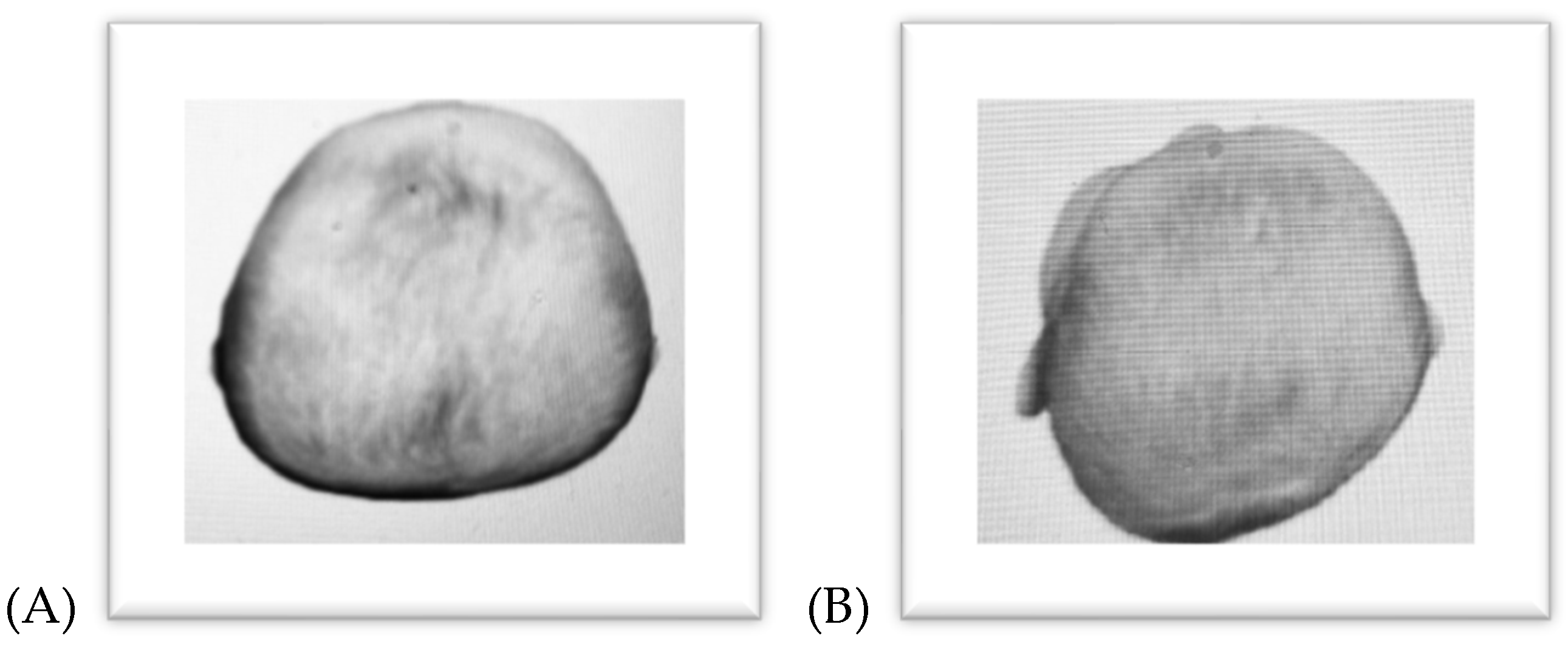
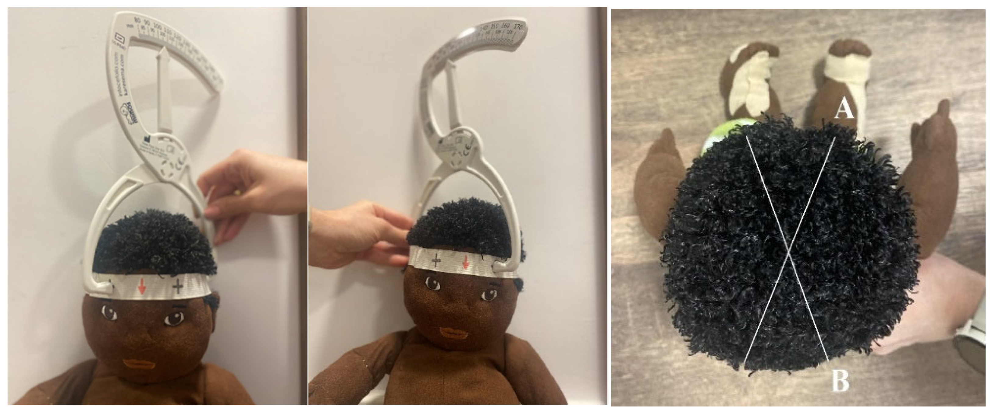
4.1. Cranial Measurements
4.2. Assessment of Psychomotor Development in Infants
4.3. Statistical Methods
5. Results
6. Discussion
7. Conclusions
- Asymmetry and reduced or increased muscle tone are associated with cranial shape abnormalities in infants up to 5 months of age.
- Reduced and increased muscle tone correlate most strongly with brachycephaly, with mean degrees of flattening increasing with age.
- The sex of the infants is not a differentiating factor in the level of either brachycephaly or plagiocephaly.
Funding
Conflicts of Interest
References
- Hadders-Algra, M. Early human motor development: From variation to the ability to vary and adapt. Neurosci. Biobehav. Rev. 2018, 90, 411–427. [Google Scholar] [CrossRef] [PubMed]
- Hadders-Algra, M.; Tacke, U.; Pietz, J.; Rupp, A.A.; Philippi, H. Reliability and predictive validity of the Standardized Infant NeuroDevelopmental Assessment neurological scale. Dev. Med. Child Neurol. 2019, 61, 654–660. [Google Scholar] [CrossRef] [PubMed]
- Goo, M.; Tucker, K.; Johnston, L.M. Muscle tone assessments for children aged 0 to 12 years: A systematic review. Dev. Med. Child Neurol. 2018, 60, 660–671. [Google Scholar] [CrossRef]
- Straathof, E.L.M.; Heineman, K.R.; Hamer, E.G.; Hadders-Algra, M. Patterns of atypical muscle tone in the general infant population—Prevalence and associations with perinatal risk and neurodevelopmental status. Early Hum. Dev. 2021, 152, 105276. [Google Scholar] [CrossRef]
- Michalska, A.; Szczukocki, M.; Szwilling, Z.; Wendor, J. The differential diagnosis of asymmetry in infants. Dev. Period Med. 2016, 20, 335–341. [Google Scholar]
- Beuriat, P.A.; Szathmari, A.; Di Rocco, F.; Mottolese, C. Deformational plagiocephaly: State of the art and review of the literature. Neurochirurgie 2019, 65, 322–329. [Google Scholar] [CrossRef]
- Huang, T.; Li, W.; Wang Ch Qu, F.; Yang, Q.; Pa, Q.; Pu, X.; Xiao, C.; Cai, Y.; Xia, M.; Zhang, Y. Research into the correlation between positional skull deformation and motor performance of infants aged under 4 months. BMC Pediatr. 2023, 23, 212. [Google Scholar] [CrossRef]
- Yang, W.; Chen, J.; Shen, W.; Wang, C.; Wu, Z.; Chang, Q.; Li, W.; Lv, K.; Pan, Q.; Li, H. Prevalence of positional skull deformities in 530 premature infants with a corrected age of up to 6 months: A multicenter study. BMC Pediatr. 2019, 19, 520. [Google Scholar] [CrossRef]
- Graham, T.; Gilbert, N.; Witthoff, K.; Gregory, T.; Walsh, M. Significant factors influencing the effectiveness of cranial remolding orthoses in infants with deformational plagiocephaly. J. Craniofacial Surg. 2019, 30, 1710–1713. [Google Scholar] [CrossRef]
- Panza, P.; Piarulli, F.; Rizzo, V.; Schettini, F.; Baldassarre, M.E.; Lorenzo, A.; Tafuri, S.; Laforgia, N. Positional plagiocephaly: Results of the osteopathic treatment of 424 infants. An observational retrospective cohort study. Ital. J. Pediatr. 2024, 50, 166. [Google Scholar] [CrossRef]
- Okamoto, T.; Harada, A.; Takamatsu, A.; Kyutoku, S.; Kaneko, T.; Ueda, K. Molding Helmet Therapy for Severe Deformational Brachycephaly: Position of Eurion and Therapeutic Effect. Plast. Reconstr. Surg. 2023, 152, 136–143. [Google Scholar] [CrossRef] [PubMed]
- Graham, T.; Millay, K.; Wang, J.; Adams-Huet, B.; O’Briant, E.; Oldham, M.; Smith, S. Significant Factors in Cranial Remolding Orthotic Treatment of Asymmetrical Brachycephaly. J. Clin. Med. 2020, 9, 1027. [Google Scholar] [CrossRef] [PubMed]
- Di Chiara, A.; La Rosa, E.; Ramieri, V.; Vellone, V.; Cascone, P. Treatment of deformational plagiocephaly with physiotherapy. J. Craniofacial Surg. 2019, 30, 2008–2013. [Google Scholar] [CrossRef]
- Bialocerkowski, A.E.; Vladusic, S.L.; Wei Ng, C. Prevalence, risk factors, and natural history of positional plagiocephaly: A systematic review. Dev. Med. Child Neurol. 2008, 50, 577–586. [Google Scholar] [CrossRef]
- Straathof, E.J.M.; Heineman, K.R.; Hamer, E.G.; Hadders-Algra, M. Prevailing head position to one side in early infancy—A population-based study. Acta Paediatr. 2020, 109, 1423–1429. [Google Scholar] [CrossRef]
- Rogers, G.F.; Oh, A.K.; Mulliken, J.B. The role of congenital muscular torticollis in the development of deformational plagiocephaly. Plast. Reconstr. Surg. 2009, 123, 643–652. [Google Scholar] [CrossRef]
- Öhman, A.; Nilsson, S.; Lagerkvist, A.L.; Beckung, E.V.A. Are infants with torticollis at risk of a delay in early motor milestones compared to a control group of healthy infants? Dev. Med. Child Neurol. 2009, 51, 545–550. [Google Scholar] [CrossRef]
- Marshall, J.M.; Shahzad, F. Safe Sleep, Plagiocephaly, and Brachycephaly: Assessment, Risks, Treatment, and When to Refer. Pediatr. Ann. 2020, 49, 440–447. [Google Scholar] [CrossRef]
- Pastor-Pons, I.; Hidalgo-García, C.; Lucha-Lópe, M.O.; Barrau-Lalmolda, M.; Rodes-Pastor, I.; Rodríguez-Fernández, A.L.; Tricás-Moreno, J.M. Effectiveness of pediatric integrative manual therapy in cervical movement limitation in infants with positional plagiocephaly: A randomized controlled trial. Ital. J. Pediatr. 2021, 47, 41. [Google Scholar] [CrossRef]
- Sasaki, J.; Hasegawa, S.; Yamamoto, S.; Wetenabe, S.; Miyachi, H.; Nagao, T. Relationship between facial asymmetry and positional plagiocephaly analyzed by three-dimensional computed tomography. J. Cranio-Maxillofac. Surg. 2020, 48, 193–198. [Google Scholar] [CrossRef]
- Ghizoni, E.; Denadai, R.; Raposo-Amaral, C.A.; Joaquim, A.F.; Tedeschi, H.; Raposo-Amaral, C.E. Diagnosis of infant synostotic and nonsynostotic cranial deformities: A review for pediatricians. Rev. Paul. Pediatr. 2016, 34, 495–502. [Google Scholar] [CrossRef] [PubMed]
- Kajdic, N.; Spazzapan, P.; Velnar, T. Craniosynostosis—Recognition, clinical characteristics, and treatment. Bosn. J. Basic Med. Sci. 2018, 18, 110–116. [Google Scholar] [CrossRef] [PubMed]
- Kelly, K.M.; Joganic, E.F.; Beals, S.P.; Riggs, J.A.; McGuire, M.K.; Littlefield, T.R. Helmet Treatment of Infants with Deformational Brachycephaly. Glob. Pediatr. Health 2018, 5, 2333794X18805618. [Google Scholar] [CrossRef] [PubMed]
- Knight, S. Positional plagiocephaly/brachycephaly is associated with later cognitive and academic outcomes. J. Pediatr. 2019, 210, 239–242. [Google Scholar] [CrossRef]
- Graham, J.M.; Kreutzman, J.; Earl, D.; Halberg, A.; Samayoa, C.; Guo, X. Deformational brachycephaly in supine-sleeping infants. J. Pediatr. 2005, 146, 253–257. [Google Scholar] [CrossRef]
- Kolehmainen, N.; Ramsay, C.; McKee, L.; Missiuna, C.; Owen, C.; Francis, J. Participation in physical play and leisure in children with motor impairments: Mixed-methods study to generate evidence for developing an intervention. Phys. Ther. 2015, 95, 1374–1386. [Google Scholar] [CrossRef]
- Choi, H.; Lim, S.H.; Kim, J.; Hong, B.Y. Outcome Analysis of the Eects of Helmet Therapy in Infants with Brachycephaly. J. Clin. Med. 2020, 9, 1171. [Google Scholar] [CrossRef]
- Looman, W.S.; Flannery, A.B. Evidence-based care of the child with deformational plagiocephaly, Part I: Assessment and diagnosis. J. Pediatr. Health Care 2012, 26, 242–250. [Google Scholar] [CrossRef]
- Maedomari, T.; Miyabayash, H.; Tanaka, Y.; Mukai, C.; Nakanomori, A.; Saito, K.; Kato, R.; Noto, T.; Nagano, N.; Morioka, I. Cranial Shape Measurements Obtained Using a Caliper and Elastic Bands Are Useful for Brachycephaly and Deformational Plagiocephaly Screening. J. Clin. Med. 2023, 12, 2787. [Google Scholar] [CrossRef]
- Jung, B.K.; Yun, I.S. Diagnosis and treatment of positional plagiocephaly. Arch. Craniofacial Surg. 2020, 21, 80–86. [Google Scholar] [CrossRef]
- Abboud, H.; Rifi, L.; Melhaoui, A.; Arkha, Y.; El Ouahabi, A. Diagnosis, Management, and Outcome in 9 Children with Unilateral Posterior Synostotic Plagiocephaly. World Neurosurg. 2020, 140, 169–174. [Google Scholar] [CrossRef] [PubMed]
- Unnithan AAjaya Kumar De Jesus, O. Plagiocephaly; StatPearls Publishing: Treasure Island, FL, USA, 2023. [Google Scholar] [PubMed]
- Clarren, S.K.; Smith, D.W.; Hanson, J.W. Helmet treatment for plagiocephaly and congenital muscular torticollis. J. Pediatr. 1979, 94, 43–46. [Google Scholar] [CrossRef] [PubMed]
- Tamber, M.S.; Nikas, D.; Beier, A.; Baird, L.C.; Bauer, D.F.; Durham, S.; Klimo, P.; Lin, A.Y.; Mazzola, C.; McClung-Smith, C.; et al. Congress of Neurological Surgeons Systematic Review and Evidence-Based Guideline on the Role of Cranial Molding Orthosis (Helmet) Therapy for Patients with Positional Plagiocephaly. Neurosurgery 2016, 79, 632–633. [Google Scholar] [CrossRef] [PubMed]


| Variable | N | Mean | SD | Var. Coeff. (%) |
|---|---|---|---|---|
| Age [months] | 60 | 3.03 | 1.40 | 46.21 |
| CVA [mm] | 60 | 8.10 | 5.69 | 70.20 |
| CI [%] | 60 | 88.23 | 6.74 | 7.65 |
| Variable | Subgroup | N | Mean | SD |
|---|---|---|---|---|
| Age [months] | Asymmetry | 20 | 2.90 | 1.45 |
| CVA [mm] | 20 | 15.40 | 3.19 | |
| CI [%] | 20 | 81.00 | 5.07 | |
| Age [months] | Reduced muscle tone | 20 | 3.05 | 1.39 |
| CVA [mm] | 20 | 4.45 | 1.76 | |
| CI [%] | 20 | 93.70 | 4.01 | |
| Age [months] | Increased muscle tone | 20 | 3.15 | 1.42 |
| CVA [mm] | 20 | 4.45 | 1.73 | |
| CI [%] | 20 | 90.00 | 3.13 |
| Subgroup No. | Subgroup | 1 15.40 | 2 4.45 | 3 4.45 |
|---|---|---|---|---|
| 1 | Asymmetry | 0.00 | 0.00 | |
| 2 | Reduced muscle tone | 0.00 | 1.00 | |
| 3 | Increased muscle tone | 0.00 | 1.00 |
| Subgroup No. | Subgroup | 1 81.00 | 2 93.70 | 3 90.00 |
|---|---|---|---|---|
| 1 | Asymmetry | 0.000000 | 0.000000 | |
| 2 | Reduced muscle tone | 0.000000 | 0.019703 | |
| 3 | Increased muscle tone | 0.000000 | 0.019703 |
| Variable | Subgroup = Asymmetry | ||
|---|---|---|---|
| The Correlation Coefficients Are Significant at p < 0.05 | |||
| Age | CVA | CI | |
| Age | 1.000 | 0.4886 | 0.2009 |
| p = 0.029 | p = 0.396 | ||
| CVA | 0.4886 | 1.0000 | 0.4303 |
| p = 0.029 | p = 0.058 | ||
| CI | 0.2009 | 0.4303 | 1.0000 |
| p = 0.396 | p = 0.058 | ||
| Variable | Subgroup = Reduced Muscle Tone The Correlation Coefficients Are Significant at p < 0.05 | ||
|---|---|---|---|
| Age | CVA | CI | |
| Age | 1.0000 | −0.2239 | 0.3507 |
| p = 0.343 | p = 0.130 | ||
| CVA | −0.2239 | 1.0000 | −0.0097 |
| p = 0.343 | p = 0.968 | ||
| CI | 0.3507 | −0.0097 | 1.0000 |
| p = 0.130 | p = 0.968 | ||
| Variable | Subgroup = Increased Muscle Tone The Correlation Coefficients Are Significant at p < 0.05. | ||
|---|---|---|---|
| Age | CVA | CI | |
| Age | 1.0000 | −0.5624 | 0.6613 |
| p = 0.010 | p = 0.001 | ||
| CVA | −0.5624 | 1.0000 | −0.5441 |
| p = 0.010 | p = 0.013 | ||
| CI | 0.6613 | −0.5441 | 1.0000 |
| p = 0.001 | p = 0.013 | ||
Disclaimer/Publisher’s Note: The statements, opinions and data contained in all publications are solely those of the individual author(s) and contributor(s) and not of MDPI and/or the editor(s). MDPI and/or the editor(s) disclaim responsibility for any injury to people or property resulting from any ideas, methods, instructions or products referred to in the content. |
© 2025 by the authors. Licensee MDPI, Basel, Switzerland. This article is an open access article distributed under the terms and conditions of the Creative Commons Attribution (CC BY) license (https://creativecommons.org/licenses/by/4.0/).
Share and Cite
Zielińska, N.; Górska, M.; Skrzek, A.; Dębiec-Bąk, A. The Correlation Between Infant Head Shape in Craniometric Studies and Psychomotor Development Disorders. J. Clin. Med. 2025, 14, 1985. https://doi.org/10.3390/jcm14061985
Zielińska N, Górska M, Skrzek A, Dębiec-Bąk A. The Correlation Between Infant Head Shape in Craniometric Studies and Psychomotor Development Disorders. Journal of Clinical Medicine. 2025; 14(6):1985. https://doi.org/10.3390/jcm14061985
Chicago/Turabian StyleZielińska, Natalia, Maria Górska, Anna Skrzek, and Agnieszka Dębiec-Bąk. 2025. "The Correlation Between Infant Head Shape in Craniometric Studies and Psychomotor Development Disorders" Journal of Clinical Medicine 14, no. 6: 1985. https://doi.org/10.3390/jcm14061985
APA StyleZielińska, N., Górska, M., Skrzek, A., & Dębiec-Bąk, A. (2025). The Correlation Between Infant Head Shape in Craniometric Studies and Psychomotor Development Disorders. Journal of Clinical Medicine, 14(6), 1985. https://doi.org/10.3390/jcm14061985