Article

Peak Eccentric Cycling Exercise and Cardiorespiratory Responses to Normobaric Hypoxia Versus Normobaric Normoxia in Healthy Adults: A Randomized, Controlled Crossover Trial

1
Department of Pulmonology, University Hospital Zurich, 8091 Zurich, Switzerland
2
Faculty of Medicine, University of Zurich, 8006 Zurich, Switzerland
*
Author to whom correspondence should be addressed.
These authors contributed equally to this work.
J. Clin. Med. 2025, 14(4), 1151; https://doi.org/10.3390/jcm14041151
Submission received: 6 January 2025 / Revised: 20 January 2025 / Accepted: 22 January 2025 / Published: 11 February 2025
(This article belongs to the Section Respiratory Medicine)

Abstract

:
Background/Objectives: Pulmonary rehabilitation clinics are traditionally located at higher altitudes (HAs), where lower PO2 reduces exercise capacity and blood oxygenation. Eccentric cycling exercise (ECC), with its lower cardiorespiratory demand compared to concentric cycling (CON), might therefore be a potential advantageous training modality at HAs, particularly for individuals with reduced exercise capacity. This study aimed to compare the cardiorespiratory responses of ECC while breathing normoxic versus hypoxic gas in healthy participants. Methods: This randomized, controlled crossover trial involved healthy participants performing CON in normoxia (FiO2 = 0.21), followed by two incremental ECC tests until 70–100% of peak exercise, one with normoxia and one with normobaric hypoxia (FiO2 = 0.15), in a randomized order. Oxygen uptake (V’O2) and additional outcomes were measured breath-by-breath. Endpoints were defined at rest, 50%, 70%, peak exercise, and isotime. The trial is registered on clinicaltrails.gov (NCT05185895). Results: Twelve healthy participants (age: 30 ± 11 years, six females) completed the study. During both interventions, V’O2 increased linearly with exercise intensity, with no significant differences between normoxic and hypoxic conditions. At peak exercise, SpO2 and peak work rate were significantly lowered by 5% (95%CI: 3 to 8%, p < 0.001) and by 22 W (95%CI: 8 to 36 W, p = 0.009) in hypoxia compared to normoxia. Other outcomes were unchanged. When comparing CON to ECC in normoxia, the mean differences in V’O2 increased with higher loads. Conclusions: This study demonstrated that V’O2 and other cardiopulmonary parameters remain unchanged when performing ECC in hypoxia compared to normoxia. Comparing CON to ECC in normoxia, participants achieved higher workloads and greater V’O2 consumption during CON compared to ECC at comparable watts, confirming the higher metabolic cost associated with CON. We identified that the optimal submaximal ECC intensities, with the highest difference in V’O2 between CON versus ECC, are around 40% of peak V’O2.

1. Introduction

Historically, rehabilitation centers are often located at moderate-to-high altitudes (HAs), as the mountain air is believed to offer significant health benefits. This practice continues today, supported by evidence that exercise at high altitudes induces several performance-enhancing changes in skeletal muscles, such as an increased capillary-to-fiber ratio [1,2,3,4], increased mitochondrial density [3,5], enhanced glycolytic capacity, and greater oxygen storage in muscles [6,7,8]. Further cardiovascular acclimatization such as the hypoxia-induced expression of the glycoprotein hormone erythropoietin collectively contributes to a rise in peak oxygen uptake (V’O2) [9,10,11] and improved exercise performance at low altitudes [12,13]. However, while these benefits of long-term acclimatization are notable, the reduced PO2 level at HAs produces physiological reactions, such as reduced exercise capacity, hypoxemia, and increased minute ventilation (V’E) [14,15,16,17]. Since cardiopulmonary rehabilitation centers are frequently located at HAs, these mechanisms are limiting, especially for patients with pre-existing cardiopulmonary diseases.
Eccentric cycling (ECC) has been shown to have significantly reduced cardiorespiratory and metabolic demands compared to traditional concentric cycling (CON) [18,19,20,21,22]. Specifically, for the same mechanical intensity, V’O2 during ECC is 60–80% lower than during CON at the same intensity [18,21,23], even at high exercise intensities [20,23]. A recent systematic review highlighted that ECC exercise is superior to CON exercise in improving muscle mass, knee extensor strength, and functional outcomes like six-minute walk distance in healthy volunteers and peak V’O2 in patients with cardiopulmonary diseases [24]. Exercise therapy is a crucial part of pulmonary rehabilitation, complementing pharmacological interventions [25,26]. Especially in the advanced stages of pulmonary disease, cardiopulmonary function is often so restricted that training intensity is too low for exercise on beneficial levels to sustain muscle mass [27]. ECC training presents a highly promising treatment option, especially for individuals with cardiopulmonary limitations who may struggle with traditional exercise methods at HA [18,28]. This approach has the potential to maximize the benefits of a pulmonary rehabilitation, especially at moderate-to-high altitudes.
Despite its potential, no studies have yet explored the feasibility and safety of ECC under hypoxic conditions, a critical factor for implementing this modality in HA rehabilitation settings. To address this gap, it is essential to first investigate ECC in healthy participants to establish preliminary evidence before extending research to clinical populations. To evaluate the feasibility and safety of ECC under hypoxic conditions, it is essential to first examine this in healthy participants, as no prior studies have investigated this effect. Therefore, this randomized clinical trial aimed to evaluate the cardiorespiratory responses to ECC under normoxia and normobaric hypoxia, providing preliminary data that could guide the development of tailored rehabilitation protocols for individuals with hypoxia-sensitive diseases.

2. Materials and Methods
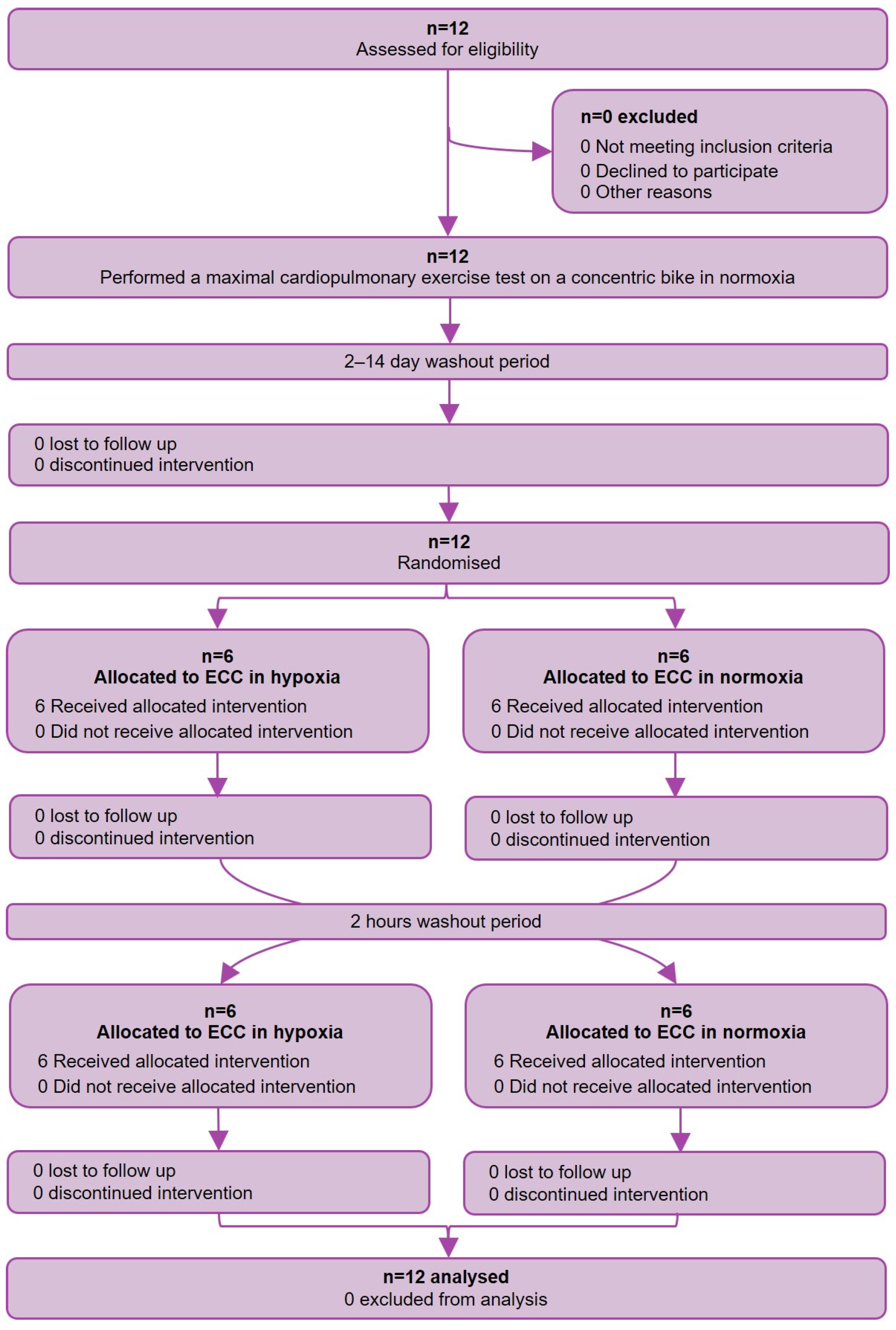
2.1. Study Design

This was a randomized, controlled crossover study. Blinding was not possible due to the nature of the condition. The measurements took place at the University Hospital of Zurich, Switzerland, from August to September 2023.

2.2. Study Population

Healthy females and males between 18 and 80 years of age without any diagnosed cardiovascular diseases were included in this study. Exclusion criteria were the inability to follow the study protocol due to language problems, psychological disorders as well as neurological or orthopedic limitations with the inability to pedal on the ergometer, and participation in another clinical study that required active treatment. Two members of the study team recruited potential participants through word-of-mouth advertising. After participants signed the informed consent, they were enrolled and subsequently randomized by the same two team members. The study was approved by the regulatory authorities (KEK Zürich, BASEC-Nr. 2021-01312) and registered at clinicaltrials.gov (NCT05185895).

2.3. Study Procedure, Assessments, and Outcomes

All participants performed two study visits. During the first visit, all subjects underwent a spirometric test to assess lung volumes, which was followed by a CON incremental ramp exercise test until exhaustion to assess peak V’O2. After a 15-minute break, all participants completed a familiarization session on the eccentric ergometer for five minutes to prevent muscle soreness and damage [29]. The second visit occurred at least two days after the first visit. At visit 2, ECC under normoxia (FiO2 = 0.21) and hypoxia (FiO2 = 0.15, equivalent to an altitude of 2500 m) was performed in a randomized order with a wash-out time of two hours between the measurements.
For both interventions, the participants were seated on the eccentric ergometer and followed the same ramp protocol as in visit 1. After a 2-minute resting period, the participants cycled at 55–65 RPM with an increasing load, aiming to reach at least 70% of their peak CON load determined during the first visit. Participants could continue beyond 70% until exhaustion, but not exceed 100%.
CON was performed on a conventional ergometer (Ergoline-Ergoselect 200” Ergoline GmbH, Bitz, Germany), while ECC was performed on an eccentric ergometer (Cyclus 2 Recumbent EccentricTrainer, RBM elektronik-automation GmbH, Leipzig, Germany). During ECC, normobaric hypoxia was provided using the Everest Summit II altitude generator (Hypoxico Altitude Training Systems, Bickenbach, Germany). V’O2 and other ventilator outcomes were measured breath-by-breath with a metabolic unit (Geratherm Medical, Gschwenda, Germany) that was calibrated before each test [30]. The exercise tests were performed in accordance with the guidelines of the American Thoracic Society [31]. Every participant had the same protocol for both tests; however, protocols varied among participants according to their individual fitness levels, which were assessed according to the weekly hours of sport (<3 h: 6 W/20 s; 3–6 h: 8 W/20 s; 6–9 h: 10 W/20 s; 9–12 h: 12 W/20 s; >12 h: 14 W/20 s).
The primary outcome of the study was the mean difference in V’O2 at peak exercise between ECC under normoxia compared to that under hypoxia.
Secondary outcomes included the mean difference in V’O2 at rest, 50% and 70% of the individual maximal capacity, and other endpoints such as heart rate (HR), oxygen saturation (SpO2), carbon dioxide output (V’CO2), minute ventilation (VE), ventilatory equivalent for CO2 (VE/V’CO2), and oxygen pulse (O2Pulse). These variables were assessed at rest, 50%, 70%, and peak exercise. Additionally, dyspnea and leg fatigue were evaluated at peak exercise using the Borg CR10 scale.

2.4. Statistical Analysis and Sample Size

The sample size calculation was performed with a two-sided significance level of 0.05, a power of 0.8, a minimal clinical important difference of 0.6 L/min, and within a standard deviation of 0.4 L/min. According to these calculations, ten participants were needed. With an assumed dropout rate of 20%, a study population of twelve participants was aspired. Randomization was conducted using block randomization with randomly permuted blocks of size 2 using the package blockrand in R (version 4.3.0). The randomization sequence was developed by a member of the study team who was not involved in recruitment and measurement.
All data were summarized as means and standard deviations (SDs). To compare differences between normoxia and normobaric hypoxia, breath-by-breath data at each endpoint were averaged over 30 s. Endpoints were defined at rest, at 50%, at 70%, at peak exercise, and at isotime, while isotime compares tests with normoxia and hypoxia at identical times corresponding to peak exercise with the shorter tests. A linear mixed model was fitted with V’O2 as the dependent variable and condition as the fixed effect, while a random intercept accounted for individual differences in baseline V’O2. The Tukey method was used to adjust for multiple comparisons. Assumptions for linear mixed models, such as homogeneity and normality of residuals, were visually assessed using Tukey–Anscombe plots and Q–Q plots.
Additional outcomes were analyzed in a similar manner. All statistical analyses were conducted using R-studio Version 4.3.2, and a p of < 0.05 was assumed to reflect statistical significance.

3. Results

3.1. Baseline Characteristics

Twelve participants were included into the study (six females). The mean ± SD age was 30 ± 12 years, and the body mass index was 22.3 ± 3.8 kg/m2. Baseline characteristics are listed in Table 1, and the study flow chart is shown in Figure 1.
Neither period nor carry-over effects were identified. In ECC under normoxia, participants reached 87 ± 8% of their normoxic CON workload, and one participant reached 100%. In ECC under hypoxia, participants reached 78 ± 16% of their hypoxic CON workload, and two participants reached 100%. Primary and secondary outcomes are presented in Table 2.

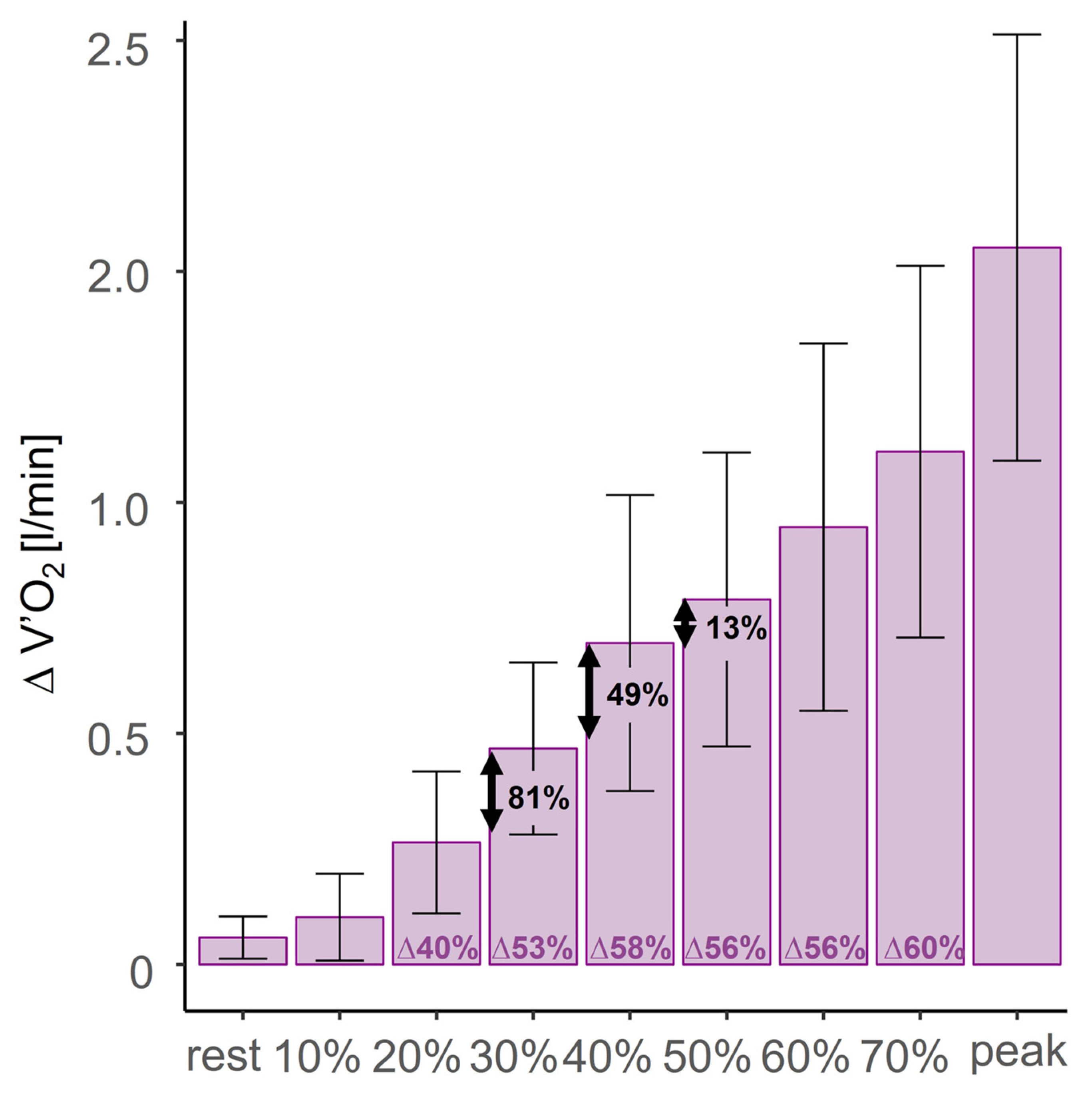
3.2. ECC Normoxia Versus Hypoxia

During both interventions, V’O2 increased linearly with exercise intensity, with no statistically significant differences between normoxia and hypoxia at any endpoint (see Figure 2).
At rest, SpO2 showed a statistically significant difference between normoxia and hypoxia by 3% (95%CI: 2 to 5%, p = 0.001). The difference increased, with the largest disparity of 6% occurring at endpoints 70% (95%CI: 3 to 9%, p < 0.001) and peak exercise (95%CI: 3 to 9%, p < 0.001).
HR increased with exercise intensity during both interventions, remaining slightly higher in hypoxic ECC at all endpoints, though without reaching statistical significance. O2 pulse was lower in hypoxia at all endpoints; however, only the endpoint isotime showed a significant difference of 0.74 mL/min (95%CI: 0.01 to 1.47 L/min, p = 0.048).
Neither V’E nor V’E/V’CO2 varied significantly between the two interventions.
BorgCR10 scale for dyspnea and leg fatigue that were assessed at peak exercise were both slightly higher in hypoxia but neither showed statistically significant differences (95%CI: −1 to 1, p = 0.592; 95%CI: −2 to 0, p = 0.251).

3.3. Normoxic CON Versus Normoxic ECC

The mean differences in V’O2 increased with a higher load (see Table 2, Figure 3). The highest difference in V’O2 was observed at peak exercise, with a mean difference of 1.55 ± 0.46 L/min (95%CI: 1.28 to 1.82 L/min, p < 0.001). This corresponded to a 63% lower V’O2 in ECC than in CON. Apart from the increase in V’O2 difference between 70% and peak exercise, the greatest rise was observed from 30% to 40%, with a mean increase in V’O2 difference of 0.23 L/min. The mathematical calculation using fractions revealed that the highest dividend (absolute mean V’O2/relative mean power) of 1.75 occurred at 40%.

4. Discussion

The results of this study demonstrate that V’O2 and other cardiopulmonary parameters remain unchanged when performing ECC in a hypoxic condition equivalent to approximately 2500 m of HA compared to normoxia. During normoxic breathing, participants achieved higher workloads and greater V’O2 responses during CON compared to ECC, indicating higher cardiopulmonary and metabolic costs associated with CON. These findings reinforce the differences in metabolic demand between ECC and CON exercises, with ECC exercise requiring significantly less V’O2 despite comparable workloads [18,19,22].
The existing studies indicate that reduced PO2, due to normobaric hypoxia, physiologically leads to a decreased peak V’O2 in healthy individuals as well as in patients with cardiopulmonary diseases [17,32,33,34]. The observed reduction in V’O2 during ECC compared to CON aligns with the existing literature [18,19,22], reflecting the inherently lower cardiorespiratory demand of ECC. This submaximal nature of ECC explains why participants achieved similar percentages of their peak V’O2 in both normoxic and hypoxic ECC conditions, suggesting that ECC relies more on mechanical efficiency and less on oxygen-dependent metabolic pathways. Supporting these findings, prior research has shown that V’O2 remains stable during submaximal CON exercise (e.g., running at 55% of peak V’O2) in acute moderate hypoxia at altitudes up to 2800 m above sea level [17]. These results further highlight that, even when performed at the same intensity in terms of power output (watts), ECC training is less affected by hypoxia due to its lower cardiopulmonary demand. Additionally, dyspnea and leg fatigue scores were comparable between normoxic and hypoxic ECC, with dyspnea consistently rated lower than leg fatigue in both conditions. This suggests that ECC effectively reduces ventilatory demands, even under hypoxic conditions. These findings, consistent with previous studies [24,28,35], underscore ECC’s potential as a rehabilitative exercise modality, particularly for individuals prone to ventilatory limitations, and especially when rehabilitation is performed at HAs.
The observed reduction in SpO2 during hypoxia, which intensified with increasing exercise intensity, is consistent with the physiological response to lower PO2 levels. Desaturation is typically accompanied by increased ventilation and cardiac output to compensate for reduced oxygen availability; however, no significant changes in ventilation or HR were detected in this study. This could be attributed to the short exposure time to hypoxia (10–15 min), as full physiological acclimatization requires longer hypoxic exposure [36,37]. The peripheral chemoreceptors are oxygen-sensitive below a certain threshold of hypoxia. HR and cardiac output are the first to adapt to hypoxia, while ventilation acclimatization occurs over a longer time [37]. This may explain the small difference in HR and the similarity in ventilation observed in this study. Additionally, the type of hypoxia plays a role, with studies showing that acute hypobaric vs. normobaric hypoxia results in significantly lower SpO2, while in long-lasting hypobaric hypoxia, the difference between the two conditions diminishes [36,38]. Ventilatory parameters are also lower in hypobaric hypoxia, while cardiovascular variables appear similar between the two types [36,38]. These factors, along with the small sample size, may explain the absence of statistically significant differences in cardiorespiratory parameters between normoxic and hypoxic ECC.
In ECC, participants generated significantly less peak power compared to CON. Reason for this might be that ECC was novel for participants, and the lack of prior experience with this type of exercise likely influenced their ability to produce power effectively. ECC involves a unique motor pattern that differs from traditional concentric exercises, requiring greater neural coordination and unfamiliar movement dynamics. This learning curve may have compounded the challenges posed by hypoxia, further reducing power production. Additionally, acute submaximal ECC exercise in untrained individuals often induces micro-damage [19]. The lower peak power output in hypoxia compared to normoxia in ECC could be attributed to two different factors. The muscle micro-damage in ECC can lead to localized vascular dysfunction, making muscles more susceptible to deoxygenation [39], an effect likely exacerbated by hypoxia. Additionally, hypoxia is associated with reduced cognitive and executive function [40,41], as highlighted in a recent systematic review [41]. Since ECC exercise relies heavily on neural components [42], these pathways may be particularly vulnerable to the effects of hypoxia, further contributing to reduced power output. These findings underscore the interplay between muscular and neural mechanisms in hypoxia, though further research is needed to fully understand these mechanisms.

4.1. Minimal Intensity for Maximal Metabolic Cost Reduction in Normoxic ECC

The largest difference between normoxic CON and normoxic ECC in V’O2 was found at peak exercise. However, training at maximal effort requires high load capacity to withstand the strain and is therefore not efficient. Descriptive and mathematical analyses revealed that the minimal difference for maximal metabolic cost reduction is expected to be at 40% of the maximal power as visually demonstrated in Figure 3. Hence, training in that range seems to be the most efficient for cardiopulmonary cost. This knowledge can help future studies to test various training protocols since, to date, there is a lack of knowledge about which training parameters are the most beneficial. Even though this range seems to be the most efficient in terms of VO2 difference and performance, it is possible that other ranges may achieve better training effects. This must be further evaluated.

4.2. Implication for ECC in HA Rehabilitation Settings

Contrary to our initial hypothesis, hypoxia did not lead to a difference in V’O2 during ECC. This suggests that ECC effectively reduces ventilatory demands, even under hypoxic conditions. However, the drop in SpO2 observed during hypoxia could pose challenges for cardiopulmonary patients, particularly those with pre-existing conditions, as reduced oxygen availability may significantly impact their exercise tolerance. Since rehabilitation stays typically extend beyond two weeks, physiological acclimatization to HAs partially restores the SpO2 levels over time [43]. This needs to be further investigated in clinical cohorts to better understand its implications for long-term safety and efficacy.
Despite this, the study findings indicate that hypoxia did not significantly impact other cardiopulmonary parameters, suggesting that ECC remains a viable exercise modality even in HA settings. Additionally, the low dyspnea scores reported during ECC, even under hypoxic conditions, highlight its potential to minimize ventilatory strain. This unique combination of maintaining low cardiopulmonary demand and perceived exertion makes ECC a promising option for safe and effective rehabilitation programs for patients with cardiopulmonary diseases with reduced exercise tolerance, such as COPD, heart failure, or pulmonary hypertension, at HAs.
These findings, derived from a healthy and relatively young population, provide preliminary data for future studies. Future research should focus on investigating the effects of ECC in individuals with cardiopulmonary pathologies and extending the duration of hypoxic exposure to further evaluate its safety and efficacy. Optimal training protocols should also be explored, aiming to achieve maximal metabolic efficiency with minimal exertion, particularly around the identified 40% of individual maximal capacity.

4.3. Limitations

Blinding was impractical due to the setup, potentially leading to subjective bias in outcomes such as felt exertion. Additionally, participants were inexperienced with cardiorespiratory exercise testing, and the brief 10-min familiarization with ECC might have been insufficient, possibly impacting their performance [44].
Moreover, the study’s findings are limited in generalizability to the investigated population. Individuals with cardiopulmonary diseases were excluded, and the relatively young study population restricts applicability to older individuals or those with cardiopulmonary conditions. Despite these limitations, this study is the first to evaluate ECC under hypoxic conditions and to propose optimal training parameters for the ECC ergometer, offering a foundation for future research in this field.

5. Conclusions

This study demonstrated for the first time that V’O2, V’E/V’CO2, HR, V’E, O2Puls, dyspnea, and leg fatigue remain unchanged at peak exercise during ECC in normoxia compared to normobaric hypoxia. These findings highlight its potential as a promising exercise modality for cardiopulmonary patients in HA rehabilitation settings. Based on a healthy and young population, this study provides preliminary data for future research to explore ECC’s effects in cardiopulmonary patients, evaluate its safety during prolonged hypoxia, and identify optimal training protocols for maximizing metabolic efficiency at around 40% of individual maximal capacity.

Author Contributions

Conceptualization, J.M. and S.U.; data curation, C.W. and E.C.; formal analysis, C.W., E.C., and J.M.; funding acquisition, S.U.; investigation, C.W. and E.C.; methodology, C.W., E.C., and J.M.; project administration, C.W.; resources, S.U.; supervision, M.L. and S.U.; validation, S.R.S., A.T., M.F., M.L., and S.U.; visualization, C.W. and J.M.; writing—original draft, C.W., E.C., S.R.S., A.T., M.F., M.L., J.M., and S.U.; writing—review and editing, C.W., E.C., S.R.S., A.T., M.F., M.L., J.M., and S.U. All authors have read and agreed to the published version of the manuscript.

Funding

This research received no external funding.

Institutional Review Board Statement

The study was conducted in accordance with the Declaration of Helsinki and approved by the Institutional Review Board (or Ethics Committee) of Kantonale Ethikkommission Zürich (BASEC-Nr. 2021-01312; date of approval: 8 August 2023).

Informed Consent Statement

Informed consent was obtained from all subjects involved in the study.

Data Availability Statement

The datasets generated and analyzed during the current study are available from the author on reasonable request.

Acknowledgments

We sincerely thank all participants for their time, effort, and invaluable contribution to this study.

Conflicts of Interest

The authors declare no conflict of interest.

Abbreviations

The following abbreviations are used in this manuscript:
BMIBody mass index
CIConfidence interval
CONConcentric cycling
CR10Clinical rating scale 10
ECCEccentric cycling
HAHigh altitude
HRHeart rate
O2Oxygen
O2PulseOxygen pulse
RPMRepetitions per minute
SDStandard deviation
SpO2Oxygen saturation
V’CO2Carbon dioxide output
V’EMinute ventilation
V’E/VO2Ventilatory equivalent for O2
V’E/V’CO2Ventilatory equivalent for CO2
V’O2Oxygen uptake
LDLinear dichroism

References

  1. Desplanches, D.; Hoppeler, H.; Linossier, M.T.; Denis, C.; Claassen, H.; Dormois, D.; Lacour, J.R.; Geyssant, A. Effects of training in normoxia and normobaric hypoxia on human muscle ultrastructure. Pflugers Arch. 1993, 425, 263–267. [Google Scholar] [CrossRef] [PubMed]
  2. Gustafsson, T.; Puntschart, A.; Sundberg, C.J.; Jansson, E. Related expression of vascular endothelial growth factor and hypoxia-inducible factor-1 mRNAs in human skeletal muscle. Acta Physiol. Scand. 1999, 165, 335–336. [Google Scholar] [CrossRef] [PubMed]
  3. Hoppeler, H.; Vogt, M.; Weibel, E.R.; Flück, M. Response of skeletal muscle mitochondria to hypoxia. Exp. Physiol. 2003, 88, 109–119. [Google Scholar] [CrossRef] [PubMed]
  4. Montero, D.; Lundby, C. Effects of exercise training in hypoxia versus normoxia on vascular health. Sports Med. 2016, 46, 1725–1736. [Google Scholar] [CrossRef] [PubMed]
  5. Geiser, J.; Vogt, M.; Billeter, R.; Zuleger, C.; Belforti, F.; Hoppeler, H. Training high—Living low: Changes of aerobic performance and muscle structure with training at simulated altitude. Int. J. Sports Med. 2001, 22, 579–585. [Google Scholar] [CrossRef] [PubMed]
  6. Hoppeler, H.; Vogt, M. Muscle tissue adaptations to hypoxia. J. Exp. Biol. 2001, 204, 3133–3139. [Google Scholar] [CrossRef] [PubMed]
  7. Vogt, M.; Puntschart, A.; Geiser, J.; Zuleger, C.; Billeter, R.; Hoppeler, H. Molecular adaptations in human skeletal muscle to endurance training under simulated hypoxic conditions. J. Appl. Physiol. 2001, 91, 173–182. [Google Scholar] [CrossRef] [PubMed]
  8. Zoll, J.; Ponsot, E.; Dufour, S.; Doutreleau, S.; Ventura-Clapier, R.; Vogt, M.; Hoppeler, H.; Richard, R.; Flück, M. Exercise training in normobaric hypoxia in endurance runners. III. Muscular adjustments of selected gene transcripts. J. Appl. Physiol. 2006, 100, 1258–1266. [Google Scholar] [CrossRef]
  9. Bahenský, P.; Bunc, V.; Tlustý, P.; Grosicki, G.J. Effect of an eleven-day altitude training program on aerobic and anaerobic performance in adolescent runners. Medicina 2020, 56, 184. [Google Scholar] [CrossRef]
  10. Bailey, D.M.; Davies, B.; Baker, J. Training in hypoxia: Modulation of metabolic and cardiovascular risk factors in men. Med. Sci. Sports Exerc. 2000, 32, 1058–1066. [Google Scholar] [CrossRef]
  11. Chen, B.; Wu, Z.; Huang, X.; Li, Z.; Wu, Q.; Chen, Z. Effect of altitude training on the aerobic capacity of athletes: A systematic review and meta-analysis. Heliyon 2023, 9, e20188. [Google Scholar] [CrossRef] [PubMed]
  12. Ponsot, E.; Dufour, S.P.; Zoll, J.; Doutrelau, S.; N’Guessan, B.; Geny, B.; Hoppeler, H.; Lampert, E.; Mettauer, B.; Ventura-Clapier, R.; et al. Exercise training in normobaric hypoxia in endurance runners. II. Improvement of mitochondrial properties in skeletal muscle. J Appl Physiol (1985) 2006, 100, 1249–1257. [Google Scholar] [CrossRef] [PubMed]
  13. Dufour, S.P.; Ponsot, E.; Zoll, J.; Doutreleau, S.; Lonsdorfer-Wolf, E.; Geny, B.; Lampert, E.; Flück, M.; Hoppeler, H.; Billat, V.; et al. Exercise training in normobaric hypoxia in endurance runners. I. Improvement in aerobic performance capacity. J Appl Physiol (1985) 2006, 100, 1238–1248. [Google Scholar] [CrossRef] [PubMed]
  14. Müller, J.; Titz, A.; Schneider, S.R.; Bauer, M.; Mayer, L.; Lüönd, L.; Ulrich, T.; Furian, M.; Forrer, A.; Schwarz, E.I.; et al. The effect of high altitude (2500 m) on incremental cycling exercise in patients with pulmonary arterial hypertension and chronic thromboembolic pulmonary hypertension: A randomised controlled crossover trial. Eur. Respir. J. 2024, 63, 2301001. [Google Scholar] [CrossRef]
  15. Furian, M.; Flueck, D.; Latshang, T.D.; Scheiwiller, P.M.; Segitz, S.D.; Mueller-Mottet, S.; Murer, C.; Steiner, A.; Ulrich, S.; Rothe, T.; et al. Exercise performance and symptoms in lowlanders with COPD ascending to moderate altitude: Randomized trial. Int. J. Chron. Obstr. Pulm. Dis. 2018, 13, 3529–3538. [Google Scholar] [CrossRef] [PubMed]
  16. Furian, M.; Hartmann, S.E.; Latshang, T.D.; Flueck, D.; Murer, C.; Scheiwiller, P.M.; Osmonov, B.; Ulrich, S.; Kohler, M.; Poulin, M.J.; et al. Exercise Performance of Lowlanders with COPD at 2,590 m: Data from a Randomized Trial. Respiration 2018, 95, 422–432. [Google Scholar] [CrossRef] [PubMed]
  17. Wehrlin, J.P.; Hallén, J. Linear decrease in .VO2max and performance with increasing altitude in endurance athletes. Eur. J. Appl. Physiol. 2006, 96, 404–412. [Google Scholar] [CrossRef]
  18. Dufour, S.P.; Lampert, E.; Doutreleau, S.; Lonsdorfer-Wolf, E.; Billat, V.L.; Piquard, F.; Richard, R. Eccentric cycle exercise: Training application of specific circulatory adjustments. Med. Sci. Sports Exerc. 2004, 36, 1900–1906. [Google Scholar] [CrossRef]
  19. Hody, S.; Croisier, J.-L.; Bury, T.; Rogister, B.; Leprince, P. Eccentric muscle contractions: Risks and benefits. Front. Physiol. 2019, 10, 536. [Google Scholar] [CrossRef] [PubMed]
  20. Meyer, K.; Steiner, R.; Lastayo, P.; Lippuner, K.; Allemann, Y.; Eberli, F.; Schmid, J.; Saner, H.; Hoppeler, H. Eccentric exercise in coronary patients: Central hemodynamic and metabolic responses. Med. Sci. Sports Exerc. 2003, 35, 1076–1082. [Google Scholar] [CrossRef]
  21. Perrey, S.; Betik, A.; Candau, R.; Rouillon, J.D.; Hughson, R.L. Comparison of oxygen uptake kinetics during concentric and eccentric cycle exercise. J. Appl. Physiol. 2001, 91, 2135–2142. [Google Scholar] [CrossRef]
  22. Touron, J.; Costes, F.; Coudeyre, E.; Perrault, H.; Richard, R. Aerobic metabolic adaptations in endurance eccentric exercise and training: From whole body to mitochondria. Front. Physiol. 2021, 11, 596351. [Google Scholar] [CrossRef]
  23. Lipski, M.; Abbiss, C.R.; Nosaka, K. Cardio-pulmonary responses to incremental eccentric and concentric cycling tests to task failure. Eur. J. Appl. Physiol. 2018, 118, 947–957. [Google Scholar] [CrossRef]
  24. Barreto, R.V.; de Lima, L.C.R.; Borszcz, F.K.; de Lucas, R.D.; Denadai, B.S. Chronic adaptations to eccentric cycling training: A systematic review and meta-analysis. Int. J. Environ. Res. Public Health 2023, 20, 2861. [Google Scholar] [CrossRef] [PubMed]
  25. Ries, A.L.; Bauldoff, G.S.; Carlin, B.W.; Casaburi, R.; Emery, C.F.; Mahler, D.A.; Make, B.; Rochester, C.L.; ZuWallack, R.; Herrerias, C. Pulmonary rehabilitation. Chest 2007, 131, 4S–42S. [Google Scholar] [CrossRef] [PubMed]
  26. Rochester, C.L.; Alison, J.A.; Carlin, B.; Jenkins, A.R.; Cox, N.S.; Bauldoff, G.; Bhatt, S.P.; Bourbeau, J.; Burtin, C.; Camp, P.G.; et al. Pulmonary rehabilitation for adults with chronic respiratory disease: An official american thoracic society clinical practice guideline. Am. J. Respir. Crit. Care Med. 2023, 208, e7–e26. [Google Scholar] [CrossRef] [PubMed]
  27. Vogiatzis, I. Strategies of muscle training in very severe COPD patients. Eur. Respir. J. 2011, 38, 971–975. [Google Scholar] [CrossRef] [PubMed]
  28. Barreto, R.V.; De Lima, L.C.R.; Denadai, B.S. Moving forward with backward pedaling: A review on eccentric cycling. Eur. J. Appl. Physiol. 2021, 121, 381–407. [Google Scholar] [CrossRef] [PubMed]
  29. LaStayo, P.C.; Pierotti, D.J.; Pifer, J.; Hoppeler, H.; Lindstedt, S.L. Eccentric ergometry: Increases in locomotor muscle size and strength at low training intensities. Am. J. Physiol.-Regul. Integr. Comp. Physiol. 2000, 278, R1282–R1288. [Google Scholar] [CrossRef] [PubMed]
  30. Beijst, C. The Accuracy and Precision of Equipment for Cardiopulmonary Exercise Testing. Master’s Thesis, Eindhoven University of Technology, Eindhoven, The Netherlands, 2011. [Google Scholar]
  31. American Thoracic Society. ATS/ACCP statement on cardiopulmonary exercise testing. Am. J. Respir. Crit. Care Med. 2003, 167, 211–277. [Google Scholar] [CrossRef] [PubMed]
  32. Naeije, R. Physiological adaptation of the cardiovascular system to high altitude. Prog. Cardiovasc. Dis. 2010, 52, 456–466. [Google Scholar] [CrossRef] [PubMed]
  33. Treml, B.; Gatterer, H.; Burtscher, J.; Kleinsasser, A.; Burtscher, M. A focused review on the maximal exercise responses in hypo- and normobaric hypoxia: Divergent oxygen uptake and ventilation responses. Int. J. Environ. Res. Public Health 2020, 17, 5239. [Google Scholar] [CrossRef] [PubMed]
  34. Ulrich, S.; Schneider, S.R.; Bloch, K.E. Effect of hypoxia and hyperoxia on exercise performance in healthy individuals and in patients with pulmonary hypertension: A systematic review. J. Appl. Physiol. 2017, 123, 1657–1670. [Google Scholar] [CrossRef] [PubMed]
  35. McLean, B.D.; Gore, C.J.; Kemp, J. Application of ‘live low-train high’ for enhancing normoxic exercise performance in team sport athletes. Sports Med. 2014, 44, 1275–1287. [Google Scholar] [CrossRef]
  36. Coppel, J.; Hennis, P.; Gilbert-Kawai, E.; Grocott, M.P. The physiological effects of hypobaric hypoxia versus normobaric hypoxia: A systematic review of crossover trials. Extrem. Physiol. Med. 2015, 4, 2. [Google Scholar] [CrossRef]
  37. Luks, A.M.; Hackett, P.H. Medical conditions and high-altitude travel. N. Engl. J. Med. 2022, 386, 364–373. [Google Scholar] [CrossRef]
  38. Schneider, S.R.; Lichtblau, M.; Furian, M.; Mayer, L.C.; Berlier, C.; Müller, J.; Saxer, S.; Schwarz, E.I.; Bloch, K.E.; Ulrich, S. Cardiorespiratory adaptation to short-term exposure to altitude vs. Normobaric hypoxia in patients with pulmonary hypertension. J. Clin. Med. 2022, 11, 2769. [Google Scholar] [CrossRef] [PubMed]
  39. Caldwell, J.T.; Wardlow, G.C.; Branch, P.A.; Ramos, M.; Black, C.D.; Ade, C.J. Effect of exercise-induced muscle damage on vascular function and skeletal muscle microvascular deoxygenation. Physiol. Rep. 2016, 4, e13032. [Google Scholar] [CrossRef]
  40. Hamacher, D.; Brennicke, M.; Behrendt, T.; Alt, P.; Törpel, A.; Schega, L. Motor-cognitive dual-tasking under hypoxia. Exp. Brain Res. 2017, 235, 2997–3001. [Google Scholar] [CrossRef]
  41. Jung, M.; Zou, L.; Yu, J.J.; Ryu, S.; Kong, Z.; Yang, L.; Kang, M.; Lin, J.; Li, H.; Smith, L.; et al. Does exercise have a protective effect on cognitive function under hypoxia? A systematic review with meta-analysis. J. Sport. Health Sci. 2020, 9, 562–577. [Google Scholar] [CrossRef] [PubMed]
  42. Douglas, J.; Pearson, S.; Ross, A.; McGuigan, M. Eccentric exercise: Physiological characteristics and acute responses. Sports Med. 2017, 47, 663–675. [Google Scholar] [CrossRef] [PubMed]
  43. Dünnwald, T.; Kienast, R.; Niederseer, D.; Burtscher, M. The use of pulse oximetry in the assessment of acclimatization to high altitude. Sensors 2021, 21, 1263. [Google Scholar] [CrossRef] [PubMed]
  44. Périard, J.D.; Travers, G.J.S.; Racinais, S.; Sawka, M.N. Cardiovascular adaptations supporting human exercise-heat acclimation. Auton. Neurosci. 2016, 196, 52–62. [Google Scholar] [CrossRef] [PubMed]
Figure 1. The flow chart of the included and analyzed patients. There were no dropouts.
Jcm 14 01151 g001
Figure 2. Mean oxygen uptake (V’O2) and standard deviation are presented for the different endpoints measured during normoxia and hypoxia. The terms “50%” and “70%” refer to 50% and 70% of each participant’s individual maximal capacity. “Isotime” corresponds to the maximum load each participant was able to sustain under normoxia and hypoxia.
Jcm 14 01151 g002
Figure 3. The absolute mean difference in oxygen uptake (V’O2) between concentric and eccentric cycling is reported alongside the standard deviation of these differences. The term “delta” represents the relative difference between concentric and eccentric cycling. The values 81%, 49%, and 13% indicate the relative increase in differences between successive steps, such as an 81% increase in the difference from 20% to 30%.
Jcm 14 01151 g003
Table 1. Baseline characteristics are presented as mean (SD) or absolute values.
CharacteristicsValue
Total Participants12
Female6
Male6
Age [years]30 (12)
Height [cm]178 (10.1)
Weight [kg]71.2 (11.1)
BMI [kg/m2]22.33 (3.78)
Peak [V’O2]2.54 (0.87)
Peak work rate [W]269 (91)
Table 2. Columns under “Eccentric Cycling” compare outcomes between normoxia and hypoxia. Columns under “Normoxia” compare outcomes between concentric cycling (CON) and eccentric cycling (ECC). True values for estimates and SDs, and model values for CIs and p-values. * indicates statistical significance at alpha <0.05.
Eccentric CyclingNormoxia
NormoxiaHypoxiaNormoxia–HypoxiaCONCON–ECC
Mean (SD)Mean (SD)Mean Change [95%CI]p-ValueMean (SD)Mean Change [95%CI]p-Value
Rest
V’O2 [L/min]0.21 (0.04)0.20 (0.01)0.01 [−0.04, 0.06]0.6190.27 (0.08)0.06 [0.03, 0.09]0.001 *
V’O2/kg [L/min/kg]2.98 (0.50)2.83 (1.44)0.15 [−0.58, 0.90]0.6613.79 (0.84)0.81 [0.45, 1.17]0.001 *
V’E/V’CO242.3 (8.10)48.1 (10.7)−5.83 [−12.99, 1.33]0.10541.42 (8.74)−0.89 [−4.78, 3.00]0.650
HR [bpm]63 (11)65 (13)−2 [−8, 6]0.67576 (9)13 [7, 18]<0.001 *
V’E [L/min]10.3 (2.23)11.8 (3.93)−1.42 [−3.75, −0.91]0.22012.25 (3.25)1.92 [0.21, 3.63]0.043 *
O2Puls [mL/beat]3.40 (0.97)3.00 (1.39)0.40 [−0.36, 1.16]0.2833.52 (0.92)0.13 [−0.04, 0.29]0.155
SpO2 [%]97 (1)94 (3)3 [2, 5]0.001 *97 (1)0 [−1, 0]0.615
50% of individual maximal capacity
V’O2 [L/min]0.60(0.24)0.65 (0.27)−0.05 [−0.12, −0.03]0.1991.38 (0.47)0.79 [0.6, 0.98]<0.001 *
V’O2/kg [L/min/kg]8.28 (2.36)9.05 (2.92)−0.77 [−1.86, 0.58]0.28919.58 (5.25)11.34 [8.8, 13.88]<0.001 *
V’E/V’CO233.5 (6.29)35.9 (8.88)−2.4 [−6.53, 1.58]0.21730.63 (5.31)−3.43 [−5.86, −1]0.015 *
HR [bpm]90 (8)97 (18)−7 [−18, 2]0.128122 (14)33 [24, 42]<0.001 *
V’E [L/min]21.3 (5.69)24.0 (8.34)−2.7 [−7.19, 1.79]0.22441.67 (12.07)20.08 [13.49, 26.68]<0.001 *
O2Puls [mL/beat]6.65 (2.50)6.53 (2.18)0.12 [−0.44, 0.98]0.43411.10 (3.01)4.49 [3.63, 5.35]<0.001 *
SpO2 [%]97 (1)92 (4)5 [3, 7]<0.001 *96 (2)−1 [−2, 0]0.095
70% of individual maximal capacity
V’O2 [L/min]0.77 (0.320)0.81 (0.352)−0.04 [−0.11, 0.08]0.7231.86 (0.62)1.11 [0.87, 1.35]<0.001 *
V’O2/kg [L/min/kg]10.7 (3.35)11.2 (3.67)−0.5 [−1.75, 1.48]0.86226.46 (7.08)16.00 [12.63, 19.37]<0.001 *
V’E/V’CO233.3 (7.18)34.9 (9.54)−1.6 [−6.01, 2.19]0.34230.91 (5.37)−2.74 [−5.95, 0.47]0.110
HR [bpm]100 (7)107 (20)−7 [−16, 3]0.147143 (15)43 [34, 52]<0.001 *
V’E [L/min]26.0 (6.89)27.7 (10.5)−1.7 [−5.08, 2.59]0.50562.58 (15.30)36.92 [29.02, 44.81]<0.001 *
O2Puls [mL/beat]7.67 (3.06)7.50 (2.73)0.17 [−0.47, 1.36]0.32212.90 (3.30)5.30 [4.25, 6.35]<0.001 *
SpO2 [%]97 (1)91 (5)6 [3, 9]<0.001 *96 (2)−1 [−2, 0]0.045 *
Peak exercise
Power [W]235 (84)213 (86)22 [8, 36]0.009 *269 (91)34 [18, 50]0.001 *
V’O2 [L/min]0.99 (0.51)0.90 (0.38)0.09 [−0.04, 0.22]0.1722.54 (0.87)1.55 [1.28, 1.82]<0.001 *
V’O2/kg [L/min/kg]13.6 (5.70)12.4 (4.35)1.2 [−0.76, 3.14]0.21735.98 (9.95)22.35 [18.44, 26.26]<0.001 *
V’E/V’CO233.3 (11.9)32.9 (7.80)0.4 [−6.46, 7.26]0.90435.32 (4.09)2.04 [−4.27, 8.35]0.523
HR [bpm]111 (15)114 (20)−3 [−11, 5]0.459167 (20)55 [43, 68]<0.001 *
V’E [L/min]30.5 (10.4)29.4 (12.0)1.1 [−4.63, 6.79]0.696116.6 (38.6) 86.08 [66.43, 105.74]<0.001 *
O2Puls [mL/beat]8.60 (3.57)7.68 (2.56)0.92 [−0.12, 1.96]0.08113.20 (4.27)5.00 [2.99, 7.66]0.001 *
SpO2 [%]96 (1)91 (5)5 [3, 8]0.001 *87 (20)−9 [−20, 2]0.115
Dyspnea [CR10]3 (2)4 (2)−1 [−1, 1]0.5927 (2)4 [3, 5]<0.001 *
Leg fatigue [CR10]6 (2)7 (2)−1 [−2, 0]0.2518 (2)2 [1, 3]0.011 *
Isotime (individual maximal workload (in watts) that a participant was able to perform under both conditions)
Power [W]213 (86)213 (86)--213 (86)--
V’O2 [L/min]0.88 (0.44)0.86 (0.38)0.02 [−0.06, 0.10]0.6502.01 (0.964)1.13 [0.77, 1.48]<0.001 *
V’O2/kg [L/min/kg]12.2 (4.88)12.0 (4.40)0.2 [−1.04, 1.51]0.70828.24 (11.8)16.04 [11.03, 21.04]<0.001 *
V’E/V’CO233.1 (7.50)33.2 (7.48)−0.1 [−4.42, 4.25]0.96938.73 (17.5)5.63 [−10.08, 7.63]0.783
HR [bpm]107 (13)114 (20)−7 [−14, 0]0.050144 (28)37 [23, 53]<0.001 *
V’E [L/min]29.7 (7.36)29.8 (11.9)−0.1 [−7.75, 7.59]0.98332.2 (4.69)2.5 [−4.22, 2.33]0.570
O2Puls [mL/beat]8.12 (3.32)7.38 (2.45)0.74 [0.01, 1.47]0.048 *12.3 (4.15)4.18 [2.82, 5.90]0.002 *
SpO2 [%]97 (1)91 (5)6 [3, 9]<0.001 *94 (3)−3 [−4, −1]0.011 *
Disclaimer/Publisher’s Note: The statements, opinions and data contained in all publications are solely those of the individual author(s) and contributor(s) and not of MDPI and/or the editor(s). MDPI and/or the editor(s) disclaim responsibility for any injury to people or property resulting from any ideas, methods, instructions or products referred to in the content.

