
Optical Coherence Tomography in Infectious Keratitis After Femtosecond Keratorefractive Surgery
 , ,
, ,
Abstract
1. Introduction
2. Material and Methods
Image Acquisition and Analysis
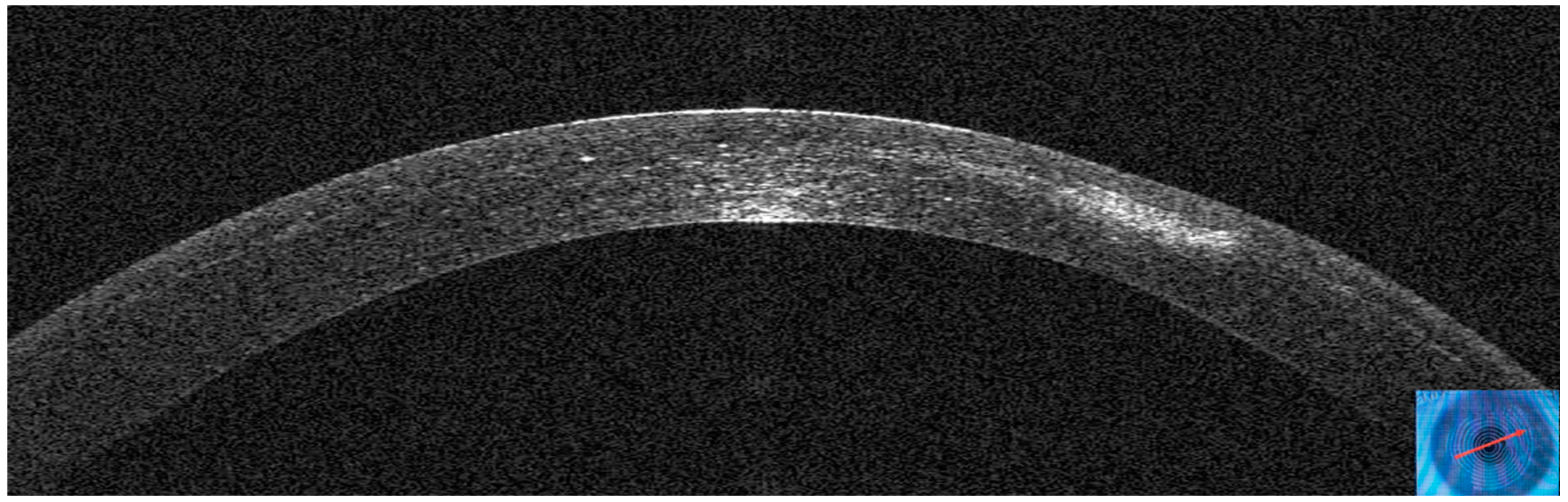
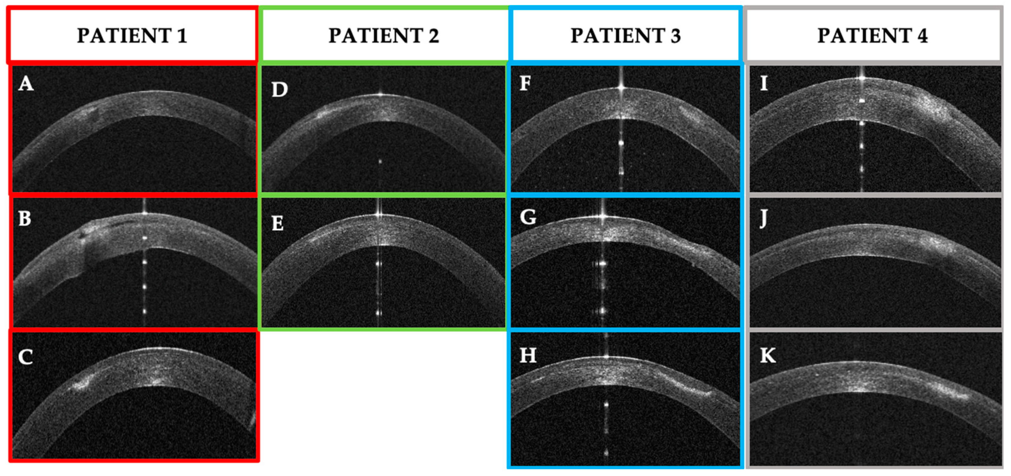
3. Results
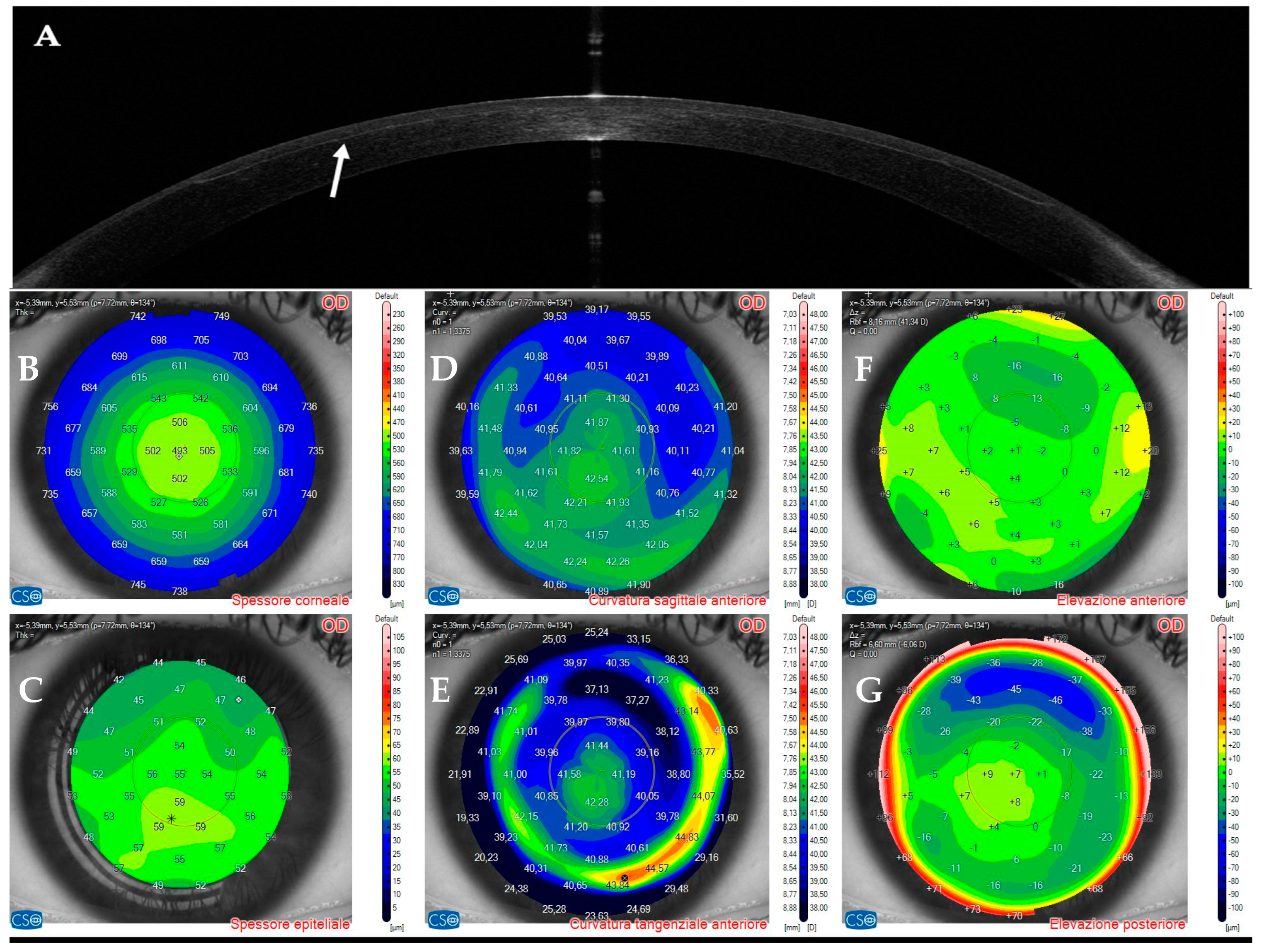
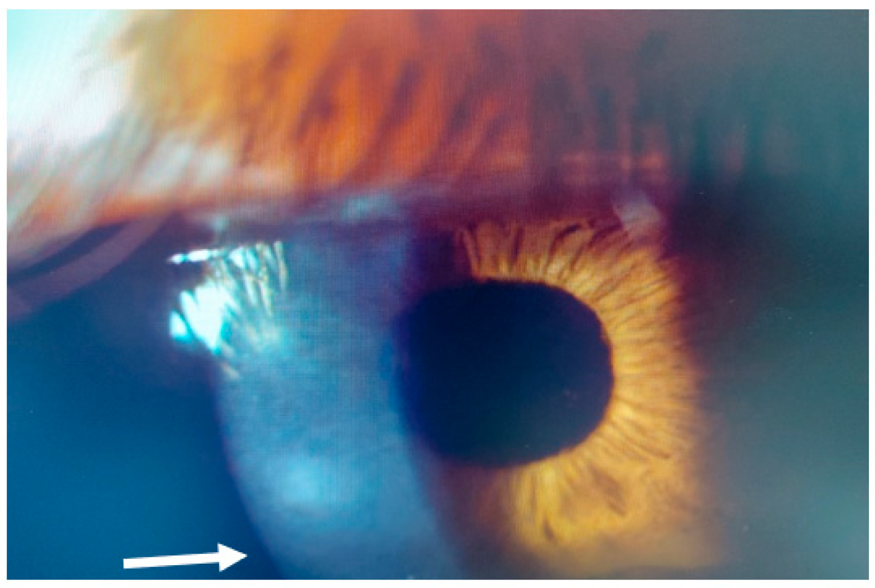
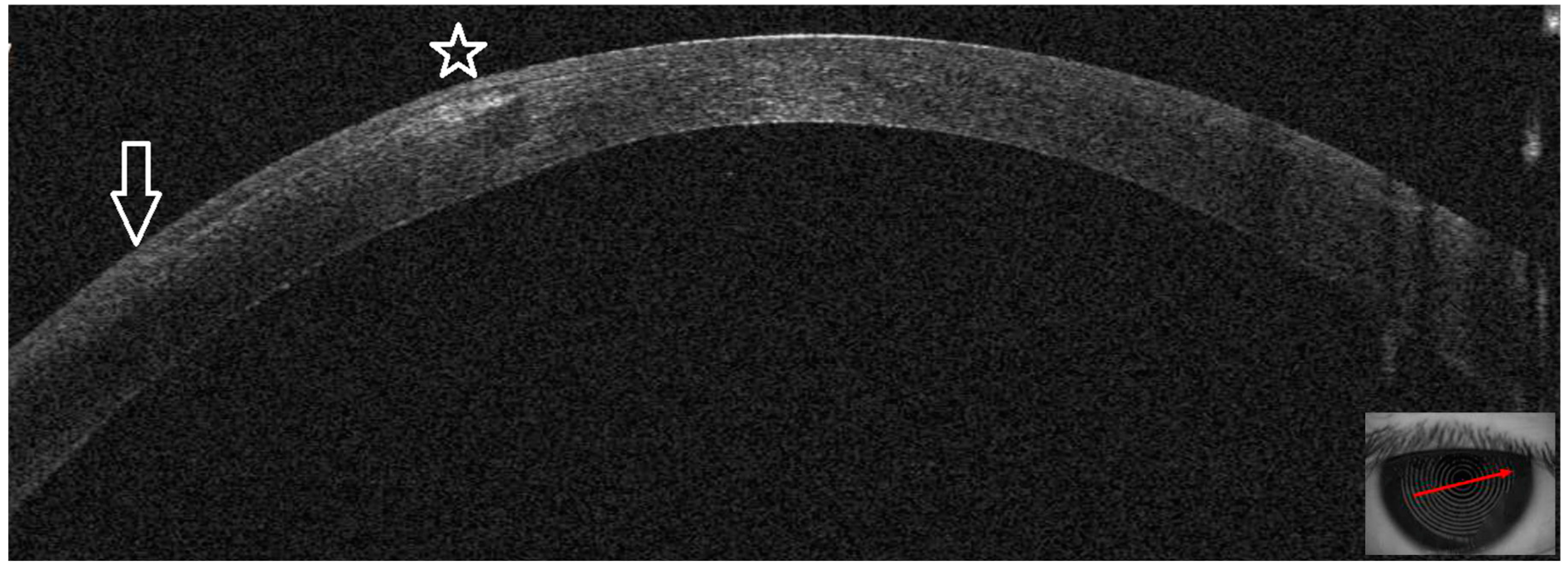
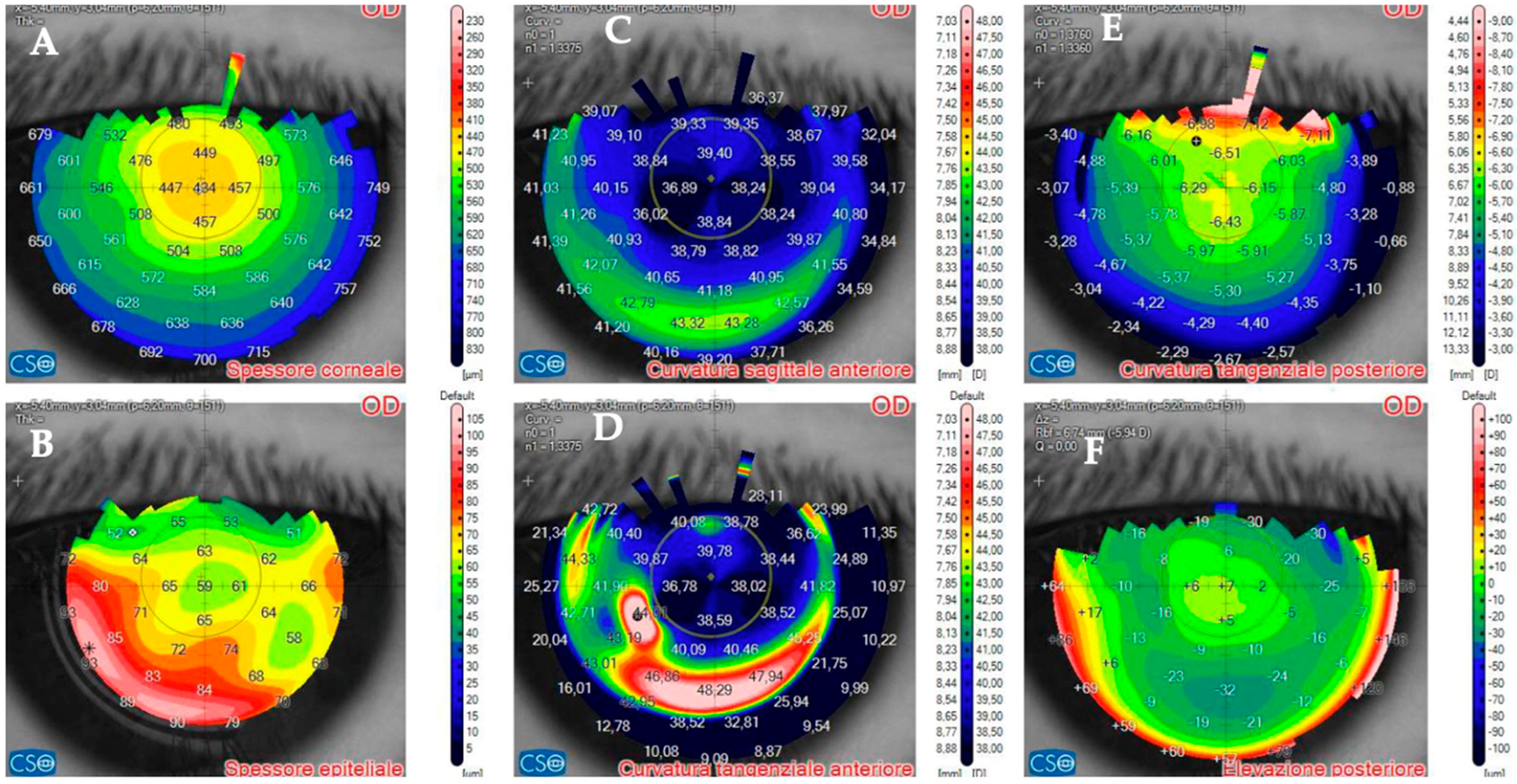
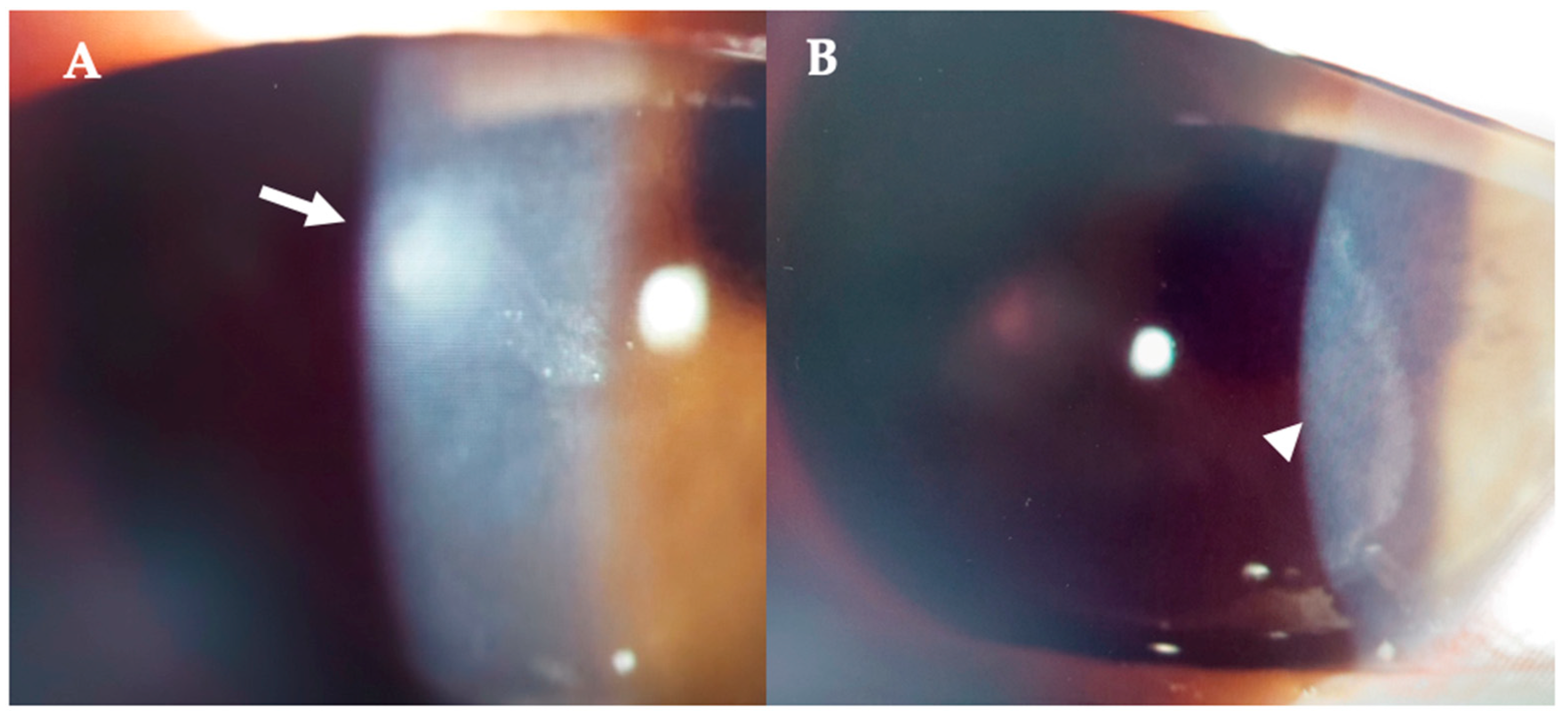
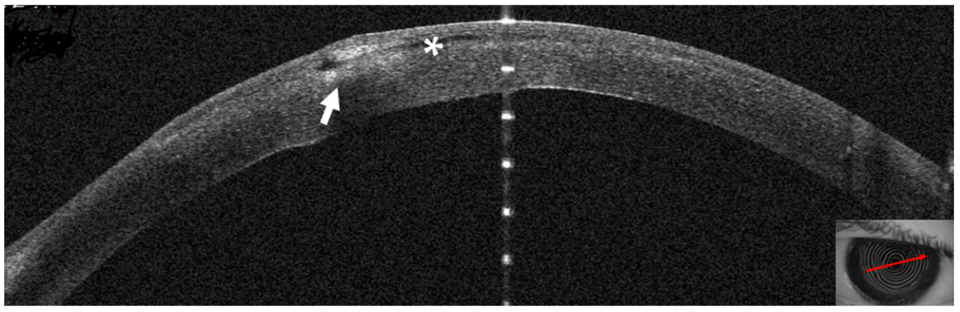
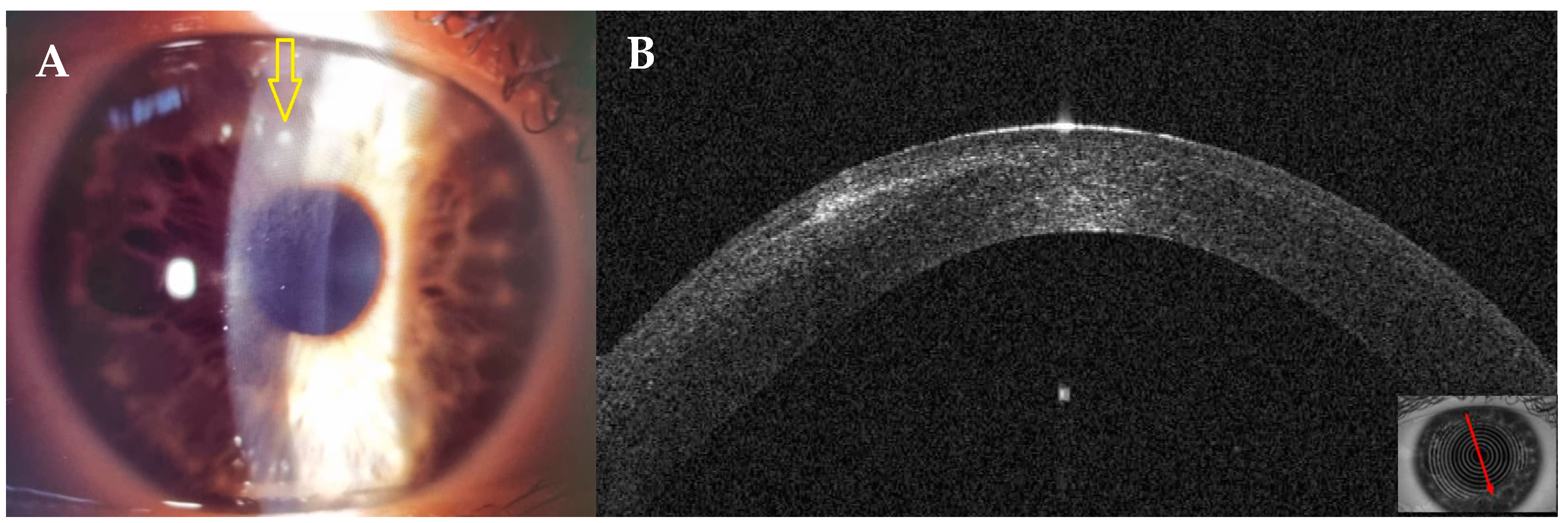
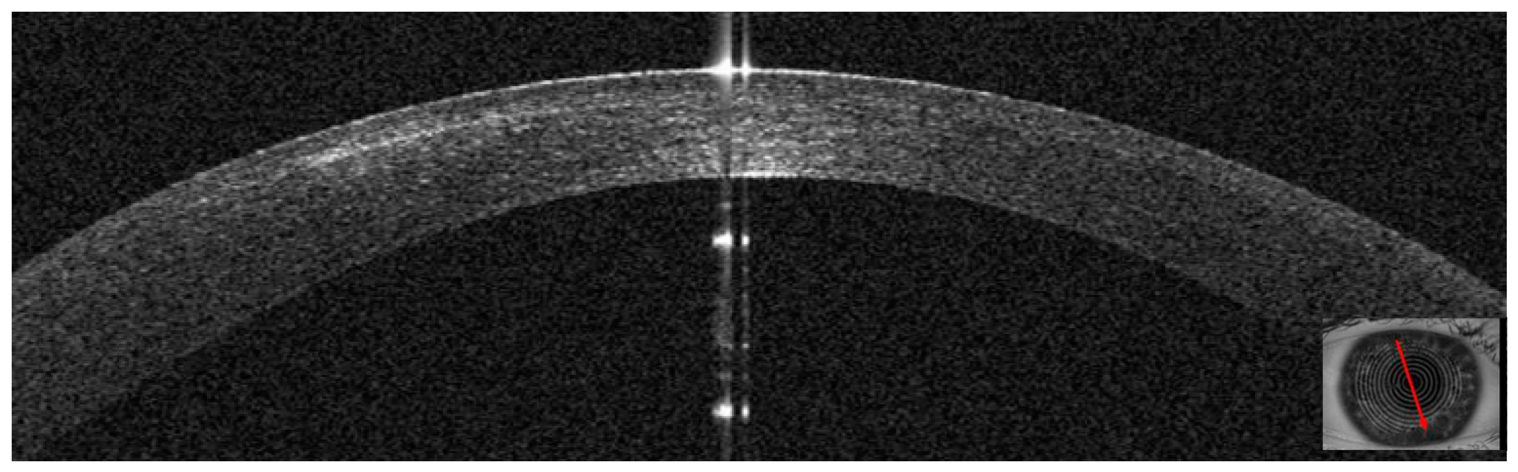
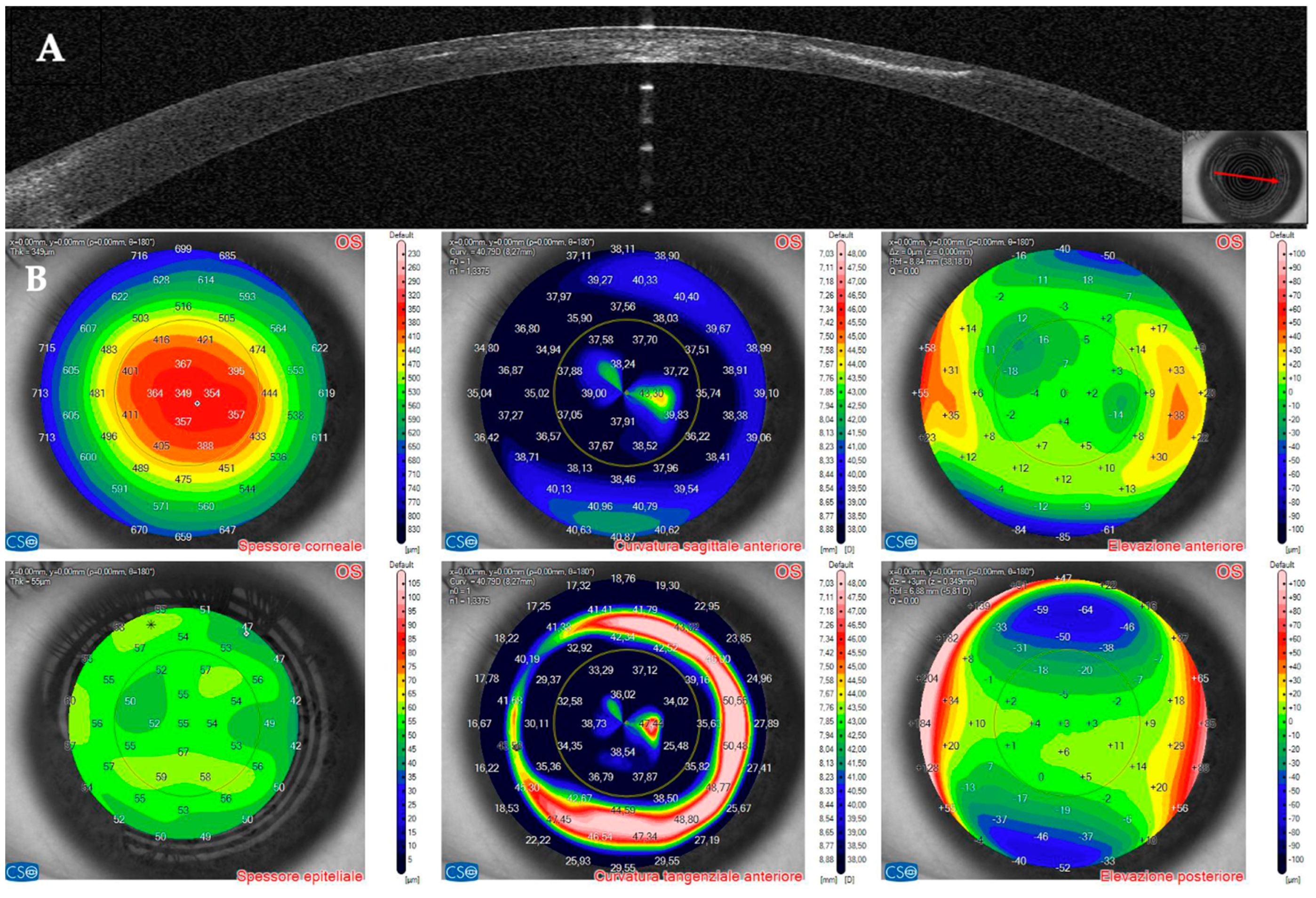
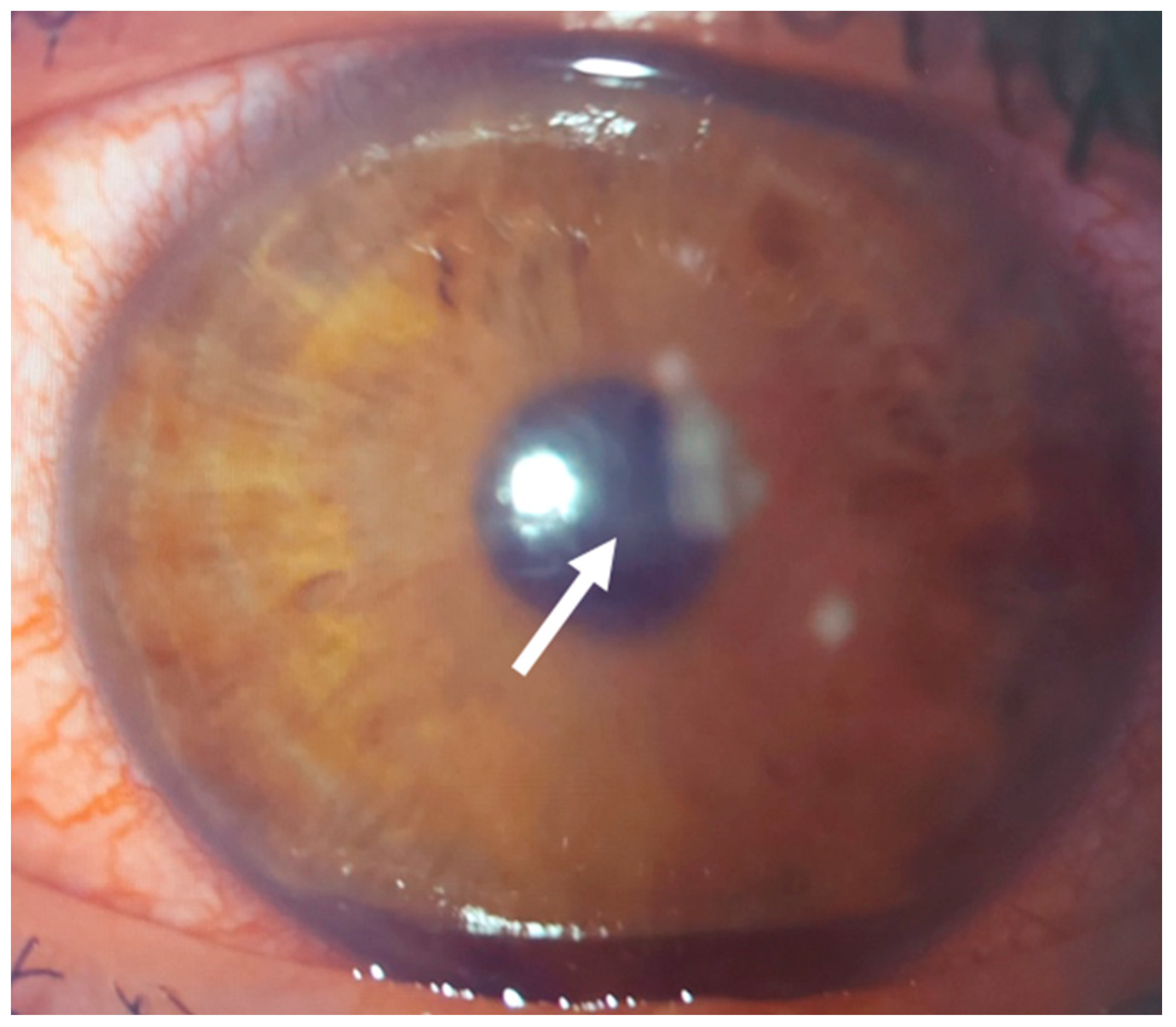
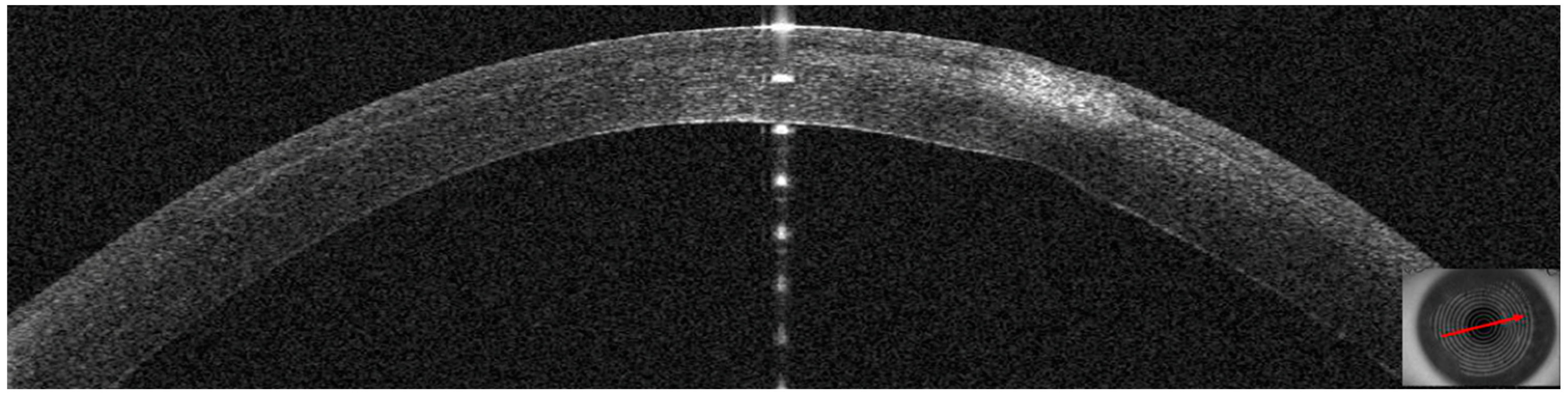
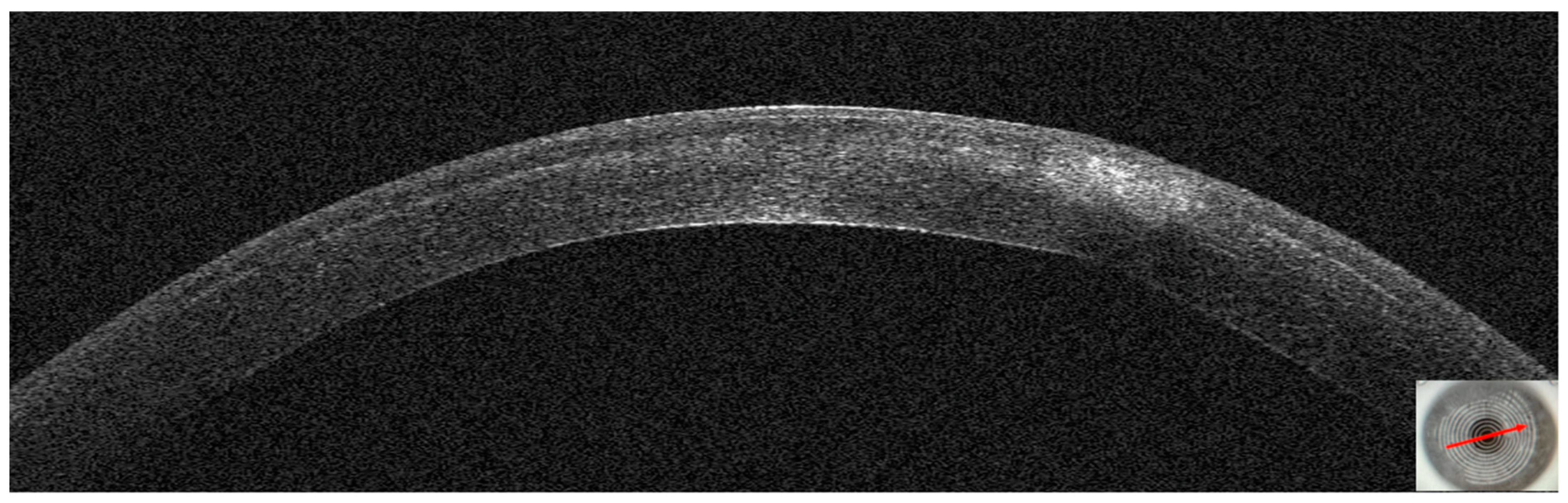
3.1. Patient #1
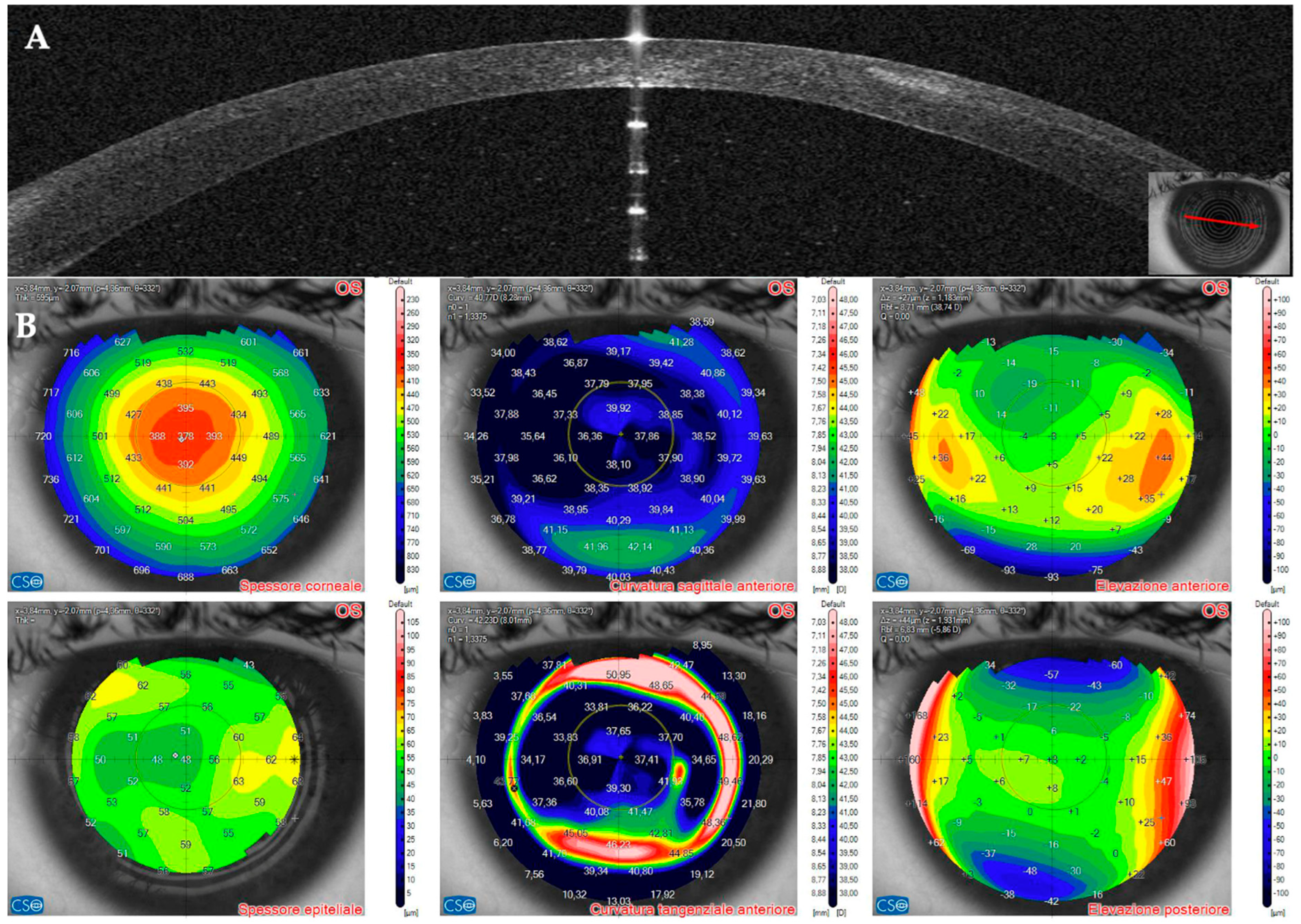
3.2. Patient #2
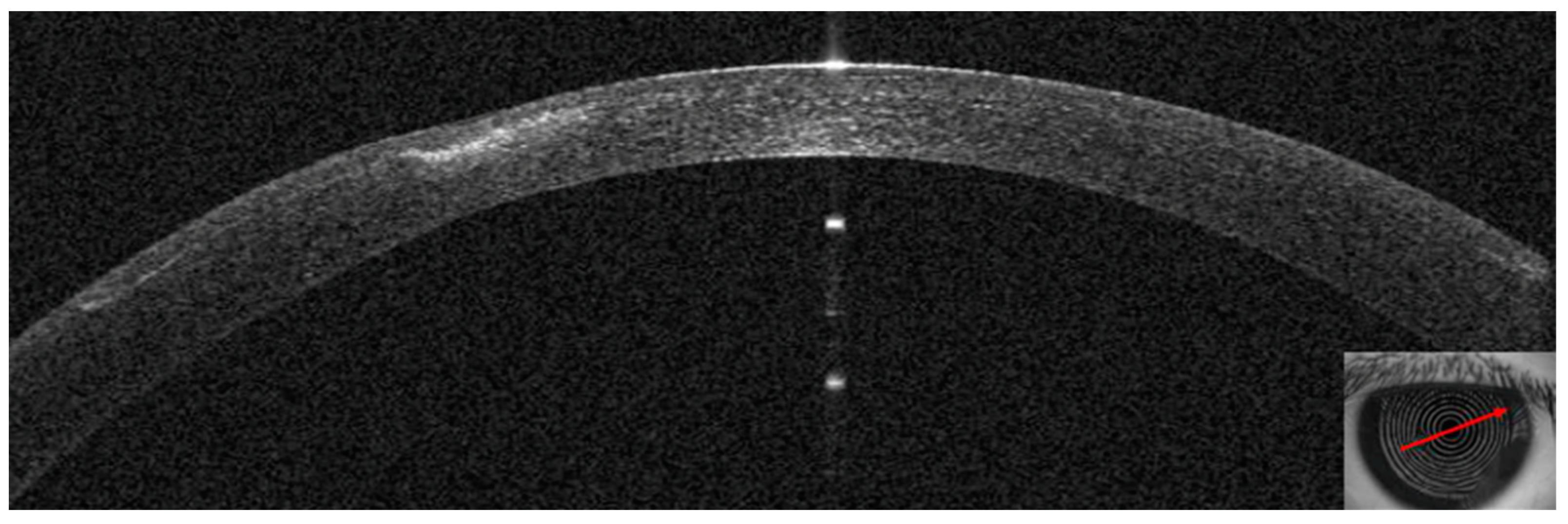
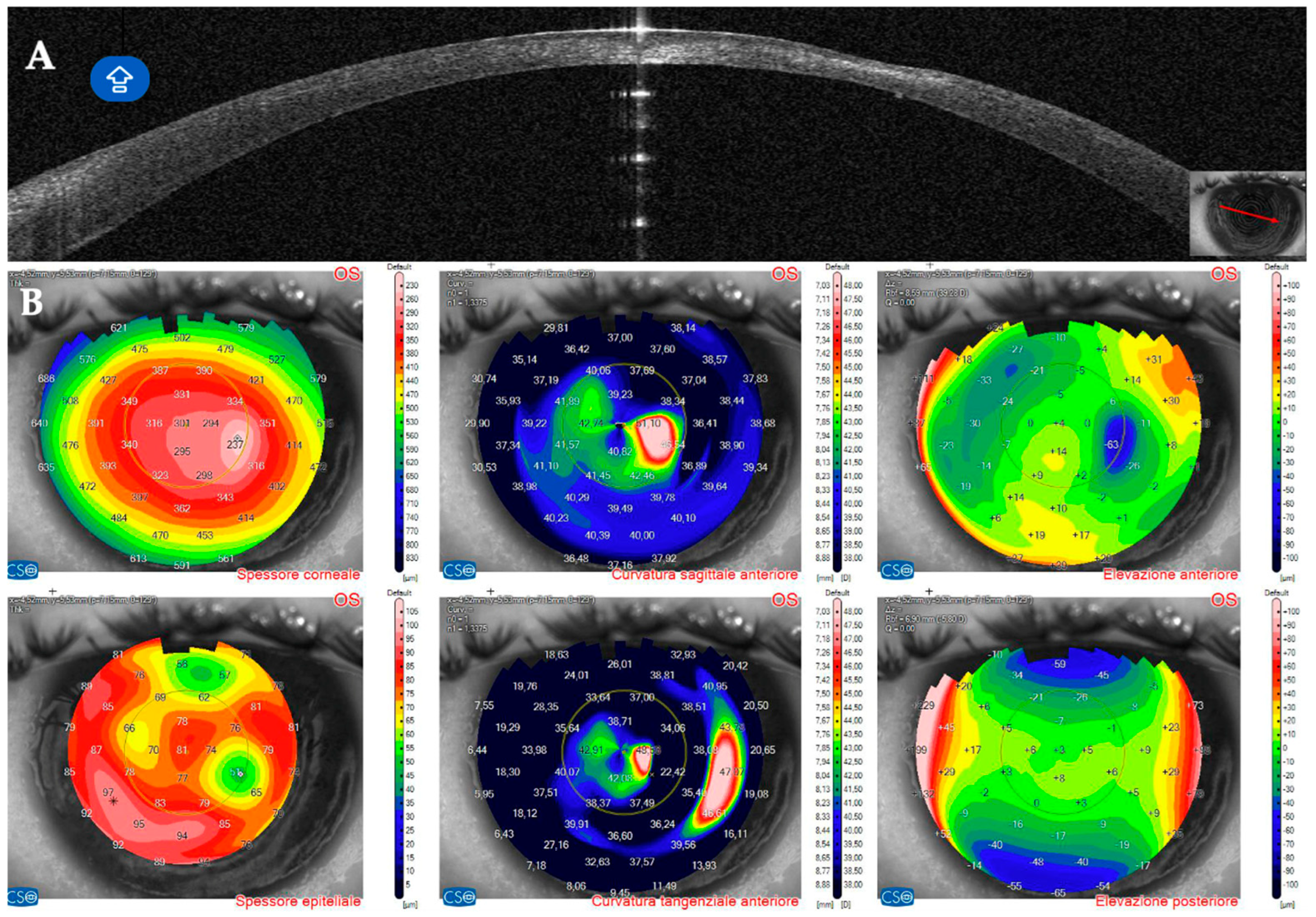
3.3. Patient #3
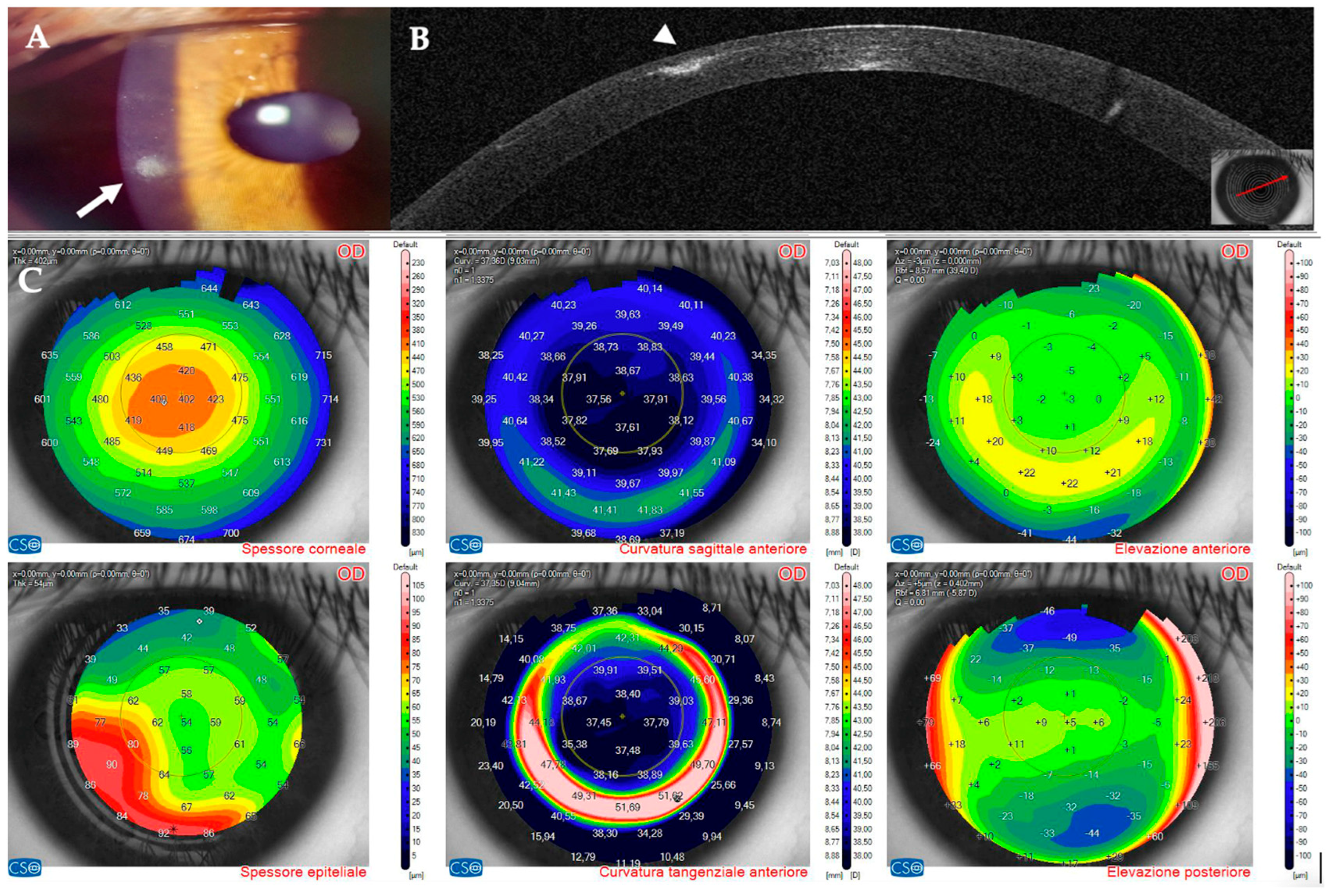
3.4. Patient #4
4. Discussion
5. Conclusions
Author Contributions
Funding
Institutional Review Board Statement
Informed Consent Statement
Data Availability Statement
Conflicts of Interest
References
- Das, S.; Garg, P.; Mullick, R.; Annavajjhala, S. Keratitis following laser refractive surgery: Clinical spectrum, prevention and management. Indian J. Ophthalmol. 2020, 68, 2813–2818. [Google Scholar] [CrossRef] [PubMed]
- Haq, Z.; Farooq, A.V.; Huang, A.J. Infections after refractive surgery. Curr. Opin. Ophthalmol. 2016, 27, 367–372. [Google Scholar] [CrossRef] [PubMed]
- Leccisotti, A.; Fields, S.V.; De Bartolo, G.; Crudale, C. Infectious keratitis after photorefractive keratectomy, femtosecond–LASIK and lenticule extraction in a 100,000–eye case series. Laser Ther. 2024, 31. [Google Scholar] [CrossRef]
- Cabrera–Aguas, M.; Khoo, P.; Watson, S.L. Infectious keratitis: A review. Clin. Exp. Ophthalmol. 2022, 50, 543–562. [Google Scholar] [CrossRef] [PubMed]
- Ganesh, S.; Brar, S.; Nagesh, B.N. Management of infectious keratitis following uneventful small–incision lenticule extraction using a multimodal approach—A case report. Indian J. Ophthalmol. 2020, 68, 3064–3066. [Google Scholar] [CrossRef] [PubMed]
- Schallhorn, J.M.; Schallhorn, S.C.; Hettinger, K.; Hannan, S. Infectious keratitis after laser vision correction: Incidence and risk factors. J. Cataract. Refract. Surg. 2017, 43, 473–479. [Google Scholar] [CrossRef] [PubMed]
- Chang, M.A.; Jain, S.; Azar, D.T. Infections following laser in situ keratomileusis: An integration of the published literature. Surv. Ophthalmol. 2004, 49, 269–280. [Google Scholar] [CrossRef]
- Cabrera–Aguas, M.; Watson, S.L. Updates in Diagnostic Imaging for Infectious Keratitis: A Review. Diagnostics 2023, 13, 3358. [Google Scholar] [CrossRef] [PubMed]
- Das, S.; Samantaray, R.; Mallick, A.; Sahu, S.K.; Sharma, S. Types of organisms and in–vitro susceptibility of bacterial isolates from patients with microbial keratitis: A trend analysis of 8 years. Indian J. Ophthalmol. 2019, 67, 49–53. [Google Scholar] [CrossRef]
- Ferreira, C.S.; Figueira, L.; Moreira–Goncalves, N.; Moreira, R.; Torrao, L.; Falcao–Reis, F. Clinical and Microbiological Profile of Bacterial Microbial Keratitis in a Portuguese Tertiary Referral Center–Where Are We in 2015? Eye Contact Lens 2018, 44, 15–20. [Google Scholar] [CrossRef] [PubMed]
- Cabrera–Aguas, M.; Khoo, P.; George, C.R.R.; Lahra, M.M.; Watson, S.L. Antimicrobial resistance trends in bacterial keratitis over 5 years in Sydney, Australia. Clin. Exp. Ophthalmol. 2020, 48, 183–191. [Google Scholar] [CrossRef] [PubMed]
- Green, M.; Carnt, N.; Apel, A.; Stapleton, F. Queensland Microbial Keratitis Database: 2005–2015. Br. J. Ophthalmol. 2019, 103, 1481–1486. [Google Scholar] [CrossRef] [PubMed]
- Ting, D.S.J.; Ho, C.S.; Cairns, J.; Elsahn, A.; Al–Aqaba, M.; Boswell, T.; Said, D.G.; Dua, H.S. 12–year analysis of incidence, microbiological profiles and in vitro antimicrobial susceptibility of infectious keratitis: The Nottingham Infectious Keratitis Study. Br. J. Ophthalmol. 2021, 105, 328–333. [Google Scholar] [CrossRef] [PubMed]
- Donnenfeld, E.D.; Kim, T.; Holland, E.J.; Azar, D.T.; Palmon, F.R.; Rubenstein, J.B.; Daya, S.; Yoo, S.H. ASCRS White Paper: Management of infectious keratitis following laser in situ keratomileusis. J. Cataract. Refract. Surg. 2005, 31, 2008–2011. [Google Scholar] [CrossRef] [PubMed]
- Yamaguchi, T.; Bissen–Miyajima, H.; Hori–Komai, Y.; Matsumoto, Y.; Ebihara, N.; Takahashi, H.; Tsubota, K.; Shimazaki, J. Infectious keratitis outbreak after laser in situ keratomileusis at a single laser center in Japan. J. Cataract. Refract. Surg. 2011, 37, 894–900. [Google Scholar] [CrossRef] [PubMed]
- Llovet, F.; de Rojas, V.; Interlandi, E.; Martín, C.; Cobo–Soriano, R.; Ortega–Usobiaga, J.; Baviera, J. Infectious keratitis in 204 586 LASIK procedures. Ophthalmology 2010, 117, 232–238.e4. [Google Scholar] [CrossRef]
- Solomon, R.; Donnenfeld, E.D.; Azar, D.T.; Holland, E.J.; Palmon, F.R.; Pflugfelder, S.C.; Rubenstein, J.B. Infectious keratitis after laser in situ keratomileusis: Results of an ASCRS survey. J. Cataract. Refract. Surg. 2003, 29, 2001–2006. [Google Scholar] [CrossRef] [PubMed]
- Johnson, J.D.; Harissi–Dagher, M.; Pineda, R.; Yoo, S.; Azar, D.T. Diffuse lamellar keratitis: Incidence, associations, outcomes, and a new classification system. J. Cataract. Refract. Surg. 2001, 27, 1560–1566. [Google Scholar] [CrossRef] [PubMed]
- Ting, D.S.J.; Gopal, B.P.; Deshmukh, R.; Seitzman, G.D.; Said, D.G.; Dua, H.S. Diagnostic armamentarium of infectious keratitis: A comprehensive review. Ocul. Surf. 2022, 23, 27–39. [Google Scholar] [CrossRef]
- Robaei, D.; Chan, U.T.; Khoo, P.; Cherepanoff, S.; Li, Y.C.; Hanrahan, J.; Watson, S. Corneal biopsy for diagnosis of recalcitrant microbial keratitis. Graefes Arch. Clin. Exp. Ophthalmol. 2018, 256, 1527–1533. [Google Scholar] [CrossRef]
- Singh, R.; Joseph, A.; Umapathy, T.; Tint, N.L.; Dua, H.S. Impression cytology of the ocular surface. Br. J. Ophthalmol. 2005, 89, 1655–1659. [Google Scholar] [CrossRef] [PubMed]
- Cruzat, A.; Qazi, Y.; Hamrah, P. In Vivo Confocal Microscopy of Corneal Nerves in Health and Disease. Ocul. Surf. 2017, 15, 15–47. [Google Scholar] [CrossRef]
- Lee, H.J.; Alipour, F.; Cruzat, A.; Posarelli, M.; Zheng, L.; Hamrah, P. Utility of In Vivo Confocal Microscopy in Diagnosis of Acanthamoeba Keratitis: A Comparison of Patient Outcomes. Cornea 2023, 42, 135–140. [Google Scholar] [CrossRef] [PubMed]
- Brasnu, E.; Bourcier, T.; Dupas, B.; Degorge, S.; Rodallec, T.; Laroche, L.; Borderie, V.; Baudouin, C. In vivo confocal microscopy in fungal keratitis. Br. J. Ophthalmol. 2007, 91, 588–591. [Google Scholar] [CrossRef]
- Kumar, R.L.; Cruzat, A.; Hamrah, P. Current state of in vivo confocal microscopy in management of microbial keratitis. Semin. Ophthalmol. 2010, 25, 166–170. [Google Scholar] [CrossRef] [PubMed]
- Abdelghany, A.A.; D’Oria, F.; Alio Del Barrio, J.; Alio, J.L. The Value of Anterior Segment Optical Coherence Tomography in Different Types of Corneal Infections: An Update. J. Clin. Med. 2021, 10, 2841. [Google Scholar] [CrossRef] [PubMed]
- Soliman, W.; Fathalla, A.M.; El–Sebaity, D.M.; Al–Hussaini, A.K. Spectral domain anterior segment optical coherence tomography in microbial keratitis. Graefes Arch. Clin. Exp. Ophthalmol. 2013, 251, 549–553. [Google Scholar] [CrossRef]
- Konstantopoulos, A.; Yadegarfar, G.; Fievez, M.; Anderson, D.F.; Hossain, P. In vivo quantification of bacterial keratitis with optical coherence tomography. Investig. Ophthalmol. Vis. Sci. 2011, 52, 1093–1097. [Google Scholar] [CrossRef]
- Konstantopoulos, A.; Kuo, J.; Anderson, D.; Hossain, P. Assessment of the use of anterior segment optical coherence tomography in microbial keratitis. Am. J. Ophthalmol. 2008, 146, 534–542. [Google Scholar] [CrossRef]
- Chong, Y.J.; Azzopardi, M.; Hussain, G.; Recchioni, A.; Gandhewar, J.; Loizou, C.; Giachos, I.; Barua, A.; Ting, D.S.J. Clinical Applications of Anterior Segment Optical Coherence Tomography: An Updated Review. Diagnostics 2024, 14, 122. [Google Scholar] [CrossRef] [PubMed]
- Kim, H.; Lim, M.C.; Mannis, M.J.; Kim, E.S. Epithelial downgrowth after femtosecond laser–assisted cataract surgery. Am. J. Ophthalmol. Case Rep. 2019, 15, 100507. [Google Scholar] [CrossRef] [PubMed]
- Ting, D.S.J.; Danjoux, J.P. Late–onset traumatic dislocation of laser in situ keratomileusis corneal flaps: A case series with many clinical lessons. Int. Ophthalmol. 2019, 39, 1397–1403. [Google Scholar] [CrossRef] [PubMed]
- Ramos, J.L.; Li, Y.; Huang, D. Clinical and research applications of anterior segment optical coherence tomography—A review. Clin. Exp. Ophthalmol. 2009, 37, 81–89. [Google Scholar] [CrossRef] [PubMed]
- Reinstein, D.Z.; Archer, T.J.; Vida, R.S. Applications of epithelial thickness mapping in corneal refractive surgery. Saudi J. Ophthalmol. 2022, 36, 25–35. [Google Scholar] [CrossRef] [PubMed]
- Chao, C.W.; Azar, D.T. Lamellar keratitis following laser–assisted in situ keratomileusis. Ophthalmol. Clin. N. Am. 2002, 15, 35–40. [Google Scholar] [CrossRef]
- Liu, H.Y.; Chu, H.S.; Chen, W.L.; Hu, F.R.; Wang, I.J. Bilateral Non–tuberculous Mycobacterial Keratitis After Small Incision Lenticule Extraction. J. Refract. Surg. 2018, 34, 633–636. [Google Scholar] [CrossRef]
- Ko, J.; Kim, S.K.; Yong, D.E.; Kim, T.I.; Kim, E.K. Delayed onset Mycobacterium intracellulare keratitis after laser in situ keratomileusis: A case report and literature review. Medicine 2017, 96, e9356. [Google Scholar] [CrossRef]
- Li, J.; Ren, S.W.; Dai, L.J.; Zhang, B.; Gu, Y.W.; Pang, C.J.; Wang, Y. Bacterial Keratitis Following Small Incision Lenticule Extraction. Infect. Drug Resist. 2022, 15, 4585–4593. [Google Scholar] [CrossRef] [PubMed]
- Moshirfar, M.; Welling, J.D.; Feiz, V.; Holz, H.; Clinch, T.E. Infectious and noninfectious keratitis after laser in situ keratomileusis Occurrence, management, and visual outcomes. J. Cataract. Refract. Surg. 2007, 33, 474–483. [Google Scholar] [CrossRef] [PubMed]
- Wang, H.; Zhu, L.S.; Pang, C.J.; Fan, Q. Repeatability assessment of anterior segment measurements in myopic patients using an anterior segment OCT with placido corneal topography and agreement with a swept–source OCT. BMC Ophthalmol. 2024, 24, 182. [Google Scholar] [CrossRef]
- Oliveira, M.A.; Rosa, A.; Soares, M.; Gil, J.; Costa, E.; Quadrado, M.J.; Murta, J. Anterior Segment Optical Coherence Tomography in the Early Management of Microbial Keratitis: A Cross–Sectional Study. Acta Med. Port. 2020, 33, 318–325. [Google Scholar] [CrossRef] [PubMed]
- Keino, H.; Aman, T.; Furuya, R.; Nakayama, M.; Okada, A.A.; Sunayama, W.; Hatanaka, Y. Automated Quantitative Analysis of Anterior Segment Inflammation Using Swept–Source Anterior Segment Optical Coherence Tomography: A Pilot Study. Diagnostics 2022, 12, 2703. [Google Scholar] [CrossRef] [PubMed]
- Maring, M.; Saraf, S.S.; Blazes, M.; Sharma, S.; Srivastava, S.; Pepple, K.L.; Lee, C.S. Grading Anterior Chamber Inflammation with Anterior Segment Optical Coherence Tomography: An Overview. Ocul. Immunol. Inflamm. 2022, 30, 357–363. [Google Scholar] [CrossRef] [PubMed]
- Patel, R.P.; Petrushkin, H.; Etherton, K.; Terence, K.; Dick, A.D.; Rahi, J.S.; Solebo, A.L. Quality assessment of anterior segment OCT images: Development and validation of quality criteria. Photodiagnosis Photodyn. Ther. 2024, 45, 103886. [Google Scholar] [CrossRef] [PubMed]
- Solomon, R.; Donnenfeld, E.D.; Holland, E.J.; Yoo, S.H.; Daya, S.; Guell, J.L.; Mah, F.S.; Scoper, S.V.; Kim, T. Microbial keratitis trends following refractive surgery: Results of the ASCRS infectious keratitis survey and comparisons with prior ASCRS surveys of infectious keratitis following keratorefractive procedures. J. Cataract. Refract. Surg. 2011, 37, 1343–1350. [Google Scholar] [CrossRef] [PubMed]
- Janiszewska–Bil, D.; Czarnota–Nowakowska, B.; Grabarek, B.O.; Dobrowolski, D.; Wylęgała, E.; Lyssek–Boroń, A. Comparison of Vision Correction and Corneal Thickness at 180–Day Follow–Up After Femtosecond Laser–Assisted In–Situ Keratomileusis (FS–LASIK), Photorefractive Keratectomy (PRK), and Small Incision Lenticule Extraction (SMILE): A Study from a Single Center in Poland of 120 Patients with Myopia. Med. Sci. Monit. 2023, 29, e939099. [Google Scholar] [CrossRef]
- Natarajan, R.; Balaji, J.J.; Pandey, S. Double standards in corneal epithelial compensation. Indian J. Ophthalmol.—Case Rep. 2022, 2, 604. [Google Scholar] [CrossRef]
- Randag, A.C.; van Rooij, J.; van Goor, A.T.; Verkerk, S.; Wisse, R.P.L.; Saelens, I.E.Y.; Stoutenbeek, R.; van Dooren, B.T.H.; Cheng, Y.Y.Y.; Eggink, C.A. The rising incidence of Acanthamoeba keratitis: A 7–year nationwide survey and clinical assessment of risk factors and functional outcomes. PLoS ONE 2019, 14, e0222092. [Google Scholar] [CrossRef]
- Posarelli, M.; Hamrah, P. Efficacy of Topical 2% Cyclosporine in Controlling the Inflammation and Improving the Treatment Outcomes in Patients with Acanthamoeba Keratitis. Investig. Ophthalmol. Vis. Sci. 2023, 64, 601. [Google Scholar]
- Yamazaki, N.; Kobayashi, A.; Yokogawa, H.; Ishibashi, Y.; Oikawa, Y.; Tokoro, M.; Sugiyama, K. In vivo imaging of radial keratoneuritis in patients with Acanthamoeba keratitis by anterior–segment optical coherence tomography. Ophthalmology 2014, 121, 2153–2158. [Google Scholar] [CrossRef] [PubMed]
- Leccisotti, A.; Fields, S.V. Diffuse lamellar keratitis after LASIK with low–energy femtosecond laser. J. Cataract. Refract. Surg. 2021, 47, 233–237. [Google Scholar] [CrossRef]
- Stuart, A.; Reinstein, D.Z.; Vida, R.S.; Archer, T.J.; Carp, G. Atypical presentation of diffuse lamellar keratitis after small–incision lenticule extraction: Sterile multifocal inflammatory keratitis. J. Cataract. Refract. Surg. 2018, 44, 774–779. [Google Scholar] [CrossRef] [PubMed]
- Santhiago, M.R.; Wilson, S.E. Cellular effects after laser in situ keratomileusis flap formation with femtosecond lasers: A review. Cornea 2012, 31, 198–205. [Google Scholar] [CrossRef]
- Grassmeyer, J.J.; Goertz, J.G.; Baartman, B.J. Diffuse Lamellar Keratitis in a Patient Undergoing Collagen Corneal Cross–Linking 18 Years After Laser In Situ Keratomileusis Surgery. Cornea 2021, 40, 917–920. [Google Scholar] [CrossRef] [PubMed]


| Patient | Baseline | Follow–Up 1 | Follow–Up 2 | Follow–Up 3 |
|---|---|---|---|---|
| Patient #1 thickness (days) | 433 microns | 567 microns (5) | 398 microns (12) | 407 microns (30) |
| Patient #4 thickness (days) | 433 microns | 424 microns (21) | 417 microns (20) | |
| Patient #3 thickness (days) | 233 microns | 346 microns (56) | ||
| Patient #4 thickness (days) | 497 microns | 597 microns (3) | 443 microns (10) |
Disclaimer/Publisher’s Note: The statements, opinions and data contained in all publications are solely those of the individual author(s) and contributor(s) and not of MDPI and/or the editor(s). MDPI and/or the editor(s) disclaim responsibility for any injury to people or property resulting from any ideas, methods, instructions or products referred to in the content. |
© 2025 by the authors. Licensee MDPI, Basel, Switzerland. This article is an open access article distributed under the terms and conditions of the Creative Commons Attribution (CC BY) license (https://creativecommons.org/licenses/by/4.0/).
Share and Cite
Leccisotti, A.; Fields, S.V.; De Bartolo, G.; Crudale, C.; Posarelli, M. Optical Coherence Tomography in Infectious Keratitis After Femtosecond Keratorefractive Surgery. J. Clin. Med. 2025, 14, 1067. https://doi.org/10.3390/jcm14041067
Leccisotti A, Fields SV, De Bartolo G, Crudale C, Posarelli M. Optical Coherence Tomography in Infectious Keratitis After Femtosecond Keratorefractive Surgery. Journal of Clinical Medicine. 2025; 14(4):1067. https://doi.org/10.3390/jcm14041067
Chicago/Turabian StyleLeccisotti, Antonio, Stefania V. Fields, Giuseppe De Bartolo, Christian Crudale, and Matteo Posarelli. 2025. "Optical Coherence Tomography in Infectious Keratitis After Femtosecond Keratorefractive Surgery" Journal of Clinical Medicine 14, no. 4: 1067. https://doi.org/10.3390/jcm14041067
APA StyleLeccisotti, A., Fields, S. V., De Bartolo, G., Crudale, C., & Posarelli, M. (2025). Optical Coherence Tomography in Infectious Keratitis After Femtosecond Keratorefractive Surgery. Journal of Clinical Medicine, 14(4), 1067. https://doi.org/10.3390/jcm14041067