INTegrated Assessment of intERmediate Coronary Stenoses by Fractional Flow rEserve and Near-infraREd Spectroscopy: The INTERFERE Study
Abstract
1. Introduction
2. Material and Methods
Statistical Analysis
3. Results
4. Discussion
Limitations
5. Conclusions
Author Contributions
Funding
Institutional Review Board Statement
Informed Consent Statement
Data Availability Statement
Conflicts of Interest
References
- De Bruyne, B.; Pijls, N.H.J.; Kalesan, B.; Barbato, E.; Tonino, P.A.L.; Piroth, Z.; Jagic, N.; Mobius-Winkler, S.; Rioufol, G.; Witt, N.; et al. Fractional flow reserve-guided PCI versus medical therapy in stable coronary disease. N. Engl. J. Med. 2012, 367, 991–1001. [Google Scholar] [CrossRef]
- Lee, J.M.; Kim, H.K.; Park, K.H.; Choo, E.H.; Kim, C.J.; Lee, S.H.; Kim, M.C.; Hong, Y.J.; Ahn, S.G.; Doh, J.-H.; et al. FRAME-AMI Investigators. Fractional flow reserve versus angiography-guided strategy in acute myocardial infarction with multivessel disease: A randomized trial. Eur. Heart J. 2023, 44, 473–484. [Google Scholar] [CrossRef] [PubMed]
- Biscaglia, S.; Guiducci, V.; Escaned, J.; Moreno, R.; Lanzilotti, V.; Santarelli, A.; Cerrato, E.; Sacchetta, G.; Jurado-Roman, A.; Menozzi, A.; et al. FIRE Trial Investigators. Complete or Culprit-Only PCI in Older Patients with Myocardial Infarction. N. Engl. J. Med. 2023, 389, 889–898. [Google Scholar] [CrossRef]
- Zimmermann, F.M.; Omerovic, E.; Fournier, S.; Kelbæk, H.; Johnson, N.P.; Rothenbühler, M.; Xaplanteris, P.; Abdel-Wahab, M.; Barbato, E.; Høfsten, D.E.; et al. Fractional flow reserve-guided percutaneous coronary intervention vs. medical therapy for patients with stable coronary lesions: Meta-analysis of individual patient data. Eur. Heart J. 2019, 40, 180–186. [Google Scholar] [CrossRef]
- Prati, F.; Romagnoli, E.; Gatto, L.; La Manna, A.; Burzotta, F.; Ozaki, Y.; Marco, V.; Boi, A.; Fineschi, M.; Fabbiocchi, F.; et al. Relationship Relationship between coronary plaque morphology of the left anterior descending artery and 12 months clinical outcome: The CLIMA study. Eur. Heart J. 2020, 41, 383–391. [Google Scholar] [CrossRef]
- Stone, G.W.; Maehara, A.; Lansky, A.J.; de Bruyne, B.; Cristea, E.; Mintz, G.S.; Mehran, R.; McPherson, J.; Farhat, N.; Marso, S.P.; et al. A prospective natural-history study of coronary atherosclerosis. N. Engl. J. Med. 2011, 364, 226–235. [Google Scholar] [CrossRef] [PubMed]
- Kedhi, E.; Berta, B.; Roleder, T.; Hermanides, R.S.; Fabris, E.; IJsselmuiden, A.J.J.; Kauer, F.; Alfonso, F.; von Birgelen, C.; Escaned, J.; et al. Thin-cap fibroatheroma predicts clinical events in diabetic patients with normal fractional flow reserve: The COMBINE OCT-FFR trial. Eur. Heart J. 2021, 42, 4671–4679. [Google Scholar] [CrossRef] [PubMed]
- Mol, J.Q.; Volleberg, R.H.J.A.; Belkacemi, A.; Hermanides, R.S.; Meuwissen, M.; Protopopov, A.V.; Laanmets, P.; Krestyaninov, O.V.; Dennert, R.; Oemrawsingh, R.M.; et al. Fractional Flow Reserve-Negative High-Risk Plaques and Clinical Outcomes After Myocardial Infarction. JAMA Cardiol. 2023, 8, 1013–1021. [Google Scholar] [CrossRef]
- Waxman, S.; Dixon, S.R.; L’Allier, P.; Moses, J.W.; Petersen, J.L.; Cutlip, D.; Tardif, J.C.; Nesto, R.W.; Muller, J.E. In vivo validation of a catheter-based near-infrared spectroscopy system for detection of lipid core coronary plaques: Initial results of the SPECTACL study. JACC Cardiovasc. Imaging 2009, 2, 858–868. [Google Scholar] [CrossRef]
- Erlinge, D.; Maehara, A.; Ben-Yehuda, O.; Bøtker, H.E.; Maeng, M.; Kjøller-Hansen, L.; Engstrøm, T.; Matsumura, M.; Crowley, A.; Dressler, O.; et al. Identification of vulnerable plaques and patients by intracoronary near-infrared spectroscopy and ultrasound (PROSPECT II): A prospective natural history study. Lancet 2021, 397, 985–995. [Google Scholar] [CrossRef]
- Waksman, R.; Di Mario, C.; Torguson, R.; Ali, Z.A.; Singh, V.; Skinner, W.H.; Artis, A.K.; Cate, T.T.; Powers, E.; Kim, C.; et al. Identification of patients and plaques vulnerable to future coronary events with near-infrared spectroscopy intravascular ultrasound imaging: A prospective, cohort study. Lancet 2019, 394, 1629–1637. [Google Scholar] [CrossRef] [PubMed]
- Park, S.J.; Ahn, J.M.; Kang, D.Y.; Yun, S.C.; Ahn, Y.K.; Kim, W.J.; Nam, C.W.; Jeong, J.O.; Chae, I.H.; Shiomi, H.; et al. Preventive percutaneous coronary intervention versus optimal medical therapy alone for the treatment of vulnerable atherosclerotic coronary plaques (PREVENT): A multicentre, open-label, randomised controlled trial. Lancet, 2024; epub ahead of print. [Google Scholar] [CrossRef] [PubMed]
- Task Force Members; Kolh, P.; Windecker, S.; Alfonso, F.; Collet, J.P.; Cremer, J.; Falk, V.; Filippatos, G.; Hamm, C.; Head, S.J.; et al. 2014 ESC/EACTS Guidelines on myocardial revascularization: The Task Force on Myocardial Revascularization of the European Society of Cardiology (ESC) and the European Association for Cardio-Thoracic Surgery (EACTS)Developed with the special contribution of the European Association of Percutaneous Cardiovascular Interventions (EAPCI). Eur. Heart J. 2014, 35, 2541–2619. [Google Scholar] [CrossRef] [PubMed]
- Gardner, C.M.; Tan, H.; Hull, E.L.; Lisauskas, J.B.; Sum, S.T.; Meese, T.M.; Jiang, C.; Madden, S.P.; Caplan, J.D.; Burke, A.P.; et al. Detection of lipid core coronary plaques in autopsy specimens with a novel catheter-based near-infrared spectroscopy system. JACC Cardiovasc. Imaging 2008, 1, 638–648. [Google Scholar] [CrossRef] [PubMed]
- Räber, L.; Ueki, Y.; Otsuka, T.; Losdat, S.; Häner, J.D.; Lonborg, J.; Fahrni, G.; Iglesias, J.F.; van Geuns, R.J.; Ondracek, A.S.; et al. Effect of Alirocumab Added to High-Intensity Statin Therapy on Coronary Atherosclerosis in Patients with Acute Myocardial Infarction: The PACMAN-AMI Randomized Clinical Trial. JAMA 2022, 327, 1771–1781. [Google Scholar] [CrossRef] [PubMed] [PubMed Central]
- Nicholls, S.J.; Kataoka, Y.; Nissen, S.E.; Prati, F.; Windecker, S.; Puri, R.; Hucko, T.; Aradi, D.; Herrman, J.R.; Hermanides, R.S.; et al. Effect of Evolocumab on Coronary Plaque Phenotype and Burden in Statin-Treated Patients Following Myocardial Infarction. JACC Cardiovasc. Imaging 2022, 15, 1308–1321. [Google Scholar] [CrossRef] [PubMed]
- Kannel, W.B.; Higgins, M. Smoking and hypertension as predictors of cardiovascular risk in population studies. J. Hypertens. Suppl. 1990, 8, S3–S8. [Google Scholar] [PubMed]
- Lehmann, N.; Erbel, R.; Mahabadi, A.A.; Kälsch, H.; Möhlenkamp, S.; Moebus, S.; Stang, A.; Roggenbuck, U.; Strucksberg, K.H.; Führer-Sakel, D.; et al. Accelerated progression of coronary artery calcification in hypertension but also prehypertension. J. Hypertens. 2016, 34, 2233–2242. [Google Scholar] [CrossRef] [PubMed]
- Hou, Z.H.; Lu, B.; Li, Z.N.; An, Y.Q.; Gao, Y.; Yin, W.H. Coronary Atherosclerotic Plaque Volume Quantified by Computed Tomographic Angiography in Smokers Compared to Nonsmokers. Acad. Radiol. 2019, 26, 1581–1588. [Google Scholar] [CrossRef] [PubMed]
- Kumagai, S.; Amano, T.; Takashima, H.; Waseda, K.; Kurita, A.; Ando, H.; Maeda, K.; Ito, Y.; Ishii, H.; Hayashi, M.; et al. Impact of cigarette smoking on coronary plaque composition. Coron. Artery Dis. 2015, 26, 60–65. [Google Scholar] [CrossRef] [PubMed]
- Dell’Aversana, S.; Ascione, R.; Vitale, R.A.; Cavaliere, F.; Porcaro, P.; Basile, L.; Napolitano, G.; Boccalatte, M.; Sibilio, G.; Esposito, G.; et al. CT Coronary Angiography: Technical Approach and Atherosclerotic Plaque Characterization. J. Clin. Med. 2023, 12, 7615. [Google Scholar] [CrossRef] [PubMed] [PubMed Central]
- Oikonomou, E.K.; Marwan, M.; Desai, M.Y.; Mancio, J.; Alashi, A.; Hutt Centeno, E.; Thomas, S.; Herdman, L.; Kotanidis, C.P.; Thomas, K.E.; et al. Non-invasive detection of coronary inflammation using computed tomography and prediction of residual cardiovascular risk (the CRISP CT study): A post-hoc analysis of prospective outcome data. Lancet 2018, 392, 929–939. [Google Scholar] [CrossRef] [PubMed] [PubMed Central]
- Nørgaard, B.L.; Leipsic, J.; Gaur, S.; Seneviratne, S.; Ko, B.S.; Ito, H.; Jensen, J.M.; Mauri, L.; De Bruyne, B.; Bezerra, H.; et al. Diagnostic performance of noninvasive fractional flow reserve derived from coronary computed tomography angiography in suspected coronary artery disease: The NXT trial (Analysis of Coronary Blood Flow Using CT Angiography: Next Steps). J. Am. Coll. Cardiol. 2014, 63, 1145–1155. [Google Scholar] [CrossRef] [PubMed]


| Total | FFR < 0.80 | FFR > 0.80 | p-Value | |
|---|---|---|---|---|
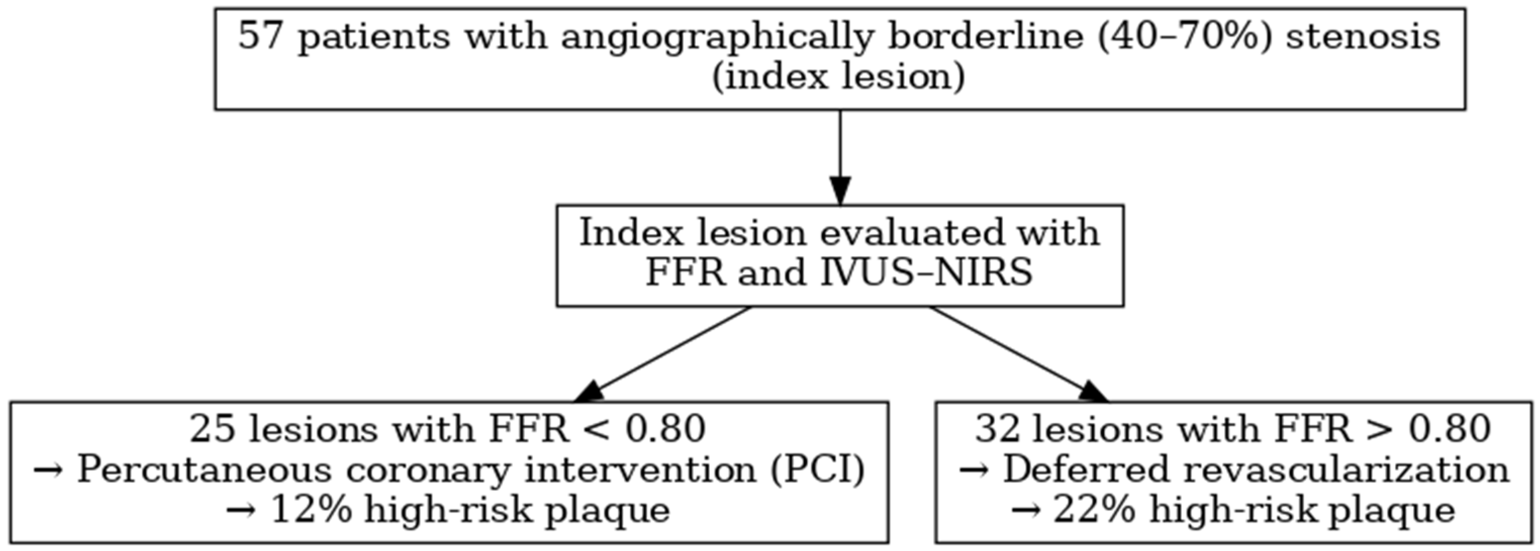
| N = 57 | N = 25 | N = 32 | ||
| Age | 65.7 ± 10 | 67.1 ± 11 | 64.7 ± 10 | 0.51 |
| Male_gender N (%) | 47 (82) | 22 (88) | 25 (78) | 0.33 |
| Diabetes | 16 (28%) | 8 (32%) | 8 (25%) | 0.57 |
| Previous ACS | 14 (25%) | 9 (36%) | 5 (16%) | 0.12 |
| Previous PCI | 19 (33%) | 11 (44%) | 8 (25%) | 0.16 |
| Previous stroke | 3 (5%) | 0 (0%) | 3 (9%) | 0.25 |
| PAD | 6 (11%) | 4 (16%) | 2 (6%) | 0.39 |
| Presentation | ||||
| STEMI | 5 (9%) | 1 (4%) | 4 (12%) | 0.37 |
| NSTEMI | 21 (37%) | 10 (40%) | 11 (34%) | 0.78 |
| Unstable angina | 15 (26%) | 7 (28%) | 8 (25%) | 0.99 |
| Stable CAD | 16 (28%) | 7 (28%) | 9 (28%) | 0.99 |
| BMI (kg/m2) | 25.5 ± 6.3 | 24.9 ± 6.7 | 26.1 ± 5.9 | 0.48 |
| Hypertension | 41 (72%) | 23 (92%) | 18 (56%) | 0.003 |
| Hypercholesterolemia | 34 (60%) | 18 (72%) | 16 (50%) | 0.11 |
| Family history of CAD | 12 (21%) | 4 (16%) | 8 (25%) | 0.52 |
| Active smoker | 12 (21%) | 9 (36%) | 3 (9%) | 0.02 |
| Past smoker | 20 (35%) | 8 (32%) | 12 (37%) | 0.78 |
| Diabetes | 16 (28%) | 8 (32%) | 8 (25%) | 0.57 |
| ASA | 55 (96%) | 25 (100%) | 30 (94%) | 0.50 |
| Clopidogrel | 18 (32%) | 11 (44%) | 7 (22%) | 0.09 |
| Prasugrel | 1 (2%) | 0 (0%) | 1 (3%) | 0.99 |
| Ticagrelor | 27 (47%) | 12 (48%) | 15 (47%) | 0.99 |
| Betablocker | 42 (74%) | 18 (72%) | 24 (75%) | 0.99 |
| ACEI or ARB | 52 (91%) | 23 (92%) | 29 (91%) | 0.99 |
| Statin | 54 (95%) | 23 (92%) | 31 (97%) | 0.58 |
| Total cholesterol (mg/dL) | 170.8 ± 49.7 | 173.1 ± 50.1 | 169.8 ± 48.5 | 0.83 |
| LDL cholesterol(mg/dL) | 98.8 ± 46.7 | 97.3 ± 48.2 | 100 ± 46.6 | 0.86 |
| HDL cholesterol (mg/dL) | 45.1 ± 11.3 | 44.6 ± 10.2 | 45.4 ± 12.3 | 0.82 |
| Triglyceride (mg/dL) | 136.3 ± 85.4 | 158.5 ± 110.9 | 119.2 ± 55.8 | 0.16 |
| Creatinine (mg/dL) | 1.09 ± 0.50 | 1.04 ± 0.4 | 1.13 ± 0.57 | 0.55 |
| GFR (mL/min) | 79.9 ± 29.3 | 80.1 ± 33.3 | 79.0 ± 26.0 | 0.83 |
| Number of diseased vessels | ||||
| 1 | 24 (42%) | 6 (24%) | 18 (56%) | 0.02 |
| 2 | 21 (37%) | 11 (44%) | 10 (31%) | 0.41 |
| 3 | 12 (21%) | 8 (32%) | 4 (12%) | 0.10 |
| SYNTAX score | 12.0 ± 5.4 | 13.8 ± 5.6 | 10.6 ± 4.8 | 0.03 |
| Index lesion LAD | 39 (68%) | 20 (80%) | 19 (59%) | 0.15 |
| Index lesion Cx | 7 (12%) | 5 (20%) | 2 (6%) | 0.22 |
| Index lesion RCA | 11 (19%) | 1 (4%) | 10 (31%) | 0.02 |
| Index lesion MLA (mm2) | 3.6 ± 1.2 | 3.6 ± 1.1 | 3.6 ± 1.2 | 0.93 |
| Plaque burden at MLA (%) | 73.3 ± 7.9 | 74.2 ± 7.2 | 72.6 ± 8.4 | 0.44 |
| LCBImax 4mm around index lesion | 204 ± 160 | 195.2 ± 140.9 | 210.9 ± 175.3 | 0.72 |
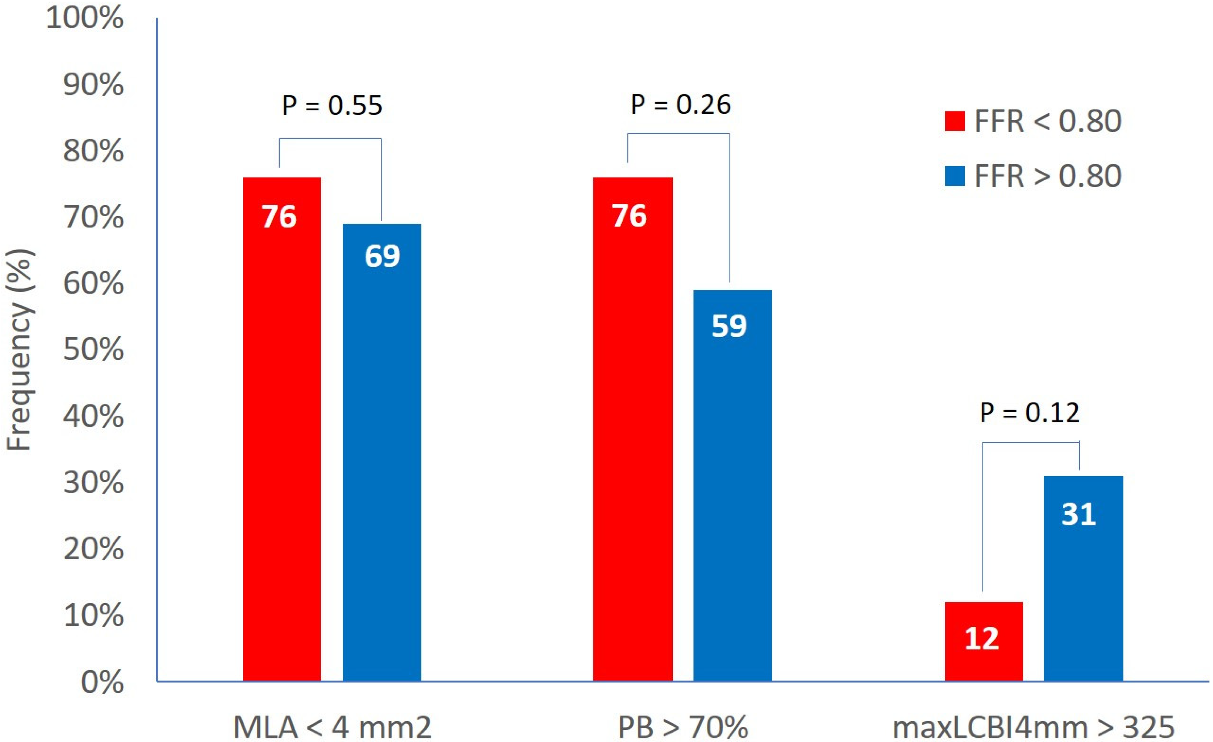
| MLA < 4 mm2 | 41 (72%) | 19 (76%) | 22(69%) | 0.55 |
| Plaque burden at MLA > 70% | 38 (67%) | 19 (76%) | 19 (59%) | 0.26 |
| LCBImax 4 mm around index lesion > 325 | 13 (23%) | 3 (12%) | 10 (31%) | 0.12 |
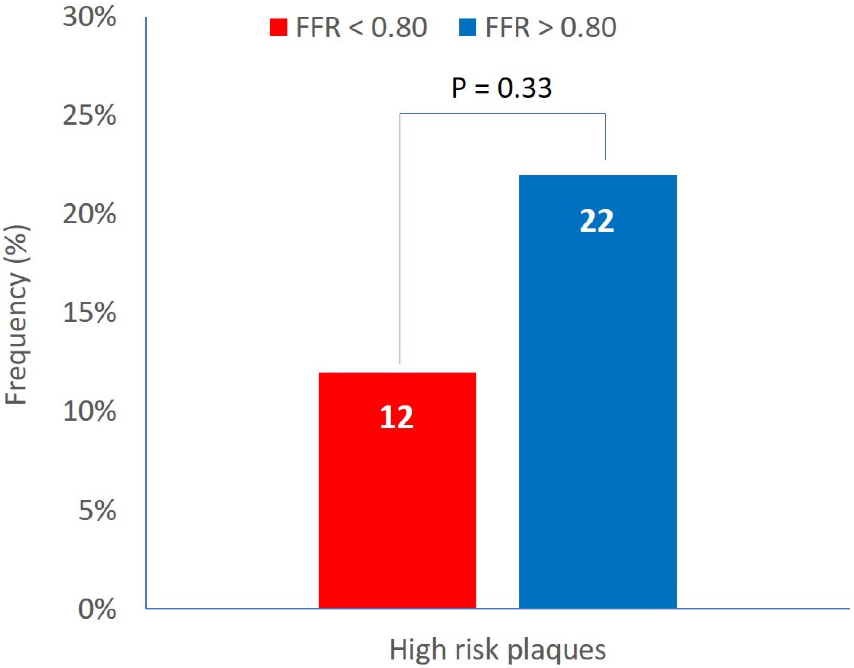
| High-risk plaques (MLA < 4 mm2 + PB > 70% + LCBImax 4 mm around index lesion > 325) | 10 (18%) | 3 (12%) | 7 (22%) | 0.33 |
| General Population | Patients with High-Risk Lesions | Patients Without High-Risk Lesions | p-Value | |
|---|---|---|---|---|
| N = 57 | N = 10 | N = 47 | ||
| All-cause death | 10 (18%) | 2 (20%) | 8 (17%) | 0.82 |
| Myocardial infarction | 1 (2%) | 0 (0%) | 1 (2%) | 0.64 |
| Unplanned revascularization | 2 (4%) | 0 (0%) | 2 (4%) | 0.51 |
| Composite endpoint: all-cause death, myocardial infarction, unplanned revascularization | 12 (21%) | 2 (20%) | 10 (21%) | 0.93 |
| General Population | Patients with High-Risk Lesions | Patients Without High-Risk Lesions | p-Value | |
| N = 32 | N = 7 | N = 25 | ||
| All-cause death | 6 (19%) | 2 (29%) | 4 (16%) | 0.45 |
| Myocardial infarction | 0 (0%) | 0 (0%) | 0 (0%) | |
| Unplanned revascularization | 1 (3%) | 0 (0%) | 1 (4%) | 0.59 |
| Composite endpoint: all-cause death, myocardial infarction, unplanned revascularization | 7 (22%) | 2 (29%) | 5 (20%) | 0.63 |
| Predictor | OR (95% CI) | p-Value |
|---|---|---|
| Age | 1.05 (0.98–1.12) | 0.135 |
| Hypercholesterolemia | 0.38 (0.10–1.47) | 0.161 |
| Creatinine (mg/dL) | 1.83 (0.41–8.16) | 0.425 |
| Male gender | 1.76 (0.36–8.63) | 0.487 |
| Active smoker | 0.69 (0.16–2.96) | 0.617 |
| Hypertension | 1.41 (0.34–5.84) | 0.638 |
| Diabetes | 0.72 (0.17–3.06) | 0.657 |
| High-risk lesions | 0.89 (0.18–4.42) | 0.889 |
Disclaimer/Publisher’s Note: The statements, opinions and data contained in all publications are solely those of the individual author(s) and contributor(s) and not of MDPI and/or the editor(s). MDPI and/or the editor(s) disclaim responsibility for any injury to people or property resulting from any ideas, methods, instructions or products referred to in the content. |
© 2025 by the authors. Licensee MDPI, Basel, Switzerland. This article is an open access article distributed under the terms and conditions of the Creative Commons Attribution (CC BY) license (https://creativecommons.org/licenses/by/4.0/).
Share and Cite
Picchi, A.; Campo, G.; Misuraca, L.; Baratta, P.; Biancofiore, A.; Calabria, P.; Massoni, A.; Limbruno, U. INTegrated Assessment of intERmediate Coronary Stenoses by Fractional Flow rEserve and Near-infraREd Spectroscopy: The INTERFERE Study. J. Clin. Med. 2025, 14, 6769. https://doi.org/10.3390/jcm14196769
Picchi A, Campo G, Misuraca L, Baratta P, Biancofiore A, Calabria P, Massoni A, Limbruno U. INTegrated Assessment of intERmediate Coronary Stenoses by Fractional Flow rEserve and Near-infraREd Spectroscopy: The INTERFERE Study. Journal of Clinical Medicine. 2025; 14(19):6769. https://doi.org/10.3390/jcm14196769
Chicago/Turabian StylePicchi, Andrea, Gianluca Campo, Leonardo Misuraca, Pasquale Baratta, Antonio Biancofiore, Paolo Calabria, Alberto Massoni, and Ugo Limbruno. 2025. "INTegrated Assessment of intERmediate Coronary Stenoses by Fractional Flow rEserve and Near-infraREd Spectroscopy: The INTERFERE Study" Journal of Clinical Medicine 14, no. 19: 6769. https://doi.org/10.3390/jcm14196769
APA StylePicchi, A., Campo, G., Misuraca, L., Baratta, P., Biancofiore, A., Calabria, P., Massoni, A., & Limbruno, U. (2025). INTegrated Assessment of intERmediate Coronary Stenoses by Fractional Flow rEserve and Near-infraREd Spectroscopy: The INTERFERE Study. Journal of Clinical Medicine, 14(19), 6769. https://doi.org/10.3390/jcm14196769


