
Impact of Tafamidis on Delaying Clinical, Functional, and Structural Cardiac Changes in Patients with Wild-Type Transthyretin Amyloid Cardiomyopathy
 , ,
, ,  , ,
, ,  , and
, and 
Abstract
1. Introduction
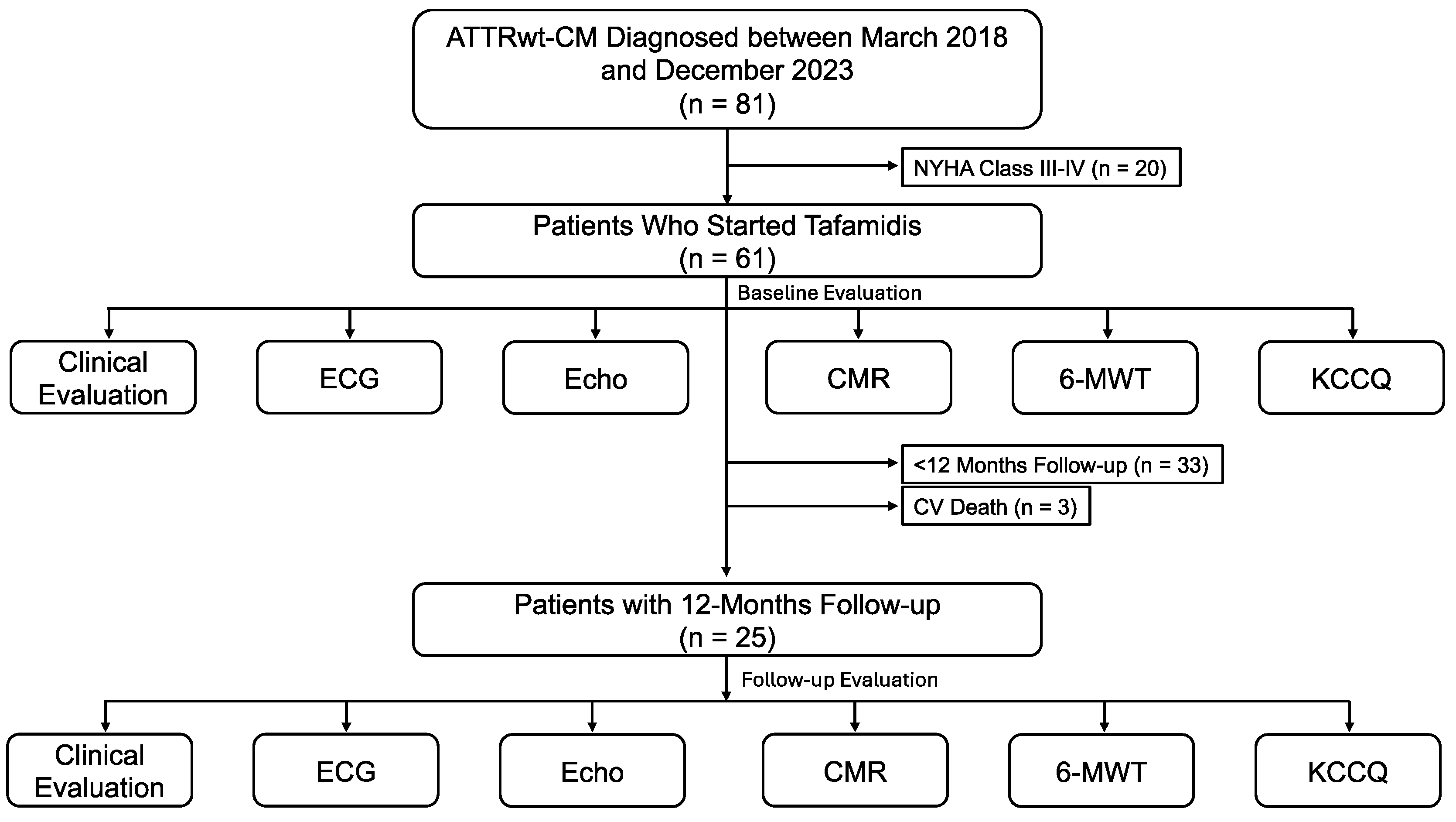
2. Methods
2.1. Study Population
2.2. Eligibility Criteria
2.3. Clinical Investigation and Data Collection
2.4. Echocardiography
2.5. Cardiac Magnetic Resonance
2.6. Laboratory Assessment
2.7. Quality of Life and Functional Capacity
2.8. Assessment of Disease Progression
2.9. Statistical Analysis
3. Results
3.1. Study Population, Functional Status, and Biochemical Parameters
3.2. Echocardiographic Parameters
3.3. CMR Parameters
3.4. Assessment of Disease Progression during 12 Months of Follow-up
4. Discussion
Study Limitations
5. Conclusions
Author Contributions
Funding
Institutional Review Board Statement
Informed Consent Statement
Data Availability Statement
Conflicts of Interest
References
- Ruberg, F.L.; Grogan, M.; Hanna, M.; Kelly, J.W.; Maurer, M.S. Transthyretin Amyloid Cardiomyopathy. J. Am. Coll. Cardiol. 2019, 73, 2872–2891. [Google Scholar] [CrossRef] [PubMed]
- Lioncino, M.; Monda, E.; Palmiero, G.; Caiazza, M.; Vetrano, E.; Rubino, M.; Esposito, A.; Salerno, G.; Dongiglio, F.; D’Onofrio, B.; et al. Cardiovascular Involvement in Transthyretin Cardiac Amyloidosis. Heart Fail. Clin. 2022, 18, 73–87. [Google Scholar] [CrossRef] [PubMed]
- Witteles, R.M.; Bokhari, S.; Damy, T.; Elliott, P.M.; Falk, R.H.; Fine, N.M.; Gospodinova, M.; Obici, L.; Rapezzi, C.; Garcia-Pavia, P. Screening for Transthyretin Amyloid Cardiomyopathy in Everyday Practice. JACC Heart Fail. 2019, 7, 709–716. [Google Scholar] [CrossRef]
- Lane, T.; Fontana, M.; Martinez-Naharro, A.; Quarta, C.C.; Whelan, C.J.; Petrie, A.; Rowczenio, D.M.; Gilbertson, J.A.; Hutt, D.F.; Rezk, T.; et al. Natural History, Quality of Life, and Outcome in Cardiac Transthyretin Amyloidosis. Circulation 2019, 140, 16–26. [Google Scholar] [CrossRef] [PubMed]
- Monda, E.; Bakalakos, A.; Rubino, M.; Verrillo, F.; Diana, G.; De Michele, G.; Altobelli, I.; Lioncino, M.; Perna, A.; Falco, L.; et al. Targeted Therapies in Pediatric and Adult Patients with Hypertrophic Heart Disease: From Molecular Pathophysiology to Personalized Medicine. Circ. Heart Fail. 2023, 16, E010687. [Google Scholar] [CrossRef]
- González-López, E.; Gallego-Delgado, M.; Guzzo-Merello, G.; De Haro-del Moral, F.J.; Cobo-Marcos, M.; Robles, C.; Bornstein, B.; Salas, C.; Lara-Pezzi, E.; Alonso-Pulpon, L.; et al. Wild-Type Transthyretin Amyloidosis as a Cause of Heart Failure with Preserved Ejection Fraction. Eur. Heart J. 2015, 36, 2585–2594. [Google Scholar] [CrossRef] [PubMed]
- Bakalakos, A.; Monda, E.; Elliott, P.M. The Diagnostic and Therapeutic Implications of Phenocopies and Mimics of Hypertrophic Cardiomyopathy. Can. J. Cardiol. 2024, 40, 754–765. [Google Scholar] [CrossRef] [PubMed]
- Maurer, M.S.; Schwartz, J.H.; Gundapaneni, B.; Elliott, P.M.; Merlini, G.; Waddington-Cruz, M.; Kristen, A.V.; Grogan, M.; Witteles, R.; Damy, T.; et al. Tafamidis Treatment for Patients with Transthyretin Amyloid Cardiomyopathy. N. Engl. J. Med. 2018, 379, 1007–1016. [Google Scholar] [CrossRef] [PubMed]
- Shah, S.J.; Fine, N.; Garcia-Pavia, P.; Klein, A.L.; Fernandes, F.; Weissman, N.J.; Maurer, M.S.; Boman, K.; Gundapaneni, B.; Sultan, M.B.; et al. Effect of Tafamidis on Cardiac Function in Patients with Transthyretin Amyloid Cardiomyopathy: A Post Hoc Analysis of the ATTR-ACT Randomized Clinical Trial. JAMA Cardiol. 2024, 9, 25. [Google Scholar] [CrossRef]
- Rettl, R.; Mann, C.; Duca, F.; Dachs, T.-M.; Binder, C.; Ligios, L.C.; Schrutka, L.; Dalos, D.; Koschutnik, M.; Donà, C.; et al. Tafamidis Treatment Delays Structural and Functional Changes of the Left Ventricle in Patients with Transthyretin Amyloid Cardiomyopathy. Eur. Heart J.-Cardiovasc. Imaging 2022, 23, 767–780. [Google Scholar] [CrossRef]
- Rettl, R.; Duca, F.; Binder, C.; Dachs, T.-M.; Cherouny, B.; Camuz Ligios, L.; Mann, C.; Schrutka, L.; Dalos, D.; Charwat-Resl, S.; et al. Impact of Tafamidis on Myocardial Strain in Transthyretin Amyloid Cardiomyopathy. Amyloid 2023, 30, 127–137. [Google Scholar] [CrossRef] [PubMed]
- Giblin, G.T.; Cuddy, S.A.M.; González-López, E.; Sewell, A.; Murphy, A.; Dorbala, S.; Falk, R.H. Effect of Tafamidis on Global Longitudinal Strain and Myocardial Work in Transthyretin Cardiac Amyloidosis. Eur. Heart J.-Cardiovasc. Imaging 2022, 23, 1029–1039. [Google Scholar] [CrossRef] [PubMed]
- Garcia-Pavia, P.; Bengel, F.; Brito, D.; Damy, T.; Duca, F.; Dorbala, S.; Nativi-Nicolau, J.; Obici, L.; Rapezzi, C.; Sekijima, Y.; et al. Expert Consensus on the Monitoring of Transthyretin Amyloid Cardiomyopathy. Eur. J. Heart Fail. 2021, 23, 895–905. [Google Scholar] [CrossRef] [PubMed]
- Garcia-Pavia, P.; Rapezzi, C.; Adler, Y.; Arad, M.; Basso, C.; Brucato, A.; Burazor, I.; Caforio, A.L.P.; Damy, T.; Eriksson, U.; et al. Diagnosis and Treatment of Cardiac Amyloidosis. A Position Statement of the European Society of Cardiology Working Group on Myocardial and Pericardial Diseases. Eur. J. Heart Fail. 2021, 23, 512–526. [Google Scholar] [CrossRef] [PubMed]
- Lang, R.M.; Badano, L.P.; Mor-Avi, V.; Afilalo, J.; Armstrong, A.; Ernande, L.; Flachskampf, F.A.; Foster, E.; Goldstein, S.A.; Kuznetsova, T.; et al. Recommendations for Cardiac Chamber Quantification by Echocardiography in Adults: An Update from the American Society of Echocardiography and the European Association of Cardiovascular Imaging. J. Am. Soc. Echocardiogr. 2015, 28, 1–39. [Google Scholar] [CrossRef]
- Matthews, S.D.; Rubin, J.; Cohen, L.P.; Maurer, M.S. Myocardial Contraction Fraction: A Volumetric Measure of Myocardial Shortening Analogous to Strain. J. Am. Coll. Cardiol. 2018, 71, 255–256. [Google Scholar] [CrossRef] [PubMed]
- Voigt, J.-U.; Pedrizzetti, G.; Lysyansky, P.; Marwick, T.H.; Houle, H.; Baumann, R.; Pedri, S.; Ito, Y.; Abe, Y.; Metz, S.; et al. Definitions for a Common Standard for 2D Speckle Tracking Echocardiography: Consensus Document of the EACVI/ASE/Industry Task Force to Standardize Deformation Imaging. J. Am. Soc. Echocardiogr. 2015, 28, 183–193. [Google Scholar] [CrossRef] [PubMed]
- Senapati, A.; Sperry, B.W.; Grodin, J.L.; Kusunose, K.; Thavendiranathan, P.; Jaber, W.; Collier, P.; Hanna, M.; Popovic, Z.B.; Phelan, D. Prognostic Implication of Relative Regional Strain Ratio in Cardiac Amyloidosis. Heart 2016, 102, 748–754. [Google Scholar] [CrossRef]
- Manganaro, R.; Marchetta, S.; Dulgheru, R.; Ilardi, F.; Sugimoto, T.; Robinet, S.; Cimino, S.; Go, Y.Y.; Bernard, A.; Kacharava, G.; et al. Echocardiographic Reference Ranges for Normal Non-Invasive Myocardial Work Indices: Results from the EACVI NORRE Study. Eur. Heart J. Cardiovasc. Imaging 2019, 20, 582–590. [Google Scholar] [CrossRef]
- Nagueh, S.F.; Smiseth, O.A.; Appleton, C.P.; Byrd, B.F.; Dokainish, H.; Edvardsen, T.; Flachskampf, F.A.; Gillebert, T.C.; Klein, A.L.; Lancellotti, P.; et al. Recommendations for the Evaluation of Left Ventricular Diastolic Function by Echocardiography: An Update from the American Society of Echocardiography and the European Association of Cardiovascular Imaging. Eur. Heart J. Cardiovasc. Imaging 2016, 17, 1321–1360. [Google Scholar] [CrossRef]
- Palmiero, G.; Monda, E.; Verrillo, F.; Dongiglio, F.; Caiazza, M.; Rubino, M.; Lioncino, M.; Diana, G.; Vetrano, E.; Fusco, A.; et al. Prevalence and Clinical Significance of Right Ventricular Pulmonary Arterial Uncoupling in Cardiac Amyloidosis. Int. J. Cardiol. 2023, 388, 131147. [Google Scholar] [CrossRef] [PubMed]
- Rudski, L.G.; Lai, W.W.; Afilalo, J.; Hua, L.; Handschumacher, M.D.; Chandrasekaran, K.; Solomon, S.D.; Louie, E.K.; Schiller, N.B. Guidelines for the Echocardiographic Assessment of the Right Heart in Adults: A Report from the American Society of Echocardiography Endorsed by the European Association of Echocardiography, a Registered Branch of the European Society of Cardiology, and the Canadian Society of Echocardiography. J. Am. Soc. Echocardiogr. 2010, 23, 685–713. [Google Scholar] [CrossRef]
- Messroghli, D.R.; Moon, J.C.; Ferreira, V.M.; Grosse-Wortmann, L.; He, T.; Kellman, P.; Mascherbauer, J.; Nezafat, R.; Salerno, M.; Schelbert, E.B.; et al. Clinical Recommendations for Cardiovascular Magnetic Resonance Mapping of T1, T2, T2* and Extracellular Volume: A Consensus Statement by the Society for Cardiovascular Magnetic Resonance (SCMR) Endorsed by the European Association for Cardiovascular Imaging (EACVI). J. Cardiovasc. Magn. Reson. 2017, 19, 75. [Google Scholar] [CrossRef] [PubMed]
- Levey, A.S.; Stevens, L.A.; Schmid, C.H.; Zhang, Y.L.; Castro, A.F.; Feldman, H.I.; Kusek, J.W.; Eggers, P.; Van Lente, F.; Greene, T.; et al. A New Equation to Estimate Glomerular Filtration Rate. Ann. Intern. Med. 2009, 150, 604–612. [Google Scholar] [CrossRef]
- Gillmore, J.D.; Damy, T.; Fontana, M.; Hutchinson, M.; Lachmann, H.J.; Martinez-Naharro, A.; Quarta, C.C.; Rezk, T.; Whelan, C.J.; Gonzalez-Lopez, E.; et al. A New Staging System for Cardiac Transthyretin Amyloidosis. Eur. Heart J. 2018, 39, 2799–2806. [Google Scholar] [CrossRef] [PubMed]
- Spertus, J.A.; Jones, P.G.; Sandhu, A.T.; Arnold, S.V. Interpreting the Kansas City Cardiomyopathy Questionnaire in Clinical Trials and Clinical Care. J. Am. Coll. Cardiol. 2020, 76, 2379–2390. [Google Scholar] [CrossRef] [PubMed]
- Ichikawa, Y.; Oota, E.; Odajima, S.; Kintsu, M.; Todo, S.; Takeuchi, K.; Yamauchi, Y.; Shiraki, H.; Yamashita, K.; Fukuda, T.; et al. Impact of Tafamidis on Echocardiographic Cardiac Function of Patients with Transthyretin Cardiac Amyloidosis. Circ. J. 2023, 87, 508–516. [Google Scholar] [CrossRef]
- Chamling, B.; Bietenbeck, M.; Korthals, D.; Drakos, S.; Vehof, V.; Stalling, P.; Meier, C.; Yilmaz, A. Therapeutic Value of Tafamidis in Patients with Wild-Type Transthyretin Amyloidosis (ATTRwt) with Cardiomyopathy Based on Cardiovascular Magnetic Resonance (CMR) Imaging. Clin. Res. Cardiol. 2023, 112, 353–362. [Google Scholar] [CrossRef]

| Clinical Features | Overall Cohort (n = 28) | Patients Experiencing CV Death (n = 3) | Final Cohort (n = 25) |
|---|---|---|---|
| Demographic data | |||
| Age, years | 76.5 ± 6.0 | 81.0 ± 2.0 | 75.9 ± 6.1 |
| Male sex | 26 (93%) | 3 (100%) | 23 (92%) |
| BMI, kg/m2 | 27.8 ± 3.9 | 26.1 ± 4.6 | 28.0 ± 3.9 |
| BSA, m2 | 1.81 ± 0.17 | 1.70 ± 0.10 | 1.82 ± 0.17 |
| Systolic BP, mmHg | 124 ± 14 | 111 ± 24 | 126 ± 13 |
| Diastolic BP, mmHg | 71 ± 8 | 66 ± 8 | 71 ± 8 |
| Atrial fibrillation | 10 (36) | 3 (100) | 7 (28) |
| Carpal tunnel syndrome | 21 (75) | 0 (0) | 21 (84) |
| Spinal stenosis | 7 (25) | 0 (0) | 7 (28) |
| Functional parameters | |||
| NYHA functional class | |||
| I | 5 (20) | 0 (0) | 5 (20) |
| II | 23 (82) | 3 (100) | 20 (80) |
| KCCQ | 64 ± 20 | 63 ± 24 | 64 ± 20 |
| 6MWT | 310 ± 104 | 220 ± 105 | 321 ± 101 |
| NAC stage | |||
| 1 | 19 (68) | 2 (67) | 17 (68) |
| 2 | 8 (29) | 1 (33) | 7 (28) |
| 3 | 1 (3) | 0 (0) | 1 (4) |
| Biochemical parameters | |||
| Creatinine, mg/dL | 1.2 ± 0.3 | 1.4 ± 0.4 | 1.2 ± 0.3 |
| eGFR, mL/min/1.73 m2 | 59 ± 15 | 51 ± 22 | 60 ± 14 |
| K+, mEq/L | 4.4 ± 0.4 | 4.8 ± 0.6 | 4.4 ± 0.3 |
| Na+, mEq/L | 137 ± 14 | 135 ± 2 | 137 ± 15 |
| HS-cTnI, pg/mL | 89 ± 75 | 83 ± 35 | 89 ± 79 |
| NT-proBNP, pg/mL | 2172 ± 1566 | 3765 ± 4892 | 2045 ± 1196 |
| Albumin, g/dL | 4.2 ± 0.4 | 3.6 ± 0.9 | 4.3 ± 0.3 |
| Echocardiographic parameters | |||
| Left ventricle | |||
| LVEDD, mm | 48 ± 5 | 49 ± 3 | 48 ± 6 |
| LVESD, mm | 35 ± 6 | 41 ± 2 | 34 ± 6 |
| IVSD, mm | 16 ± 2 | 18 ± 5 | 16 ± 2 |
| PWD, mm | 14 ± 2 | 15 ± 3 | 14 ± 2 |
| RWT | 0.59 ± 0.12 | 0.61 ± 0.16 | 0.59 ± 0.11 |
| LVMi, g/m2 | 174 ± 41 | 218 ± 65 | 169 ± 36 |
| LVEDV, mL | 101 ± 28 | 95 ± 37 | 102 ± 29 |
| LVESV, mL | 59 ± 21 | 61 ± 30 | 58 ± 21 |
| LVEF, % | 43 ± 9 | 37 ± 8 | 44 ± 9 |
| MCF, % | 15.0 ± 4.8 | 10.4 ± 4.9 | 15.5 ± 4.5 |
| 3D-LVEDV, mL | 108 ± 29 | 107 ± 26 | 109 ± 29 |
| 3D-LVESV, mL | 61 ± 21 | 70 ± 24 | 61 ± 21 |
| 3D-LVEF, % | 44 ± 9 | 36 ± 8 | 45 ± 8 |
| 3D-LVSV, mL | 47 ± 13 | 37 ± 4 | 48 ± 13 |
| 3D-LMV indexed, g/m2 | 112 ± 24 | 126 ± 26 | 110 ± 24 |
| Ea, mmHg | 2.4 ± 0.7 | 2.7 ± 0.3 | 2.4 ± 0.7 |
| Ees, mmHg | 1.6 ± 0.5 | 1.7 ± 0.7 | 1.5 ± 0.4 |
| LV GLS, % | −10.5 ± 3.1 | −7.5 ± 3.2 | −10.8 ± 2.9 |
| EFSR | 4.3 ± 0.8 | 5.4 ± 1.4 | 4.1 ± 0.6 |
| RRSR | 0.9 ± 0.3 | 1.0 ± 0.2 | 0.9 ± 0.3 |
| GWI, mmHg% | 1080 ± 434 | 648 ± 457 | 1128 ± 415 |
| GCW, mmHg% | 1256 ± 461 | 821 ± 573 | 1307 ± 431 |
| GWW, mmHg% | 95 ± 43 | 98 ± 38 | 95 ± 44 |
| GWE, % | 89 ± 5 | 85 ± 6 | 90 ± 4 |
| E wave, cm/s | 78 ± 27 | 116 ± 29 | 73 ± 24 |
| A wave, cm/s | 46 ± 20 | 55 ± 47 | 46 ± 18 |
| E/A ratio | 1.87 ± 1.07 | 2.65 ± 1.77 | 1.78 ± 1.01 |
| DecT, ms | 188 ± 47 | 209 ± 55 | 186 ± 48 |
| E/E average ratio | 14 ± 6 | 20 ± 9 | 13 ± 5 |
| Left atrium | |||
| LAD, mm | 48 ± 5 | 45 ± 3 | 48 ± 5 |
| LAVI, mL/m2 | 49 ± 13 | 46 ± 11 | 49 ± 13 |
| LAV pre-P, mL | 71 ± 21 | 61 ± 18 | 72 ± 22 |
| LAV min, mL | 65 ± 21 | 55 ± 10 | 66 ± 22 |
| LAPEF, % | 16 ± 8 | 25 ± 2 | 15 ± 8 |
| LAAEF, % | 18 ± 11 | 7 ± 4 | 20 ± 10 |
| LAEI, % | 39 ± 21 | 41 ± 4 | 39 ± 22 |
| TLAEF, % | 27 ± 11 | 29 ± 2 | 27 ± 11 |
| LACI ratio | 3.4 ± 5.5 | 2.4 ± 0.3 | 3.5 ± 6.1 |
| LAr_strain (reservoir), % | 8.9 ± 5.6 | 9.3 ± 0.6 | 8.8 ± 6.0 |
| LAcd_strain (conduit), % | −6.8 ± 3.9 | −9.0 ± 1.0 | −6.6 ± 4.1 |
| LAc_strain (booster), % | −2.0 ± 2.7 | 0.3 ± 0.6 | −2.3 ± 2.7 |
| Right atrium | |||
| RAA, cm2 | 21.6 ± 5.7 | 20.4 ± 5.1 | 21.8 ± 5.8 |
| RAV max, mL | 82 ± 29 | 65 ± 13 | 84 ± 29 |
| RAV pre-P, mL | 60 ± 15 | 57 ± 22 | 61 ± 15 |
| RAV min, mL | 59 ± 26 | 54 ± 20 | 59 ± 27 |
| RAPEF, % | 14 ± 7 | 12 ± 9 | 14 ± 7 |
| RAAEF, % | 24 ± 13 | 12 ± 15 | 25 ± 13 |
| RAEI, % | 47 ± 29 | 26 ± 25 | 50 ± 29 |
| TRAEF, % | 29 ± 13 | 19 ± 15 | 31 ± 13 |
| RAr_strain (reservoir), % | 13 ± 7 | 8 ± 3 | 14 ± 7 |
| RAcd_strain (conduit), % | −9 ± 5 | −8 ± 4 | −9 ± 5 |
| RAc_strain (booster), % | −5 ± 6 | −1 ± 0 | −5 ± 6 |
| Right ventricle | |||
| RVD1, cm | 47 ± 5 | 46 ± 8 | 47 ± 5 |
| RVD2, cm | 35 ± 6 | 34 ± 11 | 35 ± 6 |
| RDV3, cm | 74 ± 9 | 71 ± 14 | 74 ± 9 |
| 3D-RVEDV, mL | 109 ± 31 | 110 ± 42 | 109 ± 30 |
| 3D-RVESV, mL | 65 ± 21 | 74 ± 30 | 63 ± 21 |
| 3D-RVEF, % | 41 ± 7 | 33 ± 4 | 42 ± 7 |
| 3D-RVSV, mL | 44 ± 13 | 36 ± 13 | 46 ± 13 |
| FAC, % | 36 ± 8 | 31 ± 5 | 37 ± 8 |
| TAPSE, mm | 17 ± 4 | 14 ± 1 | 17 ± 4 |
| Tricuspid S wave, cm/s | 11 ± 3 | 12 ± 6 | 11 ± 3 |
| RV GLS, % | −12 ± 4 | −10 ± 4 | −13 ± 4 |
| RV FWLS, % | −16 ± 6 | −14 ± 6 | −16 ± 6 |
| Pulmonary AcT, ms | 110 ± 29 | 96 ± 15 | 112 ± 30 |
| SPAP, mmHg | 35 ± 11 | 43 ± 14 | 34 ± 10 |
| TAPSE/SPAP ratio | 0.53 ± 0.24 | 0.33 ± 0.11 | 0.56 ± 0.24 |
| Clinical Features | Baseline (n = 25) | Follow-Up (n = 25) | Mean Difference | p-Value |
|---|---|---|---|---|
| Demographic data | ||||
| Age, years | 75.9 ± 6.1 | 77.1 ± 6.1 | 1.2 ± 0.1 | <0.001 |
| Male sex | 23 (92%) | - | - | - |
| BMI, kg/m2 | 28.0 ± 3.9 | 27.4 ± 3.6 | −0.5 ± 0.4 | 0.254 |
| BSA, m2 | 1.82 ± 0.17 | 1.81 ± 0.18 | −0.02 ± 0.01 | 0.256 |
| Systolic BP, mmHg | 126 ± 13 | 128 ± 12 | 3 ± 2 | 0.191 |
| Diastolic BP, mmHg | 71 ± 8 | 72 ± 5 | 1 ± 2 | 0.613 |
| Atrial fibrillation | 7 (28) | 10 (40) | - | 0.370 |
| Carpal tunnel syndrome | 21 (84) | 21 (84) | - | 1.000 |
| Spinal stenosis | 7 (28) | 7 (28) | - | 1.000 |
| Functional parameters | ||||
| NYHA functional class | ||||
| I | 5 (20) | 7 (28) | - | 0.574 |
| II | 20 (80) | 18 (72) | - | |
| KCCQ | 64 ± 20 | 75 ± 20 | 11 ± 3 | 0.002 |
| 6MWT | 321 ±101 | 343 ± 116 | 23 ± 16 | 0.180 |
| NAC stage | ||||
| 1 | 17 (68) | 14 (56) | - | 0.507 |
| 2 | 7 (28) | 8 (32) | - | |
| 3 | 1 (4) | 3 (12) | - | |
| Biochemical parameters | ||||
| Creatinine, mg/dL | 1.2 ± 0.3 | 1.4 ± 0.4 | 0.2 ± 0.1 | 0.054 |
| eGFR, mL/min/1.73 m2 | 60 ± 14 | 55 ± 17 | −4 ± 3 | 0.115 |
| K+, mEq/L | 4.4 ± 0.3 | 4.4 ± 0.4 | 0.0 ± 0.1 | 0.947 |
| Na+, mEq/L | 137 ± 15 | 140 ± 3 | 4 ± 3 | 0.247 |
| HS-cTnI, pg/mL | 89 ± 79 | 83 ± 70 | −6 ± 21 | 0.780 |
| NT-proBNP, pg/mL | 2045 ± 1196 | 2575 ± 1711 | 530 ± 276 | 0.067 |
| Albumin, g/dL | 4.3 ± 0.3 | 4.3 ± 0.3 | −0.0 ± 0.1 | 0.500 |
| Medical therapy | ||||
| Beta-Blockers | 5 (20) | 10 (40) | - | 0.123 |
| ACEi/ARBs | 7 (28) | 5 (20) | - | 0.508 |
| MRA | 13 (52) | 15 (60) | - | 0.568 |
| Furosemide | 20 (80) | 23 (92) | - | 0.221 |
| Furosemide dosage, mg | 25 (IQR 13–50) | 25 (IQR 25–50) | - | 0.187 |
| Baseline (n = 25) | Follow-Up (n = 25) | Mean Difference | p-Value | |
|---|---|---|---|---|
| Left ventricle | ||||
| LVEDD, mm | 48 ± 6 | 48 ± 5 | 0 ± 0 | 0.337 |
| LVESD, mm | 34 ± 6 | 33 ± 6 | −2 ± 1 | 0.056 |
| IVSD, mm | 16 ± 2 | 16 ± 2 | 0 ± 0 | 1.000 |
| PWD, mm | 14 ± 2 | 14 ± 2 | 0 ± 0 | 0.063 |
| RWT | 0.59 ± 0.11 | 0.58 ± 0.11 | −0.01 ± 0.01 | 0.388 |
| LVMi, g/m2 | 169 ± 36 | 167 ± 32 | −2 ± 2 | 0.524 |
| LVEDV, mL | 102 ± 29 | 103 ± 33 | 1 ± 4 | 0.795 |
| LVESV, mL | 58 ± 21 | 57 ± 24 | −1 ± 3 | 0.774 |
| LVEF, % | 44 ± 9 | 45 ± 10 | 1 ± 1 | 0.295 |
| MCF, % | 15.5 ± 4.5 | 16.5 ± 5.8 | 1.0 ± 0.5 | 0.051 |
| 3D-LVEDV, mL | 109 ± 29 | 108 ± 34 | 1 ± 5 | 0.870 |
| 3D-LVESV, mL | 61 ± 21 | 58 ± 22 | −3 ± 7 | 0.708 |
| 3D-LVEF, % | 45 ± 8 | 46 ± 10 | 1 ± 1 | 0.357 |
| 3D-LVSV, mL | 48 ± 13 | 50 ± 18 | 2 ± 3 | 0.486 |
| 3D-LMV indexed, g/m2 | 110 ± 24 | 109 ± 26 | −1 ± 2 | 0.427 |
| Ea, mmHg | 2.4 ± 0.7 | 2.5 ± 0.7 | 0.2 ± 0.1 | 0.099 |
| Ees, mmHg | 1.5 ± 0.4 | 1.7 ± 0.4 | +0.1 ± 0.1 | 0.077 |
| LV GLS, % | −10.8 ± 2.9 | −10.9 ± 3.0 | −0.1 ± 0.3 | 0.873 |
| EFSR | 4.1 ± 0.6 | 4.3 ± 0.9 | 0.2 ± 0.1 | 0.262 |
| RRSR | 0.9 ± 0.3 | 1.0 ± 0.7 | 0.1 ± 0.1 | 0.139 |
| GWI, mmHg% | 1128 ± 415 | 1087 ± 340 | −40 ± 46 | 0.391 |
| GCW, mmHg% | 1307 ± 431 | 1289 ± 348 | −18 ± 51 | 0.716 |
| GWW, mmHg% | 95 ± 44 | 96 ± 43 | 1 ± 7 | 0.886 |
| GWE, % | 90 ± 4 | 89 ± 4 | 0 ± 1 | 0.814 |
| E wave, cm/s | 73 ± 24 | 73 ± 17 | 0 ± 4 | 0.933 |
| A wave, cm/s | 46 ± 18 | 48 ± 21 | 0 ± 3 | 0.983 |
| E/A ratio | 1.78 ± 1.01 | 1.76 ± 0.92 | 0.16 ± 0.24 | 0.502 |
| DecT, ms | 186 ± 48 | 178 ± 53 | −8 ± 11 | 0.505 |
| E/E average ratio | 13 ± 5 | 12 ± 4 | −1 ± 1 | 0.833 |
| Left atrium | ||||
| LAD, mm | 48 ± 5 | 48 ± 5 | 0 ± 0 | 0.685 |
| LAVI, mL/m2 | 49 ± 13 | 49 ± 13 | 0 ± 2 | 0.796 |
| LAV pre-P, mL | 72 ± 22 | 69 ± 28 | −2 ± 4 | 0.708 |
| LAV min, mL | 66 ± 22 | 65 ± 26 | −1 ± 4 | 0.772 |
| LAPEF, % | 15 ± 8 | 19 ± 7 | 3 ± 2 | 0.114 |
| LAAEF, % | 20 ± 10 | 15 ± 10 | −6 ± 3 | 0.055 |
| LAEI, % | 39 ± 22 | 45 ± 28 | 6 ± 6 | 0.336 |
| TLAEF, % | 27 ± 11 | 29 ± 12 | 2 ± 3 | 0.387 |
| LACI ratio | 3.5 ± 6.1 | 3.2 ± 4.2 | −0.3 ± 0.5 | 0.628 |
| LAr_strain (reservoir), % | 8.8 ± 6.0 | 9.8 ± 5.9 | 0.9 ± 0.5 | 0.054 |
| LAcd_strain (conduit), % | −6.6 ± 4.1 | −7.4 ± 3.9 | −0.9 ± 0.4 | 0.053 |
| LAc_strain (booster), % | −2.3 ± 2.7 | −2.4 ± 2.9 | −0.1 ± 0.5 | 0.872 |
| Right atrium | ||||
| RAA, cm2 | 21.8 ± 5.8 | 21.8 ± 4.6 | 0.1 ± 0.7 | 0.890 |
| RAV max, mL | 84 ± 29 | 81 ± 25 | −3 ± 3 | 0.333 |
| RAV pre-P, mL | 61 ± 15 | 59 ± 15 | 1 ± 1 | 0.547 |
| RAV min, mL | 59 ± 27 | 55 ± 25 | −4 ± 3 | 0.125 |
| RAPEF, % | 14 ± 7 | 15 ± 9 | 0 ± 1 | 0.935 |
| RAAEF, % | 25 ± 13 | 32 ± 11 | 6 ± 3 | 0.062 |
| RAEI, % | 50 ± 29 | 58 ± 35 | 9 ± 5 | 0.066 |
| TRAEF, % | 31 ± 13 | 34 ± 14 | 3 ± 2 | 0.085 |
| RAr_strain (reservoir), % | 14 ± 7 | 11 ± 10 | −2 ± 2 | 0.304 |
| RAcd_strain (conduit), % | −9 ± 5 | −8 ± 4 | 1 ± 1 | 0.579 |
| RAc_strain (booster), % | −5 ± 6 | −5 ± 6 | 0 ± 1 | 0.962 |
| Right ventricle | ||||
| RVD1, cm | 47 ± 5 | 46 ± 5 | 1 ± 1 | 0.265 |
| RVD2, cm | 35 ± 6 | 34 ± 5 | 0 ± 1 | 0.575 |
| RDV3, cm | 74 ± 9 | 72 ± 8 | −2 ± 1 | 0.150 |
| 3D-RVEDV, mL | 109 ± 30 | 113 ± 29 | 4 ± 4 | 0.381 |
| 3D-RVESV, mL | 63 ± 21 | 66 ± 20 | 3 ± 3 | 0.381 |
| 3D-RVEF, % | 42 ± 7 | 41 ± 9 | −1 ± 1 | 0.579 |
| 3D-RVSV, mL | 46 ± 13 | 47 ± 16 | 1 ± 2 | 0.603 |
| FAC, % | 37 ± 8 | 37 ± 9 | 1 ± 1 | 0.561 |
| TAPSE, mm | 17 ± 4 | 18 ± 5 | 1 ± 1 | 0.216 |
| Tricuspid S wave, cm/s | 11 ± 3 | 12 ± 2 | 0 ± 0 | 0.737 |
| RV GLS, % | −13 ± 4 | −13 ± 4 | 1 ± 0 | 0.335 |
| RV FWLS, % | −16 ± 6 | −17 ± 5 | 1 ± 1 | 0.184 |
| Pulmonary AcT, ms | 112 ± 30 | 115 ± 28 | 3 ± 2 | 0.210 |
| SPAP, mmHg | 34 ± 10 | 30 ± 5 | −5 ± 2 | 0.008 |
| TAPSE/SPAP ratio | 0.56 ± 0.24 | 0.63 ± 0.27 | −0.07 ± 0.02 | 0.005 |
| Baseline (n = 17) | Follow-Up (n = 17) | Mean Difference | p-Value | |
|---|---|---|---|---|
| LVEDV indexed, mL/m2 | 75 ± 25 | 71 ± 19 | −3 ± 2 | 0.258 |
| LVESV indexed, mL/m2 | 33 ± 20 | 28 ± 13 | −5 ± 2 | 0.056 |
| LVSV index, mL/m2 | 42 ± 11 | 44 ± 11 | 2 ± 2 | 0.236 |
| LVEF, % | 57 ± 12 | 62 ± 11 | 4 ± 2 | 0.040 |
| LV MWT, mm | 19 ± 3 | 19 ± 3 | 0 ± 0 | 0.886 |
| T1-time, ms | 1162 ± 66 | 1116 ± 52 | −43 ± 11 | 0.001 |
| ECV, % | 51 ± 8 | 50 ± 10 | 0 ± 0 | 0.886 |
| LAA, cm2 | 31 ± 6 | 30 ± 6 | −1 ± 1 | 0.170 |
| RVEDV indexed, mL/m2 | 68 ± 14 | 60 ± 13 | −8 ± 2 | 0.004 |
| RVESV indexed, mL/m2 | 26 ± 10 | 22 ± 7 | −4 ± 1 | 0.017 |
| RVSV indexed, mL/m2 | 42 ± 10 | 38 ± 12 | −4 ± 2 | 0.039 |
| RVEF, % | 62 ± 10 | 63 ± 11 | 1 ± 2 | 0.456 |
| RAA, cm2 | 24 ± 7 | 23 ± 8 | 0 ± 1 | 0.637 |
| Tool and Domain | Clinical Feature | Threshold Indicating Disease Progression | Disease Progression |
|---|---|---|---|
| Clinical progression (clinical and functional parameters) | 6 (24) | ||
| Clinical and medical history | Cardiovascular-related hospitalization | Heart failure-related hospitalization | 2 (8) |
| NYHA class | Stepwise class change | One class increase in NYHA | 1 (4) |
| KCCQ | Description of measurements | 10-point decrease in KCCQ | 0 (0) |
| Functional capacity | 6MWT | Decrease of 30–40 meters (in the absence of obvious non-cardiovascular causes) | 5 (20) |
| Biochemical progression (biomarkers and laboratory markers) | 13 (52) | ||
| Biomarkers and laboratory markers | NT-proBNP | 30% increase with 300 pg/mL cut-off | 7 (28) |
| Troponin (high-sensitivity) assay | 30% increase | 3 (12) | |
| Clinical staging system | Advance in NAC staging score | 9 (36) | |
| Structural progression (imaging parameters and ECG) | 7 (28) | ||
| Echocardiography | LV measures wall thickness/mass | ≥2 mm increase in LV MWT | 1 (4) |
| Systolic function measurements | ≥5% decrease in LVEF | 2 (8) | |
| ≥5 mL decrease in SV and ≥1% increase in LV GLS | 0 (0) | ||
| Diastolic dysfunction worsening | Stepwise increase in diastolic functioning grade | 5 (20) | |
| Electrocardiography/ Holter ECG | New onset of arrhythmic/conduction disturbances | New onset of bundle branch block | 0 (0) |
| New onset of atrioventricular block | 0 (0) | ||
| New onset of arrhythmias with an indication of permanent pacing (sinus node dysfunction or atrial fibrillation with a very slow ventricular response without pharmacological treatment) | 3 (12) | ||
| Overall disease progression (at least one criterion from each domain: clinical; biochemical; and structural) | 2 (8) | ||
| Clinical and Functional Endpoints | Biomarkers and Laboratory Markers | Imaging and Electrocardiographic Parameters | |||||||||||
|---|---|---|---|---|---|---|---|---|---|---|---|---|---|
| CV-RH | NYHA Class Increase | KCCQ Score Decrease | 6MWT Decrease | NT-proBNP Increase | HS-cTn Increase | Advance in NAC Score | LVWT Increase | LVEF Decrease | LV SV Decrease/GLS Increase | Advance in Diastolic Functioning Grade | New Onset of Arrhythmic or Conduction Disorders | Progression | |
| ID1 | 0 | 0 | 0 | 0 | 0 | 0 | 1 | 0 | 0 | 0 | 0 | 0 | 0 |
| ID2 | 0 | 0 | 0 | 1 | 0 | 0 | 0 | 0 | 1 | 0 | 0 | 0 | 0 |
| ID3 | 0 | 0 | 0 | 0 | 1 | 0 | 1 | 0 | 0 | 0 | 0 | 0 | 0 |
| ID4 | 0 | 0 | 0 | 0 | 1 | 0 | 0 | 0 | 0 | 0 | 0 | 0 | 0 |
| ID5 | 0 | 0 | 0 | 0 | 1 | 0 | 1 | 0 | 0 | 0 | 0 | 0 | 0 |
| ID6 | 0 | 0 | 0 | 0 | 0 | 0 | 0 | 0 | 0 | 0 | 0 | 0 | 0 |
| ID7 | 1 | 0 | 0 | 1 | 0 | 0 | 1 | 1 | 0 | 0 | 0 | 0 | 1 |
| ID8 | 0 | 0 | 0 | 0 | 0 | 0 | 0 | 0 | 0 | 0 | 1 | 1 | 0 |
| ID9 | 0 | 0 | 0 | 0 | 0 | 0 | 1 | 0 | 0 | 0 | 1 | 0 | 0 |
| ID10 | 0 | 0 | 0 | 0 | 0 | 0 | 1 | 0 | 0 | 0 | 0 | 0 | 0 |
| ID11 | 0 | 0 | 0 | 0 | 0 | 0 | 1 | 0 | 0 | 0 | 1 | 0 | 0 |
| ID12 | 0 | 0 | 0 | 1 | 0 | 0 | 0 | 0 | 0 | 0 | 0 | 0 | 0 |
| ID13 | 0 | 0 | 0 | 0 | 0 | 0 | 0 | 0 | 0 | 0 | 0 | 0 | 0 |
| ID14 | 0 | 0 | 0 | 0 | 0 | 0 | 0 | 0 | 0 | 0 | 0 | 1 | 0 |
| ID15 | 0 | 0 | 0 | 0 | 0 | 0 | 0 | 0 | 0 | 0 | 0 | 0 | 0 |
| ID16 | 0 | 0 | 0 | 0 | 0 | 0 | 0 | 0 | 0 | 0 | 0 | 0 | 0 |
| ID17 | 0 | 0 | 0 | 0 | 0 | 0 | 0 | 0 | 0 | 0 | 0 | 0 | 0 |
| ID18 | 0 | 0 | 0 | 1 | 0 | 0 | 0 | 0 | 0 | 0 | 0 | 0 | 0 |
| ID19 | 0 | 0 | 0 | 1 | 1 | 0 | 0 | 0 | 0 | 0 | 0 | 0 | 0 |
| ID20 | 1 | 1 | 0 | 0 | 1 | 1 | 1 | 0 | 0 | 0 | 1 | 0 | 1 |
| ID21 | 0 | 0 | 0 | 0 | 0 | 0 | 0 | 0 | 1 | 0 | 1 | 1 | 0 |
| ID22 | 0 | 0 | 0 | 0 | 0 | 1 | 0 | 0 | 0 | 0 | 0 | 0 | 0 |
| ID23 | 0 | 0 | 0 | 0 | 1 | 0 | 1 | 0 | 0 | 0 | 0 | 0 | 0 |
| ID24 | Deceased | ||||||||||||
| ID25 | Deceased | ||||||||||||
| ID26 | 0 | 0 | 0 | 0 | 1 | 1 | 0 | 0 | 0 | 0 | 0 | 0 | 0 |
| ID27 | 0 | 0 | 0 | 0 | 0 | 0 | 0 | 0 | 0 | 0 | 0 | 0 | 0 |
| ID28 | Deceased | ||||||||||||
Disclaimer/Publisher’s Note: The statements, opinions and data contained in all publications are solely those of the individual author(s) and contributor(s) and not of MDPI and/or the editor(s). MDPI and/or the editor(s) disclaim responsibility for any injury to people or property resulting from any ideas, methods, instructions or products referred to in the content. |
© 2024 by the authors. Licensee MDPI, Basel, Switzerland. This article is an open access article distributed under the terms and conditions of the Creative Commons Attribution (CC BY) license (https://creativecommons.org/licenses/by/4.0/).
Share and Cite
Palmiero, G.; Monda, E.; Verrillo, F.; Dongiglio, F.; Cirillo, C.; Caiazza, M.; Rubino, M.; Cirillo, A.; Fusco, A.; Diana, G.; et al. Impact of Tafamidis on Delaying Clinical, Functional, and Structural Cardiac Changes in Patients with Wild-Type Transthyretin Amyloid Cardiomyopathy. J. Clin. Med. 2024, 13, 3730. https://doi.org/10.3390/jcm13133730
Palmiero G, Monda E, Verrillo F, Dongiglio F, Cirillo C, Caiazza M, Rubino M, Cirillo A, Fusco A, Diana G, et al. Impact of Tafamidis on Delaying Clinical, Functional, and Structural Cardiac Changes in Patients with Wild-Type Transthyretin Amyloid Cardiomyopathy. Journal of Clinical Medicine. 2024; 13(13):3730. https://doi.org/10.3390/jcm13133730
Chicago/Turabian StylePalmiero, Giuseppe, Emanuele Monda, Federica Verrillo, Francesca Dongiglio, Chiara Cirillo, Martina Caiazza, Marta Rubino, Annapaola Cirillo, Adelaide Fusco, Gaetano Diana, and et al. 2024. "Impact of Tafamidis on Delaying Clinical, Functional, and Structural Cardiac Changes in Patients with Wild-Type Transthyretin Amyloid Cardiomyopathy" Journal of Clinical Medicine 13, no. 13: 3730. https://doi.org/10.3390/jcm13133730
APA StylePalmiero, G., Monda, E., Verrillo, F., Dongiglio, F., Cirillo, C., Caiazza, M., Rubino, M., Cirillo, A., Fusco, A., Diana, G., Ciccarelli, G., Dellegrottaglie, S., Calabrò, P., Golino, P., & Limongelli, G. (2024). Impact of Tafamidis on Delaying Clinical, Functional, and Structural Cardiac Changes in Patients with Wild-Type Transthyretin Amyloid Cardiomyopathy. Journal of Clinical Medicine, 13(13), 3730. https://doi.org/10.3390/jcm13133730