
Identification of Clinical Response Predictors of Tocilizumab Treatment in Patients with Severe COVID-19 Based on Single-Center Experience

Abstract
1. Introduction
2. Materials and Methods
2.1. Study Setting, Design, and Population
- (1)
- Positive real-time reverse transcription–polymerase chain reaction test (RT-PCR) for SARS-CoV-2 from nasopharyngeal swab on admission;
- (2)
- Interstitial pneumonia defined as the presence of ground-glass opacities present in lung chest computed tomography (CT) scans;
- (3)
- Hypoxemia defined as peripheral blood saturation measured with pulse oximeter (SpO2) < 94%.
- (1)
- No improvement in SpO2/fraction of inspired oxygen (FiO2) and
- (2)
- No decrease in oxygen supplementation demand (measured as oxygen flow, QO2, L/min).
2.2. Clinical, Radiological, and Laboratory Assessment
- Baseline data—from Emergency Department (ED) or from ward admission;
- On TCZ initiation;
- On 2nd day after TCZ first dose;
- On 5th day after TCZ first dose;
- Last reported data—before discharge or death.
- Arterial blood gas with arterial lactate concentration;
- Fasting glucose concentration in venous blood;
- Thyrotropic hormone (TSH) serum activity;
- Calcifediol (25(OH)D3) serum concentration.
- Complete blood count (CBC) with automated differential and complex indices derived from it: neutrocyte-to-lymphocyte ratio (NLR) and neutrocyte-to-monocyte ratio (NMR), and immature neutrocyte count (INC) analysis;
- Inflammatory parameters: C-reactive protein (CRP), interleukin-6 (IL-6) and procalcitionin (PCT) serum concentration;
- Electrolytes and renal parameters: sodium and potassium serum concentration, blood urea nitrogen (BUN), serum creatinine (SCr);
- Serum liver enzymes: aspartate aminotransferase (AST), alanine aminotransferase (ALT), total bilirubin;
- Organ damage indices: D-dimer serum concentration, lactate dehydrogenase (LDH) serum activity.
- Fibrinogen serum concentration;
- Organ damage indices: high sensitive troponin I (hs-TnI);
- Coagulation parameters: international normalized ratio (INR), prothrombin time (PT), activated partial thromboplastin time (aPTT).
- Ferritin serum concentration;
- Serum liver enzyme activity: alkaline phosphatase (ALP), gamma-glutamyl transferase (GGT);
- High sensitive troponin I (hs-TnI).
- Electrolytes and renal parameters: urinalysis, chloride;
- Organ damage indices: creatine kinase (CK) serum activity, B-natriuretic peptide (BNP) serum concentration;
- Total protein (TP) and albumin serum concentration.
2.3. Outcomes
- (1)
- Respiratory improvement on 5th day after first TCZ infusion compared to the day of treatment initiation, defined by elevation of SpO2/FiO2 of at least 20;
- (2)
- No further respiratory deterioration after 5th day from TCZ initiation until hospital discharge, and no progression to ICU or death during hospitalization.
- Death;
- Intensive care unit (ICU) transfer due to acute respiratory distress syndrome (ARDS) progression [56];
- Respiratory deterioration (based on SpO2/FiO2 index);
- Cardiovascular manifestations: major adverse cardiovascular events (MACE) [63] including acute myocardial injury (AMI) [64], acute coronary syndromes (ACS), myocarditis, new onset of atrial fibrillation (AF), chronic heart failure decompensation (HF) or HF de novo, stroke and embolism; hypotension and hemodynamic instability;
- Secondary infections, including sepsis and septic shock [68];
- Kidney outcomes: acute kidney injury (AKI) [69], AKI demanding renal replacement therapy (RRT), acute tubulointerstitial nephritis, proteinuria, sterile pyuria;
- Neuropsychiatric manifestations: acute confusional state, COVID-19 encephalopathy [70];
2.4. Statistical Analysis
3. Results
3.1. Demographic and Clinical Characteristics
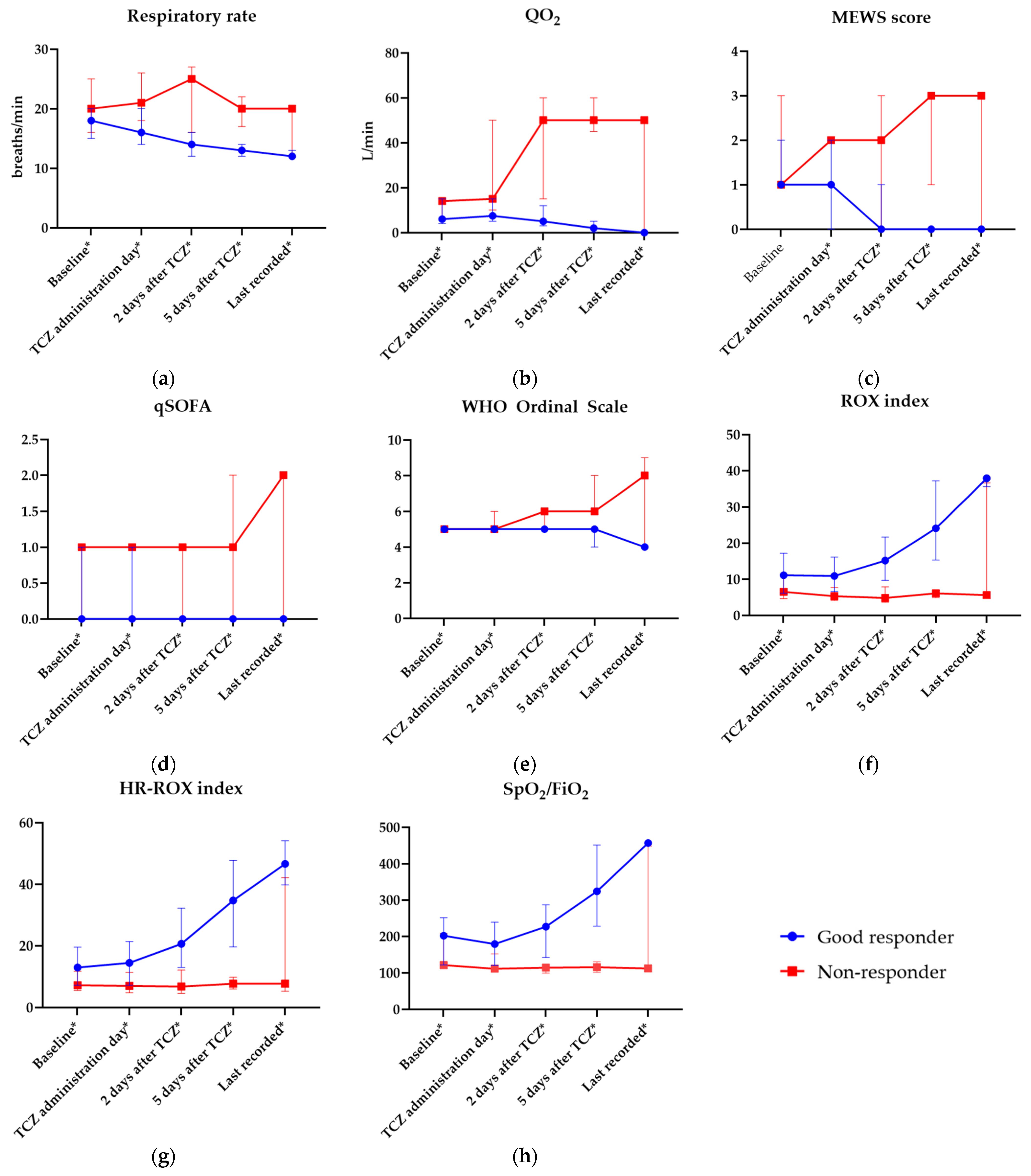
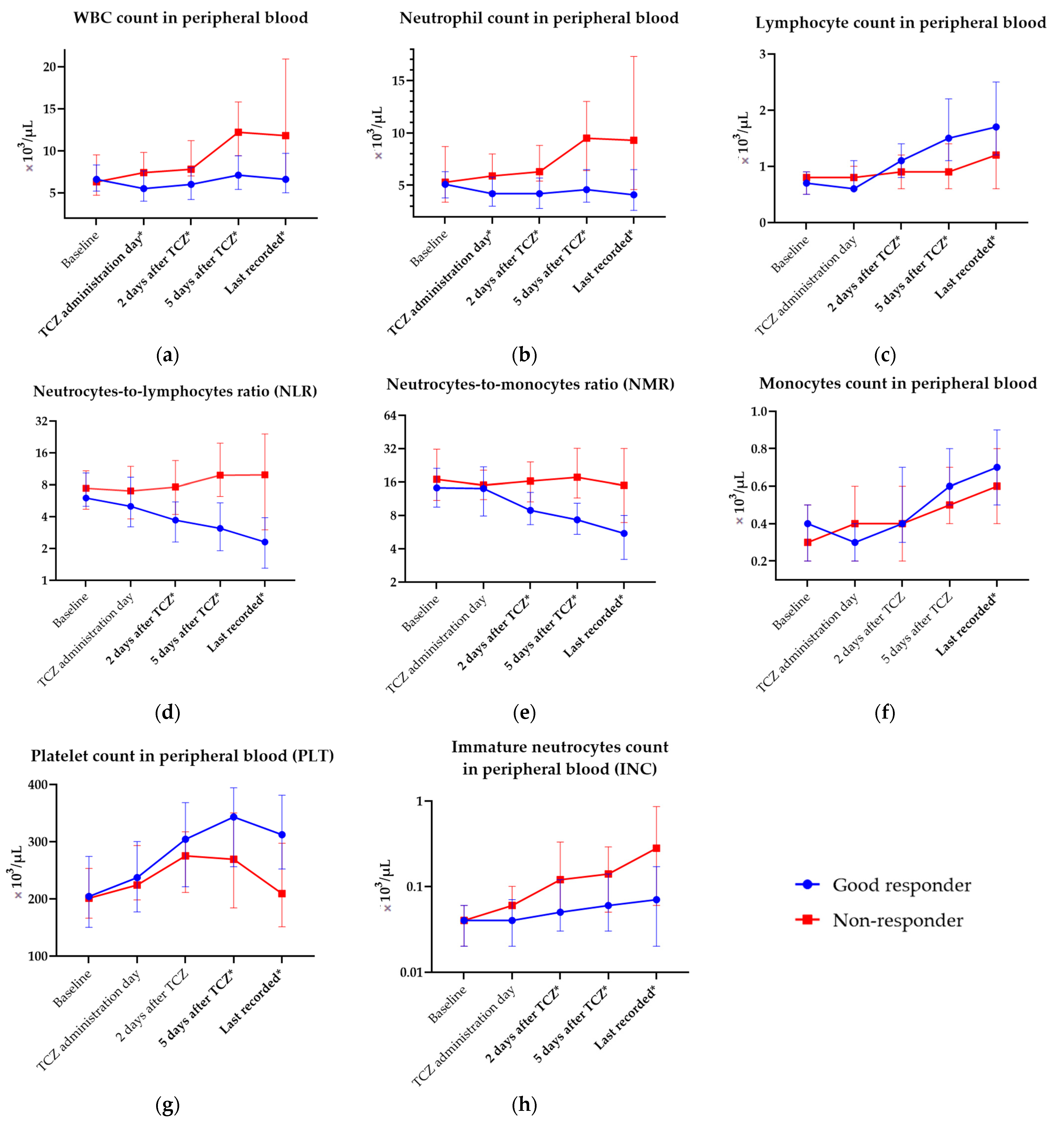
3.2. Laboratory Analysis
3.3. Early Prediction of Good Outcome after Tocilizumab Initiation and Mortality Prediction
3.4. Additional Outcomes and Safety
4. Discussion
5. Conclusions
Supplementary Materials
Author Contributions
Funding
Institutional Review Board Statement
Informed Consent Statement
Data Availability Statement
Acknowledgments
Conflicts of Interest
References
- Knight, J.S.; Caricchio, R.; Casanova, J.-L.; Combes, A.J.; Diamond, B.; Fox, S.E.; Hanauer, D.A.; James, J.A.; Kanthi, Y.; Ladd, V.; et al. The intersection of COVID-19 and autoimmunity. J. Clin. Investig. 2021, 131, e154886. [Google Scholar] [CrossRef] [PubMed]
- Fajgenbaum, D.C.; June, C.H. Cytokine Storm. N. Engl. J. Med. 2020, 383, 2255–2273. [Google Scholar] [CrossRef] [PubMed]
- Henderson, L.A.; Canna, S.W.; Schulert, G.S.; Volpi, S.; Lee, P.Y.; Kernan, K.F.; Caricchio, R.; Mahmud, S.; Hazen, M.M.; Halyabar, O.; et al. On the Alert for Cytokine Storm: Immunopathology in COVID-19. Arthritis Rheumatol. 2020, 72, 1059–1063. [Google Scholar] [CrossRef]
- Ramos-Casals, M.; Brito-Zerón, P.; López-Guillermo, A.; Khamashta, M.A.; Bosch, X. Adult haemophagocytic syndrome. Lancet 2014, 383, 1503–1516. [Google Scholar] [CrossRef] [PubMed]
- Huang, C.; Wang, Y.; Li, X.; Ren, L.; Zhao, J.; Hu, Y.; Zhang, L.; Fan, G.; Xu, J.; Gu, X.; et al. Clinical features of patients infected with 2019 novel coronavirus in Wuhan, China. Lancet 2020, 395, 497–506. [Google Scholar] [CrossRef]
- Ruan, Q.; Yang, K.; Wang, W.; Jiang, L.; Song, J. Clinical predictors of mortality due to COVID-19 based on an analysis of data of 150 patients from Wuhan, China. Intensive Care Med. 2020, 46, 846–848. [Google Scholar] [CrossRef]
- Schmidt, W.; Jóźwiak, B.; Czabajska, Z.; Pawlak-Buś, K.; Leszczynski, P. On-admission laboratory predictors for developing critical COVID-19 during hospitalization—A multivariable logistic regression model. Ann. Agric. Environ. Med. 2022, 29, 274–280. [Google Scholar] [CrossRef]
- Borczuk, A.C.; Salvatore, S.P.; Seshan, S.V.; Patel, S.S.; Bussel, J.B.; Mostyka, M.; Elsoukkary, S.; He, B.; Del Vecchio, C.; Fortarezza, F.; et al. COVID-19 pulmonary pathology: A multi-institutional autopsy cohort from Italy and New York City. Mod. Pathol. 2020, 33, 2156–2168. [Google Scholar] [CrossRef] [PubMed]
- Nienhold, R.; Ciani, Y.; Koelzer, V.H.; Tzankov, A.; Haslbauer, J.D.; Menter, T.; Schwab, N.; Henkel, M.; Frank, A.; Zsikla, V.; et al. Two distinct immunopathological profiles in autopsy lungs of COVID-19. Nat. Commun. 2020, 11, 5086. [Google Scholar] [CrossRef]
- Varga, Z.; Flammer, A.J.; Steiger, P.; Haberecker, M.; Andermatt, R.; Zinkernagel, A.S.; Mehra, M.R.; Schuepbach, R.A.; Ruschitzka, F.; Moch, H. Endothelial cell infection and endotheliitis in COVID-19. Lancet 2020, 395, 1417–1418. [Google Scholar] [CrossRef]
- El Jamal, S.M.; Pujadas, E.; Ramos, I.; Bryce, C.; Grimes, Z.M.; Amanat, F.; Tsankova, N.M.; Mussa, Z.; Olson, S.; Salem, F.; et al. Tissue-based SARS-CoV-2 detection in fatal COVID-19 infections: Sustained direct viral-induced damage is not necessary to drive disease progression. Hum. Pathol. 2021, 114, 110–119. [Google Scholar] [CrossRef] [PubMed]
- Bryce, C.; Grimes, Z.; Pujadas, E.; Ahuja, S.; Beasley, M.B.; Albrecht, R.; Hernandez, T.; Stock, A.; Zhao, Z.; AlRasheed, M.R.; et al. Pathophysiology of SARS-CoV-2: The Mount Sinai COVID-19 autopsy experience. Mod. Pathol. 2021, 34, 1456–1467. [Google Scholar] [CrossRef] [PubMed]
- Mehta, P.; Fajgenbaum, D.C. Is severe COVID-19 a cytokine storm syndrome: A hyperinflammatory debate. Curr. Opin. Rheumatol. 2021, 33, 419–430. [Google Scholar] [CrossRef] [PubMed]
- Leisman, D.E.; Ronner, L.; Pinotti, R.; Taylor, M.D.; Sinha, P.; Calfee, C.S.; Hirayama, A.V.; Mastroiani, F.; Turtle, C.J.; Harhay, M.O.; et al. Cytokine elevation in severe and critical COVID-19: A rapid systematic review, meta-analysis, and comparison with other inflammatory syndromes. Lancet Respir. Med. 2020, 8, 1233–1244. [Google Scholar] [CrossRef]
- Belen-Apak, F.B.; Sarıalioğlu, F. Pulmonary intravascular coagulation in COVID-19: Possible pathogenesis and recommendations on anticoagulant/thrombolytic therapy. J. Thromb. Thrombolysis 2020, 50, 278–280. [Google Scholar] [CrossRef]
- Webb, B.J.; Peltan, I.D.; Jensen, P.; Hoda, D.; Hunter, B.; Silver, A.; Starr, N.; Buckel, W.; Grisel, N.; Hummel, E.; et al. Clinical criteria for COVID-19-associated hyperinflammatory syndrome: A cohort study. Lancet Rheumatol. 2020, 2, e754–e763. [Google Scholar] [CrossRef]
- McGonagle, D.; Kearney, M.F.; O’Regan, A.; O’Donnell, J.S.; Quartuccio, L.; Watad, A.; Bridgewood, C. Therapeutic implications of ongoing alveolar viral replication in COVID-19. Lancet Rheumatol. 2022, 4, e135–e144. [Google Scholar] [CrossRef]
- Patel, S.; Saxena, B.; Mehta, P. Recent updates in the clinical trials of therapeutic monoclonal antibodies targeting cytokine storm for the management of COVID-19. Heliyon 2021, 7, e06158. [Google Scholar] [CrossRef]
- Xu, X.; Han, M.; Li, T.; Sun, W.; Wang, D.; Fu, B.; Zhou, Y.; Zheng, X.; Yang, Y.; Li, X.; et al. Effective Treatment of Severe COVID-19 Patients with Tocilizumab. Proc. Natl. Acad. Sci. USA 2020, 117, 10970–10975. [Google Scholar] [CrossRef]
- Da Van Kraaij, T.; Mostard, R.L.; Ramiro, S.; Checa, C.M.; Van Dongen, C.M.; Van Haren, E.H.; Buijs, J.; Landewé, R.B. Tocilizumab in Severe COVID-19 Pneumonia and Concomitant Cytokine Release Syndrome. Eur. J. Case Rep. Intern. Med. 2020, 7, 001675. [Google Scholar] [CrossRef]
- Campins, L.; Boixeda, R.; Perez-Cordon, L.; Aranega, R.; Lopera, C.; Force, L. Early tocilizumab treatment could improve survival among COVID-19 patients. Clin. Exp. Rheumatol. 2020, 38, 578. [Google Scholar]
- Viswanatha, G.L.; Male, C.A.; Shylaja, H. Efficacy and safety of tocilizumab in the management of COVID-19: A systematic review and meta-analysis of observational studies. Ann. Rheum. Dis. 2022, 40, 634–646. [Google Scholar] [CrossRef] [PubMed]
- Vela, D.; Vela-Gaxha, Z.; Rexhepi, M.; Olloni, R.; Hyseni, V.; Nallbani, R. Efficacy and safety of tocilizumab versus standard care/placebo in patients with COVID-19; A systematic review and meta-analysis of randomized clinical trials. Br. J. Clin. Pharmacol. 2022, 88, 1955–1963. [Google Scholar] [CrossRef]
- Zhang, J.; Chen, C.; Yang, Y.; Yang, J. Effectiveness of tocilizumab in the treatment of hospitalized adults COVID-19: A Systematic Review and Meta-Analysis. Medicine 2022, 101, e28967. [Google Scholar] [CrossRef] [PubMed]
- The WHO Rapid Evidence Appraisal for COVID-19 Therapies (REACT) Working Group; Shankar-Hari, M.; Vale, C.L.; Godolphin, P.J.; Fisher, D.; Higgins, J.P.T.; Spiga, F.; Savović, J.; Tierney, J.; Baron, G.; et al. Association Between Administration of IL-6 Antagonists and Mortality Among Patients Hospitalized for COVID-19: A Meta-analysis. JAMA 2021, 326, 499–518. [Google Scholar] [CrossRef] [PubMed]
- Petrelli, F.; Cherri, S.; Ghidini, M.; Perego, G.; Ghidini, A.; Zaniboni, A. Tocilizumab as treatment for COVID-19: A systematic review and meta-analysis. World J. Methodol. 2021, 11, 95–109. [Google Scholar] [CrossRef]
- Broman, N.; Feuth, T.; Vuorinen, T.; Valtonen, M.; Hohenthal, U.; Löyttyniemi, E.; Hirvioja, T.; Jalava-Karvinen, P.; Marttila, H.; Nordberg, M.; et al. Early administration of tocilizumab in hospitalized COVID-19 patients with elevated inflammatory markers; COVIDSTORM—A prospective, randomized, single-centre, open-label study. Clin. Microbiol. Infect. 2022, 28, 844–851. [Google Scholar] [CrossRef]
- Rutgers, A.; Westerweel, P.E.; van der Holt, B.; Postma, S.; van Vonderen, M.G.A.; Piersma, D.P.; Postma, D.; van den Berge, M.; Jong, E.; de Vries, M.; et al. Timely administration of tocilizumab improves outcome of hospitalized COVID-19 patients. PLoS ONE 2022, 17, e0271807. [Google Scholar] [CrossRef]
- Hermine, O.; Mariette, X.; Tharaux, P.-L.; Resche-Rigon, M.; Porcher, R.; Ravaud, P.; Bureau, S.; Dougados, M.; Tibi, A.; CORIMUNO-19 Collaborative Group; et al. Effect of Tocilizumab vs Usual Care in Adults Hospitalized With COVID-19 and Moderate or Severe Pneumonia: A Randomized Clinical Trial. JAMA Intern. Med. 2021, 181, 32–40. [Google Scholar] [CrossRef]
- Salvarani, C.; Dolci, G.; Massari, M.; Merlo, D.F.; Cavuto, S.; Savoldi, L.; Bruzzi, P.; Boni, F.; Braglia, L.; Turrà, C.; et al. Effect of Tocilizumab vs Standard Care on Clinical Worsening in Patients Hospitalized with COVID-19 Pneumonia: A Randomized Clinical Trial. JAMA Intern. Med. 2021, 181, 24–31. [Google Scholar] [CrossRef]
- Rosas, I.O.; Diaz, G.; Gottlieb, R.L.; Lobo, S.M.; Robinson, P.; Hunter, B.D.; Cavalcante, A.W.; Overcash, J.S.; Hanania, N.A.; Skarbnik, A.; et al. Tocilizumab and remdesivir in hospitalized patients with severe COVID-19 pneumonia: A randomized clinical trial. Intensiv. Care Med. 2021, 47, 1258–1270. [Google Scholar] [CrossRef]
- Stone, J.H.; Frigault, M.J.; Serling-Boyd, N.J.; Fernandes, A.D.; Harvey, L.; Foulkes, A.S.; Horick, N.K.; Healy, B.C.; Shah, R.; Bensaci, A.M.; et al. Efficacy of Tocilizumab in Patients Hospitalized with COVID-19. N. Engl. J. Med. 2020, 383, 2333–2344. [Google Scholar] [CrossRef]
- Le, R.Q.; Li, L.; Yuan, W.; Shord, S.S.; Nie, L.; Habtemariam, B.A.; Przepiorka, D.; Farrell, A.T.; Pazdur, R. FDA Approval Summary: Tocilizumab for Treatment of Chimeric Antigen Receptor T Cell-Induced Severe or Life-Threatening Cytokine Release Syndrome. Oncologist 2018, 23, 943–947. [Google Scholar] [CrossRef]
- Kaneko, Y.; Kameda, H.; Ikeda, K.; Ishii, T.; Murakami, K.; Takamatsu, H.; Tanaka, Y.; Abe, T.; Takeuchi, T. Tocilizumab in patients with adult-onset still’s disease refractory to glucocorticoid treatment: A randomised, double-blind, placebo-controlled phase III trial. Ann. Rheum. Dis. 2018, 77, 1720–1729. [Google Scholar] [CrossRef]
- Yokota, S.; Tanaka, T.; Kishimoto, T. Efficacy, safety and tolerability of tocilizumab in patients with systemic juvenile idiopathic arthritis. Ther. Adv. Musculoskelet. Dis. 2012, 4, 387–397. [Google Scholar] [CrossRef] [PubMed]
- Flisiak, R.; Horban, A.; Jaroszewicz, J.; Kozielewicz, D.; Pawłowska, M.; Parczewski, M.; Piekarska, A.; Simon, K.; Tomasiewicz, K.; Zarębska-Michaluk, D. Recommendations of management in SARS-CoV-2 infection of the Polish Association of Epidemiologists and Infectiologists. Pol. Arch. Intern. Med. 2020, 130, 352–357. [Google Scholar] [CrossRef]
- Flisiak, R.; Horban, A.; Jaroszewicz, J.; Kozielewicz, D.; Mastalerz-Migas, A.; Owczuk, R.; Parczewski, M.; Pawłowska, M.; Piekarska, A.; Simon, K.; et al. Management of SARS-CoV-2 infection: Recommendations of the Polish Association of Epidemiologists and Infectiologists as of 23 February 2022. Pol. Arch. Intern. Med. 2022, 132, 16230. [Google Scholar] [CrossRef] [PubMed]
- Leszczyński, P. COVID-19: A short message to rheumatologists. Rheumatology 2020, 58, 130–133. [Google Scholar] [CrossRef] [PubMed]
- Berlin, D.A.; Gulick, R.M.; Martinez, F.J. Severe COVID-19. N. Engl. J. Med. 2020, 383, 2451–2460. [Google Scholar] [CrossRef]
- Wu, Z.; McGoogan, J.M. Characteristics of and Important Lessons from the Coronavirus Disease 2019 (COVID-19) Outbreak in China: Summary of a Report of 72 314 Cases from the Chinese Center for Disease Control and Prevention. JAMA 2020, 323, 1239–1242. [Google Scholar] [CrossRef]
- Kosior, D.A.; Undas, A.; Kopeć, G.; Hryniewiecki, T.; Torbicki, A.; Mularek-Kubzdela, T.; Windyga, J.; Pruszczyk, P. Guidance for anticoagulation management in venous thromboembolism during the coronavirus disease 2019 pandemic in Poland: An expert opinion of the Section on Pulmonary Circulation of the Polish Cardiac Society. Kardiologia Polska 2020, 78, 642–646. [Google Scholar] [CrossRef]
- Von Elm, E.; Altman, D.G.; Egger, M.; Pocock, S.J.; Gøtzsche, P.C.; Vandenbroucke, J.P. The Strengthening the Reporting of Observational Studies in Epidemiology (STROBE) Statement: Guidelines for Reporting Observational Studies. Ann. Intern. Med. 2007, 147, 573–577. [Google Scholar] [CrossRef] [PubMed]
- Uhlig, K.; Menon, V.; Schmid, C.H. Recommendations for Reporting of Clinical Research Studies. Am. J. Kidney Dis. 2007, 49, 3–7. [Google Scholar] [CrossRef]
- Charlson, M.E.; Pompei, P.; Ales, K.L.; MacKenzie, C.R. A new method of classifying prognostic comorbidity in longitudinal studies: Development and validation. J. Chronic Dis. 1987, 40, 373–383. [Google Scholar] [CrossRef] [PubMed]
- Wettstein, R.B.; Shelledy, D.C.; Peters, J.I. Delivered oxygen concentrations using low-flow and high-flow nasal cannulas. Respir. Care 2005, 50, 604–609. [Google Scholar] [PubMed]
- McNarry, A.F.; Goldhill, D.R. Simple bedside assessment of level of consciousness: Comparison of two simple assessment scales with the Glasgow Coma scale. Anaesthesia 2004, 59, 34–37. [Google Scholar] [CrossRef]
- Subbe, C.P.; Kruger, M.; Rutherford, P.; Gemmel, L. Validation of a modified Early Warning Score in medical admissions. QJM 2001, 94, 521–526. [Google Scholar] [CrossRef]
- Seymour, C.W.; Liu, V.X.; Kahn, J.M.; Shankar-Hari, M.; Singer, M.; Deutschman, C.S.; Escobar, G.J.; Angus, D.C.; Iwashyna, T.J.; Brunkhorst, F.M.; et al. Assessment of Clinical Criteria for Sepsis: For the Third International Consensus Definitions for Sepsis and Septic Shock (Sepsis-3). JAMA 2016, 315, 762–774. [Google Scholar] [CrossRef]
- Freund, Y.; Lemachatti, N.; Krastinova, E.; Van Laer, M.; Claessens, Y.-E.; Avondo, A.; Occelli, C.; Feral-Pierssens, A.-L.; Truchot, J.; Ortega, M.; et al. Prognostic Accuracy of Sepsis-3 Criteria for In-Hospital Mortality Among Patients with Suspected Infection Presenting to the Emergency Department. JAMA 2017, 317, 301–308. [Google Scholar] [CrossRef]
- Marshall, J.C.; Murthy, S.; Diaz, J.; Adhikari, N.K.; Angus, D.C.; Arabi, Y.M.; Baillie, K.; Bauer, M.; Berry, S.; Blackwood, B.; et al. WHO Working Group on the Clinical Characterisation and Management of COVID-19 infection A Minimal Common Outcome Measure Set for COVID-19 Clinical Research. Lancet Infect. Dis. 2020, 20, e192–e197. [Google Scholar] [CrossRef]
- Roca, O.; Messika, J.; Caralt, B.; García-De-Acilu, M.; Sztrymf, B.; Ricard, J.-D.; Masclans, J.R. Predicting success of high-flow nasal cannula in pneumonia patients with hypoxemic respiratory failure: The utility of the ROX index. J. Crit. Care 2016, 35, 200–205. [Google Scholar] [CrossRef]
- Goh, K.J.; Chai, H.Z.; Ong, T.H.; Sewa, D.W.; Phua, G.C.; Tan, Q.L. Early prediction of high flow nasal cannula therapy outcomes using a modified ROX index incorporating heart rate. J. Intensiv. Care 2020, 8, 41. [Google Scholar] [CrossRef] [PubMed]
- Catoire, P.; Tellier, E.; de la Rivière, C.; Beauvieux, M.-C.; Valdenaire, G.; Galinski, M.; Revel, P.; Combes, X.; Gil-Jardiné, C. Assessment of the SpO2/FiO2 ratio as a tool for hypoxemia screening in the emergency department. Am. J. Emerg. Med. 2021, 44, 116–120. [Google Scholar] [CrossRef] [PubMed]
- Hansell, D.M.; Bankier, A.A.; MacMahon, H.; McLoud, T.C.; Müller, N.L.; Remy, J. Fleischner Society: Glossary of Terms for Thoracic Imaging. Radiology 2008, 246, 697–722. [Google Scholar] [CrossRef] [PubMed]
- Schmidt, W.; Pawlak-Buś, K.; Jóźwiak, B.; Katulska, K.; Leszczyński, P. Development and Validation of COVID-19 Radiological Risk Score (COVID-RRS): A Multivariable Radiological Score to Estimate the in-Hospital Mortality Risk in COVID-19 Patients. Eur. Rev. Med. Pharmacol. Sci. 2023, 27, 384–394. [Google Scholar] [CrossRef]
- ARDS Definition of Task Force; Ranieri, V.M.; Rubenfeld, G.D.; Thompson, B.T.; Ferguson, N.D.; Caldwell, E.; Fan, E.; Camporota, L.; Slutsky, A.S. Acute Respiratory Distress Syndrome: The Berlin Definition. JAMA 2012, 307, 2526–2533. [Google Scholar] [CrossRef] [PubMed]
- Konstantinides, S.V.; Meyer, G.; Becattini, C.; Bueno, H.; Geersing, G.J.; Harjola, V.-P.; Huisman, M.V.; Humbert, M.; Jennings, C.S.; Jiménez, D.; et al. 2019 ESC Guidelines for the diagnosis and management of acute pulmonary embolism developed in collaboration with the European Respiratory Society (ERS). Eur. Heart J. 2020, 41, 543–603. [Google Scholar] [CrossRef]
- Mazzolai, L.; Aboyans, V.; Ageno, W.; Agnelli, G.; Alatri, A.; Bauersachs, R.; Brekelmans, M.P.; Büller, H.R.; Elias, A.; Farge, D.; et al. Diagnosis and management of acute deep vein thrombosis: A joint consensus document from the European Society of Cardiology working groups of aorta and peripheral vascular diseases and pulmonary circulation and right ventricular function. Eur. Heart J. 2017, 39, 4208–4218. [Google Scholar] [CrossRef] [PubMed]
- Iba, T.E.; Warkentin, T.; Thachil, J.; Levi, M.; Levy, J.H. Proposal of the Definition for COVID-19-Associated Coagulopathy. J. Clin. Med. 2021, 10, 191. [Google Scholar] [CrossRef]
- Taylor, F.B.; Toh, C.H.; Hoots, W.K.; Wada, H.; Levi, M. Scientific Subcommittee on Disseminated Intravascular Coagulation (DIC) of the International Society on Thrombosis and Haemostasis (ISTH) Towards Definition, Clinical and Laboratory Criteria, and a Scoring System for Disseminated Intravascular Coagulation. Thromb. Haemost. 2001, 86, 1327–1330. [Google Scholar]
- Toh, C.H.; Hoots, W.K.; SSC on Disseminated Intravascular Coagulation of the ISTH. The scoring system of the Scientific and Standardisation Committee on Disseminated Intravascular Coagulation of the International Society on Thrombosis and Haemostasis: A 5-year overview. J. Thromb. Haemost. 2007, 5, 604–606. [Google Scholar] [CrossRef] [PubMed]
- Schulman, S.; Kearon, C.; The Subcommittee on Control of Anticoagulation of the Scientific and Standardization Committee of the International Society on Thrombosis and Haemostasis. Definition of major bleeding in clinical investigations of antihemostatic medicinal products in non-surgical patients. J. Thromb. Haemost. 2005, 3, 692–694. [Google Scholar] [CrossRef]
- Nauffal, V.; Achanta, A.; Goldhaber, S.Z.; Piazza, G. Association of ABO blood group type with cardiovascular events in COVID-19. J. Thromb. Thrombolysis 2021, 51, 584–586. [Google Scholar] [CrossRef] [PubMed]
- Thygesen, K.; Alpert, J.S.; Jaffe, A.S.; Chaitman, B.R.; Bax, J.J.; Morrow, D.A.; White, H.D. Executive Group on behalf of the Joint European Society of Cardiology (ESC)/American College of Cardiology (ACC)/American Heart Association (AHA)/World Heart Federation (WHF) Task Force for the Universal Definition of Myocardial Infarction Fourth Universal Definition of Myocardial Infarction. J. Am. Coll. Cardiol. 2018, 72, 2231–2264. [Google Scholar] [CrossRef] [PubMed]
- Kwo, P.Y.; Cohen, S.M.; Lim, J.K. ACG Clinical Guideline: Evaluation of Abnormal Liver Chemistries. Off. J. Am. Coll. Gastroenterol. 2017, 112, 18–35. [Google Scholar] [CrossRef] [PubMed]
- Aithal, G.P.; Watkins, P.B.; Andrade, R.J.; Larrey, D.; Molokhia, M.; Takikawa, H.; Hunt, C.M.; A Wilke, R.; Avigan, M.; Kaplowitz, N.; et al. Case Definition and Phenotype Standardization in Drug-Induced Liver Injury. Clin. Pharmacol. Ther. 2011, 89, 806–815. [Google Scholar] [CrossRef]
- Roedl, K.; Jarczak, D.; Drolz, A.; Wichmann, D.; Boenisch, O.; de Heer, G.; Burdelski, C.; Frings, D.; Sensen, B.; Nierhaus, A.; et al. Severe liver dysfunction complicating course of COVID-19 in the critically ill: Multifactorial cause or direct viral effect? Ann. Intensiv. Care 2021, 11, 44. [Google Scholar] [CrossRef]
- Singer, M.; Deutschman, C.S.; Seymour, C.W.; Shankar-Hari, M.; Annane, D.; Bauer, M.; Bellomo, R.; Bernard, G.R.; Chiche, J.-D.; Coopersmith, C.M.; et al. The Third International Consensus Definitions for Sepsis and Septic Shock (Sepsis-3). JAMA 2016, 315, 801–810. [Google Scholar] [CrossRef]
- Khwaja, A. KDIGO Clinical Practice Guidelines for Acute Kidney Injury. Nephron Clin. Pr. 2012, 120, c179–c184. [Google Scholar] [CrossRef]
- Michael, B.D.; Walton, D.; Westenberg, E.; García-Azorín, D.; Singh, B.; Tamborska, A.A.; Netravathi, M.; Chomba, M.; Wood, G.K.; Easton, A.; et al. Consensus Clinical Guidance for Diagnosis and Management of Adult COVID-19 Encephalopathy Patients. J. Neuropsychiatry 2022, 35, 12–27. [Google Scholar] [CrossRef]
- Manson, J.J.; Crooks, C.; Naja, M.; Ledlie, A.; Goulden, B.; Liddle, T.; Khan, E.; Mehta, P.; Martin-Gutierrez, L.; E Waddington, K.; et al. COVID-19-associated hyperinflammation and escalation of patient care: A retrospective longitudinal cohort study. Lancet Rheumatol. 2020, 2, e594–e602. [Google Scholar] [CrossRef] [PubMed]
- Caricchio, R.; Gallucci, M.; Dass, C.; Zhang, X.; Gallucci, S.; Fleece, D.; Bromberg, M.; Criner, G.J. Preliminary predictive criteria for COVID-19 cytokine storm. Ann. Rheum. Dis. 2020, 80, 88–95. [Google Scholar] [CrossRef] [PubMed]
- Zeng, J.; Xie, M.-H.; Yang, J.; Chao, S.-W.; Xu, E.-L. Clinical efficacy of tocilizumab treatment in severe and critical COVID-19 patients. World J. Clin. Cases 2020, 8, 3763–3773. [Google Scholar] [CrossRef]
- Çelik, İ.; Öztürk, R. From asymptomatic to critical illness: Decoding various clinical stages of Covid-19. Turk. J. Med. Sci. 2021, 51, 3284–3300. [Google Scholar] [CrossRef] [PubMed]
- Fisher, M.J.; Raymundo, L.A.M.; Monteforte, M.; Taub, E.M.; Go, R. Tocilizumab in the treatment of critical COVID-19 pneumonia: A retrospective cohort study of mechanically ventilated patients. Int. J. Infect. Dis. 2020, 103, 536–539. [Google Scholar] [CrossRef] [PubMed]
- Minihan, B.; McAuliffe, E.; Powell, J.; Wong, S.; Wilkie, K.; Murphy, C.; Maher, A.; Power, L.; O’Connell, N.; Dunne, C. Association between tocilizumab treatment of hyperinflammatory patients with COVID-19 in a critical care setting and elevated incidence of hospital-acquired bacterial and invasive fungal infections. J. Hosp. Infect. 2022, 126, 29–36. [Google Scholar] [CrossRef] [PubMed]
- Mehta, P.; Haskard, D.O.; A Laffan, M.; Chambers, R.C.; Hunt, B.J. Thromboses and COVID-19: Reducing inflammation in addition to thromboprophylaxis. Lancet Rheumatol. 2021, 3, e171–e172. [Google Scholar] [CrossRef]
- Siddiqi, H.K.; Mehra, M.R. COVID-19 illness in native and immunosuppressed states: A clinical–therapeutic staging proposal. J. Heart Lung Transplant. 2020, 39, 405–407. [Google Scholar] [CrossRef]
- Fathi, M.; Vakili, K.; Sayehmiri, F.; Mohamadkhani, A.; Hajiesmaeili, M.; Rezaei-Tavirani, M.; Eilami, O. The prognostic value of comorbidity for the severity of COVID-19: A systematic review and meta-analysis study. PLoS ONE 2021, 16, e0246190. [Google Scholar] [CrossRef]
- Millán, M.A.D.; Mesa-Plaza, N.; Guerrero-Santillán, M.; Morales-Ortega, A.; Bernal-Bello, D.; Sedano, A.I.F.; de Viedma-García, V.G.; Velázquez-Ríos, L.; Frutos-Pérez, B.; De Ancos-Aracil, C.L.; et al. Prognostic factors and combined use of tocilizumab and corticosteroids in a Spanish cohort of elderly COVID-19 patients. J. Med. Virol. 2021, 94, 1540–1549. [Google Scholar] [CrossRef]
- Williamson, E.J.; Walker, A.J.; Bhaskaran, K.; Bacon, S.; Bates, C.; Morton, C.E.; Curtis, H.J.; Mehrkar, A.; Evans, D.; Inglesby, P.; et al. Factors associated with COVID-19-related death using OpenSAFELY. Nature 2020, 584, 430–436. [Google Scholar] [CrossRef] [PubMed]
- Fialek, B.; Pruc, M.; Smereka, J.; Jas, R.; Rahnama-Hezavah, M.; Denegri, A.; Szarpak, A.; Jaguszewski, M.J.; Peacock, F.W.; Szarpak, L. Diagnostic value of lactate dehydrogenase in COVID-19: A systematic review and meta-analysis. Cardiol. J. 2022, 29, 751–758. [Google Scholar] [CrossRef] [PubMed]
- Medina-Hernández, E.; Pérez-Navarro, L.M.; Hernández-Ruiz, J.; Villalobos-Osnaya, A.; Hernández-Medel, M.L.; Casillas-Suárez, C.; Pérez-García, A. Changes in lactate dehydrogenase on admission throughout the COVID-19 pandemic and possible impacts on prognostic capability. Biomarkers Med. 2022, 16, 1019–1028. [Google Scholar] [CrossRef] [PubMed]
- Henry, B.M.; Aggarwal, G.; Wong, J.; Benoit, S.; Vikse, J.; Plebani, M.; Lippi, G. Lactate dehydrogenase levels predict coronavirus disease 2019 (COVID-19) severity and mortality: A pooled analysis. Am. J. Emerg. Med. 2020, 38, 1722–1726. [Google Scholar] [CrossRef] [PubMed]
- Olewicz-Gawlik, A.; Ginter-Matuszewska, B.; Kamiński, M.; Adamek, A.; Bura, M.; Mozer-Lisewska, I.; Kowala-Piaskowska, A. Changes in Inflammatory Markers after Administration of Tocilizumab in COVID-19: A Single-Center Retrospective Study. J. Clin. Med. 2022, 11, 107. [Google Scholar] [CrossRef] [PubMed]
- Selçuk, M.; Keskin, M.; Çınar, T.; Günay, N.; Doğan, S.; Çiçek, V.; Kılıç, Ş.; Asal, S.; Yavuz, S.; Keser, N.; et al. Prognostic significance of N-Terminal Pro-BNP in patients with COVID-19 pneumonia without previous history of heart failure. J. Cardiovasc. Thorac. Res. 2021, 13, 141–145. [Google Scholar] [CrossRef]
- Zinellu, A.; Sotgia, S.; Carru, C.; Mangoni, A.A. B-Type Natriuretic Peptide Concentrations, COVID-19 Severity, and Mortality: A Systematic Review and Meta-Analysis with Meta-Regression. Front. Cardiovasc. Med. 2021, 8, 690790. [Google Scholar] [CrossRef] [PubMed]
- Cordeanu, E.-M.; Duthil, N.; Severac, F.; Lambach, H.; Tousch, J.; Jambert, L.; Mirea, C.; Delatte, A.; Younes, W.; Frantz, A.-S.; et al. Prognostic Value of Troponin Elevation in COVID-19 Hospitalized Patients. J. Clin. Med. 2020, 9, 4078. [Google Scholar] [CrossRef]
- Kumar, A.; Karn, E.; Trivedi, K.; Kumar, P.; Chauhan, G.; Kumari, A.; Pant, P.; Munisamy, M.; Prakash, J.; Sarkar, P.G.; et al. Procalcitonin as a predictive marker in COVID-19: A systematic review and meta-analysis. PLoS ONE 2022, 17, e0272840. [Google Scholar] [CrossRef]
- Liu, Y.-M.; Xie, J.; Chen, M.-M.; Zhang, X.; Cheng, X.; Li, H.; Zhou, F.; Qin, J.-J.; Lei, F.; Chen, Z.; et al. Kidney Function Indicators Predict Adverse Outcomes of COVID-19. Med 2021, 2, 38–48.e2. [Google Scholar] [CrossRef]
- Gungor, B.; Atici, A.; Baycan, O.F.; Alici, G.; Ozturk, F.; Tugrul, S.; Asoglu, R.; Cevik, E.; Sahin, I.; Barman, H.A. Elevated D-dimer levels on admission are associated with severity and increased risk of mortality in COVID-19: A systematic review and meta-analysis. Am. J. Emerg. Med. 2021, 39, 173–179. [Google Scholar] [CrossRef] [PubMed]
- D’Ecclesiis, O.; Gavioli, C.; Martinoli, C.; Raimondi, S.; Chiocca, S.; Miccolo, C.; Bossi, P.; Cortinovis, D.; Chiaradonna, F.; Palorini, R.; et al. Vitamin D and SARS-CoV2 infection, severity and mortality: A systematic review and meta-analysis. PLoS ONE 2022, 17, e0268396. [Google Scholar] [CrossRef] [PubMed]
- Rastogi, A.; Bhansali, A.; Khare, N.; Suri, V.; Yaddanapudi, N.; Sachdeva, N.; Puri, G.D.; Malhotra, P. Short term, high-dose vitamin D supplementation for COVID-19 disease: A randomised, placebo-controlled, study (SHADE study). Postgrad. Med. J. 2022, 98, 87–90. [Google Scholar] [CrossRef] [PubMed]
- Long, W.; Yang, J.; Li, Z.; Li, J.; Chen, S.; Chen, D.; Wang, S.; Li, Q.; Hu, D.; Huang, J.; et al. Abnormal Fibrinogen Level as a Prognostic Indicator in Coronavirus Disease Patients: A Retrospective Cohort Study. Front. Med. 2021, 8, 687220. [Google Scholar] [CrossRef] [PubMed]
- Di Micco, P.; Russo, V.; Carannante, N.; Imparato, M.; Cardillo, G.; Lodigiani, C. Prognostic Value of Fibrinogen among COVID-19 Patients Admitted to an Emergency Department: An Italian Cohort Study. J. Clin. Med. 2020, 9, 4134. [Google Scholar] [CrossRef]
- Flisiak, R.; Jaroszewicz, J.; Rogalska, M.; Łapiński, T.; Berkan-Kawińska, A.; Bolewska, B.; Tudrujek-Zdunek, M.; Kozielewicz, D.; Rorat, M.; Leszczyński, P.; et al. Tocilizumab Improves the Prognosis of COVID-19 in Patients with High IL-6. J. Clin. Med. 2021, 10, 1583. [Google Scholar] [CrossRef]
- Piscoya, A.; del Riego, A.P.; Cerna-Viacava, R.; Rocco, J.; Roman, Y.M.; Escobedo, A.A.; Pasupuleti, V.; White, C.M.; Hernandez, A.V. Efficacy and harms of tocilizumab for the treatment of COVID-19 patients: A systematic review and meta-analysis. PLoS ONE 2022, 17, e0269368. [Google Scholar] [CrossRef] [PubMed]
- Peng, J.; Fu, M.; Mei, H.; Zheng, H.; Liang, G.; She, X.; Wang, Q.; Liu, W. Efficacy and secondary infection risk of tocilizumab, sarilumab and anakinra in COVID-19 patients: A systematic review and meta-analysis. Rev. Med. Virol. 2022, 32, e2295. [Google Scholar] [CrossRef]
- Mutua, V.; Henry, B.M.; von Csefalvay, C.; Cheruiyot, I.; Vikse, J.; Lippi, G.; Bundi, B.; Mong’are, N. Tocilizumab in Addition to Standard of Care in the Management of COVID-19: A Meta-Analysis of RCTs. Acta Biomed. 2022, 93, e2022014. [Google Scholar] [CrossRef] [PubMed]
- Naik, N.B.; Puri, G.D.; Kajal, K.; Mahajan, V.; Bhalla, A.; Kataria, S.; Singla, K.; Panigrahi, P.; Singh, A.; Lazar, M.; et al. High-Dose Dexamethasone Versus Tocilizumab in Moderate to Severe COVID-19 Pneumonia: A Randomized Controlled Trial. Cureus 2021, 13, e20353. [Google Scholar] [CrossRef] [PubMed]
- De Benedetto, I.; Lupia, T.; Shbaklo, N.; Bianchi, A.; Concialdi, E.; Penna, M.; Corcione, S.; De Rosa, F.G. Prognostic evaluation of Acinetobacter baumannii ventilator associated pneumonia in COVID-19. Infez. Med. 2022, 30, 570–576. [Google Scholar] [CrossRef] [PubMed]
- Zarębska-Michaluk, D.; Jaroszewicz, J.; Rogalska, M.; Martonik, D.; Pabjan, P.; Berkan-Kawińska, A.; Bolewska, B.; Oczko-Grzesik, B.; Kozielewicz, D.; Tudrujek-Zdunek, M.; et al. Effectiveness of Tocilizumab with and without Dexamethasone in Patients with Severe COVID-19: A Retrospective Study. J. Inflamm. Res. 2021, ume 14, 3359–3366. [Google Scholar] [CrossRef]
- AlQahtani, H.; AlBilal, S.; Mahmoud, E.; Aldibasi, O.; Alharbi, A.; Shamas, N.; Alsaedy, A.; Owaidah, K.; Alqahtani, F.Y.; Aleanizy, F.S.; et al. Outcomes associated with tocilizumab with or without corticosteroid versus dexamethasone for treatment of patients with severe to critical COVID-19 pneumonia. J. Infect. Public Health 2021, 15, 36–41. [Google Scholar] [CrossRef] [PubMed]
- Wolfe, C.R.; Tomashek, K.M.; Patterson, T.F.; Gomez, C.A.; Marconi, V.C.; Jain, M.K.; O Yang, O.; Paules, C.I.; Palacios, G.M.R.; Grossberg, R.; et al. Baricitinib versus dexamethasone for adults hospitalised with COVID-19 (ACTT-4): A randomised, double-blind, double placebo-controlled trial. Lancet Respir. Med. 2022, 10, 888–899. [Google Scholar] [CrossRef]
- Wild, J.M.; Porter, J.C.; Molyneaux, P.L.; George, P.M.; Stewart, I.; Allen, R.J.; Aul, R.; Baillie, J.K.; Barratt, S.L.; Beirne, P.; et al. Understanding the burden of interstitial lung disease post-COVID-19: The UK Interstitial Lung Disease-Long COVID Study (UKILD-Long COVID). BMJ Open Respir. Res. 2021, 8, e001049. [Google Scholar] [CrossRef] [PubMed]
- Saleki, K.; Shirzad, M.; Javanian, M.; Mohammadkhani, S.; Alijani, M.H.; Miri, N.; Oladnabi, M.; Azadmehr, A. Serum soluble Fas ligand is a severity and mortality prognostic marker for COVID-19 patients. Front. Immunol. 2022, 13, 947401. [Google Scholar] [CrossRef] [PubMed]
- Kessel, C.; Vollenberg, R.; Masjosthusmann, K.; Hinze, C.; Wittkowski, H.; Debaugnies, F.; Nagant, C.; Corazza, F.; Vély, F.; Kaplanski, G.; et al. Discrimination of COVID-19 From Inflammation-Induced Cytokine Storm Syndromes Using Disease-Related Blood Biomarkers. Arthritis Rheumatol. 2021, 73, 1791–1799. [Google Scholar] [CrossRef] [PubMed]
- Peker, B.O.; Şener, A.G.; Aydoğmuş, F.K. Antinuclear antibodies (ANAs) detected by indirect immunofluorescence (IIF) method in acute COVID-19 infection; future roadmap for laboratory diagnosis. J. Immunol. Methods 2021, 499, 113174. [Google Scholar] [CrossRef]


| Characteristic | Clinical Responders | Non-Responders | p-Value |
|---|---|---|---|
| Patients | 86 (71.7%) | 34 (28.3%) | - |
| Sex, male | 59 (68.6%) | 19 (55.9%) | 0.188 a |
| Age (years) * | 56.1 (±13.2) | 63.5 (±12.5) | 0.006 b |
| Body mass index (kg/m2) | 27.8 (24.7–32.3) | 30.6 (26.1–33.2) | 0.157 c |
| Charlson Comorbidity Index * | 2 (1–3) | 3 (2–4) | 0.004 c |
| Comorbidities: | |||
| atrial fibrillation * | 5 (5.8%) | 7 (20.6%) | 0.024 d |
| chronic coronary syndrome * | 9 (10.5%) | 10 (29.4%) | 0.010 d |
| asthma and/or COPD * | 4 (4.7%) | 6 (17.7%) | 0.029 d |
| Time from symptom onset to admission (days) | 11 (8–12) | 9 (8–13) | 0.315 c |
| Time from dyspnea onset to admission (days) | 2 (0–4) | 2 (1–4) | 0.136 c |
| Duration of hospitalization (days) | 13 (11–17) | 14 (10–22) | 0.553 c |
| Time from symptom onset to TCZ (days) | 12 (9–13) | 10 (9–14) | 0.337 c |
| Time from dyspnea onset to TCZ (days) | 3 (2–6) | 4.5 (3–7) | 0.086 c |
| Lung parenchyma involvement in CT (%) * | 50 (35–60) | 70 (60–85) | <0.001 c |
| COVID-RRS * | 5.0 (4.0–6.0) | 7.3 (5.6–8.5) | <0.001 c |
| Baseline PaO2/FiO2 * | 203 (136–277) | 106 (80–177) | <0.001 c |
| SpO2/FiO2 on TCZ initiation * | 179 (120–239) | 111 (102–152) | <0.001 c |
| WHO clinical progression scale on TCZ initiation * | |||
| 5 | 75 (87.2%) | 23 (67.6%) | 0.013 a |
| 6 | 11 (12.7%) | 11 (32.4%) | |
| Concomitant treatment: | |||
| remdesivir | 71 (82.6%) | 27 (79.4%) | 0.688 a |
| convalescent plasma | 61 (70.9%) | 24 (70.5%) | 0.970 a |
| LMWH | 84 (97.7%) | 32 (94.1%) | 0.318 d |
| Predictor | OR | 95%CI | p Value |
|---|---|---|---|
| COVID-RRS ≤ 6.5 | 7.37 | 3.06–17.79 | <0.001 |
| <70% of lung parenchyma involvement | 6.76 | 2.63–17.36 | <0.001 |
| 9–12 days from symptom onset (vs. <9 and >12) | 6.43 | 1.82–22.73 | 0.004 |
| ROX index ≥ 8.51 | 5.77 | 2.35–14.27 | <0.001 |
| RR < 20 breaths/min | 5.40 | 2.29–12.75 | <0.001 |
| No indices of AMI | 5.39 | 1.87–15.51 | 0.002 |
| HR-ROX index ≥ 11.59 | 4.97 | 2.02–12.59 | <0.001 |
| No CCS on administration day | 4.70 | 1.81–12.23 | 0.001 |
| qSOFA = 0 | 4.55 | 2.03–10.22 | <0.001 |
| SpO2/FiO2 > 122 | 4.47 | 1.92–10.40 | <0.001 |
| cHIS score < 3 | 4.44 | 1.43–13.77 | 0.009 |
| Lack of asthma/COPD | 4.39 | 1.55–16.71 | 0.030 |
| Lack of atrial fibrillation | 4.20 | 1.23–14.33 | 0.022 |
| Baseline PaO2/FiO2 > 200 mmHg | 4.04 | 1.59–10.27 | 0.003 |
| Age < 65 years | 3.69 | 1.60–8.46 | 0.002 |
| Lack of ischemic heart disease | 3.56 | 1.29–9.79 | 0.014 |
| No HFNOT (WHO Ordinal Scale = 5) | 3.26 | 1.25–8.49 | 0.016 |
| CCI < 4 | 2.96 | 1.31–6.71 | 0.009 |
| ≤5 days from onset of dyspnea | 2.43 | 1.06–5.56 | 0.035 |
| Predictor | OR | 95%CI | p-Value |
|---|---|---|---|
| LDH < 447 U/L | 12.67 | 4.42–36.31 | <0.001 |
| BNP < 50.5 pg/mL | 6.57 | 2.73–15.83 | <0.001 |
| hs-TnI < 26 ng/L | 4.80 | 1.55–14.81 | 0.006 |
| fibrinogen ≥ 490 mg/dL | 4.46 | 1.86–10.72 | <0.001 |
| BUN < 22.2 mg/dL | 4.17 | 2.02–10.99 | 0.017 |
| PCT 0.06–0.12 ng/mL | 3.98 | 1.40–11.28 | 0.009 |
| 25(OH)D3 ≥ 30 ng/mL | 3.20 | 1.20–8.54 | 0.020 |
| D-Dimer ≤ 1.28 µg/mL | 3.12 | 1.37–7.09 | 0.006 |
| IL-6 47.4–137.0 pg/mL | 3.07 | 1.90–4.98 | <0.001 |
| WBC < 7.4 G/L | 2.74 | 1.20–6.25 | 0.017 |
| CK < 151 IU/L | 2.62 | 1.16–5.91 | 0.020 |
| lack of pyuria | 2.5 | 1.04–5.31 | 0.039 |
| Predictor | OR | 95%CI | p Value |
|---|---|---|---|
| LDH < 439 IU/L | 46.55 | 10.30–210.4 | <0.001 |
| QO2 < 14 L/min | 19.83 | 7.23–54.43 | <0.001 |
| SpO2/FiO2 >132 | 17.63 | 6.34–49.06 | <0.001 |
| WHO Ordinal Scale < 6 | 13.83 | 5.20–36.73 | <0.001 |
| ROX index > 8.61 | 13.60 | 5.13–36.06 | <0.001 |
| MEWS < 2 | 12.79 | 4.14–39.54 | <0.001 |
| HR-ROX index > 12.47 | 12.73 | 4.82–33.59 | <0.001 |
| Neutrocytes < 4.8 G/L | 9.17 | 3.41–24.65 | <0.001 |
| IL-6 < 239.3 pg/mL | 9.17 | 3.41–24.65 | <0.001 |
| NLR < 5.5 | 9.07 | 3.68–22.35 | <0.001 |
| RR < 17/min | 7.92 | 3.24–19.31 | <0.001 |
| D-dimer < 4.40 µg/mL | 7.81 | 3.14–19.43 | <0.001 |
| NMR < 12.1 | 6.87 | 2.84–16.62 | <0.001 |
| qSOFA = 0 | 6.56 | 2.74–15.68 | <0.001 |
| WBC < 7.1 G/L | 4.93 | 2.04–11.88 | <0.001 |
| BUN < 20.2 mg/dL | 3.80 | 1.65–8.76 | 0.002 |
| Lymphocytes > 1.30 G/L | 3.20 | 1.20–8.54 | 0.016 |
| INC < 0.07 G/L | 2.80 | 1.23–6.40 | 0.013 |
| Predictor | Mortality | Good Response | ||||
|---|---|---|---|---|---|---|
| OR (95%CI) | Cut-Off/Range | p Value | OR (95%CI) | Cut-Off/Range | p-Value | |
| RR (/min) | 10.7 (3.3–34.4) | >2 | <0.001 | 5.4 (2.3–12.8) | <20 | <0.001 |
| ROX | 10.0 (2.77–36.09) | <8.74 | <0.001 | 5.8 (2.4–14.3) | ≥8.51 | 0.004 |
| HR-ROX | 8.8 (2.4–31.7) | <12.6 | <0.001 | 5.0 (2.0–12.6) | ≥11.59 | <0.001 |
| qSOFA | 8.9 (2.8–28.8) | ≥1 | <0.001 | 4.6 (2.0–10.2) | =0 | <0.001 |
| WHO OS | 7.3 (2.5–21.2) | >5 | <0.001 | 3.3 (1.3–8.5) | =5 | 0.016 |
| SpO2/FiO2 | 26.8 (7.7–92.6) | <111 | <0.001 | 4.5 (1.9–10.4) | >122 | 0.068 |
| age (years) | 4.9 (1.6–14.6) | >62 | 0.002 | 3.7 (1.6–8.5) | <65 | 0.002 |
| CCI | 8.5 (2.6–27.5) | ≥3.0 | 3.0 (1.3–6.7) | <4 | 0.009 | |
| days from symptom onset | NS | - | 0.499 | 6.4 (1.8–22.7) | 9–12 | 0.004 |
| days from dyspnea onset | NS | - | 0.124 | 2.4 (1.1–5.6) | ≤5 | 0.035 |
| COVID-RRS | 17.0 (4.6–62.9) | ≥6.5 | < 0.001 | 6.8 (1.8–22.7) | ≤6.5 | <0.001 |
| lung involvement (%) | 12.5 (4.4–36.1) | ≥70 | <0.001 | 7.4 (3.1–17.8) | <70 | <0.001 |
| WBC (G/L) | 14.9 (1.9–115.9) | >5.2 | <0.001 | 13 (15.1%) | <7.4 | 0.017 |
| NLR | 6.3 (2.0–20.1) | >6.2 | 0.001 | NS | - | 0.056 |
| SCr (mg/dL) | 6.3 (1.4–28.4) | ≥0.8 | 0.009 | NS | - | 0.098 |
| BUN (mg/dL) | 11.1 (3.7–33.3) | >22.2 | <0.001 | 4.2 (2.0–11.0) | <22.2 | 0.017 |
| D-dimer (µg/mL) | 4.7 (1.6–13.9) | >1.11 | 0.006 | 3.1 (1.4–7.1) | ≤1.28 | 0.006 |
| fibrinogen (mg/dL) | 3.6 (1.3–9.9) | <479 | 0.014 | 4.5 (1.9–10.7) | ≥490 | <0.001 |
| IL-6 (pg/mL) | NS | - | 0.573 | 3.1 (1.9–5.0) | 47.4–137.0 | <0.001 |
| LDH (U/L) | 18.2 (5.4–61.0) | ≥530 | <0.001 | 12.7 (4.4–36.3) | <447 | <0.001 |
| CK (IU/L) | 4.4 (1.4–13.3) | >308 | 0.009 | 2.6 (1.2–5.9) | <151 | 0.020 |
| BNP (pg/mL) | 5.0 (1.7–14.1) | >51.20 | 0.002 | 6.6 (2.7–15.8) | <50.5 | <0.001 |
| PCT (ng/mL) | 3.2 (1.2–8.5) | >0.13 | 0.004 | 4.0 (1.4–11.3) | 0.06–0.12 | 0.009 |
| hs-TnI (ng/L) | 6.2 (1.9–19.9) | >26 | 0.004 | 4.8 (1.6–14.8) | <26 | 0.006 |
| baseline PaO2/FiO2 (mmHg) | 10.5 (3.6–30.7) | <100 | <0.001 | 4.0 (1.6–10.3) | >200 | 0.003 |
| baseline 25(OH)D3 (ng/mL) | 6.9 (1.9–25.1) | <27 | 0.003 | 3.2 (1.2–8.5) | ≥30 | 0.009 |
| Outcome | Prevalence upon Administration | Occurence after Administration | ||||
|---|---|---|---|---|---|---|
| Clinical Responders | Non-Responders | p-Value | Clinical Responders | Non-Responders | p-Value | |
| death * | - | - | - | - | 20 (58.8%) | <0.001 a |
| ICU transfer due to ARDS progression * | - | - | - | - | 20 (58.8%) | <0.001 a |
| CAC * | - | 2 (5.9%) | 0.078 b | 31 (36.1%) | 22 (64.7%) | 0.004 a |
| neutropenia | 15 (17.4%) | 3 (8.8%) | 0.233 a | 13 (15.1%) | 2 (5.9%) | 0.140 b |
| agranulocytosis | - | - | - | 10 (11.6%) | 2 (5.9%) | 0.282 b |
| major bleeding | - | - | - | - | 1 (2.9%) | 0.283 b |
| MACE | 8 (9.3%) | 11 (32.4%) | 0.001 a | 3 (3.5%) | 9 (26.5%) | <0.001 b |
| liver injury | 28 (32.6%) | 9 (26.5%) | 0.515 a | 18 (20.9%) | 15 (44.1%) | 0.010 a |
| DILI | - | - | - | 1 (1.2%) | - | 0.716 b |
| patients with secondary infections | 2 (2.3%) | 4 (11.8%) | 0.033 a | 6 (7.0%) | 10 (29.4%) | 0.002 b |
| AKI | - | - | - | 6 (7.0%) | 17 (50%) | <0.001 a |
| acute tubulointerstitial nephritis | 6 (7.0%) | - | 0.182 b | 4 (4.7%) | 2 (5.9%) | 0.547 b |
| hemodynamic instability | - | - | - | 6 (7.0%) | 3 (8.8%) | 0.711 b |
| respiratory deterioration | - | - | - | 13 (15.1%) | 26 (76.5%) | <0.001 a |
| COV-HI | 50 (58.1%) | 23 (67.7%) | 0.336 a | 8 (9.3%) | 14 (41.2%) | <0.001 a |
| cHIS | 65 (75.6%) | 30 (88.3%) | 0.124 a | 4 (4.7%) | 3 (8.8%) | 0.313 b |
| CCS | 10 (11.6%) | 13 (38,2%) | <0.001 a | 4 (4.7%) | 3 (8.8%) | 0.313 b |
Disclaimer/Publisher’s Note: The statements, opinions and data contained in all publications are solely those of the individual author(s) and contributor(s) and not of MDPI and/or the editor(s). MDPI and/or the editor(s) disclaim responsibility for any injury to people or property resulting from any ideas, methods, instructions or products referred to in the content. |
© 2023 by the authors. Licensee MDPI, Basel, Switzerland. This article is an open access article distributed under the terms and conditions of the Creative Commons Attribution (CC BY) license (https://creativecommons.org/licenses/by/4.0/).
Share and Cite
Schmidt, W.; Pawlak-Buś, K.; Jóźwiak, B.; Leszczyński, P. Identification of Clinical Response Predictors of Tocilizumab Treatment in Patients with Severe COVID-19 Based on Single-Center Experience. J. Clin. Med. 2023, 12, 2429. https://doi.org/10.3390/jcm12062429
Schmidt W, Pawlak-Buś K, Jóźwiak B, Leszczyński P. Identification of Clinical Response Predictors of Tocilizumab Treatment in Patients with Severe COVID-19 Based on Single-Center Experience. Journal of Clinical Medicine. 2023; 12(6):2429. https://doi.org/10.3390/jcm12062429
Chicago/Turabian StyleSchmidt, Wiktor, Katarzyna Pawlak-Buś, Barbara Jóźwiak, and Piotr Leszczyński. 2023. "Identification of Clinical Response Predictors of Tocilizumab Treatment in Patients with Severe COVID-19 Based on Single-Center Experience" Journal of Clinical Medicine 12, no. 6: 2429. https://doi.org/10.3390/jcm12062429
APA StyleSchmidt, W., Pawlak-Buś, K., Jóźwiak, B., & Leszczyński, P. (2023). Identification of Clinical Response Predictors of Tocilizumab Treatment in Patients with Severe COVID-19 Based on Single-Center Experience. Journal of Clinical Medicine, 12(6), 2429. https://doi.org/10.3390/jcm12062429