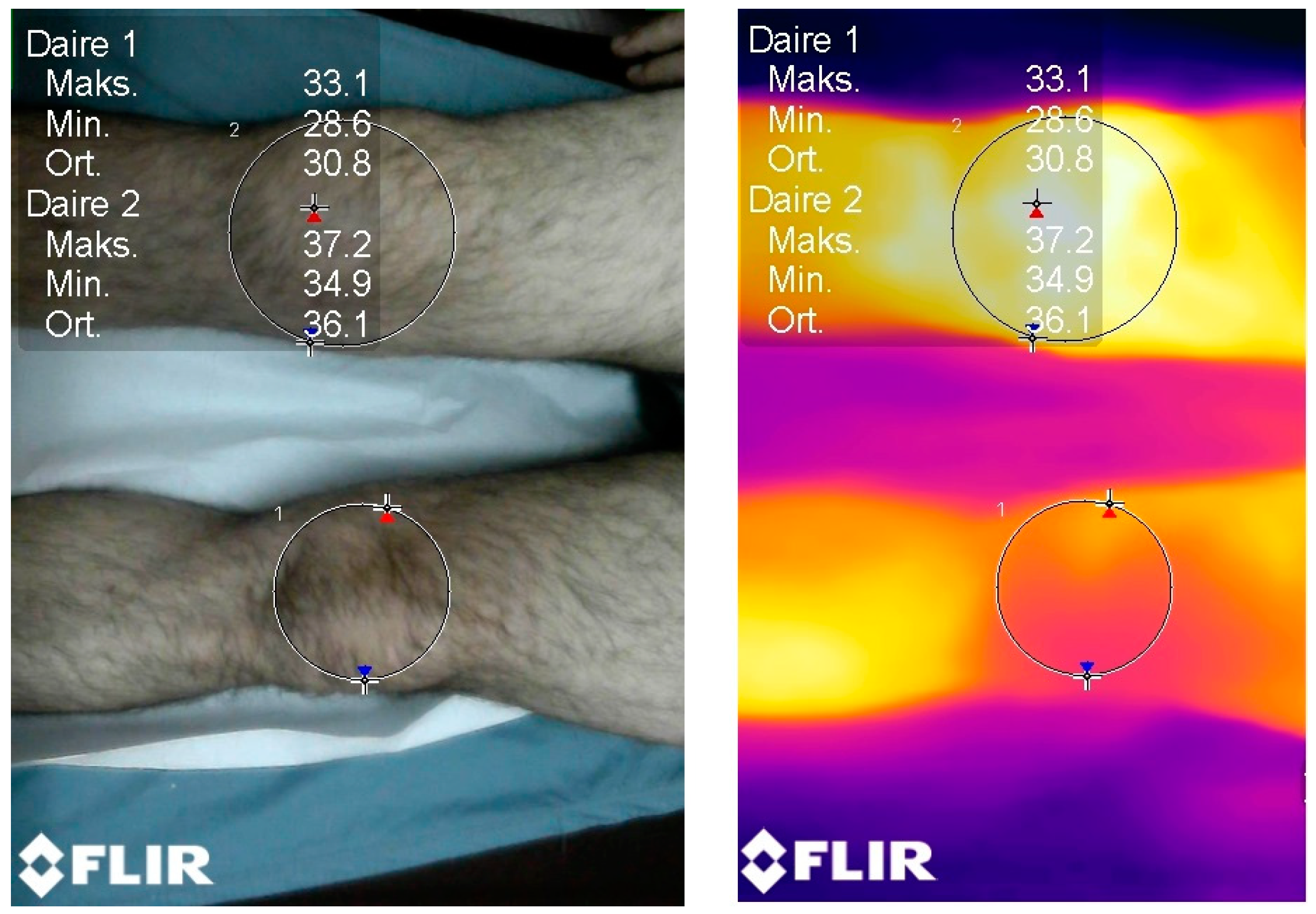
A New Perspective on the Diagnosis of Septic Arthritis: High-Resolution Thermal Imaging
Abstract
1. Introduction
2. Patients and Methods
2.1. Thermal Imaging (Standardization)
2.2. Statistical Analysis
3. Results
3.1. Relationship between Blood Values and the Hottest Spot-i
3.2. Relationship between Joint Fluid Values and Thermal Measurements
3.3. The Relationship between the Hottest Spot-i and Other Values
3.4. The Relationship between Hottest Spot-c and Other Values
3.5. The Relationship between the Hottest Spot Difference and Other Values
4. Discussion
Thermal Imagers (Camera)
- To use thermal imaging as a new additional non-invasive diagnostic tool in the differential diagnosis of septic arthritis.
- To compare the temperature increase and changes between septic and non-septic arthritis.
- To assign a quantitative value for the local temperature increase in the infected joint, in addition to palpation.
- In the diagnosis of septic arthritis, a high-resolution thermal imager can be used as an auxiliary non-invasive diagnostic tool.
- A temperature increase, which is an important sign of septic arthritis, can be detected by using the thermal imaging, in addition to palpation.
- Specially designed thermal devices with special software for septic arthritis can be developed.
- Although the imaging in this study was performed with a high-resolution and sensitive thermal camera, there is a minimal level of uncertainty regarding its accuracy due to internal and external factors, such as humidity. Further research should be directed at whether skin surface temperature differences can be used to differentiate between various mimicking clinical diseases.
- This cohort study’s findings are not sufficient to exclude the necessity of an invasive intra-articular aspiration. More comprehensive research studies are needed.
Author Contributions
Funding
Institutional Review Board Statement
Informed Consent Statement
Data Availability Statement
Conflicts of Interest
References
- Matan, A.J.; Smith, J.T. Pediatric septic arthritis. Orthopedics 1997, 20, 630–635. [Google Scholar] [CrossRef] [PubMed]
- Kocher, M.S.; Mandiga, R.; Zurakowski, D.; Barnewolt, C.; Kasser, J.R. Validation of a Clinical Prediction Rule for the Differentiation Between Septic Arthritis and Transient Synovitis of the Hip in Children. J. Bone Jt. Surg. 2004, 86, 1629–1635. [Google Scholar] [CrossRef] [PubMed]
- Kocher, M.S.; Zurakowski, D.; Kasser, J.R. Differentiating between septic arthritis and transient synovitis of the hip in children: An evidence-based clinical prediction algorithm. JBJS 1999, 81, 1662–1670. [Google Scholar] [CrossRef] [PubMed]
- Caird, M.S.; Flynn, J.M.; Leung, Y.L.; Millman, J.E.; Joann, G.D.; Dormans, J.P. Factors distinguishing septic arthritis from transient synovitis of the hip in children: A prospective study. JBJS 2006, 88, 1251–1257. [Google Scholar] [CrossRef]
- Kocher, M.S.; Mandiga, R.; Murphy, J.; Goldmann, N.; Harper, M.; Sundel, R.; Ecklund, K.; Kasser, J.R. A clinical practice guideline for treatment of septic arthritis in children: Efficacy in improving process of care and effect on outcome of septic arthritis of the hip. J. Bone Jt. Surg. 2003, 85, 994–999. [Google Scholar] [CrossRef]
- Simpson, R.C.; McEvoy, H.C.; Machin, G.; Howell, K.; Naeem, M.; Plassmann, P.; Ring, F.; Campbell, P.; Song, C.; Tavener, J.; et al. In-Field-of-View Thermal Image Calibration System for Medical Thermography Applications. Int. J. Thermophys. 2008, 29, 1123–1130. [Google Scholar] [CrossRef]
- Ammer, K. The Glamorgan Protocol for recording and evaluation of thermal images of the human body. Thermol Int. 2008, 18, 125–144. [Google Scholar]
- Jones, B.F.; Plassmann, P. Digital infrared thermal imaging of human skin. IEEE Eng. Med. Biol. Mag. 2002, 21, 41–48. [Google Scholar] [CrossRef]
- Farokhzad, S.; Motlagh, A.M.; Moghadam, P.A.; Honarmand, S.J.; Kheiralipour, K. Application of infrared thermal imaging technique and discriminant analysis methods for non-destructive identification of fungal infection of potato tubers. J. Food Meas. Charact. 2019, 14, 88–94. [Google Scholar] [CrossRef]
- Ring, E.F.J.; Ammer, K. Infrared thermal imaging in medicine. Physiol. Meas. 2012, 33, R33–R46. [Google Scholar] [CrossRef]
- Elhamahmy, M.; Mahmoud, M.; Bayoumi, T. The Effect of Applying Exogenous Salicylic Acid on Aphid Infection and its Influence on Histo-Physiological Traits and Thermal Imaging of Canola. Cercet. Agron. Mold. 2016, 49, 67–85. [Google Scholar] [CrossRef]
- Schollemann, F.; Kunczik, J.; Dohmeier, H.; Pereira, C.B.; Follmann, A.; Czaplik, M. Infection Probability Index: Implementation of an Automated Chronic Wound Infection Marker. J. Clin. Med. 2021, 11, 169. [Google Scholar] [CrossRef] [PubMed]
- Ko, L.N.; Raff, A.B.; Garza-Mayers, A.C.; Dobry, A.S.; Ortega-Martinez, A.; Anderson, R.R.; Kroshinsky, D. Skin Surface Temperatures Measured by Thermal Imaging Aid in the Diagnosis of Cellulitis. J. Investig. Dermatol. 2018, 138, 520–526. [Google Scholar] [CrossRef] [PubMed]
- Fiz, J.A.; Lozano, M.; Monte-Moreno, E.; Gonzalez-Martinez, A.; Faundez-Zanuy, M.; Becker, C.; Pons-Rodriguez, L.; Manzano, J.R. Tuberculine reaction measured by infrared thermography. Comput. Methods Programs Biomed. 2015, 122, 199–206. [Google Scholar] [CrossRef]
- van Netten, J.J.; van Baal, J.G.; Liu, C.; van Der Heijden, F.; Bus, S.A. Infrared thermal imaging for automated detection of diabetic foot complications. J. Diabetes Sci. Technol. 2013, 7, 1122–1129. [Google Scholar] [CrossRef]
- Spalding, S.J.; Kwoh, C.K.; Boudreau, R.; Enama, J.; Lunich, J.; Huber, D.; Denes, L.; Hirsch, R. Three-dimensional and thermal surface imaging produces reliable measures of joint shape and temperature: A potential tool for quantifying arthritis. Arthritis Res. Ther. 2008, 10, 1–9. [Google Scholar] [CrossRef]
- Lasanen, R.; Piippo-Savolainen, E.; Remes-Pakarinen, T.; Kröger, L.; Heikkilä, A.; Julkunen, P.; Karhu, J.; Töyräs, J. Thermal imaging in screening of joint inflammation and rheumatoid arthritis in children. Physiol. Meas. 2015, 36, 273–282. [Google Scholar] [CrossRef]
- Zhao, Y.; Iyer, R.S.; Reichley, L.; Oron, A.P.; Gove, N.E.; Kitsch, A.E.; Biswas, D.; Friedman, S.; Partridge, S.C.; Wallace, C.A. A Pilot Study of Infrared Thermal Imaging to Detect Active Bone Lesions in Children with Chronic Nonbacterial Osteomyelitis. Arthritis Care Res. 2018, 71, 1430–1435. [Google Scholar] [CrossRef]
- Owen, R.; Ramlakhan, S.; Saatchi, R.; Burke, D. Development of a high-resolution infrared thermographic imaging method as a diagnostic tool for acute undifferentiated limp in young children. Med. Biol. Eng. Comput. 2017, 56, 1115–1125. [Google Scholar] [CrossRef]
- Yusuf, E.; Hügle, T.; Daikeler, T.; Voide, C.; Borens, O.; Trampuz, A. The potential use of microcalorimetry in rapid differentiation between septic arthritis and other causes of arthritis. Eur. J. Clin. Microbiol. Infect. Dis. 2014, 34, 461–465. [Google Scholar] [CrossRef]
- Dandé, A.; Nöt, L.G.; Wiegand, N.; Kocsis, B.; Lőrinczy, D. DSC analysis of human synovial fluid samples in the diagnostics of non-septic and septic arthritis. J. Therm. Anal. Calorim. 2017, 130, 1249–1252. [Google Scholar] [CrossRef]


| n | % | ||
|---|---|---|---|
| Gender | Male | 27 | 55.1 |
| Woman | 22 | 44.9 | |
| Localization | Right knee | 26 | 53.1 |
| Left knee | 23 | 46.9 | |
| Reproduction in the joint | No | 34 | 69.4 |
| Yes | 15 | 30.6 | |
| Septic arthritis | No | 34 | 69.4 |
| yes | 15 | 30.6 | |
| Minimum | Maximum | Mean | ss | |
|---|---|---|---|---|
| Temperature | 36.00 | 39.50 | 37.02 | 0.94 |
| Wbc | 5.19 | 20.99 | 9.49 | 4.30 |
| Aso | 51.90 | 720.00 | 253.49 | 136.33 |
| Crp | 0.10 | 293.00 | 20.01 | 50.03 |
| Sedimentation | 1.00 | 168.00 | 73.57 | 41.41 |
| Leukocyte count in joint fluid (mm3) | 800.00 | 120,000.00 | 38,463.27 | 35,764.33 |
| Coldest spot | 29.20 | 38.40 | 34.64 | 2.19 |
| Hottest spot-i | 33.50 | 40.60 | 37.14 | 1.68 |
| Coldest spot-c | 28.70 | 37.90 | 33.00 | 2.10 |
| Hottest spot-c | 31.60 | 39.60 | 35.50 | 1.77 |
| Hottest spot difference | −1.20 | 7.00 | 1.64 | 1.60 |
| Coldest spot difference | −2.70 | 7.10 | 1.64 | 2.11 |
| Average-i | 32.80 | 39.50 | 36.26 | 1.69 |
| Average-c | 30.30 | 38.50 | 34.57 | 1.93 |
| Average difference | −1.40 | 7.30 | 1.69 | 1.78 |
| Septic Arthritis | Test Statistics | p | ||||
|---|---|---|---|---|---|---|
| No | Yes | |||||
| Mean | ss | Mean | ss | |||
| Temperature | 36.98 | 0.89 | 37.12 | 1.07 | −0.467 | 0.642 |
| Wbc | 9.22 | 3.97 | 10.09 | 5.09 | −0.650 | 0.519 |
| Aso | 208.47 | 105.13 | 355.53 | 146.93 | −3.983 | 0.000 |
| a Crp | 18.33 | 53.03 | 23.81 | 43.96 | 165.000 | 0.051 |
| Sedimentation | 62.24 | 37.83 | 99.27 | 38.58 | −3.140 | 0.003 |
| Leukocyte count in joint fluid mm3 | 22,026.47 | 22,317.71 | 75,720.00 | 32,681.21 | −6.703 | 0.000 |
| Coldest spot | 34.14 | 2.24 | 35.77 | 1.62 | −2.533 | 0.015 |
| Hottest spot-i | 36.79 | 1.71 | 37.93 | 1.34 | −2.272 | 0.028 |
| Coldest spot-c | 33.36 | 1.71 | 32.19 | 2.68 | 1.557 | 0.136 |
| Hottest spot-c | 35.82 | 1.51 | 34.79 | 2.15 | 1.917 | 0.061 |
| Hottest spot difference | 0.98 | 1.03 | 3.13 | 1.67 | −5.540 | 0.000 |
| Coldest spot difference | 0.78 | 1.60 | 3.58 | 1.84 | −5.398 | 0.000 |
| Average-i | 35.89 | 1.70 | 37.10 | 1.38 | −2.417 | 0.020 |
| Average-c | 34.95 | 1.60 | 33.70 | 2.38 | 1.864 | 0.077 |
| Average difference | 0.94 | 1.16 | 3.40 | 1.80 | −4.867 | 0.000 |
| Septic Arthritis | Age | Test Statistics | p | ||||
|---|---|---|---|---|---|---|---|
| Under 18 | Over 18 | ||||||
| Mean | ss | Mean | ss | ||||
| No | Temperature | 36.72 | 0.79 | 37.15 | 0.93 | −1.389 | 0.174 |
| Wbc | 10.11 | 4.22 | 8.67 | 3.80 | 1.024 | 0.313 | |
| Aso | 219.45 | 128.36 | 201.67 | 90.68 | 0.474 | 0.639 | |
| a Crp | 35.30 | 84.69 | 7.82 | 5.76 | 96.000 | 0.151 | |
| Sedimentation | 64.38 | 43.67 | 60.90 | 34.81 | 0.257 | 0.799 | |
| Leukocyte count in joint fluid mm3 | 21,592.31 | 17,803.30 | 22,295.24 | 25,128.86 | −0.088 | 0.931 | |
| Coldest spot-i | 33.12 | 2.41 | 34.78 | 1.92 | −2.230 | 0.033 | |
| Hottest spot-i | 36.53 | 1.48 | 36.96 | 1.86 | −0.701 | 0.489 | |
| Coldest spot-c | 33.02 | 1.59 | 33.57 | 1.78 | −0.906 | 0.371 | |
| Hottest spot-c | 36.11 | 1.15 | 35.64 | 1.69 | 0.881 | 0.385 | |
| Hottest spot difference | 0.42 | 0.65 | 1.32 | 1.09 | −2.677 | 0.012 | |
| Coldest spot difference | 0.09 | 1.44 | 1.21 | 1.57 | −2.079 | 0.046 | |
| Average-i | 35.67 | 1.49 | 36.03 | 1.83 | −0.602 | 0.551 | |
| Average-c | 35.22 | 1.32 | 34.79 | 1.75 | 0.772 | 0.446 | |
| Average difference | 0.45 | 0.75 | 1.25 | 1.27 | −2.059 | 0.048 | |
| Yes | Temperature | 37.26 | 1.18 | 37.05 | 1.07 | 0.347 | 0.734 |
| Wbc | 10.53 | 7.45 | 9.88 | 3.93 | 0.225 | 0.825 | |
| Aso | 347.80 | 216.21 | 359.40 | 112.95 | −0.139 | 0.892 | |
| a Crp | 10.82 | 10.77 | 30.31 | 53.05 | 18.000 | 0.391 | |
| Sedimentation | 71.60 | 31.91 | 113.10 | 34.99 | −2.224 | 0.045 | |
| Leukocyte count in joint fluid mm3 | 55,560.00 | 44,088.18 | 85,800.00 | 21,420.65 | −1.451 | 0.207 | |
| Coldest spot-i | 36.74 | 1.42 | 35.29 | 1.55 | 1.749 | 0.104 | |
| Hottest spot-i | 38.52 | 1.64 | 37.63 | 1.14 | 1.239 | 0.237 | |
| Coldest spot-c | 33.10 | 3.22 | 31.74 | 2.42 | 0.923 | 0.373 | |
| Hottest spot-c | 35.56 | 2.68 | 34.41 | 1.88 | 0.973 | 0.348 | |
| Hottest spot difference | 2.96 | 2.49 | 3.22 | 1.24 | −0.275 | 0.788 | |
| Coldest spot difference | 3.64 | 2.62 | 3.55 | 1.48 | 0.086 | 0.933 | |
| Average-i | 37.82 | 1.58 | 36.74 | 1.20 | 1.489 | 0.160 | |
| Average-c | 34.64 | 2.63 | 33.23 | 2.23 | 1.090 | 0.296 | |
| Average difference | 3.18 | 2.44 | 3.51 | 1.53 | −0.324 | 0.751 | |
| Temp. | Wbc | Aso | Crp | Sedimentation | Leukocyte Count in Joint Fluid mm3 | Coldest Spot-i | Hottest Spot-i | Coldest Spot-c | Hottest Spot-c | Hottest Spot Difference | Coldest Spot Difference | Average-i | Average-c | Average Difference | |
|---|---|---|---|---|---|---|---|---|---|---|---|---|---|---|---|
| Temperature | 1 | 0.116 | 0.295 | 0.022 | 0.040 | 0.018 | 0.338 | 0.252 | 0.237 | 0.117 | 0.135 | 0.115 | 0.243 | 0.116 | 0.105 |
| Wbc | 1 | −0.095 | 0.181 | 0.043 | 0.121 | −0.073 | −0.055 | −0.172 | −0.275 | 0.247 | 0.095 | −0.114 | −0.320 | 0.240 | |
| Aso | 1 | 0.492 | 0.420 | 0.424 | 0.367 | 0.419 | 0.104 | 0.144 | 0.280 | 0.278 | 0.419 | 0.092 | 0.297 | ||
| Crp | 1.000 | 0.418 | 0.225 | 0.427 | 0.325 | 0.126 | −0.061 | 0.410 | 0.367 | 0.305 | −0.034 | 0.376 | |||
| Sedimentation | 1 | 0.491 | 0.284 | 0.284 | −0.065 | −0.071 | 0.377 | 0.360 | 0.274 | −0.090 | 0.358 | ||||
| Leukocyte count in joint fluid mm3 | 1 | 0.198 | 0.171 | −0.361 | −0.367 | 0.587 | 0.565 | 0.219 | −0.346 | 0.583 | |||||
| Coldest spot-i | 1 | 0.796 | 0.518 | 0.380 | 0.414 | 0.524 | 0.840 | 0.396 | 0.366 | ||||||
| Hottest spot-i | 1 | 0.438 | 0.572 | 0.416 | 0.391 | 0.969 | 0.528 | 0.345 | |||||||
| Coldest spot-c | 1 | 0.811 | −0.439 | −0.457 | 0.426 | 0.855 | −0.524 | ||||||||
| Hottest spot-c | 1 | −0.509 | −0.412 | 0.541 | 0.969 | −0.540 | |||||||||
| Hottest spot difference | 1 | 0.867 | 0.417 | −0.520 | 0.960 | ||||||||||
| Coldest spot difference | 1 | 0.449 | −0.439 | 0.902 | |||||||||||
| Average-i | 1 | 0.523 | 0.380 | ||||||||||||
| Average-c | 1 | −0.590 | |||||||||||||
| Average difference | 1 |
Disclaimer/Publisher’s Note: The statements, opinions and data contained in all publications are solely those of the individual author(s) and contributor(s) and not of MDPI and/or the editor(s). MDPI and/or the editor(s) disclaim responsibility for any injury to people or property resulting from any ideas, methods, instructions or products referred to in the content. |
© 2023 by the authors. Licensee MDPI, Basel, Switzerland. This article is an open access article distributed under the terms and conditions of the Creative Commons Attribution (CC BY) license (https://creativecommons.org/licenses/by/4.0/).
Share and Cite
Gunay, H.; Bakan, O.M.; Mirzazade, J.; Sozbilen, M.C. A New Perspective on the Diagnosis of Septic Arthritis: High-Resolution Thermal Imaging. J. Clin. Med. 2023, 12, 1573. https://doi.org/10.3390/jcm12041573
Gunay H, Bakan OM, Mirzazade J, Sozbilen MC. A New Perspective on the Diagnosis of Septic Arthritis: High-Resolution Thermal Imaging. Journal of Clinical Medicine. 2023; 12(4):1573. https://doi.org/10.3390/jcm12041573
Chicago/Turabian StyleGunay, Huseyin, Ozgur Mert Bakan, Javad Mirzazade, and Murat Celal Sozbilen. 2023. "A New Perspective on the Diagnosis of Septic Arthritis: High-Resolution Thermal Imaging" Journal of Clinical Medicine 12, no. 4: 1573. https://doi.org/10.3390/jcm12041573
APA StyleGunay, H., Bakan, O. M., Mirzazade, J., & Sozbilen, M. C. (2023). A New Perspective on the Diagnosis of Septic Arthritis: High-Resolution Thermal Imaging. Journal of Clinical Medicine, 12(4), 1573. https://doi.org/10.3390/jcm12041573


