

Absence of Oligoclonal Bands in Multiple Sclerosis: A Call for Differential Diagnosis

 , ,
, ,
Abstract
1. Introduction
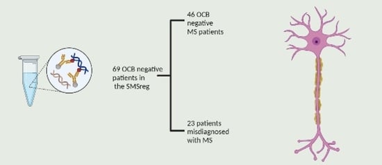
2. Methods
Statistical Analysis
3. Results
4. Discussion
4.1. The McDonald Criteria and Radiology
4.2. OCB as Biomarkers
4.3. PPMS Diagnosis—A Challenge
4.4. Benign MS
4.5. Criteria for Treatment
5. Conclusions
Author Contributions
Funding
Institutional Review Board Statement
Informed Consent Statement
Data Availability Statement
Conflicts of Interest
References
- Brownlee, W.J.; Hardy, T.A.; Fazekas, F.; Miller, D.H. Diagnosis of multiple sclerosis: Progress and challenges. Lancet 2017, 389, 1336–1346. [Google Scholar] [CrossRef]
- Dobson, R.; Ramagopalan, S.; Davis, A.; Giovannoni, G. Cerebrospinal fluid oligoclonal bands in multiple sclerosis and clinically isolated syndromes: A meta-analysis of prevalence, prognosis and effect of latitude. J. Neurol. Neurosurg. Psychiatry 2013, 84, 909–914. [Google Scholar] [CrossRef] [PubMed]
- Jin, H.; Lu, Q.; Gao, F.; Hao, H. Application of oligoclonal bands and other cerebrospinal fluid variables in multiple sclerosis and other neuroimmunological diseases: A narrative review. Ann. Transl. Med. 2023, 11, 282. [Google Scholar] [CrossRef] [PubMed]
- Imrell, K.; Landtblom, A.M.; Hillert, J.; Masterman, T. Multiple sclerosis with and without CSF bands: Clinically indistinguishable but immunogenetically distinct. Neurology 2006, 67, 1062–1064. [Google Scholar] [CrossRef]
- Vrethem, M.; Kvarnstrom, M.; Stenstam, J.; Cassel, P.; Gustafsson, M.; Landtblom, A.M.; Ernerudh, J. Cytokine mapping in cerebrospinal fluid and blood in multiple sclerosis patients without oligoclonal bands. Mult. Scler. J. 2012, 18, 669–673. [Google Scholar] [CrossRef]
- Karrenbauer, V.D.; Bedri, S.K.; Hillert, J.; Manouchehrinia, A. Cerebrospinal fluid oligoclonal immunoglobulin gamma bands and long-term disability progression in multiple sclerosis: A retrospective cohort study. Sci. Rep. 2021, 11, 14987. [Google Scholar] [CrossRef]
- Ferreira, D.; Voevodskaya, O.; Imrell, K.; Stawiarz, L.; Spulber, G.; Wahlund, L.O.; Hillert, J.; Westman, E.; Karrenbauer, V.D. Multiple sclerosis patients lacking oligoclonal bands in the cerebrospinal fluid have less global and regional brain atrophy. J. Neuroimmunol. 2014, 274, 149–154. [Google Scholar] [CrossRef]
- Solomon, A.J.; Naismith, R.T.; Cross, A.H. Misdiagnosis of multiple sclerosis: Impact of the 2017 McDonald criteria on clinical practice. Neurology 2019, 92, 26–33. [Google Scholar] [CrossRef]
- Deisenhammer, F.; Zetterberg, H.; Fitzner, B.; Zettl, U.K. The Cerebrospinal Fluid in Multiple Sclerosis. Front. Immunol. 2019, 10, 726. [Google Scholar] [CrossRef] [PubMed]
- Callander, M.; Haghighi, S.; Landtblom, A.M.; Ahlgren, C.E.; Nilsson, S.I.; Rydberg, L.; Al Khoury, H.; Rosegren, L.; Andersen, O. Multiple sclerosis immunopathic trait and HLA-DR(2)15 as independent risk factors in multiple sclerosis. Mult. Scler. J. 2007, 13, 441–445. [Google Scholar] [CrossRef] [PubMed]
- Gobbin, F.; Zanoni, M.; Marangi, A.; Orlandi, R.; Crestani, L.; Benedetti, M.D.; Gajofatto, A. 2017 McDonald criteria for multiple sclerosis: Earlier diagnosis with reduced specificity? Mult. Scler. Relat. Disord. 2019, 29, 23–25. [Google Scholar] [CrossRef] [PubMed]
- Tintore, M.; Vidal-Jordana, A.; Sastre-Garriga, J. Treatment of multiple sclerosis—Success from bench to bedside. Nat. Rev. Neurol. 2019, 15, 53–58. [Google Scholar] [CrossRef] [PubMed]
- Filippi, M.; Rocca, M.A.; Ciccarelli, O.; De Stefano, N.; Evangelou, N.; Kappos, L.; Rovira, A.; Sastre-Garriga, J.; Tintore, M.; Frederiksen, J.L.; et al. MRI criteria for the diagnosis of multiple sclerosis: MAGNIMS consensus guidelines. Lancet Neurol. 2016, 15, 292–303. [Google Scholar] [CrossRef] [PubMed]
- Thompson, A.J.; Banwell, B.L.; Barkhof, F.; Carroll, W.M.; Coetzee, T.; Comi, G.; Correale, J.; Fazekas, F.; Filippi, M.; Freedman, M.S.; et al. Diagnosis of multiple sclerosis: 2017 revisions of the McDonald criteria. Lancet Neurol. 2018, 17, 162–173. [Google Scholar] [CrossRef]
- Filippi, M.; Preziosa, P.; Banwell, B.L.; Barkhof, F.; Ciccarelli, O.; De Stefano, N.; Geurts, J.J.G.; Paul, F.; Reich, D.S.; Toosy, A.T.; et al. Assessment of lesions on magnetic resonance imaging in multiple sclerosis: Practical guidelines. Brain 2019, 142, 1858–1875. [Google Scholar] [CrossRef]
- Sarbu, N.; Shih, R.Y.; Jones, R.V.; Horkayne-Szakaly, I.; Oleaga, L.; Smirniotopoulos, J.G. White Matter Diseases with Radiologic-Pathologic Correlation. Radiographics 2016, 36, 1426–1447. [Google Scholar] [CrossRef]
- Kaisey, M.; Solomon, A.J.; Luu, M.; Giesser, B.S.; Sicotte, N.L. Incidence of multiple sclerosis misdiagnosis in referrals to two academic centers. Mult. Scler. Relat. Disord. 2019, 30, 51–56. [Google Scholar] [CrossRef]
- Midaglia, L.; Sastre-Garriga, J.; Pappolla, A.; Quibus, L.; Carvajal, R.; Vidal-Jordana, A.; Arrambide, G.; Rio, J.; Comabella, M.; Nos, C.; et al. The frequency and characteristics of MS misdiagnosis in patients referred to the multiple sclerosis centre of Catalonia. Mult. Scler. J. 2021, 27, 913–921. [Google Scholar] [CrossRef]
- Yamout, B.I.; Khoury, S.J.; Ayyoubi, N.; Doumiati, H.; Fakhreddine, M.; Ahmed, S.F.; Tamim, H.; Al-Hashel, J.Y.; Behbehani, R.; Alroughani, R. Alternative diagnoses in patients referred to specialized centers for suspected MS. Mult. Scler. Relat. Disord. 2017, 18, 85–89. [Google Scholar] [CrossRef]
- The Swedish MS Registry (MS-Register). Available online: www.neuroreg.se (accessed on 20 June 2020).
- Arrambide, G.; Tintore, M.; Espejo, C.; Auger, C.; Castillo, M.; Rio, J.; Castillo, J.; Vidal-Jordana, A.; Galan, I.; Nos, C.; et al. The value of oligoclonal bands in the multiple sclerosis diagnostic criteria. Brain 2018, 141, 1075–1084. [Google Scholar] [CrossRef]
- Mattson, D.H. Update on the diagnosis of multiple sclerosis. Expert Rev. Neurother. 2002, 2, 319–328. [Google Scholar] [CrossRef]
- Hobart, J.; Freeman, J.; Thompson, A. Kurtzke scales revisited: The application of psychometric methods to clinical intuition. Brain 2000, 123 Pt 5, 1027–1040. [Google Scholar] [CrossRef]
- Calabrese, M.; Gasperini, C.; Tortorella, C.; Schiavi, G.; Frisullo, G.; Ragonese, P.; Fantozzi, R.; Prosperini, L.; Annovazzi, P.; Cordioli, C.; et al. “Better explanations” in multiple sclerosis diagnostic workup: A 3-year longitudinal study. Neurology 2019, 92, e2527–e2537. [Google Scholar] [CrossRef] [PubMed]
- Tintore, M.; Rovira, A.; Rio, J.; Otero-Romero, S.; Arrambide, G.; Tur, C.; Comabella, M.; Nos, C.; Arevalo, M.J.; Negrotto, L.; et al. Defining high, medium and low impact prognostic factors for developing multiple sclerosis. Brain 2015, 138, 1863–1874. [Google Scholar] [CrossRef] [PubMed]
- Brownlee, W.J. Use (and misuse) of the McDonald criteria to diagnose multiple sclerosis. Eur. J. Neurol. 2018, 25, 209–210. [Google Scholar] [CrossRef] [PubMed]
- Lynch, D.S.; Wade, C.; Paiva, A.R.B.; John, N.; Kinsella, J.A.; Merwick, A.; Ahmed, R.M.; Warren, J.D.; Mummery, C.J.; Schott, J.M.; et al. Practical approach to the diagnosis of adult-onset leukodystrophies: An updated guide in the genomic era. J. Neurol. Neurosurg. Psychiatry 2019, 90, 543–554. [Google Scholar] [CrossRef]
- Wolf, N.I.; Ffrench-Constant, C.; van der Knaap, M.S. Hypomyelinating leukodystrophies—Unravelling myelin biology. Nat. Rev. Neurol. 2021, 17, 88–103. [Google Scholar] [CrossRef]
- Kelly, S.B.; Chaila, E.; Kinsella, K.; Duggan, M.; Walsh, C.; Tubridy, N.; Hutchinson, M. Using atypical symptoms and red flags to identify non-demyelinating disease. J. Neurol. Neurosurg. Psychiatry 2012, 83, 44–48. [Google Scholar] [CrossRef]
- Milo, R.; Korczyn, A.D.; Manouchehri, N.; Stuve, O. The temporal and causal relationship between inflammation and neurodegeneration in multiple sclerosis. Mult. Scler. J. 2020, 26, 876–886. [Google Scholar] [CrossRef]
- Stangel, M.; Fredrikson, S.; Meinl, E.; Petzold, A.; Stuve, O.; Tumani, H. The utility of cerebrospinal fluid analysis in patients with multiple sclerosis. Nat. Rev. Neurol. 2013, 9, 267–276. [Google Scholar] [CrossRef]
- Munoz, U.; Sebal, C.; Escudero, E.; Esiri, M.; Tzartos, J.; Sloan, C.; Sadaba, M.C. Main Role of Antibodies in Demyelination and Axonal Damage in Multiple Sclerosis. Cell Mol. Neurobiol. 2022, 42, 1809–1827. [Google Scholar] [CrossRef] [PubMed]
- Magliozzi, R.; Howell, O.; Vora, A.; Serafini, B.; Nicholas, R.; Puopolo, M.; Reynolds, R.; Aloisi, F. Meningeal B-cell follicles in secondary progressive multiple sclerosis associate with early onset of disease and severe cortical pathology. Brain 2007, 130, 1089–1104. [Google Scholar] [CrossRef] [PubMed]
- Zheng, Y.; Cai, M.T.; Yang, F.; Zhou, J.P.; Fang, W.; Shen, C.H.; Zhang, Y.X.; Ding, M.P. IgG Index Revisited: Diagnostic Utility and Prognostic Value in Multiple Sclerosis. Front. Immunol. 2020, 11, 1799. [Google Scholar] [CrossRef]
- DiSano, K.D.; Gilli, F.; Pachner, A.R. Intrathecally produced CXCL13: A predictive biomarker in multiple sclerosis. Mult. Scler. J. Exp. Transl. Clin. 2020, 6, 2055217320981396. [Google Scholar] [CrossRef] [PubMed]
- Vecchio, D.; Bellomo, G.; Serino, R.; Virgilio, E.; Lamonaca, M.; Dianzani, U.; Cantello, R.; Comi, C.; Crespi, I. Intrathecal kappa free light chains as markers for multiple sclerosis. Sci. Rep. 2020, 10, 20329. [Google Scholar] [CrossRef] [PubMed]
- Wolinsky, J.S.; PROMiSe Trial Study Group. The PROMiSe trial: Baseline data review and progress report. Mult. Scler. J. 2004, 10 (Suppl. 1), S65–S71; Discussion S62–S71. [Google Scholar] [CrossRef]
- Solomon, A.J.; Klein, E.P.; Bourdette, D. “Undiagnosing” multiple sclerosis: The challenge of misdiagnosis in MS. Neurology 2012, 78, 1986–1991. [Google Scholar] [CrossRef]
- Mathey, G.; Pische, G.; Soudant, M.; Pittion-Vouyovitch, S.; Guillemin, F.; Debouverie, M.; Epstein, J. Long-term analysis of patients with benign multiple sclerosis: New insights about the disability course. J. Neurol. 2021, 268, 3817–3825. [Google Scholar] [CrossRef]
- Berntsson, S.G.; Kristoffersson, A.; Bostrom, I.; Feresiadou, A.; Burman, J.; Landtblom, A.M. Rapidly increasing off-label use of rituximab in multiple sclerosis in Sweden—Outlier or predecessor? Acta Neurol. Scand. 2018, 138, 327–331. [Google Scholar] [CrossRef]

| Variables | MS | Non-MS | p-Value |
|---|---|---|---|
| Patients (n = 69) | 46 (66%) | 23 (34%) | |
| Age at onset (year), mean (SD) | 35.9 (9.8) | 39.8 (11.3) | 0.55 |
| Female/male | 32/14 | 18/5 | 0.11 |
| Presenting symptoms | |||
| Sensory (%) | 15 (32) | 11 (48) | |
| Myelitis (%) | 1 (2) | 1 (4) | |
| Brainstem (%) | 4 (9) | 1 (4) | |
| Optic neuritis (%) | 9 (19) | 4 (17) | |
| Ataxia (%) | 2 (4) | 2 (8) | |
| Motor (%) | 14 (30) | 4 (17) | |
| Phenotype | MS | Non-MS | 0.84 |
| RR (%) | 21 (45) | 15 (65) | |
| PP (%) | 14 (30) | 6 (26) | |
| SP (%) | 11 (24) | 2 (9) | |
| EDSS, mean (SD) | 4.5 (3.2) | 2.5 (2.3) | 0.001 |
| Reached EDSS 3 (%) | 27 (58) | 7 (30) | |
| Reached EDSS 6 (%) | 21 (45) | 2 (9) | |
| CSF | |||
| Age at CSF examination, mean (SD) | 39.8 (9.9) | 42.7 (11.6) | 0.42 |
| IgG Index, mean (SD) | |||
| Ref (0.36−0.56) mg/dL | 0.65 (0.3) | 0.53 (0.16) | 0.12 |
| Treatment | |||
| None (%) | 24 (52) | 18 (78) | |
| Platform DMT (%) | 15 (32) | 4 (17) | |
| More potent DMT (%) | 7 (15) | 1 (4) | |
| Case | Age at Onset, y/Sex | MS-Subtype in SMSreg | Presenting Symptoms | EDSS | DMT | Time to Alternative Diagnosis | Revised Diagnosis |
|---|---|---|---|---|---|---|---|
| 1 | 54/F | SPMS | Motor | 5.5 | No | 24 | Normal pressure hydrocephalus |
| 2 | 66/F | RRMS | Myelitis | 1.0 | No | - | Non-inflammatory myelitis |
| 3 | 33/F | RRMS | Dizziness | 1.0 | No | Vertigo NUD | |
| 4 | 43/M | PPMS | Brainstem | 3.0 | No | 10 | Arnold-Chiari malformation |
| 5 | 48/F | PPMS | Motor | 7.5 | IFN | - | Gait difficulties |
| 6 | 59/M | RRMS | Sensory | 1.0 | No | - | Cervical disc herniation |
| 7 | 59/M | PPMS | Gait | 5.5 | No | 5 | Mild cognitive impairment |
| 8 | 35/F | RRMS | Sensory | 2.5 | INF | Paresthesia | |
| 9 | 30/F | RRMS | Sensory | 4.5 | No | 33 | Leukodystrophy |
| 10 | 30/F | RRMS | Sensory | 0.0 | No | - | Vertigo |
| 11 | 35/F | PPMS | Sensory | 2.0 | No | 21 | Adrenoleuko-dystrophy |
| 12 | 36/F | RRMS | ON | 0.0 | No | - | Optic neuritis |
| 13 | 41/F | PPMS | Sensory, fatigue | 1.5 | No | - | Fatigue |
| 14 | 26/F | RRMS | ON | 2.5 | IFN | - | Optic neuritis |
| 15 | 30/F | RRMS | Sensory | 0.0 | No | - | Numbness of the limbs |
| 16 | 22/F | RRMS | Sensory | 0.0 | No | - | Numbness in the face |
| 17 | 28/M | RRMS | Motor | 3.5 | No | - | Hemiparesis NUD |
| 18 | 40/F | RRMS | Sensory | 0 | No | - | Possible NMO |
| 19 | 39/F | RRMS | Sensory | 1.5 | No | - | Myelitis |
| 20 | 49/F | RRMS | Brainstem | 5.0 | NTZ | 2 | NMO |
| 21 | 42/M | RRMS | ON | 1.5 | No | - | Optic neuritis |
| 22 | 35/F | PPMS | Sensory | 6.5 | No | - | Hereditary spastic paraparesis |
| 23 | 37/F | RRMS | ON | 0 | INF | - | Optic neuritis |
| Case | Age at Onset (y)/Sex | MRI of the Brain | MRI of the Spinal Cord | Revised Diagnosis |
|---|---|---|---|---|
| 1 | 54/F | Supra- and infratentorial WM lesions (WML) suggest small vessel disease. Compressed parasagittal sulci, dilated ventricles, fissure sylvii, and convexity sulci suggestive of NPH | Normal | Normal pressure hydrocephalus |
| 2 | 66/F | Left single frontal WML | 8 mm C2- lesion | Non-inflammatory myelitis |
| 3 | 33/F | Symmetrical periventricular WML suggestive of small vessel disease | Normal cervical MRI | Vertigo |
| n4 | 43/M | Five non-specific WML | Arnold-Chiari malformation | Arnold-Chiari malformation |
| 5 | 48/F | Multiple, partially confluent signal intensity changes in WM suggest small vessel disease. Ischemic lesions in the basal ganglia. | Cervical and thoracic spinal cord normal | Gait difficulties |
| 6 | 59/M | Symmetrical small WML. One large lesion in left temporal WM | Cervical disc herniation | Cervical disc herniation |
| 7 | 59/M | Symmetrical periventricular WML suggestive of small vessel disease and non-specific WML. Cortical infarctions parietally and in the left cerebellar hemisphere | Normal cervical MRI | Mild cognitive impairment |
| 8 | 35/F | Symmetrical periventricular WML suggestive of small vessel disease and non-specific WML | Normal cervical MRI | Paresthesia |
| 9 | 30/F | Non-specific WML. Pons and cerebellar oedema | Cervical spine oedema | Leukodystrophy |
| 10 | 30/F | Normal | Missing | vertigo |
| 11 | 35/F | Non-specific WML. Corona radiata gliosis | Stripe-formed lesion in the posterior cervical medulla. | Adrenoleuko-dystrophy |
| 12 | 36/F | 7 non-specific supratentorial WML | Missing | Optic neuritis |
| 13 | 41/F | Symmetrical confluent WML. Gliosis | Normal cervical MRI | Fatigue |
| 14 | 26/F | Normal | Normal cervical MRI. Artefact at C3 level | Optic neuritis |
| 15 | 30/F | Normal | Normal cervical and thoracical MRI | Numbness in the limbs |
| 16 | 22/F | Normal | Normal cervical and thoracic MRI. | Numbness in the face |
| 17 | 28/M | 2 periventricular WML | Normal cervical and thoracical MRI | Hemiparesis NUD |
| 18 | 40/F | Normal | C3, C4 lesions. GD+ C3 lesion | Possible NMO |
| 19 | 39/F | Non-specific WML | C2, C4–5, C5–6 lesions. GD+ C2, C5-C6 lesion | Myelitis |
| 20 | 49/F | GD+ medulla obongata lesion | GD+ C1–2 lesion | NMO |
| 21 | 42/M | 2–3 non-specific WML | Missing | Optic neuritis |
| 22 | 35/F | Normal | Normal | Hereditary spastic paraparesis |
| 23 | 37/F | 10 non-specific WML | Normal cervical and thoracical MRI | Optic neuritis |
Disclaimer/Publisher’s Note: The statements, opinions and data contained in all publications are solely those of the individual author(s) and contributor(s) and not of MDPI and/or the editor(s). MDPI and/or the editor(s) disclaim responsibility for any injury to people or property resulting from any ideas, methods, instructions or products referred to in the content. |
© 2023 by the authors. Licensee MDPI, Basel, Switzerland. This article is an open access article distributed under the terms and conditions of the Creative Commons Attribution (CC BY) license (https://creativecommons.org/licenses/by/4.0/).
Share and Cite
Katsarogiannis, E.; Landtblom, A.-M.; Kristoffersson, A.; Wikström, J.; Semnic, R.; Berntsson, S.G. Absence of Oligoclonal Bands in Multiple Sclerosis: A Call for Differential Diagnosis. J. Clin. Med. 2023, 12, 4656. https://doi.org/10.3390/jcm12144656
Katsarogiannis E, Landtblom A-M, Kristoffersson A, Wikström J, Semnic R, Berntsson SG. Absence of Oligoclonal Bands in Multiple Sclerosis: A Call for Differential Diagnosis. Journal of Clinical Medicine. 2023; 12(14):4656. https://doi.org/10.3390/jcm12144656
Chicago/Turabian StyleKatsarogiannis, Evangelos, Anne-Marie Landtblom, Anna Kristoffersson, Johan Wikström, Robert Semnic, and Shala G. Berntsson. 2023. "Absence of Oligoclonal Bands in Multiple Sclerosis: A Call for Differential Diagnosis" Journal of Clinical Medicine 12, no. 14: 4656. https://doi.org/10.3390/jcm12144656
APA StyleKatsarogiannis, E., Landtblom, A.-M., Kristoffersson, A., Wikström, J., Semnic, R., & Berntsson, S. G. (2023). Absence of Oligoclonal Bands in Multiple Sclerosis: A Call for Differential Diagnosis. Journal of Clinical Medicine, 12(14), 4656. https://doi.org/10.3390/jcm12144656