Article

Local Anesthesia Onset and Pain Perception in Hemophilic and Thalassemic Conditions

1
Department of Conservative Dentistry and Endodontics, Institute of Dental Sciences, Siksha ‘O’ Anusandhan (Deemed to Be) University, Bhubaneswar 751003, Odisha, India
2
Department of Dentistry, Shadan Institute of Medical Sciences Research Centre and Teaching Hospital, Himayat Sagar, Hyderabad 500086, Telangana, India
3
Department of Dentistry, Dr.V.R.K. Women’s Medical College Teaching Hospital and Research Centre, Aziz nagar, Hyderabad 500075, Telangana, India
4
Department of Conservative Dentistry and Endodontics, Sriram Chandra Bhanja Dental College & Hospital, Cuttack 753007, Odisha, India
5
Department of Public Health Dentistry, Institute of Dental Sciences, Siksha ‘O’ Anusandhan (Deemed to Be) University, Bhubaneswar 751003, Odisha, India
*
Author to whom correspondence should be addressed.
J. Clin. Med. 2023, 12(11), 3646; https://doi.org/10.3390/jcm12113646
Submission received: 15 March 2023 / Revised: 16 April 2023 / Accepted: 19 May 2023 / Published: 24 May 2023
(This article belongs to the Special Issue New Insights into Regenerative Dentistry)

Abstract

:
The study aims to evaluate and compare the onset of local anesthesia (LA) and pain perception during endodontic treatment in hemophilic and thalassemic patients. Methods: The study included 90 patients with symptomatic irreversible pulpitis of the mandibular molars. Three groups (n = 30 in each group) were included. Group 1: hemophilic patients; group 2: thalassemic patients; and group 3: individuals without any systemic diseases. Onset of LA and visual analogue scale (VAS) scores was recorded immediately after the administration of local anesthesia, during the pulp exposure procedure, and during canal instrumentation, and were compared between the three groups. Frequency distribution, ANOVA, and linear regression analysis (p < 0.05) were applied. Results: The mean onset time was 46 ± 34 s in the hemophilic group, 42 ± 23 s in the thalassemic group, and 38 ± 12 s in controls, but the differences were statistically insignificant. After LA administration (LA-VAS), all three groups experienced a statistically significant reduction in pain (p = 0.048). On pulp exposure (PE-VAS) (p = 0.82) and during canal instrumentation (CI-VAS) (p = 0.55), there was no statistically significant difference in pain perception between the groups. The coefficients indicate a positive correlation between the VAS and onset time, indicating a positive reduction in the VAS following the administration of LA. Conclusions: Hemophilic patients exhibited a clinically longer average onset time for LA. However, the difference among the three groups with regard to the overall pain perception after LA administration, during and after pulp exposure, and during canal instrumentation was statistically insignificant.

Graphical Abstract

1. Introduction

Endodontists encounter several types of bleeding diseases in routine clinical practice. The earlier identification of the disorders and their potential systemic causes play a major part in minimizing the risk of bleeding during treatment [1]. Hemophilia is a group of inherited disorders characterized by a lack of one or more clotting factors, leading to a prolonged clotting time and potentially catastrophic bleeding tendencies [1]. Thalassemia is an inherited chronic microcytic anemia characterized by defective hemoglobin synthesis and ineffective erythropoiesis [2,3]; it is one of the most confusing hemoglobinopathies and the severity varies from minor anemia to transfusion dependence [4]. The aforementioned conditions challenge the skills of clinicians in view of the fact that severe bleeding is induced during dental treatments, which might be life threatening at times [5,6]. Patients with thalassemia and hemophilia have impaired oral hygiene as a consequence of their decreased immunity [7]. As a result, these patients tend to acquire caries and periodontal disorders [8].
Non-steroidal anti-inflammatory drugs (NSAIDs) are widely used in the management of pain [9]. However, non-selective inhibition of cyclooxygenase (COX) 1 and 2 increases the risk of bleeding from the upper gastrointestinal tract, decreases platelet aggregation by suppressing COX-1-dependent thromboxane A2, and inhibits the production of gastroprotective prostaglandins primarily formed by COX-1, limiting the use of NSAIDs for pain management in hemophilia and thalassemia patients [10,11]. Paracetamol is considered a safer alternative to NSAIDs in patients with bleeding disorders [11].
Profound pulpal anesthesia (inferior alveolar nerve block is given to achieve pulpal anesthesia for mandibular teeth) is mandatory for the success of root canal treatment [12,13]. Effective pain management facilitates a significant reduction in the anxiety and fear associated with the root canal therapy [14]. Local anesthesia is an integral part of endodontic treatment protocols pertaining to the management of pain in clinical practice [15]. As lignocaine diffuses freely through the interstitial tissues and lipid-rich nerves, it provides a rapid onset [16]. Epinephrine prolongs the duration as well as the depth of anesthesia [17]. In healthy persons, the time taken for the onset of local anesthesia is approximately two to three minutes [18]. However, the onset of local anesthesia or pain perception during endodontic treatments in hemophilic and thalassemic patients has not been discussed in the literature.
Hence, the objective of this study is to evaluate the onset of local anesthesia and pain perception during endodontic procedures in hemophilic and thalassemic patients, and to compare them with healthy individuals. The null hypothesis was that there would be no difference in the onset of local anesthesia and that the pain threshold levels would be greater or similar in hemophilic patients and thalassemic patients compared to healthy individuals.

2. Materials and Methods

This study was intended and realized according to the STROBE Statements applicable (URL http//www.strobestatement.org 2018 (accessed on March 2019). The study was approved by the Institutional Ethics Committee of the Institute of Medical Sciences and SUM Hospital, Siksha O Anusandhan (Deemed to be) University, Bhubaneswar, Odisha, India (Ref No/DMR/IMS.SH/SOA/180317), and was conducted in the Department of Conservative Dentistry and Endodontics, Institute of Dental Sciences. Written informed consent was obtained from each participant. The study was conducted during the period 2017–2020. The sample size was determined based on the results from previous studies with [13,19] using G* power software, version 3.1.9 (available at http://www.gpower.hhu.de/en.html (accessed on May 2017)). With the level of significance and the power of the test set at 5% and 80%, respectively, the individual group sample size was n = 30 [total 90, standard deviation (SD) = 0.80]. Sample selection and methodology is described in Figure 1. The inclusion criteria were as follows: thalassemic or hemophilic patients above the age of 14 years with a clinical diagnosis of symptomatic irreversible pulpitis, under moderate or severe pain that was documented using a five-point visual analog scale (VAS), (It is a numerical rating scale (NRS), 10-cm in length, marked from 0–10 at an equal spacing of one centimeter. Pain intensity has shown to be highly associated with a 5-point verbal descriptive scale “no pain”, “mild pain”, “moderate pain”, “severe pain”, and “excruciating/worst pain” [20], as shown in Figure 2), and exhibiting a positive response to the electric pulp test (DYE1204128, DENJOY DENTAL Co., Ltd., Changsha, China) and/or cold test (Endo ice, Miracold Plus, Heger Werken, Duisburg, Germany). The study excluded non-vital teeth, teeth with periapical lesions, mentally challenged patients, patients diagnosed with other systemic diseases (e.g., diabetes mellitus, cardiovascular diseases, renal disease, cancer) patients allergic to local anesthesia or sulfites, patients who were taking medications (e.g., anti-depressant, anti-coagulants, antibiotics) that would alter pain perception, pregnancy, and smokers [21].
The methodology is described in Figure 1. This study recorded the detailed case history of the participants. The hemophilic patients underwent factor replacement (Table 1) and the thalassemic patients underwent blood transfusion (Table 2) before endodontic treatment at the Department of Hematology, SUM Hospital, Bhubaneswar, India. On the basis of the inclusion and exclusion criteria, the patients were categorized into three groups (n = 30 in each group). Group 1: hemophilic patients; group 2: thalassemic patients; and group 3 (control group): individuals without any systemic diseases. “Preoperative pain” response (Pre-op VAS) was recorded using a visual analog scale (VAS). The local anesthesia allergy test (Septodont, Saint-Maur-des-Fossés, France) was performed for individuals who had no history of LA administration.
Intradermal test [22]: Test solutions were prepared for the patch test by diluting the concentrations of local anesthesia to 1:10 and 1:100 dilutions in the following manner: (a) 1:10 dilution was prepared by drawing 0.1 mL of the full-strength anesthetic solution (2% lignocaine with 1:80,000 adrenalin) into a syringe, placing it into a sterile vial, and then diluting with 0.9 mL of sterile saline; (b) 1:100 dilution was prepared by drawing 0.1 mL of the 1:10 dilution into a syringe, placing it into a sterile vial, and then diluting with 0.9 mL of sterile saline. The injection site (forearms) was cleaned with a sterile alcohol swab and dried. The site was then marked at 3 cm apart. The intradermal injection was performed by inserting a 30-gauge needle tip, bevel up, just underneath the surface of the skin and injecting 0.1 mL of the 1:100 dilution of the agent, forming a “bleb”, and the 1:10 dilution was administered next unless precluded by a significantly positive reaction to the 1:100 dilution. One milliliter of intradermal full-strength anesthetic was administered unless precluded by a significantly positive reaction to the 1:10 dilution. Each injection site was evaluated for 15–20 min for any allergic reactions. If a reaction occurred, a tourniquet was tied above the injection site, appropriate treatment was rendered [22], and the patient was excluded from the study. Non-allergic patients were included in the study.
All individuals underwent standard electric pulp and cold testing to assess tooth vitality. To verify the readings, contralateral tooth responses were recorded and then performed on the suspected tooth. The teeth were cleaned prior to testing and as an interface medium, a tooth paste (Sensodyne, Global Health Care Products, Silvasssa, India) was applied to the buccal crown surface after drying and isolating teeth with a cotton roll. An electric pulp tester (EPT) probe placed on the sound coronal third of the labial surface recorded the patient’s “tingling” sensation. Cold testing with Endo ice (Miracold Plus, Heger Werken, Duisburg, Germany) involved placing a large cotton pellet on the buccal surface of the tooth for 15 s or until the patient responded [23]. The patient response to both tests was recorded. This procedure was performed both before and after local anesthesia. The chronbach’s alpha for EPT was found to be 0.84, indicating a strong reliability of the instrument used.
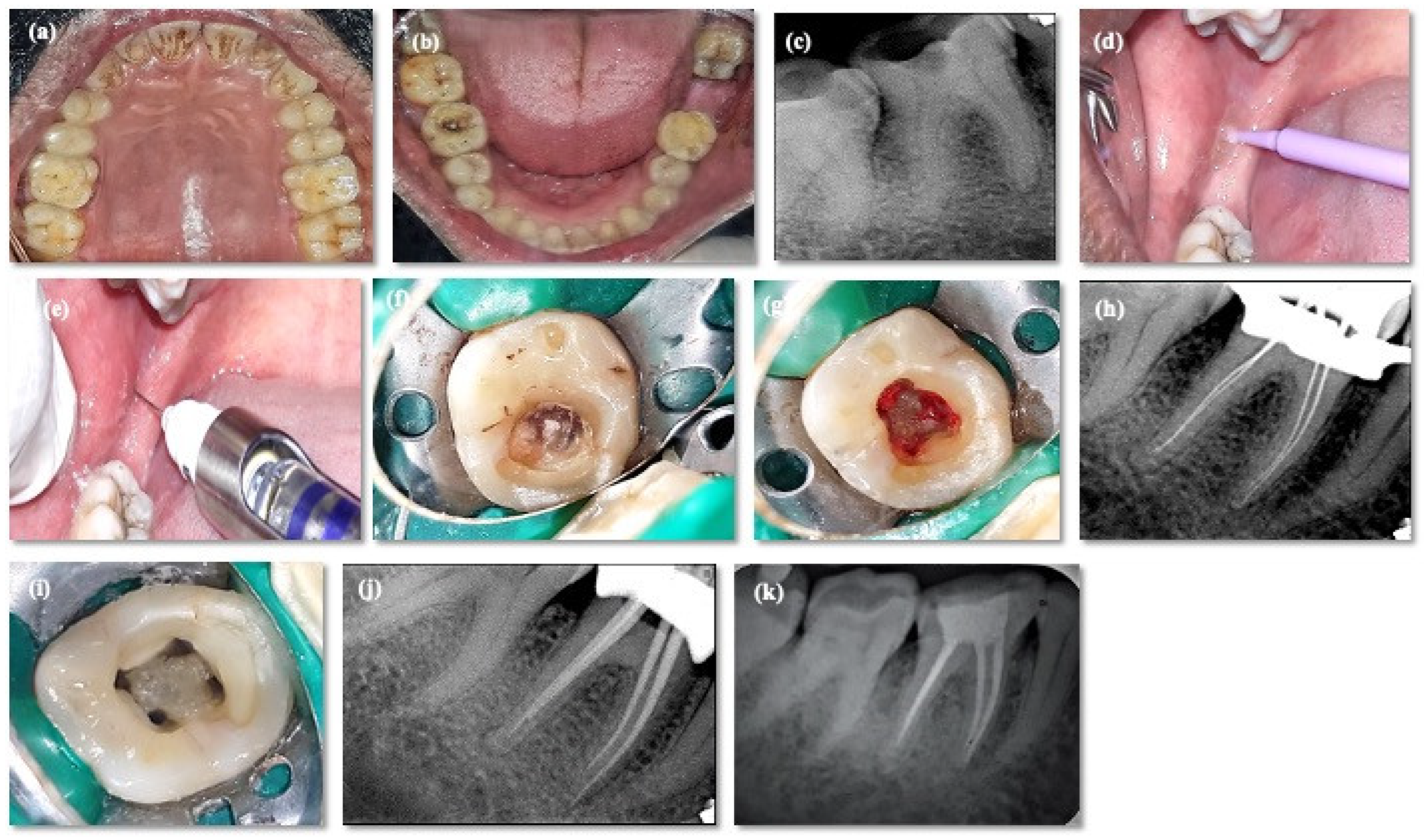
A small quantity of benzocaine gel (20% ProGel-B, Septodont, Saint-Maur-des-Fossés, France) was applied at the injection site using a microbrush applicator for 10–15 s. The aforementioned gel was left in contact with the mucosa for two minutes for effective action. Standard inferior alveolar nerve block (IANB) was administered using a solution comprised of 2% lidocaine with a 1:80,000 epinephrine concentration (Septodont, Saint-Maur-des-Fossés, France) using 1.8 mL Septodont cartridges (Septodont Fusion syringe, Septodont, Cambridge, ON, Canada) and 27-gauge needles (25 mm in length) (Septoject, Septodont, Navi Mumbai, India) over a 1-min time period. The time of onset (in seconds) of action of LA [VAS score (LA-VAS)] was recorded based on initiation of subjective symptoms (lip/tongue numb) by an independent trained evaluator who was blinded to the study protocol. Subsequently, rubber dam isolation of the tooth was performed, and the treatment was initiated within 15 min of LA administration [24]. Successively, access opening was performed using an Endo-Z bur (Dentsply, Gurugram, India), and the VAS score during pulp exposure (PE-VAS) was recorded. A complete de-roofing of the pulp chamber was performed using an Endo-Z bur (Dentsply, India). If pain persisted (VAS ≥ 4) during exposing the pulp or deroofing, the volume of the local anesthetic solution (1.8 mL) was increased using the second cartridge. The VAS score (CI-VAS) was recorded during orifice enlargement and canal instrumentation (CI). If pain continued to persist, a supplementary injection comprised of 2% lidocaine (Septocaine, Septodont, Cambridge, ON, Canada) was administered as a pulpal infiltration if needed. In the absence of pain, the procedure was continued, and the treatment was completed. Root canal procedures were performed by a single operator and were completed in 60 min. Figure 3 depicts clinical procedure of group 1.
Statistical analysis: The data were scrutinized, coded, and analyzed using IBM SPSS statistics 24.0 (SPSS South Asia Pvt Ltd., www.spss.co.in (accessed on April 2023). The descriptive statistics, including the mean and standard deviation of the scale variables, such as age, gender, and volume, were calculated using the frequency distribution procedure. A comparison of the 3 categories, LA-VAS, PE-VAS, and CI-VAS, with PRE-OP VAS and the mean time of onset of the three groups was performed using the analysis of variance (ANOVA) and linear regression analysis. p < 0.05 was considered to be statistically significant.

3. Results

The hemophilic group had a significantly higher proportion of patients in the age groups of 14–20 years (46.7%) and 21–29 years (36.7%), and the thalassemic group in the age group 30–39 years (36.7) (Table 3). The hemophilic group displayed extremely high male predominance (93.3%) (Table 4).
The comparison of the mean onset time between the groups is shown in Table 5, which implies that the hemophilic group had the longest mean onset time of 45.96 ± 34.32 s and control group had the shortest at 38.96 ± 12.85 s. However, there was no statistically significant difference in the mean onset time between the three groups of patients (p = 0.560).
During the pulp exposure procedure, 14 individuals experienced moderate pain, and a repeat volume (1.8 mL) of local anesthetic was administered. As a result, moderate pain was substantially reduced (Table 6).
The comparison of VAS at various intervals is presented in Table 7. The VAS scores between the groups at different interval were statistically insignificant. Pain perception of the three groups at Pre-VAS, LA-VAS, PE-VAS, and CI-VAS is summarized in Table 8. In the preoperative phase (Pre-VAS), the hemophilic, thalassemic, and control groups showed severe pain; however, LA administration (LA-VAS) resulted in a statistically significant reduction (p = 0.048) in all three groups. On pulp exposure (PE-VAS) (p = 0.82) and during canal instrumentation (CI-VAS) (p = 0.55), there was no statistically significant difference in pain perception between the groups.
The regression table (Table 9) indicates that the model is a good fit for predictor values. In LA-VAS, β suggests that for every unit change in onset time, the VAS increases by 1.37 times. In the Pre-op VAS, β is 7.3 indicating a higher VAS due to no treatment. The coefficients indicate a positive correlation between the VAS and onset time, indicating a positive reduction in VAS after LA administration.

4. Discussion

Bleeding disorders have always been associated with stigma in the field of dentistry. Hemophilia is the most common inherited bleeding disorder [25]. Conversely, thalassemia is a genetic disorder that involves abnormal hemoglobin formation [26,27]. Some mild forms of thalassemia may cause mild anemia and iron deficiency problems, which might even go unnoticed, while severe forms of thalassemia may even lead to death [2].
Studies have shown that thalassemic patients are susceptible to dental caries because their caries index is high and they have significantly low salivary phosphorous and IgA concentration [4,28]. This occurs because patients are hesitant to visit a dentist for fear of the procedure. According to studies, thalassemic patients are more concerned with their serious medical complications, neglecting their oral health. When decay is too advanced for restorative dental treatments/fillings, thalassemic patients seek dental emergency treatment [8]. Patients with hemophilia also exhibit the same symptoms. According to studies, hemophilic patients are more resistant to dental visits and fear dentists [29]. Patients fear that dental procedures will cause bleeding episodes that can only be controlled by factor infusions, resulting in both physical and financial challenges [29,30]. For the treatment of symptomatic irreversible pulpitis, there is typically no contraindication for endodontic treatment in hemophiliac patients/thalassemic patients. In fact, endodontic treatment is generally preferred over extraction whenever possible, as it does not pose a significant risk of bleeding [1].
In the present study, ≥14 years was included because adolescents currently anticipate a normal average life span and an excellent health-related standard of living [31], and from a dental perspective, complete root formation will be presumed [32]. Regarding treatment, patient comprehension, cooperation, and reciprocity were commendable, and it was observed that the hemophilic group had a significantly higher proportion of patients aged 14–20 and 21–29 years. However, it was found the average age of hemophilic patients was younger than that of thalassemic and control patients. In the studies, elderly individuals develop medical and surgical conditions (such as cancer, prostatic hypertrophy, cardiovascular disease, renal disease, pulmonary hypertension, endocrinological complication) that were not previously observed in younger individuals [33,34].
Pain measurement in these two bleeding disorders is challenging as it includes many associated systemic disorders and psychological factors. For pain measurements, the VAS/NRS are proven for quantification [35]. A study in thalassemic patients found that age increased pain regardless of the diagnosis, transfusion status, gender, bone density, chelator type, or iron overload [36], and hemophilic adolescent patients have preserved joints, acute and transient pain, and easily recognize acute bleeds that are treated earlier and resolved compared to adults [37].
In the current study, symptomatic irreversible pulpitis was considered, characterized by a sharp pain upon thermal stimulus, lingering pain (often 30 s or longer after stimulus removal), spontaneity (unprovoked pain), and referred pain. Postural changes, such as lying down or bending over, can worsen pain, and over-the-counter analgesics rarely work. Deep caries, extensive restorations, and pulpal tissue fractures are common causes. Because the inflammation has not reached the periapical tissues, symptomatic irreversible pulpitis may be difficult to diagnose. In such cases, dental history and thermal testing determine pulpal status [38]. The standard treatment for irreversible pulpitis in permanent mature teeth is non-surgical root canal treatment (NSRCT) [39].
In the present study, the oral hygiene of all groups was considerably good and minor additional clinical findings were treated in subsequent appointments. According to a study, the average onset of LA (Lidocaine) is 1–3 min for subjective symptoms and 1–4 min for objective symptoms [18]. Research shows that most patients experience pulpal anesthetic within 10–15 min of receiving conventional IANB [24,40]. To verify the effectiveness of anesthesia, the cold and EPT test was performed, followed by rubber dam isolation. This procedure was completed in 15 min, so access was opened following 15 min of LA administration. The LA allergic test was performed on three patients (one hemophilic and two in the control group) who had no history of LA administration and they all tested negative.
The local anesthetic solution comprising 2% lidocaine with 1:80,000 epinephrine is routinely used in endodontics. The use of a vasoconstrictor improves local hemostasis [41]. There are no constraints regarding the type of local anesthetic agent that can be used in hemophilic or thalassemic patients [1]. Hence, in this study, an IANB was performed using 2% lidocaine with 1:80,000 epinephrine.
Every year, about 15 million people worldwide are affected with thalassemia. In India, above 12,000 babies are born with hemoglobinopathies every year. On average, one in every twenty-five Indians is a thalassemia carrier [4]. Advances in the field of therapy for managing thalassemia major have emerged during the last two decades, resulting in a situation where patients can expect a normal life expectancy [4]. The proportion of patients in the thalassemic group who were older was considerably greater (i.e., above the age of 40 years). Four individuals in both the thalassemic and control groups experienced moderate pain during pulpal exposure. The pain was alleviated after administration of an extra 1.8 mL volume of the local anesthetic solution. This study included moderate to severe preoperative pain patients, and preoperative pain scores did not differ between the three groups.
Hemophilia is inherited X-linked recessive manner and most prevalent bleeding disorder caused by the deficiency of two coagulation factors: factor VIII (FVIII) or factor IX (FIX) [42,43]. In India, the prevalence of hemophilia A and B is 1 in 5000 and 1 in 30,000, respectively [44]. Hemophiliacs have a short life expectancy, according to past research. However, because of the development of safe and successful treatment procedures, hemophilic patients can now have a normal life expectancy [29]. Since hemophilia is a hemorrhagic ailment that usually exclusively affects male patients, this study found a substantial male predominance in the hemophilic group (X-related recessive disease) [25]. Hemophilia A is more common than hemophilia B, and accounts for 80–85 percent of all occurrences of hemophilia in India [1]. All the patients in the hemophilic group were found to have hemophilia A in the current study.
The hemophilic and thalassemic patients in this study experienced clinically longer onset of local anesthetic action than the control group, but difference was statistically insignificant. Using laser doppler flowmetry, researchers measured the pulpal blood flow in healthy individuals and found that 2% lidocaine (1:100,000 epinephrine) reduced (73%) profound pulpal blood flow at 5 min, with a gradual rebound occurring within 65 min [45]. The delay in onset of LA action may be attributable to compromised clotting proteins and the quantity and quality of hemoglobin in hemophilic and thalassemic patients, respectively, resulting in delay in vasoconstriction and blocking sodium ions flux, which inhibits nerve conduction [17].
In clinical studies involving endodontic procedures on patients with irreversible pulpitis, 19 to 56% of patients experienced mild or no discomfort during endodontic access cavity preparation or initial instrumentation on IANB [13]. According to a study, 39 percent of patients with irreversible pulpitis who received conventional IANB with 2% lidocaine remained sensitive to the cold test [46]. In cases of irreversible pulpitis, it may be difficult to achieve effective anesthesia utilizing the inferior alveolar nerve block alone, as suggested by the previous research [13,24]. Pain intensity is a crucial aspect of chronic pain and a key treatment goal, according to surveys of patients [47]. According to the present study, there was no statistically significant difference in VAS scores between the groups at various intervals, but all groups experienced pain relief after LA administration compared to preoperative levels. It was observed that 14 individuals (8 in group 1, 4 in groups 2 and 3) exhibited moderate pain during pulpal exposure; consequently, a volume of 1.8 mL LA was administered as the IANB as a first option [48]. Subsequently, those individuals were free of pain. Therefore, alternative procedures (such as buccal infiltration, interligamentary, intraosseous, and intrapulpal injections) were not required in the event of IANB failure.
In patients with bleeding disorders, avoiding instrumentation beyond the periapex is essential. As a result, extreme caution was used in the current investigation to avoid instrumentation and filling beyond the apical region of the tooth. The decreased anesthetic effects can be attributed to many reasons, including the fact that the IANB does not always result in significant pulpal anesthesia [13,16], as inflamed tissue nerves have altered resting potentials and decreased excitability thresholds [49,50], and irreversible pulpitis has enhanced sodium channel expression. Because of the lower excitability thresholds, local anesthetic agents do not impede impulse transmission, and the tetrodotoxin-resistant (TTXr) class of sodium channels is resistant to the action of local anesthetic agents [50], [51]. The current study is one of the few studies to investigate at the onset of local anesthesia and pain perception during endodontic procedures in thalassemic and hemophilic patients.
Patient–dentist variables constitute the limitations of the current study. Since pain is a subjective variable, its depiction on a VAS is affected by an individual’s emotional state and prior anesthetic administration experience [52]. The speed of the anesthetic procedure is regulated based on the dentist’s knowledge and skill. Ideally, the anesthetic should be administered slowly [17]. It has been demonstrated that nociceptors’ sensitivity is affected not only by the chemical agent employed, but also by the mechanical effects of the injection site, as well as the injection speed and volume [53]. In spite of these limitations, we were able to avoid the potential sources of bias identified by Hogan et al., 2011 [54] by generating and analyzing sufficient intervention sequences. Few clinical trials have been conducted on these individuals, and contradictory clinical results have been observed. Therefore, more research is required to determine the optimal LA solution type and concentration before it can be used consistently.

5. Conclusions

In conclusion, hemophilic patients required a clinically longer onset of LA action, followed by thalassemic patients, but this was statistically insignificant. However, the coefficients indicate a positive correlation between the VAS and onset time, indicating a positive reduction in VAS after the administration of LA. In terms of overall pain perception during and after pulp exposure and during canal instrumentation, there was no statistically significant difference. As a rule, prior to endodontic treatment, patients with hemophilia and thalassemia must undergo factor replacement therapy and a blood transfusion, followed by mandatory antibiotic prophylaxis, to prevent complications.

Author Contributions

Conceptualization, S.G.; methodology, S.D.(Supriya Das), S.G. and D.J.; soft-ware, S.D.(Supriya Das), S.G., S.P., S.C.B.B. and L.M.; validation, S.D.(Supriya Das), S.P., A.J., S.G., S.C.B.B. and D.J.; formal analysis, S.G., S.C.B.B. and S.D.(Supriya Das); investigation, S.G. and S.P.J.; resources, S.G., S.K. and S.P.J.; data curation, S.D.(Supriya Das), S.C.B.B. and S.G.; writing—original draft preparation, S.G. and D.Y.; writing—review and editing, S.G., S.P., S.K., A.J., L.M., J.K., D.Y., D.J. and S.D.(Supriya Das); visualization, S.G. and A.J.; supervision, S.D.(Sumit Dash), S.G. and A.J.; project administration, S.G., A.J., S.K., J.K. and D.J.; funding acquisition, S.D.(Sumit Dash), S.G., D.J., S.K., J.K., S.P.J., S.P., S.C.B.B. and D.Y. All authors have read and agreed to the published version of the manuscript.

Funding

This research received no external funding.

Institutional Review Board Statement

The study was conducted in accordance with the Declaration of Helsinki and approved by Institutional Ethics Committee of the Institute of Medical Sciences and SUM Hospital, Siksha ‘O’ Anusandhan (Deemed to be) University Bhubaneswar Odisha India (Ref No/DMR/IMS.SH/SOA/180317).

Informed Consent Statement

All study participants provided written informed consent for endodontic therapy.

Data Availability Statement

Requested data from corresponding author.

Acknowledgments

Priyanka Samal, Department of Hematology, SUM Hospital, and Swati Patnaik, Department of Public Health Dentistry, Institute of Dental Sciences, Siksha ‘O’ Anusandhan (Deemed to be) University Bhubaneswar Odisha, for their support in referring patients to the department and statistical analysis assistance, respectively.

Conflicts of Interest

The authors declare no conflict of interest.

References

  1. Shastry, S.P.; Kaul, R.; Baroudi, K.; Umar, D. Hemophilia A: Dental Considerations and Management. J. Int. Soc. Prev. Community Dent. 2014, 4, S147. [Google Scholar] [PubMed]
  2. Marengo-Rowe, A.J. The Thalassemias and Related Disorders. Bayl. Univ. Med. Cent. Proc. 2007, 20, 27–31. [Google Scholar] [CrossRef] [PubMed]
  3. Gosein, M.; Maharaj, P.; Balkaransingh, P.; Banfield, R.; Greene, C.; Latchman, S.; Sinanan, A. Imaging Features of Thalassaemia. Br. J. Radiol. 2019, 92, 20180658. [Google Scholar] [CrossRef]
  4. Madhok, D.S.; Madhok, D.S. Dental Considerations in Thalassemic Patients. IOSR J. Dent. Med. Sci. 2014, 13, 57–62. [Google Scholar] [CrossRef]
  5. Dudeja, P.G.; Dudeja, K.K.; Lakhanpal, M.; Ali, S. Endodontic Management of a Haemophilic Patient—A Clinical Perspective. J. Clin. Diagn. Res. JCDR 2014, 8, ZD17. [Google Scholar] [CrossRef] [PubMed]
  6. Mostafa, N.M.; Moussa, S.A. Endodontic Treatment in the Patients with Bleeding Disorders—Short Review. Int. J. Oral Health Dent. Manag. 2018, 2, 1–3. [Google Scholar] [CrossRef]
  7. Ricerca, B.M.; Di Girolamo, A.; Rund, D. Infections in Thalassemia and Hemoglobinopathies: Focus on Therapy-Related Complications. Mediterr. J. Hematol. Infect. Dis. 2009, 1, e2009028. [Google Scholar] [CrossRef]
  8. Helmi, N.; Bashir, M.; Shireen, A.; Ahmed, I.M. Thalassemia Review: Features, Dental Considerations and Management. Electron. Physician 2017, 9, 4003–4008. [Google Scholar] [CrossRef]
  9. Ong, C.K.S.; Lirk, P.; Tan, C.H.; Seymour, R.A. An Evidence-Based Update on Nonsteroidal Anti-Inflammatory Drugs. Clin. Med. Res. 2007, 5, 19–34. [Google Scholar] [CrossRef]
  10. Dolan, G. The Challenge of an Ageing Haemophilic Population. Haemoph. Off. J. World Fed. Hemoph. 2010, 16 (Suppl. S5), 11–16. [Google Scholar] [CrossRef]
  11. Arachchillage, D.R.J.; Makris, M. Choosing and Using Non-Steroidal Anti-Inflammatory Drugs in Haemophilia. Haemophilia 2016, 22, 179–187. [Google Scholar] [CrossRef] [PubMed]
  12. Chen, G.-Y.; Wu, Z.-F.; Lin, Y.-T.; Cheng, K.-I.; Huang, Y.-T.; Huang, S.-T.; Hargono, A.; Li, C.-Y. Association between General Anesthesia and Root Canal Treatment Outcomes in Patients with Mental Disability: A Retrospective Cohort Study. J. Pers. Med. 2022, 12, 213. [Google Scholar] [CrossRef] [PubMed]
  13. Nusstein, J.M.; Reader, A.; Drum, M. Local Anesthesia Strategies for the Patient with a “Hot” Tooth. Dent. Clin. N. Am. 2010, 54, 237–247. [Google Scholar] [CrossRef]
  14. Oosterink, F.M.; De Jongh, A.; Hoogstraten, J. Prevalence of Dental Fear and Phobia Relative to Other Fear and Phobia Subtypes. Eur. J. Oral Sci. 2009, 117, 135–143. [Google Scholar] [CrossRef] [PubMed]
  15. Bhatnagar, N.B.; Mantri, S.P.; Dube, K.A.; Jaiswal, N.U.; Singh, V.J. Pulpal-Anesthesia of a Mandibular First Molar with Irreversible Pulpitis by Inferior Alveolar Nerve Block plus Buccal Infiltration Using Articaine or Lignocaine. J. Conserv. Dent. JCD 2020, 23, 201–205. [Google Scholar] [CrossRef]
  16. Nair, M.; Gurunathan, D. Comparative Evaluation of the Efficacy of Two Anesthetic Gels (2% Lignocaine and 20% Benzocaine) in Reducing Pain during Administration of Local Anesthesia—A Randomized Controlled Trial. J. Anaesthesiol. Clin. Pharmacol. 2019, 35, 65. [Google Scholar]
  17. Malamed, S.F. Handbook of Local Anesthesia—E-Book; Elsevier Health Sciences: Amsterdam, The Netherlands, 2019. [Google Scholar]
  18. Kambalimath, D.H.; Dolas, R.S.; Kambalimath, H.V.; Agrawal, S.M. Efficacy of 4% Articaine and 2% Lidocaine: A Clinical Study. J. Maxillofac. Oral Surg. 2013, 12, 3–10. [Google Scholar] [CrossRef]
  19. Tortamano, I.P.; Siviero, M.; Lee, S.; Sampaio, R.M.; Simone, J.L.; Rocha, R.G. Onset and Duration Period of Pulpal Anesthesia of Articaine and Lidocaine in Inferior Alveolar Nerve Block. Braz. Dent. J. 2013, 24, 371–374. [Google Scholar] [CrossRef]
  20. Pain Intensity Threshold—Pain Management Collaboratory. Available online: https://painmanagementcollaboratory.org/pain-intensity-threshold/ (accessed on 15 April 2023).
  21. Ahmad, Z.H.; Ravikumar, H.; Karale, R.; Preethanath, R.S.; Sukumaran, A. Study of the Anesthetic Efficacy of Inferior Alveolar Nerve Block Using Articaine in Irreversible Pulpitis. J. Contemp. Dent. Pract. 2014, 15, 71–74. [Google Scholar] [CrossRef]
  22. Lee, J.; Lee, J.-Y.; Kim, H.J.; Seo, K.-S. Dental Anesthesia for Patients with Allergic Reactions to Lidocaine: Two Case Reports. J. Dent. Anesth. Pain Med. 2016, 16, 209–212. [Google Scholar] [CrossRef]
  23. Sui, H.; Lv, Y.; Xiao, M.; Zhou, L.; Qiao, F.; Zheng, J.; Sun, C.; Fu, J.; Chen, Y.; Liu, Y.; et al. Relationship between the Difference in Electric Pulp Test Values and the Diagnostic Type of Pulpitis. BMC Oral Health 2021, 21, 339. [Google Scholar] [CrossRef] [PubMed]
  24. Fernandez, C.; Reader, A.; Beck, M.; Nusstein, J. A Prospective, Randomized, Double-Blind Comparison of Bupivacaine and Lidocaine for Inferior Alveolar Nerve Blocks. J. Endod. 2005, 31, 499–503. [Google Scholar] [CrossRef] [PubMed]
  25. Bertamino, M.; Riccardi, F.; Banov, L.; Svahn, J.; Molinari, A.C. Hemophilia Care in the Pediatric Age. J. Clin. Med. 2017, 6, 54. [Google Scholar] [CrossRef] [PubMed]
  26. Vij, R.; Machado, R.F. Pulmonary Complications of Hemoglobinopathies. Chest 2010, 138, 973–983. [Google Scholar] [CrossRef]
  27. Joly, P.; Pondarre, C.; Badens, C. Beta-Thalassemias: Molecular, Epidemiological, Diagnostical and Clinical Aspects. Ann. Biol. Clin. 2014, 72, 639–668. [Google Scholar] [CrossRef]
  28. Boström, E.A.; Lira-Junior, R. Non-Malignant Blood Disorders and Their Impact on Oral Health: An Overview. Curr. Oral Health Rep. 2019, 6, 161–168. [Google Scholar] [CrossRef]
  29. Kumar, M.; Pai, K.M.; Kurien, A.; Vineetha, R. Oral Hygiene and Dentition Status in Children and Adults with Hemophilia: A Case–Control Study. Spec. Care Dent. 2018, 38, 391–394. [Google Scholar] [CrossRef]
  30. Nayak, S.; Govind, S.; Jena, A.; Samal, P.; Sahoo, N.K.; Rath, S. Evaluation of Oral Hygiene Status, Salivary Fluoride Concentration and Microbial Level in Thalassemic and Hemophilic Patients. Siriraj Med. J. 2022, 74, 314–322. [Google Scholar] [CrossRef]
  31. Gringeri, A.; Mantovani, L.; Mackensen, S.V. Quality of Life Assessment in Clinical Practice in Haemophilia Treatment. Haemoph. Off. J. World Fed. Hemoph. 2006, 12 (Suppl. S3), 22–29. [Google Scholar] [CrossRef]
  32. Logan, W.H.G.; Kronfeld, R. Development of the Human Jaws and Surrounding Structures from Birth to the Age of Fifteen Years. J. Am. Dent. Assoc. 1933, 20, 379–428. [Google Scholar] [CrossRef]
  33. Motta, I.; Mancarella, M.; Marcon, A.; Vicenzi, M.; Cappellini, M.D. Management of Age-Associated Medical Complications in Patients with β-Thalassemia. Expert Rev. Hematol. 2020, 13, 85–94. [Google Scholar] [CrossRef] [PubMed]
  34. Franchini, M.; Tagliaferri, A.; Mannucci, P.M. The Management of Hemophilia in Elderly Patients. Clin. Interv. Aging 2007, 2, 361–368. [Google Scholar] [PubMed]
  35. Stromer, W.; Pabinger, I.; Ay, C.; Crevenna, R.; Donnerer, J.; Feistritzer, C.; Hemberger, S.; Likar, R.; Sevelda, F.; Thom, K.; et al. Pain Management in Hemophilia: Expert Recommendations. Wien. Klin. Wochenschr. 2021, 133, 1042–1056. [Google Scholar] [CrossRef] [PubMed]
  36. Haines, D.; Martin, M.; Carson, S.; Oliveros, O.; Green, S.; Coates, T.; Eile, J.; Schilling, L.; Dinu, B.; Mendoza, T.; et al. Pain in Thalassaemia: The Effects of Age on Pain Frequency and Severity. Br. J. Haematol. 2013, 160, 680–687. [Google Scholar] [CrossRef]
  37. Auerswald, G.; Dolan, G.; Duffy, A.; Hermans, C.; Jiménez-Yuste, V.; Ljung, R.; Morfini, M.; Lambert, T.; Šalek, S.Z. Pain and Pain Management in Haemophilia. Blood Coagul. Fibrinolysis 2016, 27, 845. [Google Scholar] [CrossRef]
  38. Glossary of Endodontic Terms—American Association of Endodontists. Available online: https://www.aae.org/specialty/clinical-resources/glossary-endodontic-terms/ (accessed on 16 April 2023).
  39. Santos, J.M.; Pereira, J.F.; Marques, A.; Sequeira, D.B.; Friedman, S. Vital Pulp Therapy in Permanent Mature Posterior Teeth with Symptomatic Irreversible Pulpitis: A Systematic Review of Treatment Outcomes. Medicina 2021, 57, 573. [Google Scholar] [CrossRef]
  40. Boopathi, T.; Sebeena, M.; Sivakumar, K.; Harikaran, J.; Karthick, K.; Raj, A. Supplemental Pulpal Anesthesia for Mandibular Teeth. J. Pharm. Bioallied Sci. 2013, 5, S103–S108. [Google Scholar] [CrossRef]
  41. Anderson, J.A.M.; Brewer, A.; Creagh, D.; Hook, S.; Mainwaring, J.; McKernan, A.; Yee, T.T.; Yeung, C.A. Guidance on the Dental Management of Patients with Haemophilia and Congenital Bleeding Disorders. Br. Dent. J. 2013, 215, 497–504. [Google Scholar] [CrossRef]
  42. Srivastava, A.; Brewer, A.K.; Mauser-Bunschoten, E.P.; Key, N.S.; Kitchen, S.; Llinas, A.; Ludlam, C.A.; Mahlangu, J.N.; Mulder, K.; Poon, M.C.; et al. Guidelines for the Management of Hemophilia. Haemoph. Off. J. World Fed. Hemoph. 2013, 19, e1–e47. [Google Scholar] [CrossRef]
  43. Brewer, A.; Correa, M.E. Guidelines for Dental Treatment of Patients with Inherited Bleeding Disorders. Haemophilia 2005, 11, 504–509. [Google Scholar]
  44. Kar, A.; Phadnis, S.; Dharmarajan, S.; Nakade, J. Epidemiology & Social Costs of Haemophilia in India. Indian J. Med. Res. 2014, 140, 19. [Google Scholar] [PubMed]
  45. Ahn, J.; Pogrel, M.A. The Effects of 2% Lidocaine with 1:100,000 Epinephrine on Pulpal and Gingival Blood Flow. Oral Surg. Oral Med. Oral Pathol. Oral Radiol. Endodontology 1998, 85, 197–202. [Google Scholar] [CrossRef] [PubMed]
  46. Berman, L.H.; Hargreaves, K.M. Cohen’s Pathways of the Pulp Expert Consult—E-Book; Elsevier Health Sciences: Amsterdam, The Netherlands, 2015. [Google Scholar]
  47. Turk, D.C.; Dworkin, R.H.; Revicki, D.; Harding, G.; Burke, L.B.; Cella, D.; Cleeland, C.S.; Cowan, P.; Farrar, J.T.; Hertz, S.; et al. Identifying Important Outcome Domains for Chronic Pain Clinical Trials: An IMMPACT Survey of People with Pain. Pain 2008, 137, 276–285. [Google Scholar] [CrossRef]
  48. Lee, C.; Yang, H. Alternative Techniques for Failure of Conventional Inferior Alveolar Nerve Block. J. Dent. Anesth. Pain Med. 2019, 19, 125. [Google Scholar] [CrossRef] [PubMed]
  49. Wallace, J.A.; Michanowicz, A.E.; Mundell, R.D.; Wilson, E.G. A Pilot Study of the Clinical Problem of Regionally Anesthetizing the Pulp of an Acutely Inflamed Mandibular Molar. Oral Surg. Oral Med. Oral Pathol. 1985, 59, 517–521. [Google Scholar] [CrossRef]
  50. Byers, M.R.; Taylor, P.E.; Khayat, B.G.; Kimberly, C.L. Effects of Injury and Inflammation on Pulpal and Periapical Nerves. J. Endod. 1990, 16, 78–84. [Google Scholar] [CrossRef]
  51. Roy, M.L.; Narahashi, T. Differential Properties of Tetrodotoxin-Sensitive and Tetrodotoxin-Resistant Sodium Channels in Rat Dorsal Root Ganglion Neurons. J. Neurosci. 1992, 12, 2104–2111. [Google Scholar] [CrossRef]
  52. Kjeldsen, H.B.; Klausen, T.W.; Rosenberg, J. Preferred Presentation of the Visual Analog Scale for Measurement of Postoperative Pain. Pain Pract. 2016, 16, 980–984. [Google Scholar] [CrossRef]
  53. Wang, W. Tolerability of Hypertonic Injectables. Int. J. Pharm. 2015, 490, 308–315. [Google Scholar] [CrossRef]
  54. Hogan, M.-E.; Perampaladas, K.; Machado, M.; Einarson, T.R.; Taddio, A. Systematic Review and Meta-Analysis of the Effect of Warming Local Anesthetics on Injection Pain. Ann. Emerg. Med. 2011, 58, 86–98. [Google Scholar] [CrossRef]
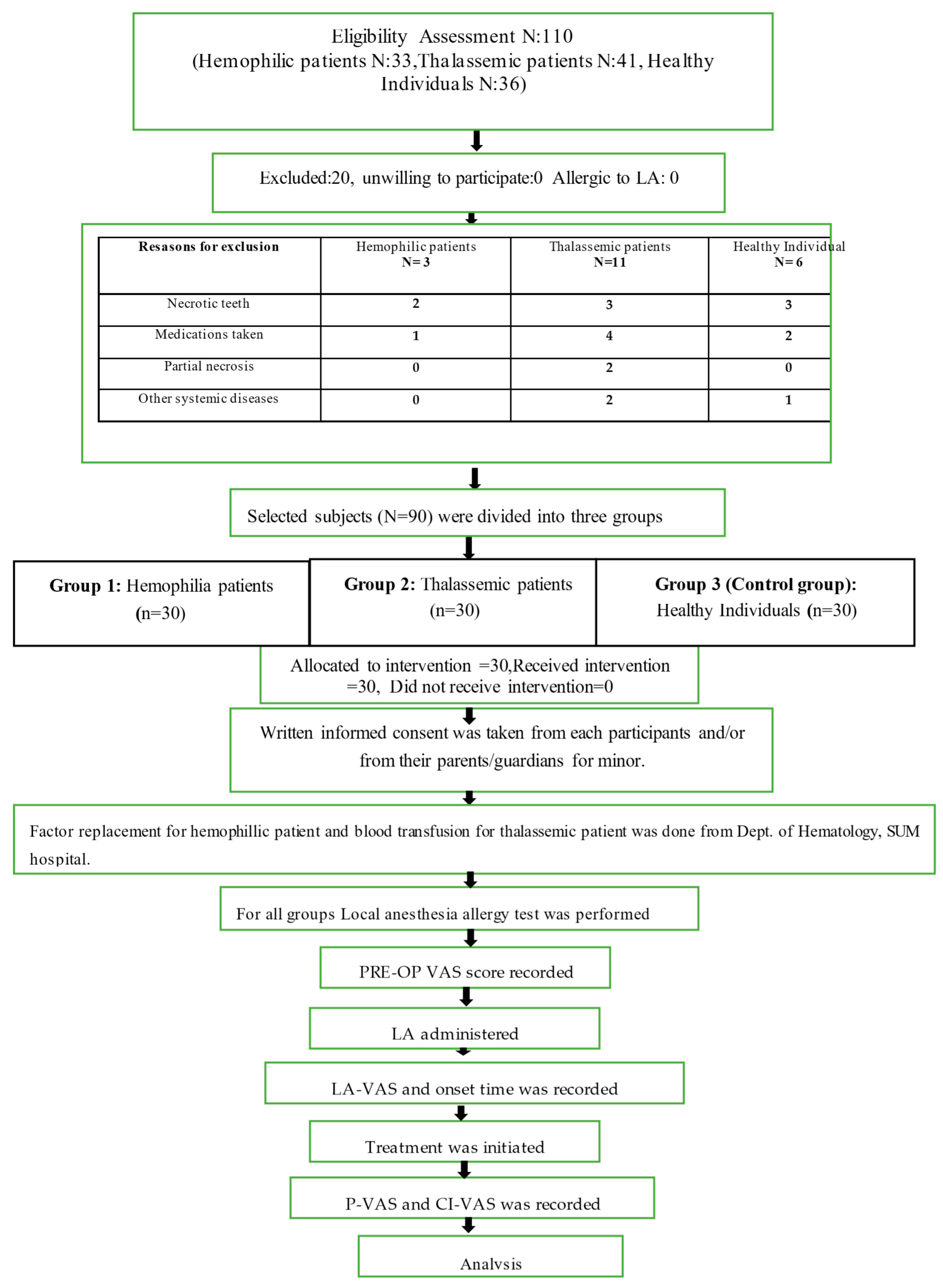
Figure 1. Sample selection and methodology flowchart.
Jcm 12 03646 g001
Figure 2. Five-point visual analog scale (NRS).
Jcm 12 03646 g002
Figure 3. Demonstrating clinical steps representing the groups (hemophilic case). (a,b) Pre-operative maxillary arch and mandibular arch; (c,f) pre-operative radiograph and clinical picture i.r.t 46; (d) topical anesthesia application; (e) inferior alveolar nerve block; (g) pulp exposure; (h) working length determination; (i) after cleaning and shaping; (j) master cone selection; and (k) post obturation radiograph.
Jcm 12 03646 g003
Table 1. Details of factor replacement in hemophilic patients.
Case
No.
Age/SexTooth
No
HemophiliaFactor
Deficient
Blood
Group
Severity of
Hemophilia
Factor Replacement
Per Appointment
[IU/dL] [Eloctate]
Frequency
120/M46AVIIIA+Severe1000 unitsOnce in every 2 days
217/M36AVIIIB+mild250 units3 days/week
315/M36AVIIIAB+mild250 units2 days/week
419/F46AVIIIAB+moderate1000 units2 days/week
525/M37AVIIIAB+moderate1000 units3 days/week
630/M37AVIIIA+Severe1500 units3 days/week
722/M47AVIIIB+Severe1500 unitsOnce in every 2 days
814/M46AVIIIA+Severe500 unitsOnce in every 2 days
916/F36AVIIIAB+moderate500 units2 days/week
1028/M37AVIIIB+moderate1000 units3 days/week
1127/M37AVIIIB+mild500 units2 days/week
1230/M47AVIIIAB+mild500 units2 days/week
1324/M46AVIIIAB+mild500 units2 days/week
1415/M37AVIIIA+mild500 units2 days/week
1524/M36AVIIIB+mild500 units2 days/week
1615/M46AVIIIAB+mild500 units2 days/week
1743/M38AVIIIAB+mild500 units3 days/week
1829/M46AVIIIB+moderate1000 units3 days/week
1928/M36AVIIIAB+Severe1500 units3 days/week
2026/M47AVIIIA+Severe1000 unitsOnce in every 2 days
2133/M46AVIIIA+Severe1000 unitsOnce in every 2 days
2235/M46AVIIIA+mild500 units2 days/week
2314/M36AVIIIA+mild500 units2 days/week
2418/M46AVIIIAB+mild500 units2 days/week
2527/M36AVIIIAB+mild1000 units2 days/week
2614/M36AVIIIB+mild500 units2 days/week
2720/M47AVIIIB+Severe1000 units3 days/week
2822/M46AVIIIB+mild500 units2 days/week
2918/M36AVIIIAB+moderate1000 units2 days/week
3020/M46AVIIIAB+mild1000 units2 days/week
The degree of severity is varied: The normal range of factor VIII is 50–100 IU/dl (severe (<1 IU/dL per kg), moderate (1–5 IU/dL per Kg), mild (6–40 IU/dL per kg), and carriers who are treated as mild hemophiliacs if the factor level is <50 IU/dL per kg).
Table 2. Details of blood transfusion and iron-chelation therapy in thalassemic patients.
S
No.
Age/SexTooth
No.
Type of
Thalassemia
Blood
Transfusion
Patient with
Iron-Chelation
Therapy
Patient without
Iron-Chelation
Therapy
SplenectomyDisease Onset
148/M47B-TIOnce a year 4 years old
250/F46B-TIOnce in 6 months 6 years old
338/M36B-TMiNo 13 years old
444/F37B-TMiNo 16 years old
529/F46B-TMEvery 2 weeks 2 years old
620/M37B-TMEvery 2 weeks At birth
735/F46B-TMEvery 2 weeks 2 monthsold
818/M48B-TMEvery 3 weeks At birth
916/M47B-TMEvery 3 weeks 6 months old
1030/M47B-TMEvery 2 weeks 5 months old
1115/M36B-TMEvery 3 weeks 6 months old
1219/M36B-TMEvery 3 weeks 2 months old
1327/M36B-TMEvery 2 weeks At birth
1426/M48B-TMEvery 2 weeks At birth
1515/M46B-TMEvery 3 weeks 2 months old
1658/F37B-TMiNo 18 years old
1714/F36B-TMEvery 3 weeks
1817/M36B-TMEvery 3 weeks 4 months old
1920/M47B-TMEvery 3 weeks 5 months old
2032/F46B-TIOnce in 6 months 10 months
2133/F36B-TIOnce in 6 months 5 years old
2238/M37B-TIOnce a year 7 years old
2341/F46B-TIOnce a year 4 years old
2433/F38B-TIOnce in 6 months 4yrs old
2530/M46B-TMEvery 2 weeks At birth
2626/F36B-TMEvery 2 weeks 2 months old
2732/F36B-TMEvery 2 weeks 5 months old
2831/M46B-TMEvery 2 weeks 8 months old
2934/M46B-TMiNo 10 years old
3022/F37B-TMEvery 2 weeks 2 months old
B-TMi: Beta-Thalassemia minor, B-TI: Beta-Thalassemic intermedia, B-TM: Beta-Thalassemia major.
Table 3. Age distribution by groups.
Age Group
Years
HemophilicThalassemicControl
No.%No.%No.%
14–201446.7930620
21–291136.7516.7723.3
30–39413.31136.71033.3
≥4013.3516.7723.3
Total301003010030100
Mean ± SD (Years)22.93 ± 7.1729.7 ± 11.2430.33 ± 10.4
Table 4. Gender distribution by groups.
GroupMaleFemaleTotal
No.%No.%No.%
Hemophilic2893.326.730100
Thalassemic1756.71343.330100
Control1343.31756.730100
Total5864.43235.690100
Table 5. Comparison of Onset time (in sec) of LA.
GroupsNMeanStd. DeviationStd. ErrorANOVA (p Value)
Onset time of LAHemophilia3045.96734.32156.26620.56
Thalassemia3042.40023.36314.2655
Control3038.96712.85322.3467
Total9042.44424.97552.6327
Table 6. Volume of anesthesia increases within the groups.
GroupYesNoTotal
No.%No.%No.%
Hemophilic620248030100
Thalassemic413.32686.730100
Control413.32686.730100
Total1415.67684.490100
Table 7. Comparison of the VAS at different intervals among the groups.
NMeanStd. DeviationStd. ErrorANOVA (p Value)
Pre-op VASHemophilia307.4671.65540.30220.17
Thalassemia308.0001.36460.2491
Control308.1331.25210.2286
Total907.8671.44720.1525
LA-VASHemophilia302.7332.57220.46960.16
Thalassemia303.3002.18380.3987
Control302.1332.28540.4173
Total902.7222.37510.2504
PE-VASHemophilia301.2672.08330.38040.82
Thalassemia301.0001.80040.3287
Control 301.0001.70190.3107
Total901.0891.85210.1952
CI-VASHemophilia300.1000.30510.05570.55
Thalassemia300.1000.30510.0557
Control 300.0330.18260.0333
Total900.0780.26930.0284
Table 8. Comparison of pain perception at various intervals.
NMeanStd. DeviationStd. ErrorANOVA (p Value)
Pre-op VASMild301.0000.00000.0000
Moderate301.0000.00000.0000
Severe301.0000.00000.0000
Total901.0000.00000.0000
LA-VASMild300.6000.49830.09100.048
Moderate300.7000.46610.0851
Severe300.8330.37900.0692
Total900.7110.45580.0480
PE-VASMild300.5670.72790.13290.926
Moderate300.5670.81720.1492
Severe300.5000.73110.1335
Total900.5440.75190.0793
CI-VASMild300.0330.18260.03330.547
Moderate300.1000.30510.0557
Severe300.1000.30510.0557
Total900.0780.26930.0284
† Significant at p < 0.05.
Table 9. Correlation between the VAS and onset time.
ModelCoefficients
Unstandardized
Coefficients
Standardized
Coefficients
tSig.
BStd. ErrorBeta
Pre-op VAS7.3000.2960.23024.6960.000
LA-VAS1.3680.4700.3353.3410.001
PE-VAS−0.2530.3520.426−0.7200.473
Disclaimer/Publisher’s Note: The statements, opinions and data contained in all publications are solely those of the individual author(s) and contributor(s) and not of MDPI and/or the editor(s). MDPI and/or the editor(s) disclaim responsibility for any injury to people or property resulting from any ideas, methods, instructions or products referred to in the content.