
Clinical and Histological Effects of Partial Blood Flow Impairment in Vascularized Lymph Node Transfer

 , ,
, ,
Abstract
:1. Introduction
2. Materials and Methods
2.1. Clinical Study
2.1.1. Patients
2.1.2. Surgical Strategy
2.1.3. Postoperative Course
2.2. Animal Study
2.2.1. Experimental Animal Models
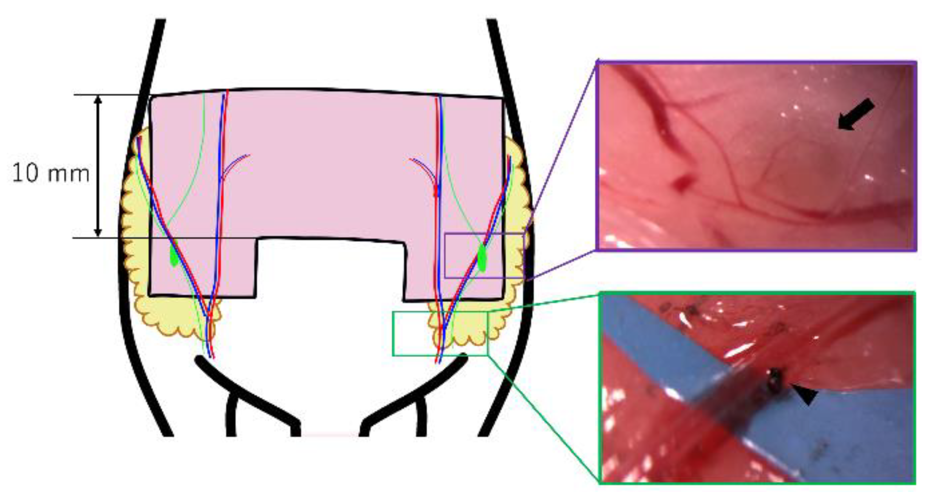
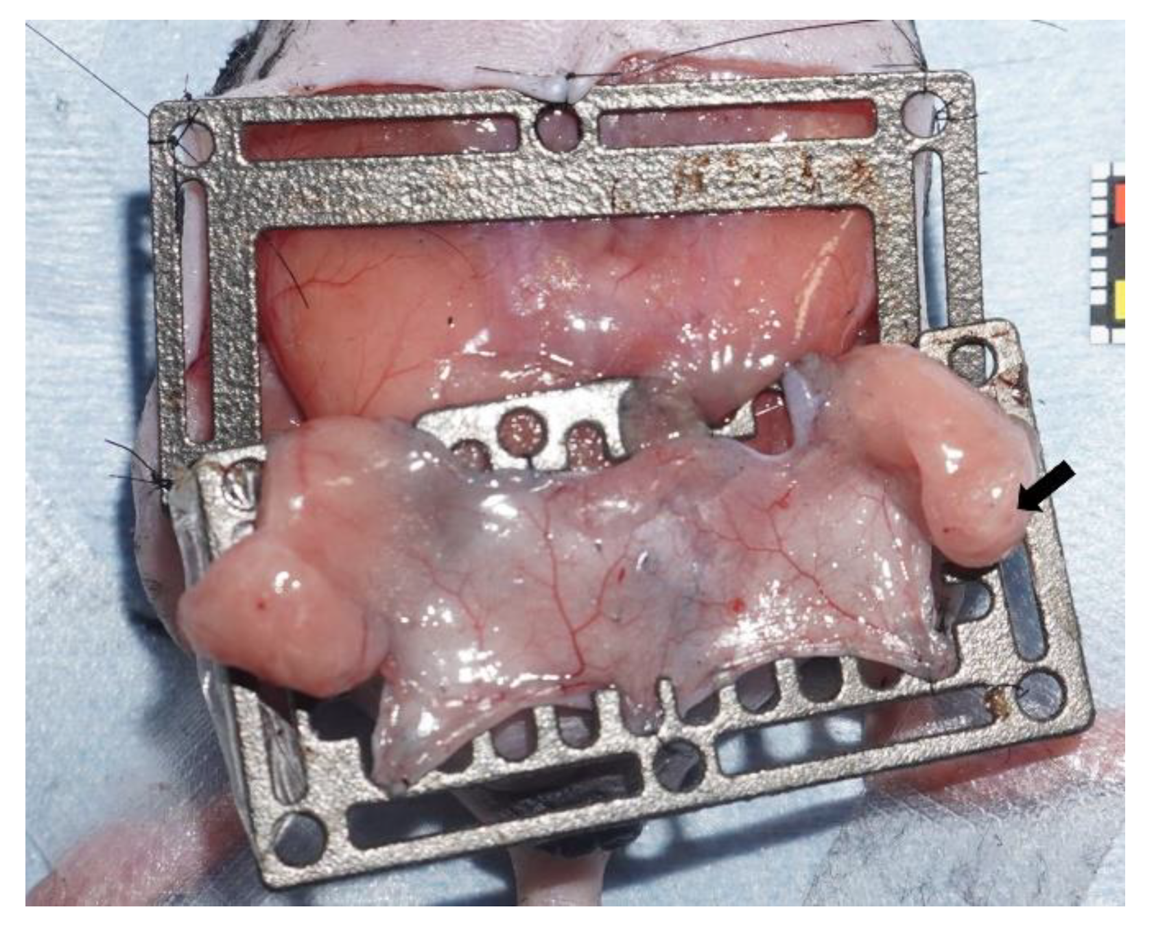
2.2.2. Murine Model of Bilateral Pedicled Abdominal Flaps with Groin LNs
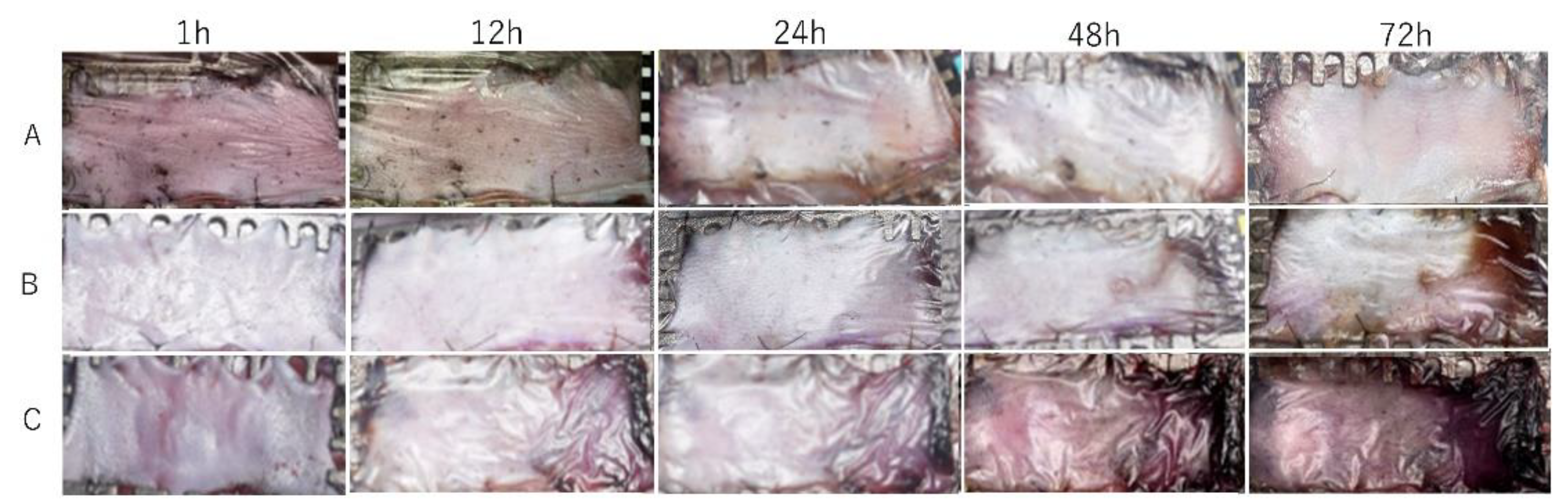
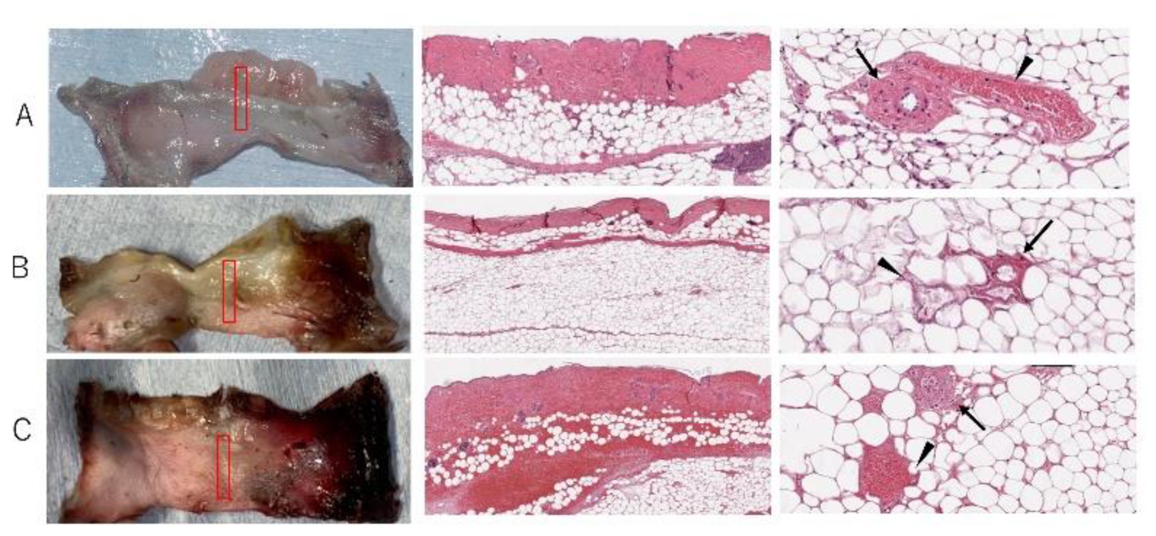
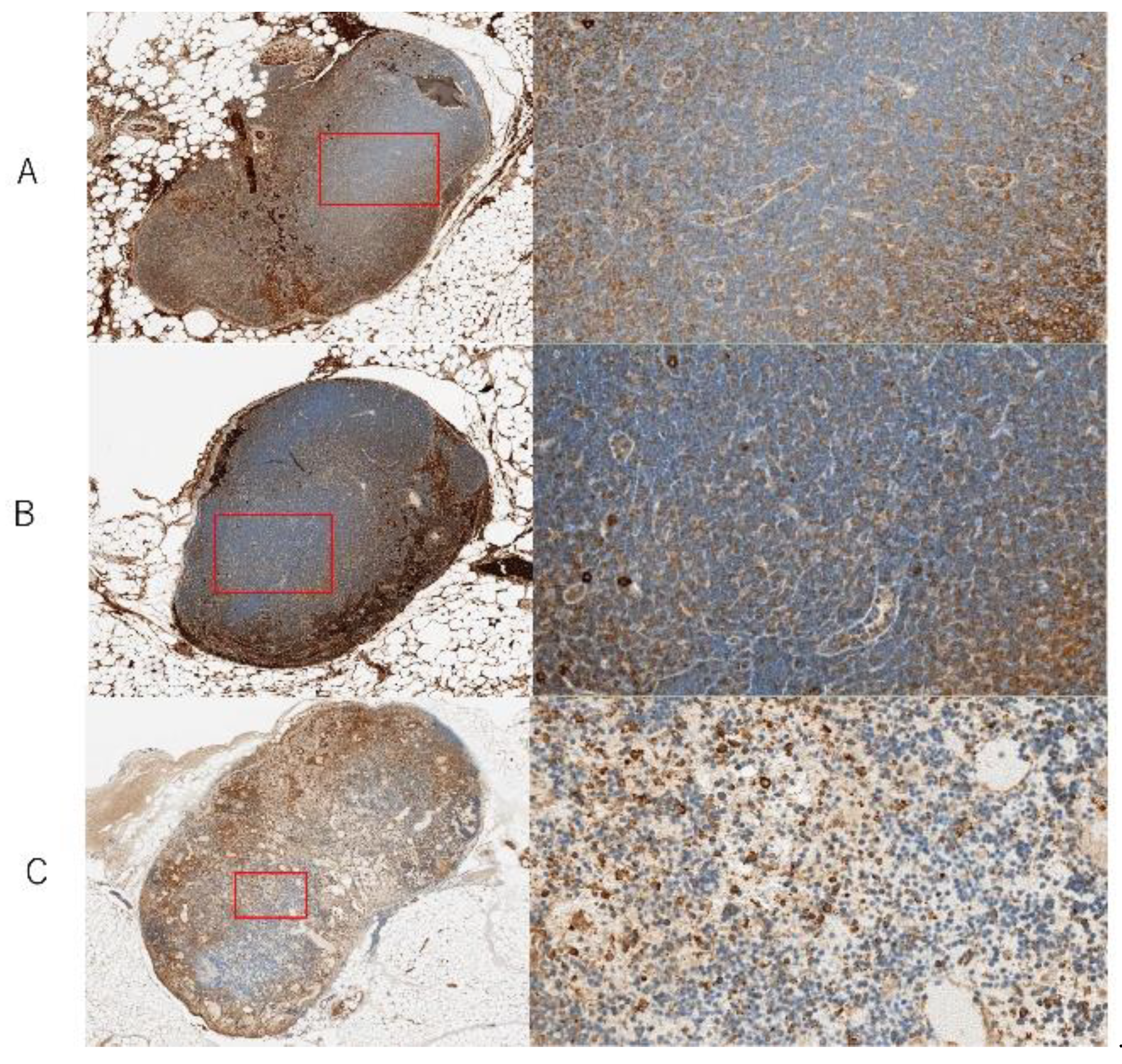
2.2.3. Pathologic Features of Ischemic and Congestive LNs
2.3. Statistical Analysis
3. Results
3.1. Clinical Study
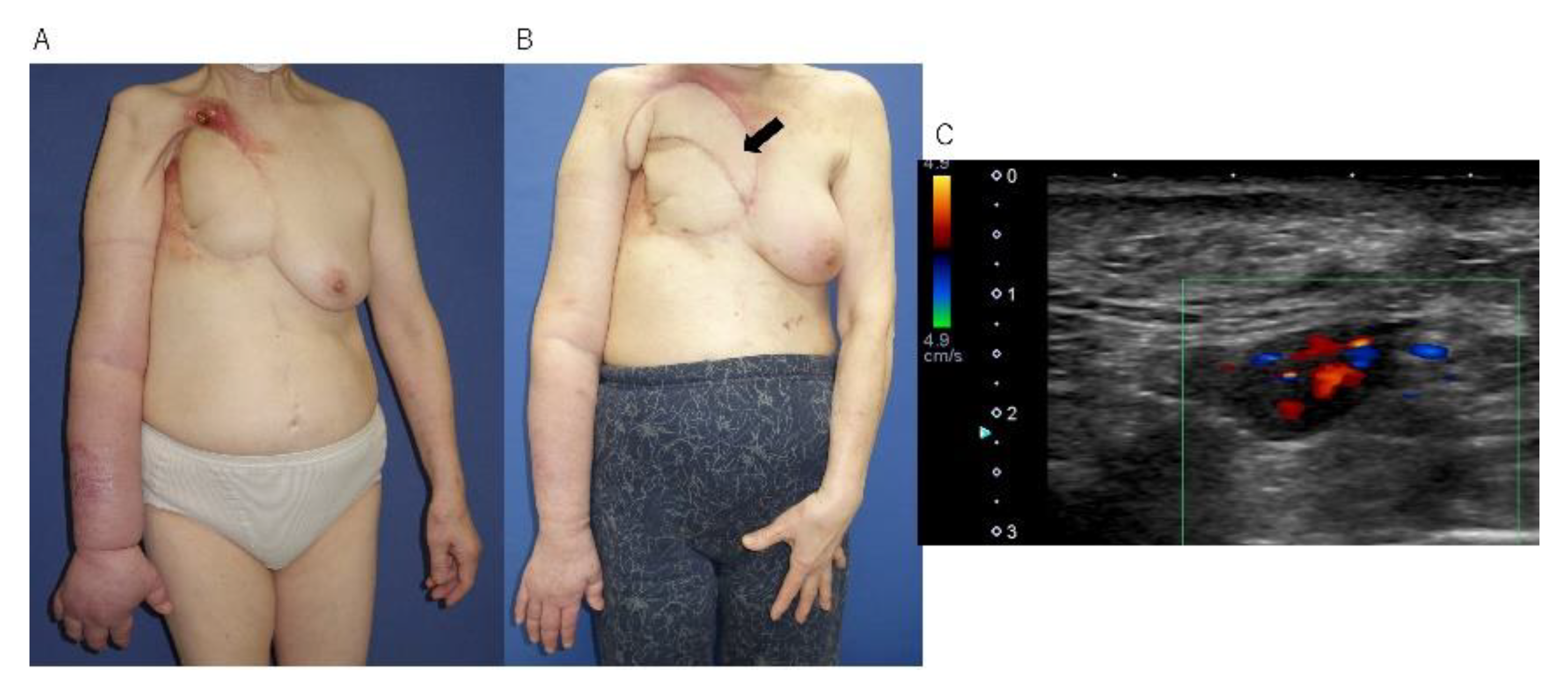
3.2. Case Report
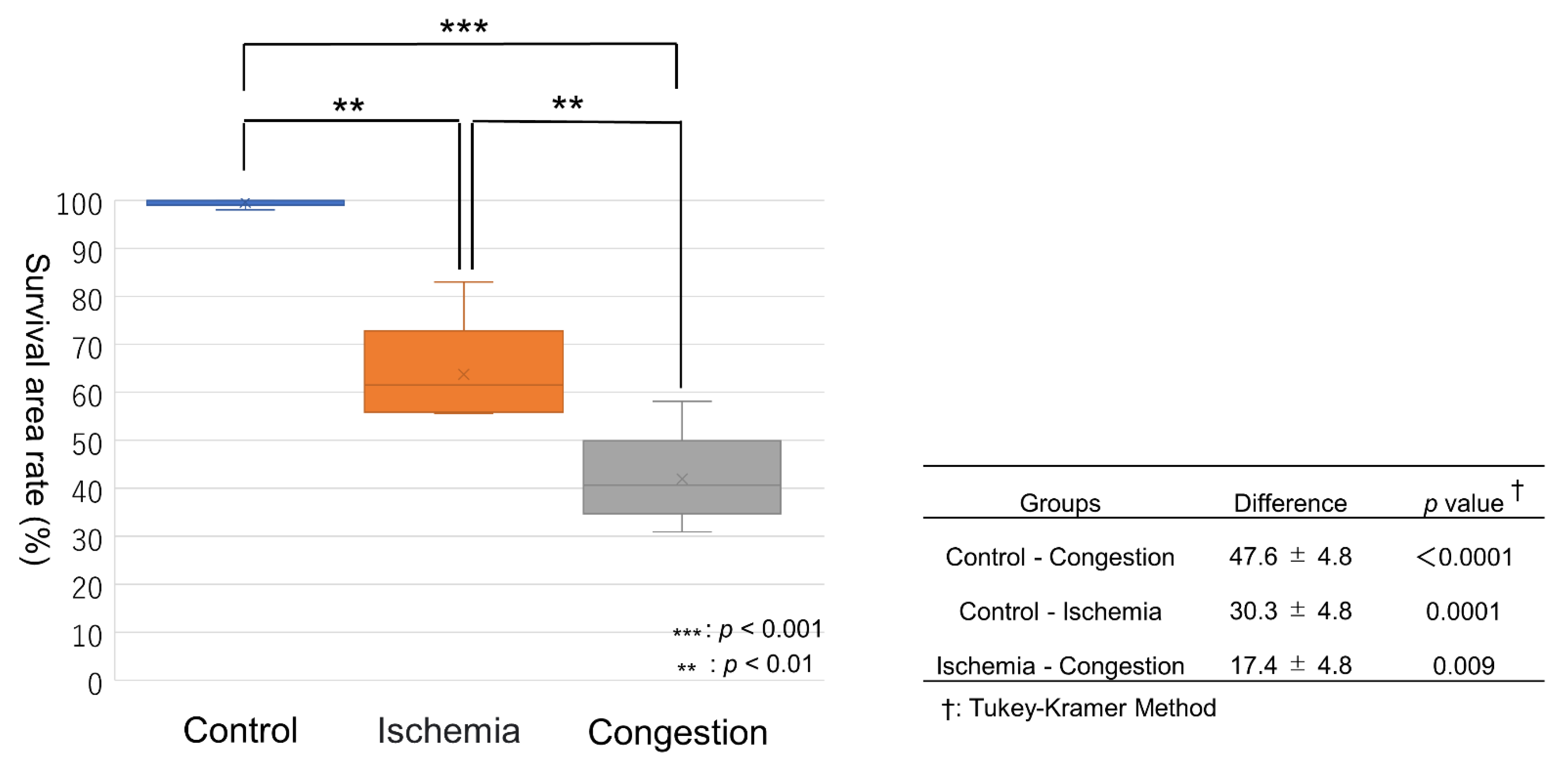
3.3. Mouse Model
4. Discussion
5. Conclusions
Author Contributions
Funding
Institutional Review Board Statement
Informed Consent Statement
Data Availability Statement
Conflicts of Interest
References
- Becker, C.; Assouad, J.; Riquet, M.; Hidden, G. Postmastectomy lymphedema: Long-term results following microsurgical lymph node transplantation. Ann. Surg. 2006, 243, 313–315. [Google Scholar] [CrossRef] [PubMed]
- Lin, C.H.; Ali, R.; Chen, S.C.; Wallace, C.; Chang, Y.C.; Chen, H.C.; Cheng, M.H. Vascularized groin lymph node transfer using the wrist as a recipient site for management of postmastectomy upper extremity lymphedema. Plast. Reconstr. Surg. 2009, 123, 1265–1275. [Google Scholar] [CrossRef] [PubMed] [Green Version]
- Cheng, M.H.; Chen, S.C.; Henry, S.L.; Tan, B.K.; Chia-Yu Lin, M.; Huang, J.J. Vascularized groin lymph node flap transfer for postmastectomy upper limb lymphedema: Flap anatomy, recipient sites, and outcomes. Plast. Reconstr. Surg. 2013, 131, 1286–1298. [Google Scholar] [CrossRef] [PubMed]
- Cheng, M.H.; Huang, J.J.; Nguyen, D.H.; Saint-Cyr, M.; Zenn, M.R.; Tan, B.K.; Lee, C.L. A novel approach to the treatment of lower extremity lymphedema by transferring a vascularized submental lymph node flap to the ankle. Gynecol. Oncol. 2012, 126, 93–98. [Google Scholar] [CrossRef]
- Honkonen, K.M.; Visuri, M.T.; Tervala, T.V.; Halonen, P.J.; Koivisto, M.; Lähteenvuo, M.T.; Alitalo, K.K.; Ylä-Herttuala, S.; Saaristo, A.M. Lymph node transfer and perinodal lymphatic growth factor treatment for lymphedema. Ann. Surg. 2013, 257, 961–967. [Google Scholar] [CrossRef]
- Raju, A.; Chang, D.W. Vascularized lymph node transfer for treatment of lymphedema: A comprehensive literature review. Ann. Surg. 2015, 261, 1013–1023. [Google Scholar] [CrossRef]
- De Brucker, B.; Zeltzer, A.; Seidenstuecker, K.; Hendrickx, B.; Adriaenssens, N.; Hamdi, M. Breast Cancer-Related Lymphedema: Quality of Life after Lymph Node Transfer. Plast. Reconstr. Surg. 2016, 137, 1673–1680. [Google Scholar] [CrossRef]
- Gratzon, A.; Schultz, J.; Secrest, K.; Lee, K.; Feiner, J.; Klein, R.D. Clinical and Psychosocial Outcomes of Vascularized Lymph Node Transfer for the Treatment of Upper Extremity Lymphedema After Breast Cancer Therapy. Ann. Surg. Oncol. 2017, 24, 1475–1481. [Google Scholar] [CrossRef]
- Akita, S.; Mitsukawa, N.; Kuriyama, M.; Kubota, Y.; Hasegawa, M.; Tokumoto, H.; Ishigaki, T.; Togawa, T.; Kuyama, J.; Satoh, K. Comparison of vascularized supraclavicular lymph node transfer and lymphaticovenular anastomosis for advanced stage lower extremity lymphedema. Ann. Plast. Surg. 2015, 74, 573–579. [Google Scholar] [CrossRef]
- Ward, J.; King, I.; Monroy-Iglesias, M.; Russell, B.; van Hemelrijck, M.; Ramsey, K.; Khan, A.A. A meta-analysis of the efficacy of vascularised lymph node transfer in reducing limb volume and cellulitis episodes in patients with cancer treatment-related lymphoedema. Eur. J. Cancer 2021, 151, 233–244. [Google Scholar] [CrossRef]
- Akita, S.; Yamaji, Y.; Tokumoto, H.; Sasahara, Y.; Kubota, Y.; Kuriyama, M.; Mitsukawa, N. Improvement of the efficacy of vascularized lymph node transfer for lower-extremity lymphedema via a prefabricated lympho-venous shunt through lymphaticovenular anastomosis between the efferent lymphatic vessel and small vein in the elevated vascularized lymph node. Microsurgery 2018, 38, 270–277. [Google Scholar]
- Tinhofer, I.E.; Yang, C.Y.; Chen, C.; Cheng, M.H. Impacts of arterial ischemia or venous occlusion on vascularized groin lymph nodes in a rat model. J. Surg. Oncol. 2020, 121, 153–162. [Google Scholar] [CrossRef] [Green Version]
- Cornelissen, A.J.; Qiu, S.S.; Lopez Penha, T.; Keuter, X.; Piatkowski de Grzymala, A.; Tuinder, S.; van der Hulst, R. Outcomes of vascularized versus non-vascularized lymph node transplant in animal models for lymphedema. Review of the literature. J. Surg. Oncol. 2017, 115, 32–36. [Google Scholar] [CrossRef]
- Tobbia, D.; Semple, J.; Baker, A.; Dumont, D.; Johnston, M. Experimental assessment of autologous lymph node transplantation as treatment of postsurgical lymphedema. Plast. Reconstr. Surg. 2009, 124, 777–786. [Google Scholar] [CrossRef] [Green Version]
- Visconti, G.; Constantinescu, T.; Chen, P.Y.; Salgarello, M.; Franceschini, G.; Masetti, R.; Chen, H.C. The Venous Lymph Node Flap: Concepts, Experimental Evidence, and Potential Clinical Implications. J. Reconstr. Microsurg. 2016, 32, 625–631. [Google Scholar]
- Ishikawa, K.; Maeda, T.; Funayama, E.; Hayashi, T.; Murao, N.; Osawa, M.; Furukawa, H.; Oyama, A.; Yamamoto, Y. Feasibility of pedicled vascularized inguinal lymph node transfer in a mouse model: A preliminary study. Microsurgery 2019, 39, 247–254. [Google Scholar] [CrossRef]
- Akita, S.; Unno, N.; Maegawa, J.; Kimata, Y.; Fukamizu, H.; Yabuki, Y.; Kitayama, S.; Shinaoka, A.; Yamada, K.; Sano, M.; et al. A phase III, multicenter, single-arm study to assess the utility of indocyanine green fluorescent lymphography in the treatment of secondary lymphedema. J. Vasc. Surg. Venous Lymphat. Disord. 2022, 10, 728–737.e3. [Google Scholar] [CrossRef]
- Yoshimatsu, H.; Visconti, G.; Karakawa, R.; Hayashi, A. Lymphatic System Transfer for Lymphedema Treatment: Transferring the Lymph Nodes with Their Lymphatic Vessels. Plast. Reconstr. Surg. Glob. Open 2020, 8, e2721. [Google Scholar] [CrossRef]
- Akita, S.; Tokumoto, H.; Yamaji, Y.; Sasahara, Y.; Kubota, Y.; Kubo, M.; Kuriyama, M.; Mitsukawa, N. Contribution of Simultaneous Breast Reconstruction by Deep Inferior Epigastric Artery Perforator Flap to the Efficacy of Vascularized Lymph Node Transfer in Patients with Breast Cancer-Related Lymphedema. J. Reconstr. Microsurg. 2017, 33, 571–578. [Google Scholar]
- Akita, S.; Mitsukawa, N.; Kubota, Y.; Sakakibara, M.; Nagashima, T.; Satoh, K. Delayed Partial Breast Reconstruction and Vascularized Lymph Node Transfer by a Superficial Circumflex Iliac Artery Perforator Flap. Plast. Reconstr. Surg. 2016, 137, 490e–491e. [Google Scholar] [CrossRef]
- Akita, S.; Mitsukawa, N.; Tokumoto, H.; Kubota, Y.; Kuriyama, M.; Sasahara, Y.; Yamaji, Y.; Satoh, K. Regional Oxygen Saturation Index: A Novel Criterion for Free Flap Assessment Using Tissue Oximetry. Plast. Reconstr. Surg. 2016, 138, 510e–518e. [Google Scholar] [CrossRef] [PubMed]
- Zoccali, G.; Molina, A.; Farhadi, J. Is long-term post-operative monitoring of microsurgical flaps still necessary? J. Plast. Reconstr. Aesthetic Surg. 2017, 70, 996–1000. [Google Scholar] [CrossRef] [PubMed]
- Nelson, J.A.; Kim, E.M.; Eftakhari, K.; Low, D.W.; Kovach, S.J.; Wu, L.C.; Serletti, J.M. Late venous thrombosis in free flap breast reconstruction: Strategies for salvage after this real entity. Plast. Reconstr. Surg. 2012, 129, 8e–15e. [Google Scholar] [CrossRef] [PubMed]
- Kochi, T.; Imai, Y.; Takeda, A.; Watanabe, Y.; Mori, S.; Tachi, M.; Kodama, T. Characterization of the arterial anatomy of the murine hindlimb: Functional role in the design and understanding of ischemia models. PLoS ONE 2013, 8, e84047. [Google Scholar]
- Matsumoto, N.M.; Aoki, M.; Nakao, J.; Peng, W.X.; Takami, Y.; Umezawa, H.; Akaishi, S.; Ohashi, R.; Naito, Z.; Ogawa, R. Experimental Rat Skin Flap Model That Distinguishes between Venous Congestion and Arterial Ischemia: The Reverse U-Shaped Bipedicled Superficial Inferior Epigastric Artery and Venous System Flap. Plast. Reconstr. Surg. 2017, 139, 79e–84e. [Google Scholar] [CrossRef] [PubMed]
- Hayashizaki, K.; Kimura, M.Y.; Tokoyoda, K.; Hosokawa, H.; Shinoda, K.; Hirahara, K.; Ichikawa, T.; Onodera, A.; Hanazawa, A.; Iwamura, C.; et al. Myosin light chains 9 and 12 are functional ligands for CD69 that regulate airway inflammation. Sci. Immunol. 2016, 1, eaaf9154. [Google Scholar] [CrossRef]
- Kimura, M.Y.; Koyama-Nasu, R.; Yagi, R.; Nakayama, T. A new therapeutic target: The CD69-Myl9 system in immune responses. Semin. Immunopathol. 2019, 41, 349–358. [Google Scholar] [CrossRef]
- Moore, J.E., Jr.; Bertram, C.D. Lymphatic System Flows. Annu. Rev. Fluid Mech. 2018, 50, 459–482. [Google Scholar] [CrossRef]
- Ohtani, O.; Ohtani, Y. Recent developments in morphology of lymphatic vessels and lymph nodes. Ann. Vasc. Dis. 2012, 5, 145–150. [Google Scholar] [CrossRef] [Green Version]
- Yamamoto, T.; Iida, T.; Yoshimatsu, H.; Fuse, Y.; Hayashi, A.; Yamamoto, N. Lymph Flow Restoration after Tissue Replantation and Transfer: Importance of Lymph Axiality and Possibility of Lymph Flow Reconstruction without Lymph Node Transfer or Lymphatic Anastomosis. Plast. Reconstr. Surg. 2018, 142, 796–804. [Google Scholar] [CrossRef]
- Yamamoto, T.; Yamamoto, N.; Kageyama, T.; Sakai, H.; Fuse, Y.; Tsukuura, R. Lymph-interpositional-flap transfer (LIFT) based on lymph-axiality concept: Simultaneous soft tissue and lymphatic reconstruction without lymph node transfer or lymphatic anastomosis. J. Plast. Reconstr. Aesthetic Surg. 2021, 74, 2604–2612. [Google Scholar] [CrossRef]


| Group | Good Blood Flow in LN | Poor Blood Flow in LN | p-Value |
|---|---|---|---|
| Number of patients | 37 | 5 | |
| Age (years) | 55.4 ± 8.8 | 57.0 ± 7.2 | 0.86 |
| BMI (kg/m2) | 22.9 ± 3.1 | 24.6 ± 3.4 | 0.24 |
| Surgical method | DIEP + VLNT (SCIA): 18 VLNT (SCIA): 12 VLNT (TDA): 7 | DIEP + VLNT (SCIA): 3 VLNT (SCIA): 1 VLNT (TDA): 1 | 0.17 |
| Volume improvement (mL) | 142.9 ± 89.4 | 62.1 ± 55.0 | 0.03 |
| Recanalization | 25 (67.6%) | 0 (0%) | 0.007 |
Publisher’s Note: MDPI stays neutral with regard to jurisdictional claims in published maps and institutional affiliations. |
© 2022 by the authors. Licensee MDPI, Basel, Switzerland. This article is an open access article distributed under the terms and conditions of the Creative Commons Attribution (CC BY) license (https://creativecommons.org/licenses/by/4.0/).
Share and Cite
Akita, S.; Ikehara, Y.; Arai, M.; Tokumoto, H.; Yamaji, Y.; Azuma, K.; Kubota, Y.; Haneishi, H.; Kimura, M.Y.; Mitsukawa, N. Clinical and Histological Effects of Partial Blood Flow Impairment in Vascularized Lymph Node Transfer. J. Clin. Med. 2022, 11, 4052. https://doi.org/10.3390/jcm11144052
Akita S, Ikehara Y, Arai M, Tokumoto H, Yamaji Y, Azuma K, Kubota Y, Haneishi H, Kimura MY, Mitsukawa N. Clinical and Histological Effects of Partial Blood Flow Impairment in Vascularized Lymph Node Transfer. Journal of Clinical Medicine. 2022; 11(14):4052. https://doi.org/10.3390/jcm11144052
Chicago/Turabian StyleAkita, Shinsuke, Yuzuru Ikehara, Minami Arai, Hideki Tokumoto, Yoshihisa Yamaji, Kazuhiko Azuma, Yoshitaka Kubota, Hideaki Haneishi, Motoko Y. Kimura, and Nobuyuki Mitsukawa. 2022. "Clinical and Histological Effects of Partial Blood Flow Impairment in Vascularized Lymph Node Transfer" Journal of Clinical Medicine 11, no. 14: 4052. https://doi.org/10.3390/jcm11144052
APA StyleAkita, S., Ikehara, Y., Arai, M., Tokumoto, H., Yamaji, Y., Azuma, K., Kubota, Y., Haneishi, H., Kimura, M. Y., & Mitsukawa, N. (2022). Clinical and Histological Effects of Partial Blood Flow Impairment in Vascularized Lymph Node Transfer. Journal of Clinical Medicine, 11(14), 4052. https://doi.org/10.3390/jcm11144052