Article

Does Concurrent Distal Tibiofibular Joint Arthrodesis Affect the Nonunion and Complication Rates of Tibiotalar Arthrodesis?

by
Carsten Schlickewei
1,*,
Julie A. Neumann
2,
Sinef Yarar-Schlickewei
1,
Helge Riepenhof
3,4,
Victor Valderrabano
5,
Karl-Heinz Frosch
1,6 and
Alexej Barg
1,2,6
1
Department of Trauma and Orthopaedic Surgery, University Medical Center Hamburg-Eppendorf, Martinistr. 52, 20251 Hamburg, Germany
2
Department of Orthopaedics, University of Utah, 590 Wakara Way, Salt Lake City, UT 84108, USA
3
Center for Trauma Rehabilitation and Sport Medicine, BG Hospital Hamburg, Bergedorfer Str. 10, 21033 Hamburg, Germany
4
Red Bull Athlete Performance Center, Brunnbachweg 71, 5303 Thalgau, Austria
5
Swiss Ortho Center, Schmerzklinik Basel, Swiss Medical Network, Hirschgässlein 15, 4010 Basel, Switzerland
6
Department of Trauma Surgery, Orthopaedics and Sports Traumatology, BG Hospital Hamburg, Bergedorfer Str. 10, 21033 Hamburg, Germany
*
Author to whom correspondence should be addressed.
J. Clin. Med. 2022, 11(12), 3387; https://doi.org/10.3390/jcm11123387
Submission received: 7 May 2022 / Revised: 4 June 2022 / Accepted: 9 June 2022 / Published: 13 June 2022
(This article belongs to the Section Orthopedics)

Abstract

:
Tibiotalar arthrodesis successfully treats ankle arthritis but carries risk of nonunion. It is unclear whether concurrent distal tibiofibular arthrodesis affects tibiotalar nonunion rate. The purpose of this study is to compare tibiotalar nonunion and complication rates in patients with versus without a distal tibiofibular arthrodesis. This is a retrospective review of 516 consecutive ankle arthrodesis performed between March 2002 and May 2016. A total of 319 ankles (312 patients) underwent primary, open tibiotalar arthrodesis (227 with distal tibiofibular arthrodesis, 92 without). Primary outcome measure was nonunion rate. Secondary outcome measures were time to tibiotalar union, rate of development of post-operative deep vein thrombosis (DVT)/pulmonary embolism (PE), rate of deep wound complications, and rate of return to operating room (OR). No differences in nonunion rates were observed in both cohorts of patients with versus without distal tibiofibular arthrodesis: 17/227 (7.5%) versus 11/92 (12%) (p = 0.2), respectively, odds ratio was 0.74, 95% CI: 0.29~2.08 (p = 0.55). There was no difference in deep wound complications (5.3% versus 10.9%, p = 0.42), time to union (3.7 months versus 4.1 months, p = 0.72), or rate of development of DVT/PE (5.2% versus 2.2%, p = 0.18) between patients with and without distal tibiofibular arthrodesis, respectively. This is the first study directly comparing nonunion and complication rates in primary, open ankle arthrodesis with and without distal tibiofibular arthrodesis. Inclusion of the distal fibular joint with the tibiotalar fusion was not associated with a change in tibiotalar nonunion rate, time to union, wound complications, or postoperative DVT/PE.

1. Introduction

Approximately 1% of the world’s population is affected with ankle osteoarthritis [1]. The posttraumatic etiology is the most common etiology of the end-stage ankle osteoarthritis [1,2,3,4,5,6,7]. After failure of nonoperative treatments for severe ankle arthritis [5] including bracing/immobilization, anti-inflammatories, shoe wear and activity modification, and corticosteroid injections, patients have two operative options—arthroplasty [8,9,10,11,12] and arthrodesis [13,14,15,16,17,18]. Tibiotalar arthrodesis still remains the current gold standard [19]. Ankle arthrodesis is generally reliable procedure in terms of postoperative pain relief and deformity correction [20], but as with any surgery there can be complications [21]. One of the most serious complications are delayed union or nonunion at the tibiotalar side [22,23,24]. Unfortunately, nonunions often results in a secondary surgery [22,25]. Several risk factor for nonunion have been identified including osteonecrosis of talus, smoking, poor bone quality, inherent positional ankle deformity, infection, major medical problems, and prior open injury, to name a few [21,22,24].
Over the years, in an attempt to improve the rate of union, ankle arthrodesis has been performed with a wide variety of fixation methods and operative techniques [26]. No matter the technique, the goal is to achieve a pain free, plantigrade foot [27]. Goldthwait from Boston was the first surgeon to describe the transfibular approach for ankle arthrodesis in patients with infantile paralysis [28]. Horwitz described this method in 1942 in more detail [29]. Since then several reports demonstrated reliable postoperative results following this procedure [30]. There are advantages and disadvantages of a transfibular technique that includes onlay grafting of the lateral malleolus to the distal tibia when compared to an anterior, posterior, or medial approach. Advantages include improved wound healing, lower procedural cost due to less expensive screws when compared to plates [31], use of localized fibular graft instead of other-site donor site morbidity [32,33,34], clear exposure of joint surfaces of both tibia and talus [30,31,35], the ability to use a stable, scaffolding fibular strut graft with a posterior soft tissue flap thus its own vascularity [30], another bony surface (medial side of fibula) for fusion [30,36], inhibition of valgus drift in cases of delayed union [36], and decreased risk to the superficial peroneal and sural nerves [35]. Disadvantages associated with transfibular approach include another joint that may go on to nonunion (distal tibiofibular joint), persistent pain at the fibular osteotomy site, difficulty with converting a tibiotalar fusion to a total ankle arthroplasty [37,38], and destabilization of the ankle theoretically increasing tibiotalar nonunion rate [36].
The purpose of this study is to evaluate whether or not the addition of a distal tibiofibular arthrodesis affects the nonunion rate in patients undergoing primary open ankle arthrodesis. The hypothesis was that the addition of a distal tibiofibular arthrodesis would decrease the nonunion rate in patients undergoing primary open ankle arthrodesis due to added stability of the construct.

2. Materials and Methods

2.1. Study Participants

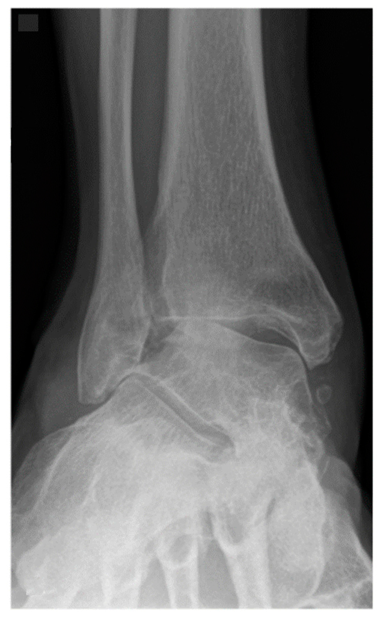
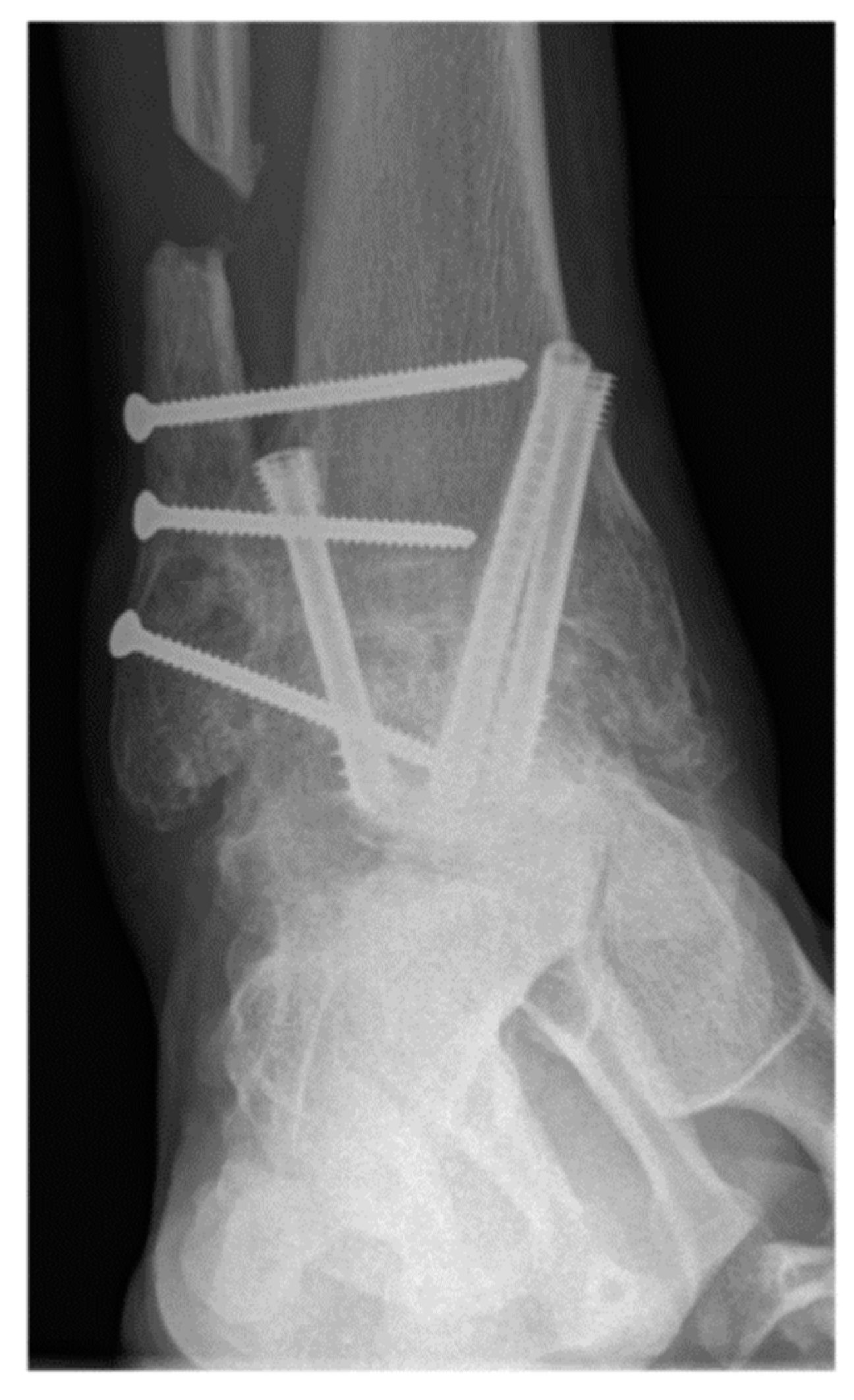
Between March 2002 and May 2016, 516 consecutive patients underwent tibiotalar arthrodesis. These patients were identified by CPT codes followed by a chart review. After retrospective review, 319 ankles from 312 patients met inclusion criteria. Inclusion criteria were failure of a minimum of 3 months of non-operative treatment [5], tibiotalar arthritis (Takakura Stage 2, 3 or 4) [39], and a minimum of 6 months follow-up (Figure 1 and Figure 2). Exclusion criteria were previous hindfoot surgery, arthroscopic tibiotalar arthrodesis, active septic arthritis, or age < 18-years-old. All patients in this study underwent primary open tibiotalar arthrodesis by one of four fellowship-trained orthopedic foot and ankle surgeons. A total of 227 of these patients underwent open tibiotalar arthrodesis with a distal tibiofibular arthrodesis and 92 patients underwent open tibiotalar arthrodesis without a distal tibiofibular arthrodesis. This study was conducted in accordance with the Declaration of Helsinki and the Guidelines for Good Clinical Practice. The data of this study were retrospectively and fully anonymized evaluated.

2.2. Surgical Technique

Operative approach, fixation method, choice of allograft/autograft/bone graft substitutes were varied based on surgeon and patient factors. Figure 2 shows radiographs of a typical case in this study in which the distal tibiofibular arthrodesis was included in the construct. In each case the foot was positioned in slight valgus, external rotation, and dorsiflexion and provisionally fixated for fluoroscopic imaging. If the foot position was deemed satisfactory, the tibiotalar joint +/− distal tibiofibular joint were fixated. Final fluoroscopic images were routinely obtained prior to closing the wounds and placing post-operative immobilization with use of a splint or boot. There was not a standardized post-operative protocol, but all patients were instructed to be non-weight bearing for a minimum of 6 weeks after surgery. Generally, patients were followed at 2, 6, and 12 weeks, 6 month, and 1-year intervals.

2.3. Outcomes Analysis

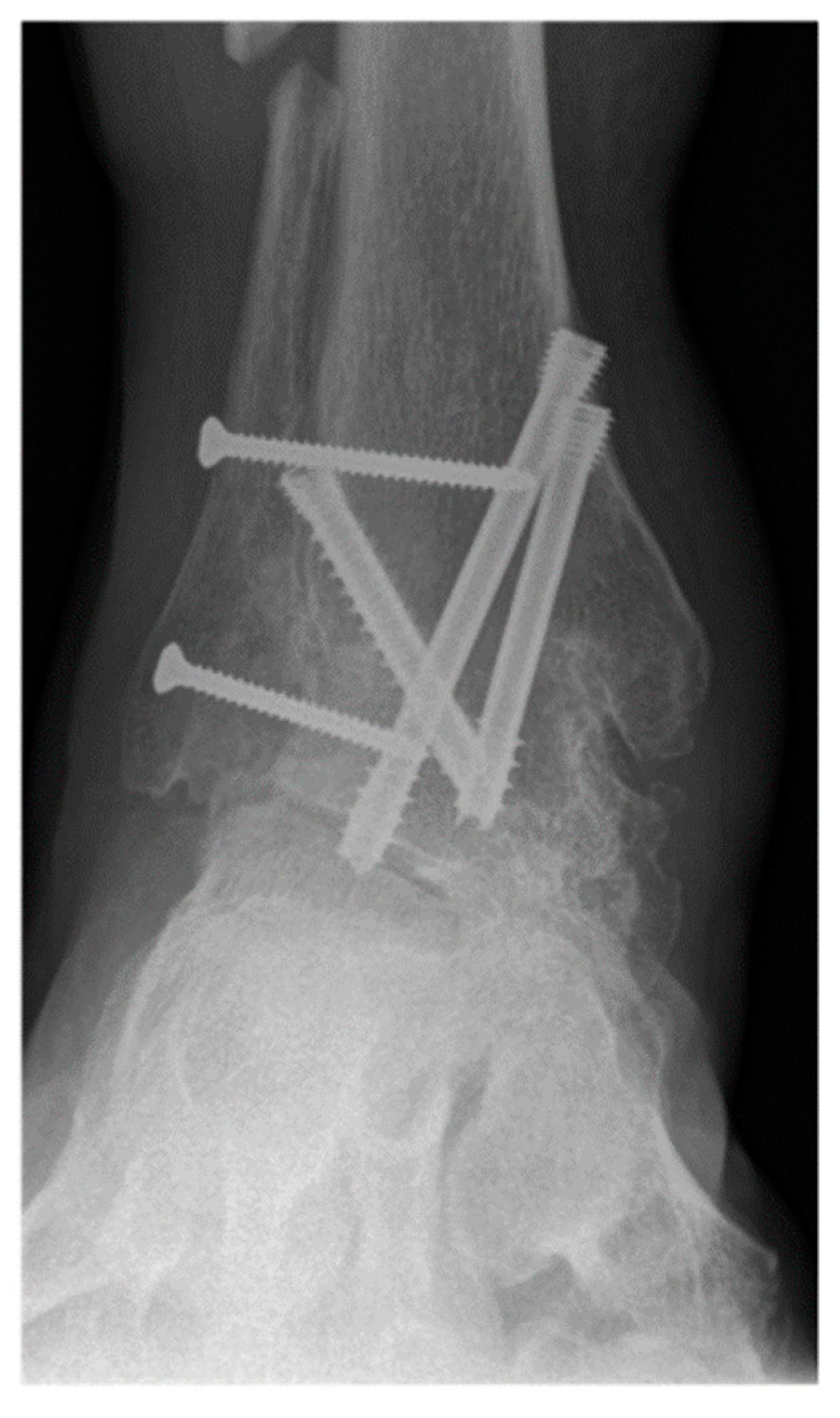
The primary outcome measure was nonunion rate of tibiotalar arthrodesis [2]. Union was defined based on patient reported symptoms and clinical physical examination criteria (no pain, no warmth, improvement in swelling, and stability to stress) and radiographic criteria (visible trabecular bridging across the arthrodesis site and no lucency around the hardware) [40]. Appropriate osseous union was defined as trabecular bridging across the tibiotalar joint (at least 80%) within 6 postoperative months [41] (Figure 3 and Figure 4). In patients with nonunion, computerized tomography (CT) was utilized for further assessment and was evaluated by independent radiologist not involved in any of surgeries. Secondary outcome measures were wound complications, return to the operating room, and rate of development of post-operative deep vein thrombosis (DVT), or pulmonary embolism (PE).

2.4. Statistical Analysis

Patient demographics and clinical characteristics were summarized descriptively for all ankle arthrodesis cases and also stratified by with or without distal tibiofibular arthrodesis. Continuous variables that were approximately normally distributed were summarized as mean and standard deviation (SD) and compared between the two groups using a t-test. Variables that were not normally distributed were summarized as median and interquartile range (IQR) and compared using Wilcoxon rank sum test. Categorical variables were summarized as frequency and percent and compared using a chi-squared test or Fisher’s exact test. Nonunion, wound complications, and DVT/PE were summarized as frequency and percentage and compared between groups using chi-squared or Fisher’s exact test. Months to union for ankles that had achieved union were summarized as median (IQR) and compared using a Wilcoxon rank sum test. Because rate of return to OR depended on patient follow-up, it was summarized as rate per person-year and a log rank test was used to compare with and without distal tibiofibular arthrodesis groups. The primary outcome, nonunion was further compared with the distal tibiofibular arthrodesis group using logistic regression adjusting for age, sex, year of surgery, smoking status, and BMP use. Odds ratio with 95% confidence interval were reported for all covariates. Statistical significance was defined as p < 0.05, and all tests were two-sided. All data were analyzed using IBM SPSS Statistics version 26.0 (IBM, Armonk, NY, USA).

3. Results

3.1. Subjects

Average age of the patients was 57.7 ± 13.5 years (range 18–88.8). Median follow-up time was 27.5 months (range 17–43). Demographic and clinical characteristics are shown in Table 1. Patients without a distal tibiofibular arthrodesis were significantly younger, but otherwise all demographic and clinical characteristics were comparable. Preoperative deformity measures in patients with and without distal tibiofibular arthrodesis were compared in Table 2, and there were no significant differences. Etiology of ankle arthritis is shown in Table 3. Again, no significant differences were seen between both cohorts. Comparison of the fixation type, operative approach, and bone graft details are demonstrated in the Table 4, Table 5 and Table 6, respectively.

3.2. Postoperative Outcomes/Complications

Outcomes by procedure type are shown in Table 7. There was no significant difference between the nonunion rate of patients who underwent distal tibiofibular arthrodesis (17/227 (7.5%)) and the nonunion rate of patients who did not undergo distal tibiofibular arthrodesis (11/92 (12%)) (p = 0.2). There was also no significant difference in the time to union, rate of deep wound complications, and rate of DVT/PE between the cohorts. Table 8 shows the rate of return to the OR. Table 9 shows the reason of return to the OR between the cohorts. Table 10 shows multivariable logistic regression predicting nonunion.

4. Discussion

This study shows that inclusion of the distal tibiofibular arthrodesis in primary open tibiotalar arthrodesis does not affect immediate complication rates including nonunion, wound complications, time to union, rate of return to the OR, and DVT/PE rate.
Patients who had a distal tibiofibular arthrodesis had autograft 208/227 (91.6%) of the time versus 64/92 (69.6%) of those who did not (p < 0.001). This is because autograft from the distal fibula was easily harvested for inclusion in the fusion mass. Not surprisingly, patients who have a distal tibiofibular arthrodesis were less likely to have utilized allograft 37/227 (16.3%) versus 23/92 (25%) of those without.
A relatively high number of 8/319 (2.5%) patients included in this study ultimately underwent a transtibial amputation (4 with distal tibiofibular arthrodesis, 4 without). Three of the four in the distal tibiofibular arthrodesis group had the tibiotalar arthrodesis for Charcot neuroarthropathy and the fourth patient underwent an amputation for an infected tibiotalar arthrodesis nonunion after prior equinovarus deformity correction. In the group without distal tibiofibular arthrodesis, one patient had a tibiotalar nonunion after attempted ankle arthrodesis after a failed total ankle arthroplasty. The second patient had a tibiotalar arthrodesis for Charcot neuroarthropathy. The third patient had chronic pain and deformity after multiple surgeries for clubfoot. The fourth patient had an opened pilon that had failed multiple open reduction, internal fixations and finally the ankle fusion with a blade plate.
The authors recognize that approximately 70% of patients suffering from the severe pain and disability associated with ankle arthritis are from a post-traumatic cause [21,36,42]. This means that patients often have previous incisions and possibly an impaired periarticular soft tissue envelope, which should be considered when planning an ankle arthrodesis. The results of this study suggest that surgeons can approach tibiotalar fusion from the approach that makes the most sense, as the inclusion of distal tibiofibular arthrodesis was not associated with significant changes in complication rates or time to fusion.
There are a number of case series describing outcomes after a transfibular approach for tibiotalar arthrodesis, but none of them directly compares the transfibular approach with an anterior approach. In 1998, Mann et al. reported an 88% fusion rate in a single surgeon series using a transfibular approach on 81 ankles at a minimum follow-up of 12 months [43]. Napiontek et al. report 23 ankles that underwent a transfibular approach, and only 1 of the 23 had a nonunion at an average of 32 months follow-up [31]. In 2007, Colman et al. evaluated 48 ankles at an average of 45 months post-operatively and showed a 96% union rate in ankles arthrodesis through a transfibular approach [30]. This study did not compare subjects to other approaches [30]. In a retrospective study on 29 patients from 2009 and 2014, Balaji et al. reported a 100% union rate at a mean of 3.8 months post-operatively where they utilized a transfibular approach with the fibula as an onlay graft [44]. They reported four superficial infections, no deep infections and they reported that patients with post-traumatic had better functional outcomes than those with rheumatoid or tuberculosis arthritis [44]. In 2016, Lee et al. reported 1 nonunion in 23 cases (92% union rate) in a heavy smoker utilizing a single lateral incision [32]. Recently, Park et al. demonstrated promising short-term results in a patient cohort consisting of 35 persons with a mean age of 66.5 years [17]. In this publication, an anterior approach was utilized in combination with transfibular approach in order to completely debride the joint surfaces including medial gutter and to increase the initial stability of the osteosynthetic fixation using anterior plate. Union was achieved in all cases with statistically significant pain relief from 6.8 (6–9) to 2.6 (0–7) using VAS [17]. Based on our experience, we routinely perform medial arthrotomy of the tibiotalar joint using separate incision to visualize and to debride the medial gutter [45]. In 2020, Suo et al. evaluated the clinical outcome of ankle arthrodesis with screw fixation through the transfibular approach for end-stage ankle arthritis in 28 ankles using a novel internal fixation screw [46]. The headless compression hollow screw was developed to have the mechanical properties of a taper, a full thread, and a change in thread pitch to better stabilize the joint complex and promote bone healing. Although the study showed satisfactory clinical results, it did not include a comparison with other methods of internal fixation.
This study has several limitations. The operative technique, fixation hardware, as well as post-operative protocols were not standardized as patients in this series were treated by one of four foot and ankle fellowship trained surgeons. Specifically, 192/227 (84.6%) of patients who had a distal tibiofibular arthrodesis had their tibiotalar fixation with screws alone versus the majority of patients 47/92 (51.1%) who did not have their distal tibiofibular arthrodesis included had a plate as their main fixation method. Therefore, one may argue that this is a study comparing nonunion rate between fixation method rather than inclusion of distal tibiofibular arthrodesis. The same could be said about the operative approach. The majority of patients 182/227 (80.2%) who had their distal tibiofibular arthrodesis included in the tibiotalar fusion had a lateral approach whereas most of the patients who had no distal tibiofibular arthrodesis had an anterior 47/92 (51.1%) or posterior 11/92 (12%) approach. One flaw in this study is neglect to account for concomitant surgeries, many of which may attribute to rate of nonunion. This study did not examine the impact of preexisting risk factors of nontraumatic ankle arthritis, such as those associated with rheumatoid arthritis, hemochromatosis, hemophilia, or Charcot neuroarthropathy [6]. These conditions present different challenges and may have different negative effects on postoperative outcomes and complications. Additionally, there is human error associated with performing chart review thus some data may be incomplete or incorrect. Lastly, in this study, tibiotalar union was assessed using conventional weight bearing radiographs. It has been demonstrated, that CT scans have higher reliability in determining the successful fusion of the hindfoot [47]. However, in our study only patients with suspected nonunion were selected for CT scan, as it would be impractical to obtain CT scans for all patients postoperatively.

5. Conclusions

This is the first study directly comparing nonunion and complication rates in primary, open ankle arthrodesis with and without distal tibiofibular arthrodesis. In this study, inclusion of the distal tibiofibular arthrodesis in tibiotalar arthrodesis fusion mass does not affect nonunion rate nor does it affect rate of wound complication, time to union, and DVT/PE rate.

Author Contributions

Conceptualization, A.B.; Data curation, A.B. and J.A.N.; Investigation, C.S., J.A.N., S.Y.-S. and H.R.; Supervision, V.V., K.-H.F. and A.B.; Writing—original draft, C.S.; Writing—review & editing, S.Y.-S. and A.B. All authors have read and agreed to the published version of the manuscript.

Funding

This research received no external funding.

Institutional Review Board Statement

Retrospective analysis of already fully anonymized data was performed, in accordance with the Institutional Review Board.

Informed Consent Statement

Obtaining informed consent was technically impossible for retrospective analysis of already fully anonymized data.

Data Availability Statement

Derived data supporting the findings of this study are available from the author upon reasonable request.

Conflicts of Interest

The authors declare no conflict of interest.

References

  1. Barg, A.; Pagenstert, G.I.; Hugle, T.; Gloyer, M.; Wiewiorski, M.; Henninger, H.B.; Valderrabano, V. Ankle osteoarthritis: Etiology, diagnostics, and classification. Foot Ankle Clin. 2013, 18, 411–426. [Google Scholar] [CrossRef] [PubMed]
  2. Gorbachova, T.; Melenevsky, Y.V.; Latt, L.D.; Weaver, J.S.; Taljanovic, M.S. Imaging and Treatment of Posttraumatic Ankle and Hindfoot Osteoarthritis. J. Clin. Med. 2021, 10, 5848. [Google Scholar] [CrossRef] [PubMed]
  3. Herrera-Pérez, M.; González-Martín, D.; Vallejo-Márquez, M.; Godoy-Santos, A.L.; Valderrabano, V.; Tejero, S. Ankle Osteoarthritis Aetiology. J. Clin. Med. 2021, 10, 4489. [Google Scholar] [CrossRef] [PubMed]
  4. Saltzman, C.L.; Salamon, M.L.; Blanchard, G.M.; Huff, T.; Hayes, A.; Buckwalter, J.A.; Amendola, A. Epidemiology of ankle arthritis: Report of a consecutive series of 639 patients from a tertiary orthopaedic center. Iowa Orthop. J. 2005, 25, 44–46. [Google Scholar] [PubMed]
  5. Tejero, S.; Prada-Chamorro, E.; González-Martín, D.; García-Guirao, A.; Galhoum, A.; Valderrabano, V.; Herrera-Pérez, M. Conservative Treatment of Ankle Osteoarthritis. J. Clin. Med. 2021, 10, 4561. [Google Scholar] [CrossRef]
  6. Valderrabano, V.; Horisberger, M.; Russell, I.; Dougall, H.; Hintermann, B. Etiology of ankle osteoarthritis. Clin. Orthop. Relat. Res. 2009, 467, 1800–1806. [Google Scholar] [CrossRef] [Green Version]
  7. Wang, B.; Saltzman, C.L.; Chalayon, O.; Barg, A. Does the subtalar joint compensate for ankle malalignment in end-stage ankle arthritis? Clin. Orthop. Relat. Res. 2015, 473, 318–325. [Google Scholar] [CrossRef] [Green Version]
  8. Alsayel, F.; Alttahir, M.; Mosca, M.; Barg, A.; Herrera-Pérez, M.; Valderrabano, V. Mobile Anatomical Total Ankle Arthroplasty-Improvement of Talus Recentralization. J. Clin. Med. 2021, 10, 554. [Google Scholar] [CrossRef]
  9. Barg, A.; Bettin, C.C.; Burstein, A.H.; Saltzman, C.L.; Gililland, J. Early Clinical and Radiographic Outcomes of Trabecular Metal Total Ankle Replacement Using a Transfibular Approach. J. Bone Jt. Surg. Am. Vol. 2018, 100, 505–515. [Google Scholar] [CrossRef]
  10. Kvarda, P.; Peterhans, U.S.; Susdorf, R.; Barg, A.; Ruiz, R.; Hintermann, B. Long-Term Survival of HINTEGRA Total Ankle Replacement in 683 Patients: A Concise 20-Year Follow-Up of a Previous Report. J. Bone Jt. Surg. Am. Vol. 2022, 104, 881–888. [Google Scholar] [CrossRef]
  11. Mosca, M.; Caravelli, S.; Vocale, E.; Massimi, S.; Censoni, D.; Di Ponte, M.; Fuiano, M.; Zaffagnini, S. Clinical Radiographical Outcomes and Complications after a Brand-New Total Ankle Replacement Design through an Anterior Approach: A Retrospective at a Short-Term Follow Up. J. Clin. Med. 2021, 10, 2258. [Google Scholar] [CrossRef] [PubMed]
  12. Stadler, C.; Stöbich, M.; Ruhs, B.; Kaufmann, C.; Pisecky, L.; Stevoska, S.; Gotterbarm, T.; Klotz, M.C. Intermediate to long-term clinical outcomes and survival analysis of the Salto Mobile Bearing total ankle prothesis. Arch. Orthop. Trauma. Surg. 2021, 1–8. [Google Scholar] [CrossRef] [PubMed]
  13. Conlin, C.; Khan, R.M.; Wilson, I.; Daniels, T.R.; Halai, M.; Pinsker, E.B. Living with Both a Total Ankle Replacement and an Ankle Fusion: A Qualitative Study from the Patients’ Perspective. Foot Ankle Int. 2021, 42, 1153–1161. [Google Scholar] [CrossRef] [PubMed]
  14. Eerdekens, M.; Deschamps, K.; Wuite, S.; Matricali, G. The Biomechanical Behavior of Distal Foot Joints in Patients with Isolated, End-Stage Tibiotalar Osteoarthritis Is Not Altered Following Tibiotalar Fusion. J. Clin. Med. 2020, 9, 2594. [Google Scholar] [CrossRef] [PubMed]
  15. Fischer, S.; Klug, A.; Faul, P.; Hoffmann, R.; Manegold, S.; Gramlich, Y. Superiority of upper ankle arthrodesis over total ankle replacement in the treatment of end-stage posttraumatic ankle arthrosis. Arch. Orthop. Trauma. Surg. 2021, 142, 435–442. [Google Scholar] [CrossRef] [PubMed]
  16. Johnson, M.D.; Shofer, J.B.; Hansen, S.T., Jr.; Ledoux, W.R.; Sangeorzan, B.J. The Impact of Coronal Plane Deformity on Ankle Arthrodesis and Arthroplasty. Foot Ankle Int. 2021, 42, 1294–1302. [Google Scholar] [CrossRef]
  17. Park, J.J.; Son, W.S.; Woo, I.H.; Park, C.H. Combined Transfibular and Anterior Approaches Increase Union Rate and Decrease Non-Weight-Bearing Periods in Ankle Arthrodesis: Combined Approaches in Ankle Arthrodesis. J. Clin. Med. 2021, 10, 5915. [Google Scholar] [CrossRef]
  18. Schieder, S.; Nemecek, E.; Schuh, R.; Kolb, A.; Windhager, R.; Willegger, M. Radiographic Sagittal Tibio-Talar Offset in Ankle Arthrodesis-Accuracy and Reliability of Measurements. J. Clin. Med. 2020, 9, 801. [Google Scholar] [CrossRef] [Green Version]
  19. Terrell, R.D.; Montgomery, S.R.; Pannell, W.C.; Sandlin, M.I.; Inoue, H.; Wang, J.C.; SooHoo, N.F. Comparison of practice patterns in total ankle replacement and ankle fusion in the United States. Foot Ankle Int. 2013, 34, 1486–1492. [Google Scholar] [CrossRef]
  20. Ahmad, J.; Raikin, S.M. Ankle arthrodesis: The simple and the complex. Foot Ankle Clin. 2008, 13, 381–400. [Google Scholar] [CrossRef]
  21. Abidi, N.A.; Gruen, G.S.; Conti, S.F. Ankle arthrodesis: Indications and techniques. J. Am. Acad. Orthop. Surg. 2000, 8, 200–209. [Google Scholar] [CrossRef] [PubMed]
  22. Chalayon, O.; Wang, B.; Blankenhorn, B.; Jackson, J.B., 3rd; Beals, T.; Nickisch, F.; Saltzman, C.L. Factors affecting the outcomes of uncomplicated primary open ankle arthrodesis. Foot Ankle Int. 2015, 36, 1170–1179. [Google Scholar] [CrossRef]
  23. Frey, C.; Halikus, N.M.; Vu-Rose, T.; Ebramzadeh, E. A review of ankle arthrodesis: Predisposing factors to nonunion. Foot Ankle Int. 1994, 15, 581–584. [Google Scholar] [CrossRef] [PubMed]
  24. Patel, S.; Baker, L.; Perez, J.; Vulcano, E.; Kaplan, J.; Aiyer, A. Risk Factors for Nonunion Following Ankle Arthrodesis: A Systematic Review and Meta-Analysis. Foot Ankle Spec. 2021, 1938640021998493. [Google Scholar] [CrossRef] [PubMed]
  25. Thordarson, D.B.; Kuehn, S. Use of demineralized bone matrix in ankle/hindfoot fusion. Foot Ankle Int. 2003, 24, 557–560. [Google Scholar] [CrossRef]
  26. Nihal, A.; Gellman, R.E.; Embil, J.M.; Trepman, E. Ankle arthrodesis. Foot Ankle Surg. Off. J. Eur. Soc. Foot Ankle Surg. 2008, 14, 1–10. [Google Scholar] [CrossRef]
  27. Yasui, Y.; Hannon, C.P.; Seow, D.; Kennedy, J.G. Ankle arthrodesis: A systematic approach and review of the literature. World J. Orthop. 2016, 7, 700–708. [Google Scholar] [CrossRef]
  28. Goldthwait, J.E. An operation for the stiffening of the ankle-joint in infantile paralysis. J. Bone Jt. Surg. Am. Vol. 1908, S2–S5, 271–275. [Google Scholar]
  29. Horwitz, T. The use of the transfibular approach in arthrodesis of the ankle join. Am. J. Surg. 1942, 55, 550–552. [Google Scholar] [CrossRef]
  30. Colman, A.B.; Pomeroy, G.C. Transfibular ankle arthrodesis with rigid internal fixation: An assessment of outcome. Foot Ankle Int. 2007, 28, 303–307. [Google Scholar] [CrossRef]
  31. Napiontek, M.; Jaszczak, T. Ankle arthrodesis from lateral transfibular approach: Analysis of treatment results of 23 feet treated by the modified Mann’s technique. Eur. J. Orthop. Surg. Traumatol. Orthop. Traumatol. 2015, 25, 1195–1199. [Google Scholar] [CrossRef] [PubMed]
  32. Lee, H.J.; Min, W.K.; Kim, J.S.; Yoon, S.D.; Kim, D.H. Transfibular ankle arthrodesis using burring, curettage, multiple drilling, and fixation with two retrograde screws through a single lateral incision. J. Orthop. Surg. 2016, 24, 101–105. [Google Scholar] [CrossRef] [PubMed] [Green Version]
  33. Paul, J.; Barg, A.; Horisberger, M.; Herrera, M.; Henninger, H.B.; Valderrabano, V. Ankle salvage surgery with autologous circular pillar fibula augmentation and intramedullary hindfoot nail. J. Foot Ankle Surg. Off. Publ. Am. Coll. Foot Ankle Surg. 2014, 53, 601–605. [Google Scholar] [CrossRef]
  34. Paul, J.; Barg, A.; Horisberger, M.; Herrera, M.; Henninger, H.B.; Valderrabano, V. Tibiotalocalcaneal arthrodesis with an intramedullary hindfoot nail and pillar fibula augmentation: Technical tip. Foot Ankle Int. 2015, 36, 984–987. [Google Scholar] [CrossRef] [PubMed]
  35. Flückiger, G.; Weber, M. The transfibular approach for ankle arthrodesis. Oper. Orthop. Traumatol. 2005, 17, 361–379. [Google Scholar] [CrossRef]
  36. Hayes, B.J.; Gonzalez, T.; Smith, J.T.; Chiodo, C.P.; Bluman, E.M. Ankle arthritis: You can’t always replace it. J. Am. Acad. Orthop. Surg. 2016, 24, e29–e38. [Google Scholar] [CrossRef]
  37. Hintermann, B.; Barg, A.; Knupp, M.; Valderrabano, V. Conversion of painful ankle arthrodesis to total ankle arthroplasty. J. Bone Jt. Surg. Am. Vol. 2009, 91, 850–858. [Google Scholar] [CrossRef]
  38. Usuelli, F.G.; de Cesar Netto, C.; Maccario, C.; Paoli, T.; D’Ambrosi, R.; Indino, C. Reconstruction of a missing or insufficient distal fibula in the setting of a total ankle replacement: The Milanese technique. Foot Ankle Surg. Off. J. Eur. Soc. Foot Ankle Surg. 2021, 28, 186–192. [Google Scholar] [CrossRef]
  39. Takakura, Y.; Tanaka, Y.; Kumai, T.; Tamai, S. Low tibial osteotomy for osteoarthritis of the ankle. Results of a new operation in 18 patients. J. Bone Jt. Surg. British Vol. 1995, 77, 50–54. [Google Scholar] [CrossRef]
  40. Gorman, T.M.; Beals, T.C.; Nickisch, F.; Saltzman, C.L.; Lyman, M.; Barg, A. Hindfoot arthrodesis with the blade plate: Increased risk of complications and nonunion in a complex patient population. Clin. Orthop. Relat. Res. 2016, 474, 2280–2299. [Google Scholar] [CrossRef] [Green Version]
  41. Herrera-Perez, M.; Andarcia-Banuelos, C.; Barg, A.; Wiewiorski, M.; Valderrabano, V.; Kapron, A.L.; De Bergua-Domingo, J.M.; Pais-Brito, J.L. Comparison of cannulated screws versus compression staples for subtalar arthrodesis fixation. Foot Ankle Int. 2015, 36, 203–210. [Google Scholar] [CrossRef] [PubMed]
  42. Boobbyer, G.N. The long-term results of ankle arthrodesis. Acta Orthop. Scand. 1981, 52, 107–110. [Google Scholar] [CrossRef] [PubMed]
  43. Mann, R.A.; Rongstad, K.M. Arthrodesis of the ankle: A critical analysis. Foot Ankle Int. 1998, 19, 3–9. [Google Scholar] [CrossRef] [PubMed]
  44. Balaji, S.M.; Selvaraj, V.; Devadoss, S.; Devadoss, A. Transfibular ankle arthrodesis: A novel method for ankle fusion—A short term retrospective study. Indian J. Orthop. 2017, 51, 75–80. [Google Scholar] [CrossRef]
  45. Sripanich, Y.; Steadman, J.; Valderrabano, V.; Barg, A. Open ankle arthrodesis: Transfibular approach. Tech. Foot Ankle 2020, 19, 26–36. [Google Scholar] [CrossRef]
  46. Suo, H.; Fu, L.; Liang, H.; Wang, Z.; Men, J.; Feng, W. End-Stage Ankle Arthritis Treated by Ankle Arthrodesis with Screw Fixation through the Transfibular Approach: A Retrospective Analysis. Orthop. Surg. 2020, 12, 1108–1119. [Google Scholar] [CrossRef]
  47. Cerrato, R.A.; Aiyer, A.A.; Campbell, J.; Jeng, C.L.; Myerson, M.S. Reproducibility of computed tomography to evaluate ankle and hindfoot fusions. Foot Ankle Int. 2014, 35, 1176–1180. [Google Scholar] [CrossRef]
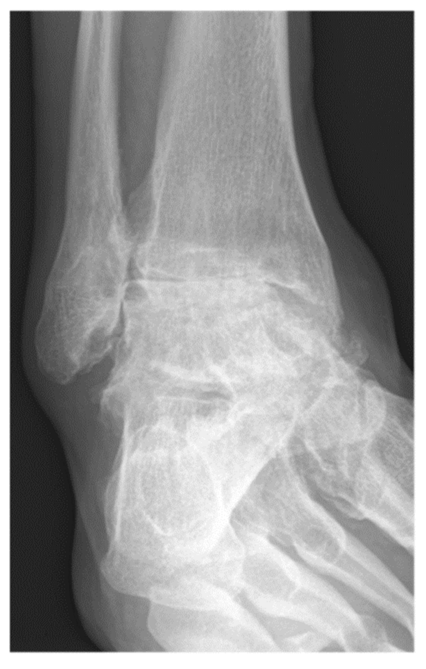
Figure 1. A 57-year-old male patient presenting end-stage varus tibiotalar osteoarthritis.
Jcm 11 03387 g001
Figure 2. A 62-year-old female patient presenting end-stage valgus tibiotalar osteoarthritis.
Jcm 11 03387 g002
Figure 3. A 57-year-old male patient presenting complete union of the tibiotar and distal tibiofibular joints at 6 months follow-up.
Jcm 11 03387 g003
Figure 4. A 62-year-old female patient presenting complete union of the tibiotar and distal tibiofibular joints at 4 months follow-up.
Jcm 11 03387 g004
Table 1. Demographic and clinical characteristics.
VariableWith Tibiofibular Arthrodesis
(n = 227)
Without Tibiofibular Arthrodesis (n = 92)All Patients
(n = 319)
p-Value
Age [years ± SD]58.8 ± 12.454.9 ± 15.757.7 ± 13.50.036 
Gender (male)133 (58.6%)51 (55.4%)184 (57.7%)0.61
BMI with range [kg/m2]29.2 (25.8–33.1)301 (26.4–34.2)29.4 (25.9–33.8)0.32 ˆ
Diabetes35 (15.4%)16 (17.4%)51 (16%)0.66
Smokers34 (15%)13 (14.1%)47 (14.7%)0.85
Right sided surgery117 (51.5%)51 (55.4%)168 (52.7%)0.53
t-test; Chi-squared test; ˆ Wilcoxon rank sum test. BMI = Body Mass Index; SD = Standard deviation.
Table 2. Pre-operative deformity measures.
Pre-Operative DeformityWith Tibiofibular Arthrodesis
(n = 227)
Without Tibiofibular Arthrodesis (n = 92)All Patients
(n = 319)
p-Value
MDTA with range [°]88° (86–91°)88° (85–90°)88° (85–91°)0.15
TTT with range [°]0° (−5–3°)0° (−2–3°)0° (−4–3°)0.21
CMA with range [°]−5.1° (−19.3–12.9°)−6.4° (−15.8–10.2°)−5.8° (−17.5–11.9°)0.98
ADTA with range [°]82° (79–87°)82° (77–86°)82° (78–86.8°)0.18
Wilcoxon rank sum test. ADTA = anterior distal tibial angle; CMA = calcaneal moment arm (negative values indicated varus malalignment); MDTA = medial distal tibial angle; TTT = tibiotalar test.
Table 3. Etiology of tibio-talar arthritis.
EtiologyWith Tibiofibular Arthrodesis
(n = 227)
Without Tibiofibular Arthrodesis
(n = 92)
All Patients
(n = 319)
p-Value
Primary7 (3.1%)5 (5.4%)12 (3.8%)
Secondary (including posttraumatic OA)220 (96.9%)87 (94.6%)307 (96.2%)0.34
Fisher’s exact test. OA = osteoarthritis.
Table 4. Type of fixation.
Type of FixationWith Tibiofibular Arthrodesis
(n = 227)
Without Tibiofibular Arthrodesis (n = 92)All Patients
(n = 319)
p-Value
Blade plate5 (2.2%)11 (12.0%)16 (5.0%)<0.001
External fixation alone4 (1.8%)13 (14.1%)17 (5.3%)
External fixation + plate1 (0.4%)0 (0%)1 (0.3%)
IM nail14 (6.2%)5 (5.4%)19 (6%)
Plate11 (4.8%)47 (51.1%)58 (18.2%)
Screws192 (84.6%)16 (17.4%)208 (65.2%)
Fisher’s exact test. IM = intramedullary.
Table 5. Operative approach.
Operative ApproachWith Tibiofibular Arthrodesis
(n = 227)
Without Tibiofibular Arthrodesis (n = 92)All Patients
(n = 319)
p-Value
Anterior8 (3.5%)47 (51.1%)55 (17.2%)<0.001
Lateral182 (80.2%)20 (21.7%)202 (63.3%)
Medial0 (0%)3 (3.3%)3 (0.9%)
Medial and anterior0 (0%)1 (1.1%)1 (0.3%)
Medial and lateral31 (13.7%)10 (10.9%)41 (12.9%)
Posterior 6 (2.6%)11 (12%)17 (5.3%)
Fisher’s exact test.
Table 6. Bone graft details.
Autograft or AllograftTypeWith Tibiofibular Arthrodesis
(n = 227)
Without Tibiofibular Arthrodesis (n = 92)All Patients
(n = 319)
p-Value
AutograftYes208 (91.6%)64 (69.6%)272 (85.3%)<0.001
Distal fibula ± distal tibia159 (70%)21 (22.8%)180 (56.4%)
Iliac crest5 (2.2%)7 (7.6%)12 (3.8%)
Proximal tibia44 (19.4%)35 (38%)79 (24.8%)
Unspecified0 (0%)1 (1.1%)1 (0.3%)
AllograftYes37 (16.3%)23 (25%)60 (18.8%)0.07
Bone block6 (2.6%)7 (7.6%)13 (4.1%)
Cancellous chips25 (11%)11 (12%)36 (11.3%)
Cortical bone powder0 (0%)1 (1.1%)1 (0.3%)
DBM6 (2.6%)4 (4.3%)10 (3.1%)0.48
BMP20 (8.8%)20 (21.7%)40 (12.5%)0.002
Fisher’s exact test; Chi-squared test. BMP = bone morphogenic protein; DBM = demineralized bone matrix.
Table 7. Outcomes.
Table 7. Outcomes.
VariableWith Tibiofibular Arthrodesis (n = 227)Without Tibiofibular Arthrodesis (n = 92)All Patients
(n = 319)
p-Value
Nonunion17 (7.5%)11 (12%)28 (8.8%)0.20
Months to union3.5 (1.4–18.2)4.1 (1.6–13.5)3.8 (2.9–6.2)0.72
Wound complications
  • Superficial
  • Deep
59 (26%)
  • 47 (20.7%)
  • 12 (5.3%)
28 (30.4%)
  • 18 (19.6%)
  • 10 (10.9%)
87 (27.3%)
  • 65 (20.4%)
  • 22 (6.9%)
0.42
Thrombosis
  • DVT
  • PE
11 (4.8%)
1 (0.4%)
1 (1.1%)
1 (1.1%)
12 (3.8%)
2 (0.6%)
0.18 *
Chi-squared; Wilcoxon rank sum test; * Fisher’s exact test; DVT = deep venous thrombosis; PE = pulmonary embolism.
Table 8. Rate of return to OR by procedure type excluding planned removal of external fixation.
Rate of Return to ORWith Tibiofibular Arthrodesis
(n = 227)
Without Tibiofibular Arthrodesis
(n = 92)
All Patients
(n = 319)
p-Value
Rate (#/yr)55 (24.2%)
0.044/person year
20 (21.7%)
0.056/ person year
86 (27%)
0.053/person year
0.89
Log-rank test. OR = operating room.
Table 9. Reason for return to OR by procedure type, excluding planned removal of external fixation.
Reason for Return to ORWith Tibiofibular Arthrodesis
(n = 227)
Without Tibiofibular Arthrodesis (n = 92)All Patients
(n = 319)
p-Value
Amputation4 (1.8%)4 (4.3%)8 (2.5%)0.12
I&D (operative site)7 (3.1%)4 (4.3%)11 (3.4%)
I&D (autograft site)1 (0.4%)0 (0%)1 (0.3%)
I&D + ROH1 (0.4%)0 (0%)1 (0.3%)
Osteotomy1 (0.4%)1 (1.1%)2 (0.6%)
Relocation of peroneus brevis tendon1 (0.4%)1 (1.1%)2 (0.6%)
Revision ankle arthrodesis14 (6.2%)3 (3.3%)17 (5.3%)
ROH20 (8.8%)7 (7.6%)26 (8.2%)
Soft tissue reconstruction2 (0.9%)0 (0%)2 (0.6%)
Subtalar arthrodesis3 (1.3%) 0 (0%) 3 (0.9%)
Fisher’s exact test. I&D = irrigation and debridement, includes hematoma evacuation and I&D for infection OR = operating room; ROH = removal of hardware.
Table 10. Multivariable logistic regression predicting nonunion.
VariablesOdds Ratiop-Value
Inclusion of distal fibular strut autograft in tibiotalar fusion0.74 (0.29~2.08)0.55
Year of surgery0.94 (0.84~1.05)0.27
Age at time of surgery1.02 (0.99~1.06)0.29
Male gender1.2 3(0.52~3.00)0.64
Smoker0.90 (0.20~2.99)0.88
+BMP0.60 (0.09~2.37)0.53
Pre-operative MDTA1.03 (0.97~1.10)0.33
BMP = bone morphogenic protein; MDTA = medial distal tibial angle.
Publisher’s Note: MDPI stays neutral with regard to jurisdictional claims in published maps and institutional affiliations.