Article

Radiological and Clinical Efficacy of Intra-Arterial 90Y-DOTATATE in Patients with Unresectable, Progressive, Liver Dominant Neuroendocrine Neoplasms

by
Agnieszka Kolasińska-Ćwikła
1,*,
Mirosław L. Nowicki
2,
Artur J. Sankowski
2,
Jakub M. Pałucki
1,
John R. Buscombe
3,
Lidia Glinka
4 and
Jarosław B. Ćwikła
4,5
1
Department of Oncology and Radiotherapy, Maria Skłodowska-Curie National Research Institute of Oncology, Wawelska 15, 02-034 Warsaw, Poland
2
Department of Radiology, Hospital Ministry of Internal Affairs, Wołoska 137, 02-507 Warsaw, Poland
3
Department of Nuclear Medicine, Barts Health, West Smithfield, London EC1A 7BE, UK
4
Department of Cardiology and Internal Medicine, School of Medicine, University of Warmia and Mazury, Warszawska 30, 10-082 Olsztyn, Poland
5
Diagnostic and Therapy Center—“Gammed” Lelechowska 5, 02-351 Warsaw, Poland
*
Author to whom correspondence should be addressed.
J. Clin. Med. 2021, 10(8), 1794; https://doi.org/10.3390/jcm10081794
Submission received: 16 March 2021 / Revised: 8 April 2021 / Accepted: 16 April 2021 / Published: 20 April 2021
(This article belongs to the Section Oncology)

Abstract

:
This study was performed to determine if intra-arterial (i.a.) administration of 90Y DOTATATE can provide an effective and safe alternative to the accepted standard for i.v. of peptide receptor radionuclide therapy (PRRT) in liver-dominant metastases of gastrointestinal pancreatic neuroendocrine neoplasm (GEP-NEN). A single site, prospective, preliminary case series study included 39 patients with histologically proven liver-dominant NEN. PRRT in the form of 1.15GBq 90Y DOTATATE was given selectively into the liver via radiological catheterization of the hepatic artery, up to four times. The endpoint was radiological response (RECIST). Secondary endpoints assessed clinical well-being post-treatment, progression-free survival (PFS), overall survival (OS), and toxicity. Partial response (PR) was noted in 13% of subjects six weeks post-therapy, increasing to 24% at six months and dropping to 13% at 36 months. Disease progression (DP) was not seen at six weeks, was 5% at six months, and 47% at 36 months. Clinical response based on PS seen in 74% of patients at six weeks, 69% at six months, and 39% at 36 months had PFS and OS, respectively, of 22.7 months and 38.2 months. There was no difference in OS/PFS between those with RECIST PR and SD. One patient had significant toxicity (3%). Use of i.a. PRRT appears to be safe and effective in treating patients with liver-dominant NEN. In addition, the best OS (51 vs. 22 months) was seen when i.a. was used as an upfront treatment of bulky GEP-NEN liver metastases and not after i.v. 90Y DOTATATE. The use of i.a. 90Y DOTATATE PRRT appears to be safe and effective in treating patients with liver-dominant NEN.

1. Introduction

The therapeutic options for patients with progressive, advanced, metastatic neuroendocrine neoplasms of gastro-entero-pancreatic origin (GEP-NENs) with extensive liver disease are often limited [1]. In tumors of pancreatic origin, there are some systemic therapy trials, in particular mTOR blockers, tyrosine kinase inhibitors (TKI), or any other antiangiogenic drugs [2,3,4]. The RADIANT-4 study showed some good results using everolimus as a second-line therapy in progressive NET [5]. Peptide receptor radionuclide therapy (PRRT) has been available to patients for several years [6,7,8,9,10]. A recent randomized control trial, NETTER-1, demonstrated that good clinical results can be obtained using 177Lu-DOTATATE PRRT in progressive midgut tumors when compared to high-dose somatostatin analogs [11]. Therefore, 177Lu-DOTATATE is seen currently as the standard of care in progressive GEP-NEN and has been presented as such in international guidelines (ENETS, NANETS, and NCCN) [12,13,14]. For example, in the current ENETS guidelines, PRRT is the best therapeutic option in progressive somatostatin (SST) receptor-positive tumors, with homogenous SST receptor (SSTR) expression after the failure of somatostatin analogs including patients with extensive liver metastases [6,7,8,9,10,12,15].
PRRT allows precise binding of the therapeutic radionuclide, normally a beta emitter, to the cell via a high-affinity ligand such as DOTATATE, because its affinity for the SSTR sub-type 2 receptor is up to nine times greater than natural somatostatin [16,17]. This choice of DOTATATE was found to increase the efficacy of PRRT compared to the previously used DOTATAOC [18]. However, the radionuclide most commonly used in PRRT is 177Lu, which has a lower energy beta emission, resulting in a more limited tissue penetration range compared to the alternative 90Y, which has a highly energetic beta particle. This high energy of 90Y and its ability to penetrate up to 2 mm of tissue could be advantageous in the treatment of patients with large-volume liver metastases. The management of such patients is complex, and curative surgery can rarely be offered, as the metastases of GEP-NENs tend to occur diffusely through both liver lobes [1,2,3,6,12,13,14,15]. However, studies performed using systemically administered 90Y DOTATATE have shown a higher level of toxicity to the kidneys and some more significant bone marrow toxic effects than reported with 177Lu-DOTATATE [6,7,8,9,10,13,17,18,19,20].
An alternate strategy would be to deliver the 90Y DOTATATE directly to the liver via radiological cannulation of the hepatic artery and relying on the significant affinity of the GEP-NEN cells for the 90Y DOTATATE, resulting in a significant “first pass” effect and thus maximizing uptake into the GEP-NEN cells and reducing the systemic bioavailability of the 90Y DOTATATE and thus reducing the toxic effects to the kidneys and bone marrow but maximizing the radiation dose delivered to the metastases within the liver [15,21,22].
We and others have published some data on the efficacy of 90Y DOTATATE/DOTAoctreotide (DOTATOC/DOTA-Lanreotide (DOTA-LAN), and though we found benefit in survival and symptom control, it was more difficult to show a significant radiological response, especially using criteria such as RECIST, which depend primarily on contrast-enhanced CT or MRI [6,7,8,9,10,15,17,18,19,20,21,22,23,24]. The radiological response consistently under-predicted the clinical and survival benefits of the 90Y or 177Lu PRRT. The updated version of RECIST v. 1.1 can be less useful in patients with extensive liver disease, as only two rather than five liver lesions are measured [25,26]. In many studies, only a small number of patients had a significant measurable radiological response such as a partial response (PR), despite showing clear clinical benefit [6,7,8,9,10,11,12,27,28,29,30,31].

The Aim of this Study

The primary aim of this study was to assess the possibility, in a preliminary study, of the possible efficacy of using two administrations of intra-arterial 90Y DOTATATE in patients with bulky unresectable liver metastases from GEP-NEN tumors. Treatment response was determined both radiologically, based on RECIST 1.0 criteria, which was felt to be appropriate for those with multiple liver lesions, and clinically, such as change in performance status—PS (ECOG), which in these patients is primarily related to symptom control. Further to this, there would be an assessment of both progression-free survival (PFS) and overall survival (OS) in patients treated with intra-arterial 90Y DOTATATE for extensive GEP-NEN liver metastases. Additional evaluation was performed of disease control rate (DCR) at selected time points in the patient’s clinical follow-up.

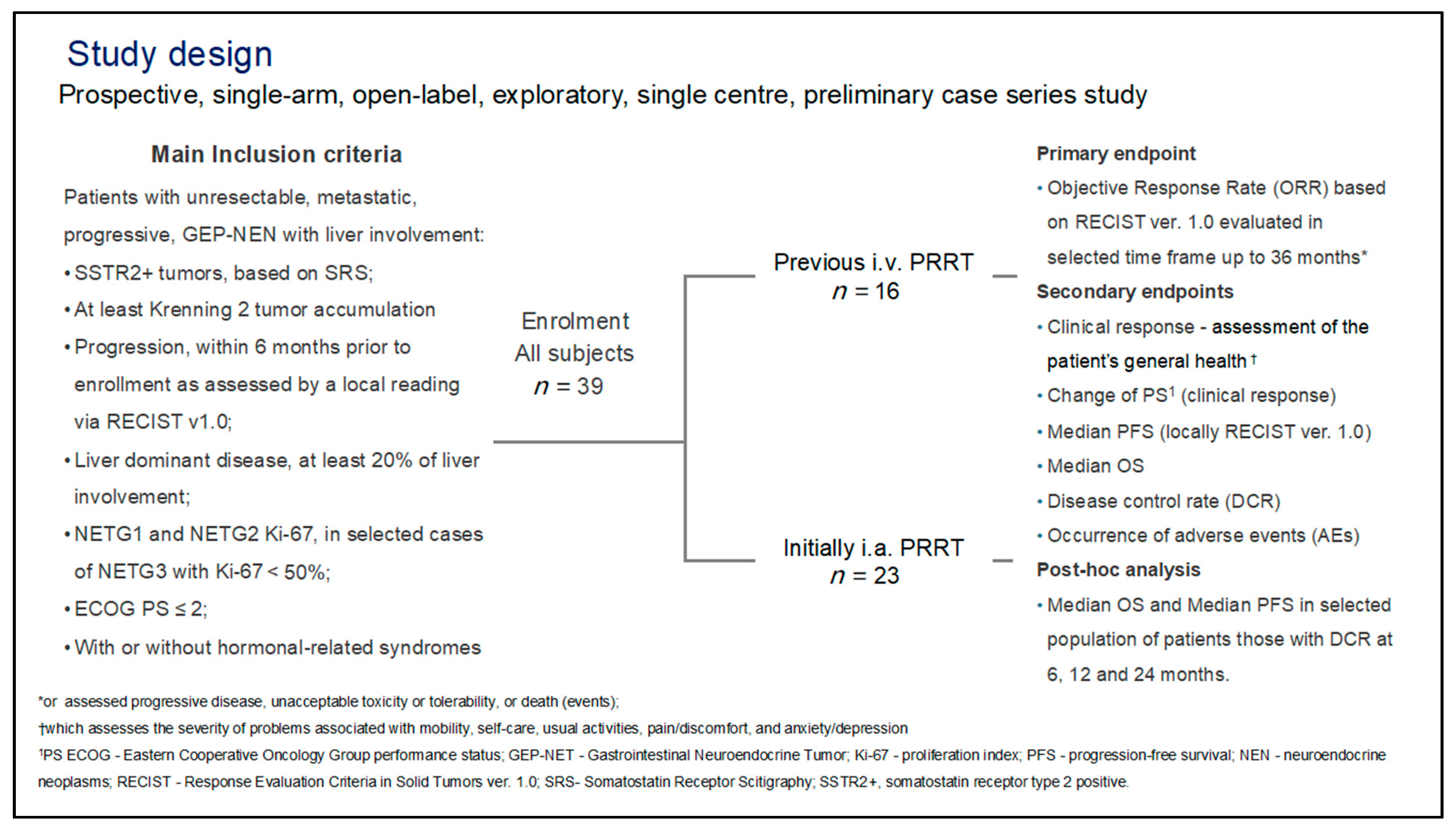
2. Materials and Methods

2.1. General

This was a prospective, open-label, interventional, single institution, preliminary case series study, which was approved by the Clinical Ethics Committee of the Central Clinical Hospital Ministry of Internal Affairs (reference 109/2004), where the study was conducted between May 2006 to December 2012 with a minimum six-year follow-up period. Prior to study inclusion, all patients understood the experimental nature of the treatment and all subjects signed their written informed consent form.

2.2. Patients

A total of 39 patients (Table 1), including 17 females (45%), with a mean age of 57.6 years (range 35–75 years) were recruited into the trial. The inclusion criteria include a histological diagnosis of gastro-entero-pancreatic neuroendocrine neoplasms (GEP-NEN): G1, G2, and in selected cases G3 (well-differentiated tumor cell with 20% < Ki-67 < 50%), [32] according to WHO and UICC/AJCC classification (TNM Classification of Malignant Tumors 8th Edition 2017). All patients had to have progressive disease noted within 12 months of entry into the study based on RECIST 1.0 criteria and unresectable liver metastases with at least 20% of liver involvement based on an increase in tumor size on CT or MRI. All patients had at least Krenning 2 uptake in the known liver lesions expressing somatostatin (SST) receptors, as seen on prior somatostatin receptor scintigraphy (SRS), using 99mTc-[HYNIC, Tyr3]octreotide (TOC) (National Centre for Nuclear Research-Radioisotope CenterPolatom; NCNR; Otwock-Świerk, Poland) [33].
Though all patients had to have unresectable bulky liver metastases, they could also have extra-hepatic SRS avid disease. Biochemical progression was defined as increase serum CgA as ULN over 10% from baseline levels. Patients were recruited if they had bulky liver metastases that had failed to respond to intravenous prior PRRT (16 patients) or had such bulky liver metastases that it was thought optimal for their first line of PRRT therapy (Figure 1).
Exclusion criteria for entry into the trial were as follows: Hb < 80 g/L, WBC < 2 × 10⁶/L, platelets < 100 × 106/L, creatinine level > 30 mg/L or GFR < 20 mL/min and poor performance status (PS), EOCG 3 or 4, and poorly differentiated NECG3 cancers. Contraindications also included pregnancy and known hypersensitivity to DOTATATE.
Previous treatments for the patient group included systemic i.v. 90Y DOTATATE PRRT in 16 patients (32%), and all but one patient had received long-acting somatostatin analog treatment, though this was suspended four weeks before the i.a. PRRT. Chemotherapy had previously been given to 12 (31%) patients, and local liver therapies such as selective trans-arterial embolization (TAE) and surgery had been performed on nine (23%) subjects (Table 2).

2.3. Therapy—Administration Protocol

An anti-emetic such as 8 mg Ondansetron (GlaxoSmithKlein, Brentford, Middlesex; UK) was administered i.v. 30 min, before the start of a 6 h i.v. infusion of 1 L of a lysine-containing amino acid solution such as Vamin 18 or Nephrotec, (Fresenius-Kabi, Bad Homburg; Germany). The amino acid infusion used to reduce any renal accumulation of the 90Y-DOTATATE was commenced 1.0–1.5 h before the PRRT.
The 90Y-DOTATATE was administered as a slow bolus injection of 10 mL over 20 min via an appropriately placed catheter or micro-catheter radiologically placed into the left or right hepatic artery. In patients with bi-lobar disease, the catheter would be placed and two infusions would be administered in the right and left hepatic arteries. Selective cannulation and infusion were performed on any aberrant hepatic arteries such as the accessory segment 4 artery.
Patients included in this analysis had at least two i.a. administrations of 90Y-DOTATATE with a mean activity of 1.15 GBq per session. The mean time between administrations of i.a.90Y-DOTATATE was 9.2 weeks (range 6–10 weeks); this primarily depended on the clinical condition of the patient and availability of angiography (Table 3).

2.4. Radiology

Any radiological response was measured via a standard multiphase contrast-enhanced CT scan with an abdomen arterial phase and a portal–venous phase of the chest, abdomen, and pelvis. The pre-treatment baseline CT had to have been performed within three months prior to the first i.a. 90Y-DOTATATE administration followed by six monthly intervals post-treatment. The initial arterial phase image was also used to help assess the hepatic vasculature pre-intervention. If CT was contraindicated, then MRI was performed with dynamic contrast enhancement (DCE) (Supplementary Materials).

2.5. Biodistribution of the Radiotracer

Between 8 and 18 h post-therapy, the biodistribution of the 90Y-DOTATATE was determined using a “Bremsstrahlung” whole body scintigraphy and SPECT images using a dual-head gamma camera (e-cam; Siemens, Erlangen, Germany) equipped with medium energy collimation with a photopeak centered on 95 keV with a 50% window.

3. Assessment of Effectiveness

3.1. Clinical Response and Performance Status

An assessment of the patient’s general health and tumor-specific symptoms was performed by three observers (AKC, MLN, and JBC) before treatment, then at six weeks, and then three months after their last therapy episode. At each visit, the patient’s general health was assessed using the standard ECOG performance status (PS) scale.
The clinical symptoms of response on i.a. PRRT assessed in this study included appetite, malaise, weight change, the presence and intensity and frequency of abdominal pain, diarrhea, flushing, nausea, vomiting, fever, wheezing, and abdominal bloating. Analgesia and somatostatin analog requirements before and after treatment were recorded in clinical files.
To assess any biochemical or endocrine toxicity, serial measurements (pre-treatment, six weeks post-treatment, and then every three months) of plasma chromogranin-A (CgA) and if relevant 24-h urinary 5-hydroxyindole acetic acid (5-HIAA) and fasting gut hormones are performed for patients with biologically functioning pancreatic neuroendocrine tumors.

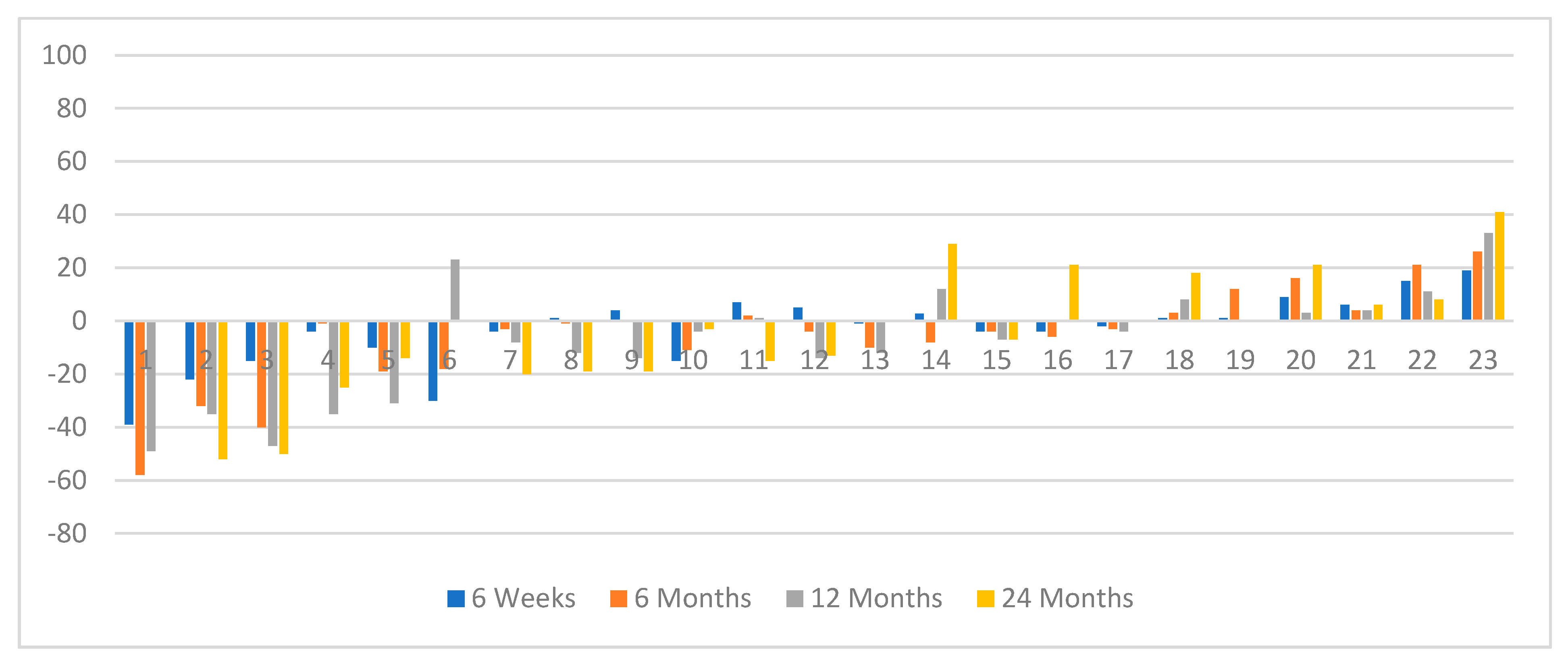
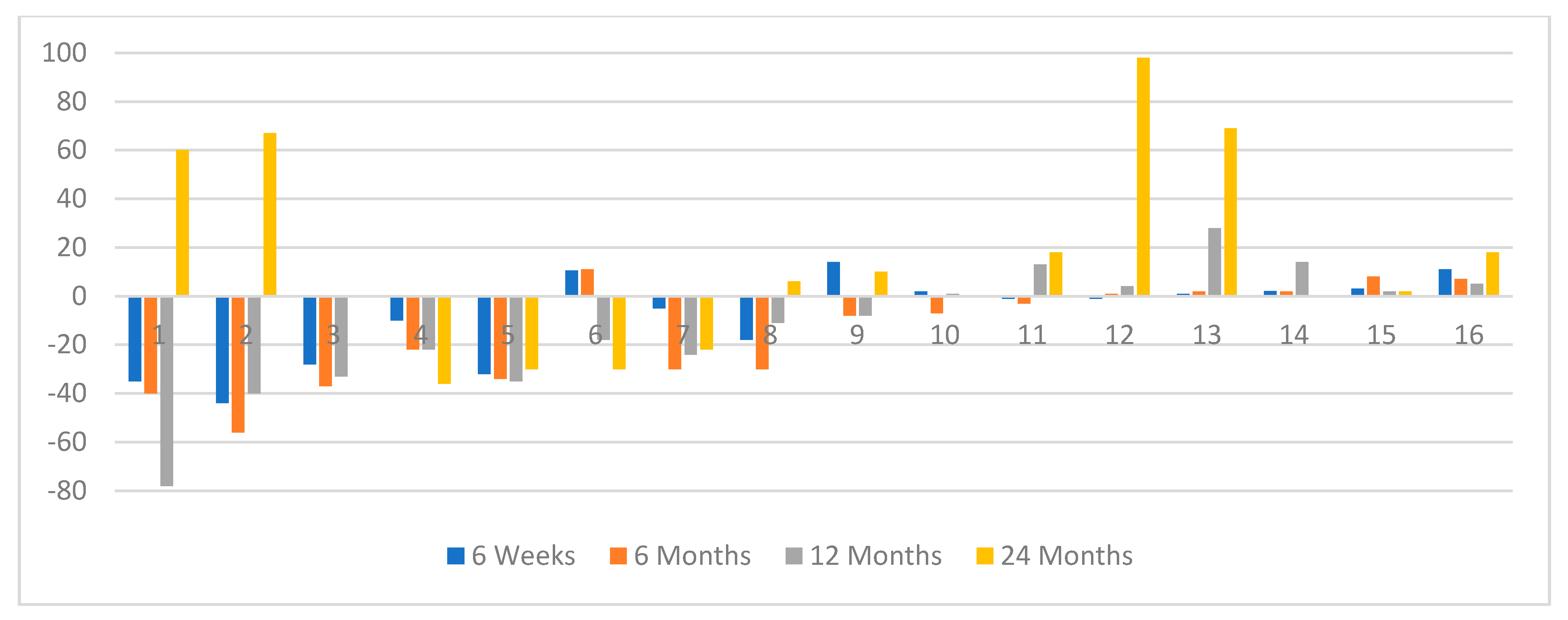
3.2. Image Analysis Radiological Response (RECIST v. 1.0)

Tumor response was determined by CT (or alternatively by MRI if CT was not possible) and scored, according to RECIST 1.0, by two independent radiologists with a special interest in GEP-NEN tumors (AJS and JMP) who were required to reach consensus on the size change of the five largest liver metastases seen on the pre-treatment liver scans. Any new liver metastases were also noted [34]. In addition, the diameters of all liver metastases greater than 10 mm were counted by each observer at each imaging time point to provide a determinate of total tumor liver metastatic load to determine the disease control rate (DCR).

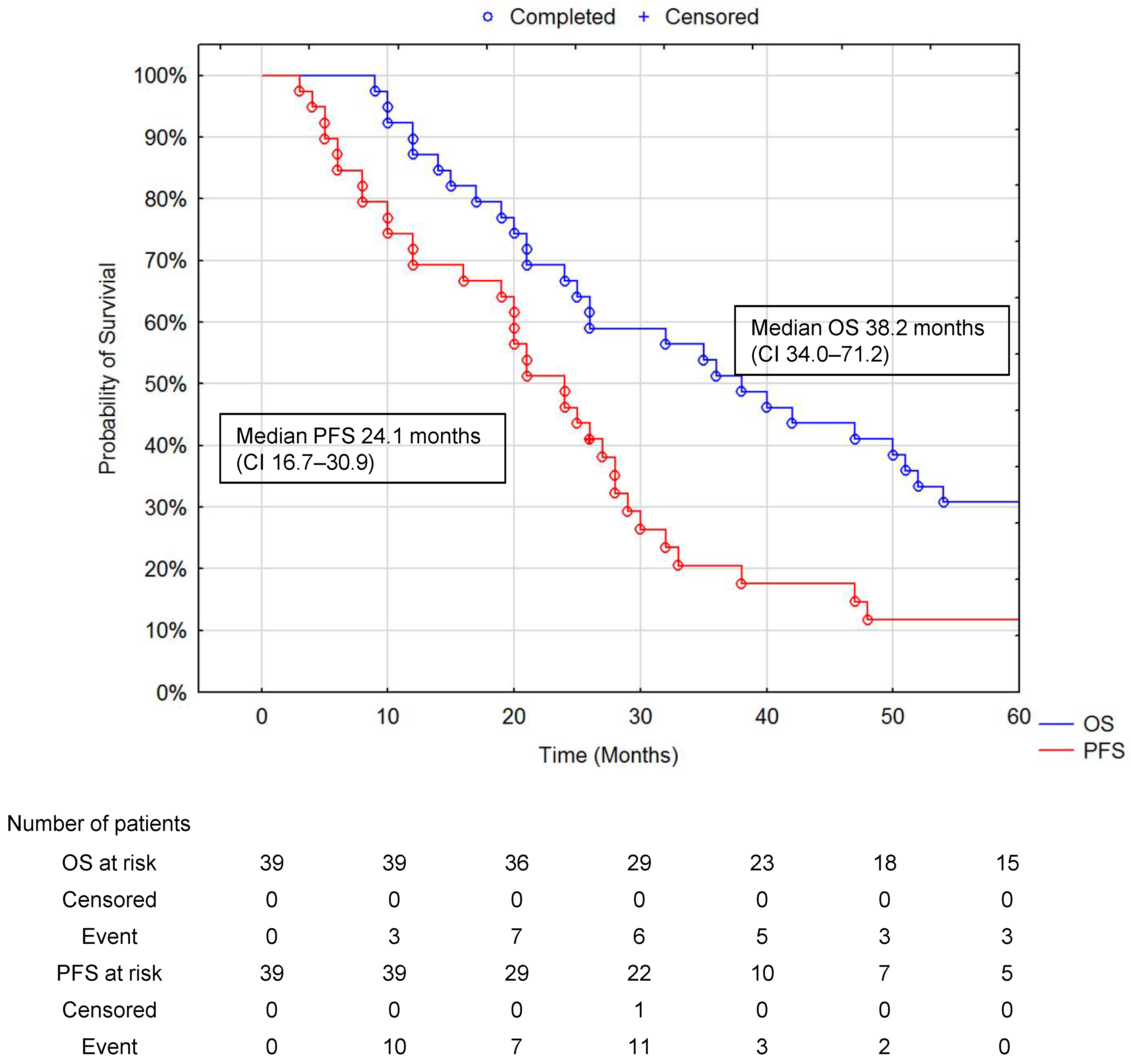
3.3. Statistical Analysis and Patient Survival Analysis

Statistical analysis was performed using Statistica v.13.1 (TIBCO Software Inc. Palo Alto, CA; USA). Differences in performance status (PS) in the ECOG scale in patients before therapy and after completed radionuclide therapy were performed using Wilcoxon’s matched pairs test. Differences between data sets of two independent samples were performed using a Mann–Wilcoxon U-test. The overall survival (OS) and progression-free survival (PFS) were estimated for the total cohort of patients using the Kaplan–Meier estimator. Overall survival (OS) was defined as the time from the first administration of i.a. therapy until death from any cause or last follow-up as censored data. PFS was calculated from the start of i.a. PRRT to recorded disease progression according to RECIST or death. Comparison of OS and PFS between different groups of patients was assessed using the Cox–Mantel test and log-rank test. The proportions of patients who had a clinical response (PS status) and CT responses were calculated separately. p < 0.05 was considered statistically significant.

4. Results

Of the 39 treated GEP-NEN patients, 14 had tumors of pancreatic origin, 13 had tumors arising from the small bowel, four has tumors arising from the large bowel/rectum, and eight had metastatic NEN but with an unknown primary (CUP—cancer of unknown primary). There were 19 of these patients with secreting tumors, nine of whom had had previous i.v. PRRT. The most common histological stage was NETG2 occurring in 28 subjects (73%) compared to NETG1 in eight patients (22%) and NETG3 in two patients (5%). At the start of treatment, 29 patients (74%) had a PS of 1, eight patients (23%) had a PS of 2, and one patient had a PS of 0. Liver metastases of over 50% of total liver volume were seen in 13 patients (33%), liver metastases involving 25–50% of the liver was seen in 18 (46%), and a metastatic volume involving less than 25% of total liver volume was seen in eight subjects (21%) (Table 1). All patients had received at least one previous line of treatment (Table 2). Patients received a mean activity of 3.13GBq normally split between two treatment cycles (Table 3). The biodistribution of the 90Y-DOTATATE post-therapy in almost all cases perfectly matched the pre-treatment diagnostic 99mTc-HYNICTOC imaging.
There was a significant clinical response as measured by the change in performance status (PS) after therapy (p < 0.01 Wilcoxon matched pair test). The PS improved in 29/39 patients (74%) at six weeks after PRRT, but in two patients, the PS had deteriorated. Of the 29 patients who had an improved PS at six weeks, 27 (93%) had maintained their improved PS; by 36 months, this improvement in PS was maintained in seven patients. The PS of four patients remained unchanged throughout the follow-up period
Based on RECIST criteria, at the six weeks post-imaging-based PRRT, no patient had suffered a DP, six months post-treatment the PR + SD (disease clinical response-DCR) was 95%, and of the 37 patients imaged at 12 months post-PRRT, the DCR was 92%. There were 15 patients still alive and imaged at 36 months after PRRT with a DCR of 53%. In those patients who had received both i.v.and i.a. PRRT, the DCR was 60% compared to just 40% in those receiving i.a. PRRT only. There were five patients with a PR at six weeks, but this had increased to nine at six months without further PRRT. At six months, two patients recorded SD. No patient had a CR (Table 4).
It was noted that 14 patients with PR during the follow-up had a mean tumor/liver ratio (T/L) of 0.28, compared to those patients who had only SD during follow-up with mean T/L = 0.48 (p < 0.002 Mann–Whitney U test). This reflected the higher tumor bulk in patients with SD than those with a PR. Four patients in whom i.a. was given without prior i.v. treatment with initial tumor shrinkage showed an increase in tumor size by 24 months (Figure 2); this was only seen in two patients who had had prior i.v.90Y-DOTATATE (Figure 3).
The median PFS and median OS for all patients were 24.1 months (CI 16.7–30.9) and 38.2 months (CI 34.0–71.2) (Figure 4).
There was a significantly different increase in median OS if patients were treated with i.a. 90Y-DOTATATE as their initial treatment at 52.1 months (CI 44.3–104.8) compared to those who had received i.v. 90Y-DOTATATE with a median OS of 22.2 months (CI 16.3–52.3) (p = 0.02); there was a borderline difference noted in PFS 12.3 months (CI 8.7–30.5) for those who had i.a. 90Y-DOTATATE compared to 28.4 months (CI 21.4–37.6) for those who received iv. 90Y-DOTATATE first (p = 0.056) (Figure 5).
In those patients with a DCR at six and 12 months, the survival advantage of treating bulky liver metastases with upfront i.a. 90Y DOTATATE was maintained. In those patients who had a DCR evaluated at six months, was 95%; at 12 months, it was 89%; and at 24 months in 30 alive patients, it was 73%. The median PFS (+/− 95% CI) at six, 12, and 24 months of DCR for all patients was, respectively, 23.9 months (18.0–32.9), 26.1 months (20.1–36.2), and 28.3 months (23.4–47.4) (Figure 6 and Figure 7).
The most common adverse event seen was Grade 1 anemia seen at six weeks post-therapy at a rate of 39%; in addition, 32% suffered a mild Grade 1 leukopenia, also peaking at six weeks post-PRRT (Table 5). There was a single patient who had significant anemia (Grade 3), which was managed conservatively. No other significant hematological or clinical biochemical or endocrine adverse events were recorded. In particular, there was no significant change in platelet count or adverse effect on liver function test results.

5. Discussion

The results of this study show that the use of i.a. 90Y DOTATATE could be an interesting strategy in which patients with bulky GEP-NEN metastases to the liver could be treated. It would appear from our preliminary findings in this small patient group that the best overall survival is obtained by the use of at least two cycles of i.a.1.1GBq 90Y DOTATATE is given upfront for such bulky disease within the liver with no apparent increase in toxicity when compared to i.v. 90Y DOTATATE.
Following the NETTER-1 trial, 177Lu DOTATATE has become the radionuclide treatment of choice in patients with metastatic GEP-NEN [11,12,13,14,35]. It has been shown to be both effective and safe. In a systematic review, the overall disease control rate (DCR) for 177Lu DOTATATE was 80%, and this was achieved without significant adverse events [36]. In the same systemic review, the DCR for i.v. 90Y DOTATATE was 92%. However, these results are not directly comparable, as the patient groups were probably not identical, and this improved efficacy was achieved with a rate of about 4% in significant adverse events (AEs).
The standard regime for systemic 90Y DOTATATE therapy was to give four cycles of 3.0-4.0 GBq, calculated to keep the renal dose below 23 Gy [20,29,35]. This, however, could lead to some significant bone marrow toxicity, with reports of late-onset myleodyspalstic syndrome (MDS) and leukemia [36]. There is no doubt that the extended path length of the 90Y beta could result in more tumor destruction, but this appeared to be handicapped by the increased adverse events rate [20,29,31,35,36]. It had been noted that liver metastases have historically been seen as a sign of a poor prognosis in GEP-NEN tumors, and untreated, they can be the cause of premature death either by the release of related hormones, loss of liver synthetic function, or pressure effects on other vital organs [37].
Therefore, it was logical to use techniques allowing local delivery of the 90Y DOTATATE to the liver. There had been some attempts to use particulates such as 90Y resin and glass to treat liver metastases in neuroendocrine tumors, but this can lead to post-treatment embolic syndrome, and its use has tended to be limited to tumors that do not express somatostatin receptors [38,39]. Comparison with purely embolic treatment such as trans-arterial embolization (TAE) and trans-arterial chemo-embolization (TACE) is more problematic, as neither of these techniques has the compounding factor of the use of high-energy beta-radiation within the liver that is seen with 90Y labeled particulates or DOTATATE.
Early reports did suggest that giving the 90Y labeled somatostatin analogs via an intra-arterial catheter in the hepatic artery can improve the response rate in liver metastases. The first reports, however, either used a regime with 90Y DOTATATE but given both systemically and intra-arterially or used a different peptide such as 90Y Lanreotide [15,22]. The largest series reporting the use of i.a. PRRT was that in a group of 23 patients receiving 1-2 cycles of just 1 GBq 90Y Lanreotide, a 79% DCR was achieved with a median overall survival (OS) of 15 months. There was no significant toxicity in this group of patients, the majority of whom had metastases representing greater than 50% of their total liver volume [15]. Lanreotide, however, has a lower affinity to GEP-NEN tumors compared with DOTATATE, so a DOTATATE-based approach may be still more efficacious [16,17,18].
Previously reported use of systemic 90Y DOTATATE in a similar group of patients from the same institutions as the patients treated in this study in which up to four cycles of 3.7 GBq each was administered showed an 87% DCR at six months post last therapy very similar to the 92% achieved in patients with liver metastases treated with at least two sessions of i.a. injection of 1.15 GBq each of 90Y DOTATATE in the current study. The mean cumulative activity administered in this study was only 3.13 GBq for all patients and was lower in those with previous i.v. PRRT vs. only i.a. 2.8 vs. 3.4 GBq, respectively. In our previous study, the mean administered activity in i.v. therapy was 11.2 GBq, which is almost four times greater than in subjects in the current trial who had initially (only) i.a. PRRT [9].
With the systemic administration of 90Y DOTATATE, the median PFS was 17 months (CI 16.4-21.2) compared to 24.1 months (CI 16.7-30.9) with the current i.a. 90Y PRRT. The median OS for those treated with systemic 90Y DOTATATE was 22 months (CI 20.4-26.7) compared to 38.2 months (CI 34.0-71.2) for those receiving i.a. 90Y DOTATATE [9]. Our study indicates additional, significant differences in median PFS and OS between when the i.a. approach of PRRT was given first; this may be due to the ability to deliver a higher therapeutic dose of the radionuclide to the liver metastases with reduced systemic side effects.
Another potential explanation of our results in both groups of patients who had current i.a. therapy was the selection of more aggressive tumor cell lines surviving treatment by previous i.v. PRRT in those patients who then relapsed and were treated with further i.a. PRRT. This phenomenon probably related to the natural history of the development of GEP-NEN, with the survival of more aggressive clones of cancer cells after anticancer therapy, like PRRT, meaning those clones of cells that survive are less sensitive to second-time beta irradiation [35].
The lack of radiological PR has been noted before in GEP-NEN patients treated with radioisotopic therapy in many others reports [7,8,9,10,15,17,18], and the results of this study are similar to a large series of over 300 patients from Rotterdam treated with 177Lu DOTATATE, where both partial response (PR) and disease stability (SD) as assessed by radiological criteria were related to a good overall prognosis [19]. In the NETTER1 randomized controlled trial, a radiological response was seen in 18% of patients, which is probably not significantly different from the 24% seen in the patients treated with i.v. 90Y DOTATATE in our previous reports [9,11]. The improved DCR at 36 months was seen in both those patients who had received previous systemic PRRT and those who were treated upfront with i.a. PRRT, indicating that i.a.90Y DOTATATE could be used safely after previous systemic PRRT, but that sequential treatment may not be the ideal scenario in this difficult group of subjects, and upfront i.a. 90Yttrium DOTATATE may be the best way forward in those patients with bulky liver metastases from GEP-NEN tumors.
In this trial, we noted a low rate of any AEs compared to the group of patients who were treated previously using i.v.90Y DOTATATE, as the reported Grade 3 and four toxicity (AEs) rate for those receiving systemic 90Y DOTATATE was 10% compared to just 3% in those treated with i.a. 90Y DOTATATE. As the patients treated by these two studies may have a number of different characteristics, the primary one being the liver-dominant disease in the i.a. group, it can be noted that it is unlikely that the i.a. 90Y DOTATATE is less effective and more toxic than the use of systemic 90Y DOTATATE. It is useful to note the Grade 3 and 4 hematological toxicity was 9% in those treated with 177Lu DOTATATE on the NETTER1, which is higher than seen in our group of patients treated with i.a. 90Y DOTATATE [11,19]. Though it is not possible to make a direct comparison between the two groups, it does suggest that low activity i.a. 90Y DOTATATE is as effective and certainly no more toxic than the standard four cycles of 7.4GBq 177Lu DOTATATE. However, the small number of patients within our study group means that further work in this area in a bigger patient group would be needed to confirm these findings.

6. Conclusions

The conclusion of this preliminary study is that treatment with two cycles of 1.1 GBq i.a. 90Y DOTATATE six weeks apart may be as effective and safe as the recommended cumulative activity of 28 GBq of 177Lu DOTATATE or 14 GBq of 90Y DOTATATE given over i.v. at eight months in those patients with bulky liver GEP-NEN metastases, with some evidence of improvement in overall response rate, including disease control rate (DCR), clinical response (PS), and also PFS and OS when given as upfront i.a.90Y DOTATATE. A larger multi-center trial should now be considered to determine if such an approach should be considered as an alternative or adjunctive form of PRRT in those with liver-dominant unresectable GEP-NEN metastases in their liver.

Supplementary Materials

The following are available online at https://www.mdpi.com/article/10.3390/jcm10081794/s1: Supplementary: preparation of radiotracer; radiology imaging-CT; MRI; SPECT imaging.

Author Contributions

Conceptualization: A.K.-Ć., J.B.Ć., M.L.N., J.R.B.; methodology: J.B.Ć., M.L.N., J.R.B.; software: J.M.P., A.J.S.; validation, A.K.-Ć., J.B.Ć., L.G.; formal analysis: A.K.-Ć., J.R.B., J.B.Ć., L.G.; investigation: A.K.-Ć., M.L.N., A.J.S., J.B.Ć.; resources: A.K.-Ć., M.L.N., A.J.S.; data curation: A.K.-Ć., L.G., J.B.Ć.; writing—original draft preparation, A.K.-Ć., J.B.Ć., J.R.B.; writing—review and editing: all authors; visualization: J.M.P., A.J.S., L.G., J.B.Ć.; supervision: J.R.B., J.B.Ć.; project Administration: A.K-Ć.; funding acquisition: J.B.Ć. All authors have read and agreed to the published version of the manuscript.

Funding

Polish Ministry of Science and Higher Education (Grant No N518 001 31/0040).

Institutional Review Board Statement

The study protocol has been approved by the Clinical Ethics Committee of the Institution where the study was conducted—Central Clinical Hospital of Ministry of Interior Affairs and Administration, (reference no 109/2004). The research was conducted ethically in accordance with the World Medical Association Declaration of Helsinki.

Informed Consent Statement

All subjects included in this study have given their written informed consent.

Data Availability Statement

This was single site, prospective, preliminary case series.

Acknowledgments

Authors wish to thank the patients’ advocacy group: “Stowarzyszenie Pacjentów i Osób Wspierających Chorych na Guzy Neuroendokrynne”. Warsaw, Poland.

Conflicts of Interest

The authors declare no conflict of interest.

References

  1. Caplin, M.E.; Buscombe, J.R.; Hilson, A.J.; Jones, A.L.; Watkinson, A.F.; Burroughs, A.K. Carcinoid tumour. Lancet 1998, 352, 799–805. [Google Scholar] [CrossRef]
  2. Caplin, M.E.; Pavel, M.; Ćwikła, J.B.; Phan, A.T.; Raderer, M.; Sedláčková, E.; Cadiot, G.; Wolin, E.M.; Capdevila, J.; Wall, L.; et al. Lanreotide in metastatic enteropancreatic neuroendocrine tumors. N. Engl. J. Med. 2014, 371, 224–233. [Google Scholar] [CrossRef]
  3. Ramage, J.K.; Ahmed, A.; Ardill, J.; Bax, N.; Breen, D.J.; Caplin, M.E.; Corrie, P.; Davar, J.; Davies, A.H.; Lewington, V.; et al. Guidelines for the management of gastroenteropancreatic neuroendocrine (including carcinoid) tumours (NETs). Gut 2011, 61, 6–32. [Google Scholar] [CrossRef]
  4. Raymond, E.; Dahan, L.; Raoul, J.L.; Bang, Y.J.; Borbath, I.; Lombard-Bohas, C.; Valle, J.; Metrakos, P.; Smith, D.; Vinik, A.; et al. Sunitinib Malate for the Treatment of Pancreatic Neuroendocrine Tumors. N. Engl. J. Med. 2011, 364, 501–513. [Google Scholar] [CrossRef] [Green Version]
  5. Yao, J.C.; Fazio, N.; Singh, S.; Buzzoni, R.; Carnaghi, C.; Wolin, E.; Tomasek, J.; Raderer, M.; Lahner, H.; Voi, M.; et al. Everolimus ofr the treatment of advanced non-functional neuroendocrine tumours of the lung or gastrointestinal tract (Radiant-4): A randomized, placebo-controlled phase 3 study. Lancet 2016, 387, 968–977. [Google Scholar] [CrossRef]
  6. Bodei, L.; Mueller-Brand, J.; Baum, R.P.; Pavel, M.E.; Hörsch, D.; O’Dorisio, M.S.; O’Dorisio, T.M.; Howe, J.R.; Cremonesi, M.; Kwekkeboom, D.J.; et al. The joint IAEA, EANM, and SNMMI practical guidance on peptide receptor radionuclide therapy (PRRNT) in neuroendocrine tumours. Eur. J. Nucl. Med. Mol. Imaging. 2013, 40, 800–816. [Google Scholar] [CrossRef]
  7. Kwekkeboom, D.J.; Teunissen, J.J.; Bakker, W.H.; Kooij, P.P.; De Herder, W.W.; Feelders, R.A.; Van Eijck, C.H.; Esser, J.-P.; Kam, B.L.; Krenning, E.P. Radiolabeled somatostatin analog [177Lu-DOTA0,Tyr3]octreotate in patients with endocrine gastroenteropancreatic tumors. J. Clin. Oncol. 2005, 23, 2754–2762. [Google Scholar] [CrossRef] [Green Version]
  8. Imhof, A.; Brunner, P.; Marincek, N.; Briel, M.; Schindler, C.; Rasch, H.; Mäcke, H.R.; Rochlitz, C.; Müller-Brand, J.; Walter, M.A. Response, survival, and long-term toxicity after therapy with the radiolabeled somatostatin analogue [90Y-DOTA]-TOC in metastasized neuroendocrine cancers. J. Clin. Oncol. 2011, 29, 2416–2423. [Google Scholar] [CrossRef]
  9. Ćwikła, J.B.; Sankowski, A.J.; Seklecka, N.; Buscombe, J.R.; Nasierowska-Guttmejer, A.; Jeziorski, K.G.; Mikołajczak, R.; Pawlak, D.; Walecki, J. Efficacy of radionuclide treatment 90Y-DOTATATE in patients with progressive metastatic gastroenteropancreatic neuroendocrine carcinomas (GEP-NET). A phase II study. Ann. Oncol. 2010, 21, 787–794. [Google Scholar] [CrossRef]
  10. Bodei, L.; Cremonesi, M.; Grana, C.M.; Fazio, N.; Iodice, S.; Baio, S.M.; Bartolomei, M.; Lombardo, D.; Ferrari, M.E.; Sansovini, M.; et al. Peptide receptor radionuclide therapy with 177Lu-DOTATATE: The IEO phase I-II study. Eur. J. Nucl. Med. Mol. Imaging 2011, 38, 2125–2135. [Google Scholar] [CrossRef]
  11. Strosberg, J.; El-Haddad, G.; Wolin, E.; Hendifar, A.; Yao, J.; Chasen, B.; Mittra, E.; Kunz, P.L.; Kulke, M.H.; Jacene, H.; et al. Phase 3 trail of 177Lu DOTATATE for midgut neuroendocrine tumors. N. Engl. J. Med. 2017, 376, 125–135. [Google Scholar] [CrossRef]
  12. Hicks, R.J.; Kwekkeboom, D.J.; Krenning, E.; Bodei, L.; Grozinsky-Glasberg, S.; Arnold, R.; Borbath, I.; Cwikla, J.; Toumpanakis, C.; Kaltsas, G.; et al. ENETS Consensus Guidelines for the Standards of Care in Neuroendocrine Tumors. Peptide Receptor Radionuclide Therapy with Radiolabeled Somatostatin Analogues. Neuroendocrinology 2017, 105, 295–309. [Google Scholar] [CrossRef] [PubMed]
  13. Hope, T.A.; Bodei, L.; Chan, J.A.; El-Haddad, G.; Fidelman, N.; Kunz, P.L.; Mailman, J.; Menda, Y.; Metz, D.C.; Mittra, E.S.; et al. NANETS/SNMMI Consensus Statement on Patient Selection and Appropriate Use of 177Lu-DOTATATE Peptide Receptor Radionuclide Therapy. J. Nucl. Med. 2020, 61, 222–227. [Google Scholar] [CrossRef] [PubMed]
  14. Shah, M.H.; Goldner, W.S.; Halfdanarson, T.R.; Bergsland, E.; Berlin, J.D.; Halperin, D.; Chan, J.; Kulke, M.H.; Benson, A.B.; Blaszkowsky, L.S.; et al. NCCN Clinical Practice Guidelines in Oncology: Neuroendocrine and Adrenal Tumors. J. Natl. Compr. Canc. Netw. 2018, 16, 693–702. [Google Scholar] [CrossRef]
  15. McStay, M.K.; Maudgil, D.; Williams, M.; Tibballs, J.M.; Watkinson, A.F.; Caplin, M.E.; Buscombe, J.R. Large-Volume Liver Metastases from Neuroendocrine Tumors: Hepatic Intraarterial 90Y-DOTA-Lanreotide as Effective Palliative Therapy. Radiology 2005, 237, 718–726. [Google Scholar] [CrossRef] [PubMed]
  16. Reubi, J.C.; Schär, J.-C.; Waser, B.; Wenger, S.; Heppeler, A.; Schmitt, J.S.; Mäcke, H.R. Affinity profiles for human somatostatin receptor subtypes SST1-SST5 of somatostatin radiotracers selected for scintigraphic and radiotherapeutic use. Eur. J. Nucl. Med. Mol. Imaging 2000, 27, 273–282. [Google Scholar] [CrossRef]
  17. Kwekkeboom, D.J.; Bakker, W.H.; Kam, B.L.; Teunissen, J.J.M.; Kooij, P.P.M.; De Herder, W.W.; Feelders, R.A.; Van Eijck, C.H.J.; De Jong, M.; Srinivasan, A.; et al. Treatment of patients with gastro-entero-pancreatic (GEP) tumours with the novel radiolabelled somatostatin analogue [177Lu-DOTA0,Tyr3]octreotate. Eur. J. Nucl. Med. Mol. Imaging 2003, 30, 417–422. [Google Scholar] [CrossRef] [PubMed] [Green Version]
  18. Esser, J.P.; Krenning, E.P.; Teunissen, J.J.M.; Kooij, P.P.M.; Van Gameren, A.L.H.; Bakker, W.H.; Kwekkeboom, D.J. Comparison of [177Lu-DOTA0,Tyr3]octreotate and [177Lu-DOTA0,Tyr3]octreotide: Which peptide is preferable for PRRT? Eur. J. Nucl. Med. Mol. Imaging 2006, 33, 1346–1351. [Google Scholar] [CrossRef] [PubMed]
  19. Kwekkeboom, D.J.; de Herder, W.W.; Kam, B.L.; van Eijck, C.H.; van Essen, M.; Kooij, P.P.; Feelders, R.A.; van Aken, M.O.; Krenning, E.P. Treatment with the Radiolabeled Somatostatine Analog [177Lu-DOTA0Tyr3]octreotate: Toxicity, Efficacy and Survival. J. Clin. Oncol. 2008, 26, 2124–2130. [Google Scholar] [CrossRef] [Green Version]
  20. Valkema, R.; Pauwels, S.A.; Kvols, L.K.; Kwekkeboom, D.J.; Jamar, F.; De Jong, M.; Barone, R.; Walrand, S.; Kooij, P.P.M.; Bakker, W.H.; et al. Long term follow-up of renal function after peptide receptor radiation therapy with 90Y-DOTA, Tyr3-Octreotide and 177Lu-DOTA, Tyr3-Octreotate. J. Nucl. Med. 2005, 46, 83S–91S. [Google Scholar]
  21. Buscombe, J.R. Interventional nuclear medicine in hepatoceullar cancers and other tumours. Nucl. Med. Commun. 2002, 23, 837–842. [Google Scholar] [CrossRef] [PubMed]
  22. Baum, R.P.; Söldner, J.; Schmücking, M.; Niesen, A. Intravenous and Intra-arterial Peptide Receptor Radionuclide Therapy (PRRT) Using 90Y-DOTA-TYR3-OCTREOTATE (90Y DOTA-TATE) in Patients with Metastatic Neuroendocrine Tumors. In Proceedings of the Annual Congress of the European-Association-of-Nuclear-Medicine, Helsinki, Finland, 5–8 September 2004. [Google Scholar]
  23. Pettersson, O.; Fross-Baron, K.; Crona, J.; Dundin, A. Tumour Contrast-Enhancement of Monitoring of PRRT 177Lu DOTATATE in Pancreatic Neuroendocrine Tumour Patients. Front. Oncol. 2020, 10, 193. [Google Scholar] [CrossRef] [PubMed]
  24. Sundin, A.; Arnold, R.; Baudin, E.; Cwikla, J.B.; Eriksson, B.; Fanti, S.; Fazio, N.; Giammarile, F.; Hicks, R.J.; Kjaer, A.; et al. ENETS Consensus Guidelines for the Standards of Care in Neuroendocrine Tumors: Radiological, Nuclear Medicine and Hybrid Imaging. Neuroendocrinology 2017, 105, 212–244. [Google Scholar] [CrossRef] [PubMed]
  25. Gehan, E.A.; Tefft, M.C. Will there be resistance to the RECIST (Response Evaluation Criteria in Solid Tumours)? J. Natl. Cancer Inst. 2000, 92, 179–181. [Google Scholar] [CrossRef]
  26. Eisenhauer, E.A.; Therasse, P.; Bogaerts, J.; Schwartz, L.H.; Sargent, D.; Ford, R.; Dancey, J.; Arbuck, S.; Gwyther, S.; Mooney, M.; et al. New response evaluation of solid tumours. Revised RECIST guidelines (RECIST1.1). Eur. J. Oncol. 2009, 45, 228–247. [Google Scholar]
  27. Waldherr, C.; Pless, M.; Maecke, H.R.; Schumacher, T.; Crazzolara, A.; Nitzsche, E.U.; Haldemann, A.; Mueller-Brand, J. Tumor response and clinical benefit in neuroendocrine tumors after 7.4 GBq 90Y-DOTATOC. J. Nucl. Med. 2002, 43, 610–616. [Google Scholar] [PubMed]
  28. Baum, R.; Wehrmann, C.; Zachert, C.; Prasad, V.; Wortmann, R. Long-term results of peptide receptor radionuclide therapy (PRRT): 5-year follow-up of 1,150 courses in 360 patients with progressive, somatostatin receptor positive neuroendocrine tumors in one clinical center. J. Nucl. Med. 2007, 48 (Suppl. S2), 37p. [Google Scholar]
  29. Chinol, M.; Bodei, L.; Cremonesi, M.; Paganelli, G. Receptor-mediated radiotherapy with 90Y-DOTA-DPhe-Tyr-octreotide: The experience of the European Institute of Oncology Group. Semin. Nucl. Med. 2002, 32, 141–147. [Google Scholar] [CrossRef]
  30. National Institute for Health and Care Excellence. Lutetium 177Lu Oxodotreotide for Treating Unresectable or Metastatic Neuroendocrine Tumours 2018. Available online: https://www.nice.org.uk/guidance/TA539 (accessed on 12 April 2021).
  31. Buscombe, J.R.; Navilkissoor, S. Molecular Radiotherapy. Clin. Med. 2012, 12, 381–386. [Google Scholar] [CrossRef] [Green Version]
  32. Perren, A.; Couvelard, A.; Scoazec, J.Y.; Costa, F.; Borbath, I.; Delle Fave, G.; Gorbounova, V.; Gross, D.; Grossma, A.; Jense, R.T.; et al. ENETS Consensus Guidelines for the Standards of Care in Neuroendocrine Tumours: Pathology Diagnosis and Prognostic Stratification. Neuroendocrinology 2017, 105, 196–200. [Google Scholar] [CrossRef] [PubMed]
  33. Cwikla, J.B.; Mikolajczak, R.; Pawlak, D.; Buscombe, J.R.; Nasierowska-Guttmejer, A.; Bator, A.; Maecke, H.R.; Walecki, J. Initial direct comparison of 99mTc-[HYNIC-TOC] and 99mTc-[HYNIC-TATE] in identifying sites of disease in patients with proven GEP-NET tumors. J. Nucl. Med. 2008, 49, 1060–1065. [Google Scholar] [CrossRef] [PubMed] [Green Version]
  34. Therasse, P.; Arbuck, S.G.; Eisenhauer, A.E.; Wanders, E.; Kaplan, J.; Rubinstein, R.; Verweij, L.; Van Glabbeke, J.; van Oosterom, M.; Christian, A.; et al. New guidelines to evaluate the response to treatment in solid tumors. J. Nat. Cancer Inst. 2000, 92, 205–216. [Google Scholar] [CrossRef] [Green Version]
  35. De Jong, M.; Valkema, R.; Jamar, F.; Kvols, L.K.; Kwekkeboom, D.J.; Breeman, W.A.; Bakker, W.H.; Smith, C.; Pauwels, S.; Krenning, E.P. Somatostatin receptor-targeted radionuclide therapy of tumors: Preclinical and clinical findings. Semin. Nucl. Med. 2002, 32, 133–140. [Google Scholar] [CrossRef]
  36. Goncalves, I.; Burbury, K.; Michael, M.; Iravani, A.; Ravi Kumar, A.S.; Akhurst, T.; Tiong, I.S.; Blombery, P.; Hofman, M.S.; Westerman, D.; et al. Characteristics and Outcomes of Therapy-Related Myeloid Neoplasms After Peptide Receptor radionuclide/chemoradionuclide Therapy (PRRT/PRCRT) for Metastatic Neuroendocrine Neoplasia: A Single-Institution Series. Eur. J. Nucl. Med. 2019, 46, 1902–1910. [Google Scholar] [CrossRef]
  37. McDermott, E.W.; Guduric, B.; Brennan, M.F. Prognostic variables in patients with gastrointestinal carcinoid tumours. Br. J. Surg. 1994, 81, 1007–1009. [Google Scholar] [CrossRef] [PubMed]
  38. Vyleta, M.; Coldwell, D. Radioembolization in the Treatment of Neuroendocrine Tumor Metastases to the Liver. Int. J. Hepatol. 2011, 2011, 785315. [Google Scholar] [CrossRef] [Green Version]
  39. Kennedy, A.; Bester, L.; Salem, R.; Sharma, R.A.; Parks, R.W.; Ruszniewski, P. Role of hepatic intra-arteria therapies in metastatic neuroendocrine tumours (NET): Guidelines from the NET-Liver -Metastases Cnsensus Conference. HPB 2015, 17, 29–37. [Google Scholar] [CrossRef] [Green Version]
Figure 1. Study design, including main inclusion criteria, selection of two groups of patients, and endpoints.
Jcm 10 01794 g001
Figure 2. The waterfall plot of ORR in selected time frame points of evaluation at six weeks, six months, 12 months, and 24 months in the group with initially i.a.PRRT (n = 23).
Jcm 10 01794 g002
Figure 3. The waterfall plot of ORR in selected time frame points of evaluation at six weeks, six months, 12 months, and 24 months in the group with previous i.v. PRRT (n = 16).
Jcm 10 01794 g003
Figure 4. OS and PFS in all patients during follow-up. Median OS 38.2 months (95% CI 34.0–71.2) and median PFS 24.1 months (95% CI 16.7–30.9). Data are presented for all patients who received at least two doses of PRRT during the study. The number of subjects remaining at risk below is 10% of cases in any group.
Jcm 10 01794 g004
Figure 5. Comparison of OS in patients with previous i.v. PRRT and initially i.a. PRRT median 22.2 (CI 16.3–52.3) vs. 52.1 (CI 44.3–104.8) (p = 0.02 Cox Mantel Test). The number of subjects remaining at risk is below 10% of cases in any group.
Jcm 10 01794 g005
Figure 6. Comparison of PFS in patients with previous i.v. PRRT at 12.3 months (95% CI 9.1–33.1) and initially i.a. PRRT at 28.4 months (95% CI 23.5–39.4) at six months of DCR. (p = 0.048 Cox–Mantel test). Disease control rate was defined as the proportion of patients with partial response (PR) or stable disease (SD), not patients with complete response (CR). The number of subjects remaining at risk was below 10% of cases in any group.
Jcm 10 01794 g006
Figure 7. Comparison of PFS in patients with previous i.v. PRRT at 19.1 months (95% CI 9.2–35.6) and initially i.a. PRRT at 29.6 months (95% CI 27.7–43.1) at 12 months of DCR (p = 0.026 Cox–Mantel test). Disease control rate was defined as the proportion of patients with partial response (PR) or stable disease (SD), not patients with complete response (CR). The number of subjects remaining at risk was below 10% of cases in any group.
Jcm 10 01794 g007
Table 1. Clinical data of all patients and those with previous i.v. PRRT and native i.a. PRRT.
All n = 39Previous i.v. PRRT
n = 16 (%)
Only i.a.
n = 23 (%)
Male to female22/1712/412/11
Age in years (mean, SD)56.4 (9.2)55.4 (8.2)57.5 (9.9)
Primary site NEN origin
Pancreas14 (36)7 (44)7 (30)
Small Bowel13 (33)5 (31)8 (35)
Large Bowel/Rectum4 (10)1 (6)3 (13)
CUP (Cancer of Unknown Primary)8 (21)3 (19)5 (22)
Secretor tumors19 (49)9 (56)10 (43)
Tumor grade
NETG1 (Ki-67 ≤ 2%)8 (22)4 (27)4 (17)
NEG2 (2 < Ki-67 ≤ 20%)28 (73)10 (67)18 (78)
NETG3 (20% < Ki-67 < 50%)2 (5)1 (6)1 (5)
Hepatic load (tuVolume/liverVolume)0.41%45%38%
≤25%8 (21)3 (19)5 (22)
< 25% x ≤ 50% 18 (46)6 (38)12 (52)
>50%13 (33)7 (43)6 (26)
WHO Performance status (PS) initialAll n = 39Previous iv. PRRT n = 16 (%)Only i.a. n = 23 (%)
0—normal activity1 (3) 1 (5)
1—restricted activity29 (74)11 (69)18 (78)
2—in bed ≤ 50% of the time9 (23)5 (31)4 (17)
Initial basic hematology and kidney creatinine levelAll
n = 39
Previous iv. PRRT
n = 16
Only i.a.
n = 23 mean value
WBC (×109/L)6.787.346.37
Hb (g/dL)12.812.013.4
Platelet (×106/L)288297282
Creatinine level mg/dL0.971.240.78
CgA x ULN; mean (95% CI)34.5 (19.8–49.2)48.8 (11.6–81.9)27.6 (13.9–41.3)
Ki-67 thresholds as per WHO and UICC/AJCC classification 2017 classification as NETG1 (Ki-67 < 3%), NETG2 (2% < Ki-67 ≤ 20%), and NETG3 (Ki-67 > 20% but below 50%). CgA—chromogranin A.
Table 2. Previous systemic therapies, considering all patients and those with previous i.v. and only i.a. PRRT.
Therapy Approach before i.a. PRRTAll n = 39 (%)Previous iv. PRRT n = 16 (%)Only i.a. PRRT n = 23 (%)
Analogs SST (long-acting)32 (82)13 (81)19 (83)
Chemotherapy any type12 (31)9 (56)3 (13)
Previous i.v. PRRT16 (41)16 (100)
Local Liver therapy TA, surgery9 (23)4 (25)5 (22)
Table 3. PRRT therapy scheme, consider all patients previous i.v. PRRT and native i.a. PRRT.
ParameterAll Subjects
n = 39
Previous i.v. PRRT
n = 16 (%)
Native i.a.
n = 23 (%)
Mean activity (GBq of 90Y DOTATATE) per session1.151.121.16
Mean Cumulative activity (GBq of 90Y) per therapy (range)3.13 (1.4–4.1)2.8 (2.0–4.1)3.4 (1.4–4.1)
Mean (range) time between each therapy sessions (weeks)9.2 (6–12)10 (8–12)8.4 (6–10)
Table 4. Objective response rate (ORR) and clinical response after six weeks (6 W) and then after six months (6 M), 12 months (12 M), 24 months (24 M), and 36 months of follow-up for all patients after i.a.PRRT (n = 39). Additional data sets in selected populations of patients after previous i.v. PRRT and in those with initially i.a. PRRT.
Response RECIST 6W,
n (%)
Clinical Response 6 WRECIST; 6 M
n (%)
Clinical Response 6 MRECIST 12 M,
n (%)
Clinical Response 12 MRECIST 24 M
n (%)
Clinical Response 24 MRECIST 36 M
n (%)
Clinical Response 36 M
All subjects n = 39
PR5 (13)29 (74)9 (24)27(70)9 (24)24 (63)5 (16)15 (46)2 (13)7 (39)
SD34 (87)8 (21)27 (71)6 (15)25 (68)4 (11)18 (58)6 (18)6 (40)4 (22)
DP 2 (5)2 (5)6 (15)3 (8)10 (26)8 (26)12 (36)7 (47)7 (39)
Previous i.v. PRRT n = 16
PR3 (19)12 (75)6 (38)10 (63)4 (25)7 (44)3 (23)3 (23)2 (40)2 (40)
SD13 (81)3 (19)10 (62)4 (25)11 (69)2 (12)6 (46)1 (8)
DP 1 (6) 2 (12)1 (6)7 (44)4 (31)9 (69)3 (60)3 (60)
Initially i.a. PRRT n = 23
PR2 (9)17 (74)3 (14)17 (74)5 (23)17 (77)2 (11)12 (23) 5 (38)
SD21 (91)5 (22)17 (77)2 (9)14 (67)2 (9)12 (67)5 (8)6 (60)4 (31)
1 (4)2 (9)4 (17)2 (10)3 (14)4 (22)3 (69)4 (40)4 (31)
M-Months; W-Weeks.
Table 5. Treatment-related hematological and kidney adverse events (AEs) in the whole group of treated patients, percentages in brackets.
Initial Week 0
n = 38
Week 6 after PRRT,
n = 38
6 Months
n = 35
12 Months
n = 29
24 Months
n = 20
WBC
Grade 1 5 (13)12 (32)9 (26)7 (24)1 (6)
Grade 2 1 (3)1 (3)
Grade 3 1 (6)
Hb
Grade 1 10 (26)15 (39)6 (21)6 (21)3 (15)
Grade 22 (5)2 (5)2 (7)2 (7)1 (5)
Grade 3 1 (3)1 (3)1 (3)
Creatinine
Grade 12 (5) 2 (6)4 (14)
Grade 22 (5)3 (8)2 (6)2 (7)
Grade 3
Publisher’s Note: MDPI stays neutral with regard to jurisdictional claims in published maps and institutional affiliations.

