Article

Disc Large Homolog 1 Is Critical for Early T Cell Receptor Micro Cluster Formation and Activation in Human T Cells

1
Laboratory of Immune Cell Biology, Center for Cancer Research, NCI, NIH, Bethesda, MD 20892, USA
2
Genome Modification Core—Laboratory Animal Sciences Program, Frederick National Laboratory for Cancer Research, NCI, NIH, Bethesda, MD 20892, USA
*
Author to whom correspondence should be addressed.
Current affiliation: Laboratory of Molecular Immunology, NIAID, NIH, Bethesda, MD 20892, USA.
Vaccines 2021, 9(12), 1446; https://doi.org/10.3390/vaccines9121446
Submission received: 19 October 2021 / Revised: 29 November 2021 / Accepted: 30 November 2021 / Published: 7 December 2021
(This article belongs to the Special Issue Research on MHC Background and TCR Diversity in Vaccinology)

Abstract

:
T cell activation by antigen involves multiple sequential steps, including T cell receptor-microcluster TCR-(MC) formation, immunological synapse formation, and phosphorylation of mediators downstream of the TCR. The adaptor protein, Disc Large Homolog 1 (DLG1), is known to regulate proximal TCR signaling and, in turn, T cell activation, acting as a molecular chaperone that organizes specific kinases downstream of antigen recognition. In this study, we used knockdown and knockout technologies in human primary T cells and a human T cell line to demonstrate the role of DLG1 in proximal T cell signaling. High-end confocal microscopy was used for pictorial representation of T cell micro-clusters and colocalization studies. From all these studies, we could demonstrate that DLG1 functions even earlier than immunological synapse formation, to regulate T cell activation by promoting TCR-MC formation. Moreover, we found that DLG1 can act as a bridge between the TCR-ζ chain and ZAP70 while inhibiting binding of the phosphatase SHP1 to TCR-ζ. Together, these effects drive dysregulation of T cell activation in DLG1-deficient T cells. Overall, the activation and survival status of T cell is a critical determinant of effective vaccine response, and DLG1-mediated T cell signaling events can be a driving factor for improving vaccine-designing strategies.

1. Introduction

The adaptive immune response is critically dependent on T cell activation and plays an important pathophysiologic role in multiple processes, from pathogen clearance to development of autoimmune disease. T cell activation starts with the recognition of cognate peptide-MHC (pMHC) complexes on antigen-presenting cells by the TCR on T cells. TCR activation triggers a downstream signal transduction cascade that equips the cell with a proper effector function, thus dictating the nature of the immune response [1].
After antigen (Ag) engagement, Lck (a SRC family kinase) is recruited to the TCR-CD3 complex, where it becomes autophosphorylated and activated to then phosphorylate the immunoreceptor tyrosine-based activation motifs (ITAMs) of the CD3 zeta chains. This, in turn, opens up a docking site for ZAP70 (a SYK family kinase), which gets recruited to the ζ chain of the TCR-CD3 complex via its SRC-homology 2 (SH2) domain [2]. Lck activates TCR-bound ZAP70, which, in turn, phosphorylates its downstream substrate LAT (linker for activation of T cell). Subsequently, LAT recruits several signaling molecules to form a multiprotein complex, termed the LAT signalosome [3]. The LAT signalosome propagates additional signal transduction events, leading to further activation of transcription factors critical for T cell growth and differentiation.
At the cellular level, interaction of the T cell with the APC leads to the formation of the Immunological Synapse (IS) at the interface of the interacting partners. In recent years, it has been shown that Ag-specific activation signals are turned on at TCR-microclusters (TCR-MC) prior to IS formation [4]. TCR-MCs are defined as interacting platforms for the TCR, kinases, phosphatases, and adaptor molecules. TCR-MCs are formed within microseconds of the TCR-pMHC interaction and become building blocks of TCR proximal signaling. They gradually segregate to the cell–cell interface along with adhesion molecules to form the Supra molecular activation complex (SMAC), which consists of a central region known as the c-SMAC and a peripheral adhesion molecule (LFA-1)-rich region known as the p-SMAC. Studies of very early activation events using Ag-specific T cells and a planar bilayer containing specific Ag peptide-MHC has revealed that TCR clustering starts immediately after its interaction with the p-MHC, an event happening much earlier than IS formation. In previous studies with Jurkat cells, TCR stimulation by immobilized anti-CD3 mAb also resulted in induction of signaling cascades [5].
Maintenance of a mature IS requires strong and continuous TCR engagement and downstream signal transduction, and this leads to maximal T cell proliferation and effector function. Adapter or scaffold proteins act as molecular chaperones linking the T cell receptor to proximal signaling moieties, thus controlling the outcome of receptor engagement [6]. The membrane-associated guanylate kinases (MAGUKs) are scaffold proteins known to have critical roles in synaptic development [7]. Members of the MAGUK protein family contain PDZ domains, SH3 (src-homology 3) domains, and a guanylate kinase-homologous region (GUK) and often regulate immunological synapse formation and T cell polarity and morphology during migration [8]. Two crucial MAGUKs implicated in T cell function are Carma1 and DLG1 [9,10]. The human homolog of Drosophila Disc Large, DLG1, translocates to the IS, interacts with Lck, and forms a molecular complex with ZAP70 and Wasp [11]. It also coordinates alternative p38 kinase activation and NFAT-driven T cell signaling [12]. Regulatory T cell activation is also influenced by DLG1 [13]. Three distinct Dlg1-deficient mouse strains have been developed, one conditional and two non-conditional. Reports suggests that germline and conditional dlg1 knockout mice had no observable defects in CD4+ or CD8+ T cell polarization and, thus, development, migration, activation, signaling, or proliferation. These data were contradictory to previous reports demonstrating a role for Dlg1 in regulating TCR-mediated actin polymerization in mature T cells. Diminished cytoskeletal reorganization was observed in primary mouse CD8+ and human Jurkat T cells; however, conditional or germline dlg1 knockout mouse failed to show such effects. Differential cytokine secretion in T cells was observed with an acute or conditional loss of Dlg1, but not a germline loss of Dlg1. All this suggests a compensatory mechanism at different stages of T cell development, which might mask DLG1 function in T cells in vivo [14]. In the present study, we aimed to further understand the role of DLG1 in proximal human T cell signaling, particularly at the very early stage of antigen recognition and TCR-MC formation.

2. Materials and Methods

2.1. Reagents and Antibodies

The rabbit antibodies used were as follows: anti-phosphorylated ZAP-70Y319 (2701), anti-phosphorylated PLC-γ1Y783 (2821), anti-phosphorylated LATY171 (3581), anti-phosphorylated ZAP-70Y493 (2704), anti-p44/42 MAPK (anti-ERK1/2; 9101), anti-phosphorylated p44/42 MAPK (anti-phosphorylated ERK1/2; 9102, at Thr202 and Tyr204), anti-ZAP-70 (clone 99F2; 2705), anti-SHP1 (clone-C14H6; 3759), anti-LAT(9166) and anti-p38 MAPK (9212) from Cell Signaling Technologies, California, USA. Anti-CD3-ζ (phosphorylated Tyr83; 68236) from Abcam, Cambridge, UK. The anti-mouse monoclonal antibodies that were used included anti TCR-ζ (6B10.2; 1239), anti-SAP97 (Dlg1; clone 2D11, sc-9961), and anti-Lck (3A5; 433), siRNAs including control siRNA-A (sc-37007), siRNA plasmid against hDlg1 (sc-36452), and hSHP1 (sc-36490) and agarose-conjugated CD3-ζ mouse monoclonal IgG1 (1239-AC) from Santa Cruz, CA, USA. Anti-human-CD3 functional-grade purified (Clone OKT3, 16-0037-85, Clone UCHT1, 16-0038-81, Clone HIT3a, 16-0039-81), anti-human-CD28 functional-grade purified (clone 28.2, 16-0289-85) were from eBiosciences, San Diego, CA, USA. Anti-human CD45 functional grade (Clone HI30, 555480) and anti-CD247 (P-Y142 CD3, clone K25-407.69, 558402) were from BD Pharmingen, San Diego, CA, USA. Goat anti-rabbit (11010, Alexa Fluor 564) and goat anti-mouse (21242, Alexa Fluor 647) were from Thermo Fisher, Waltham, MA, USA. Anti β-Actin was from Sigma, St. Louis, MO, USA. CD4-Pacific blue, CD8-APC efluor 780, IL-2-PE were from eBiosciences. Aqua-405 was purchased from Invitrogen.Anti-P-Y323 p38 from ECM biosciences, Versailles, KY, USA. Human CD4 recovery column kits were from Cedarlane Laboratories Limited, Burlington, ON, Canada. SDS sample buffer was purchased from Quality Biologicals Inc., Maryland, USA, and Restore PLUS western blot Stripping Buffer was from Thermo Scientific, Waltham, MA, USA.

2.2. Cell Line and Primary Cells

Jurkat cells were obtained from the American Type Culture Collection (ATCC). Jurkat-Zap70 (YFP), a Jurkat line overexpressing YFP-tagged ZAP70, was a kind gift from Dr. Lawrence Samelson (NIH). Human T cells were isolated from buffy coats of healthy volunteers (NIH, Department of Transfusion Medicine) using the human T cell recovery column kit (Cedarlane Laboratories Limited, Burlington, ON, Canada), according to the manufacturer’s instructions.

2.3. Isolation and Activation of T Cells and Immunoblot Analysis

Jurkat/human T cells were incubated with soluble anti-CD3 (OKT3) on ice, followed by cross-linking with anti-mouse-IgG at 37 °C for the indicated times. The cells were lysed in 1% Triton-X100 lysis buffer supplemented with protease and phosphatase inhibitors for 30 min on ice and centrifuged for 10 min at 10,000 r.p.m. The supernatant was boiled in SDS sample buffer for 5 min and subjected to SDS–PAGE, followed by immunoblot analysis.

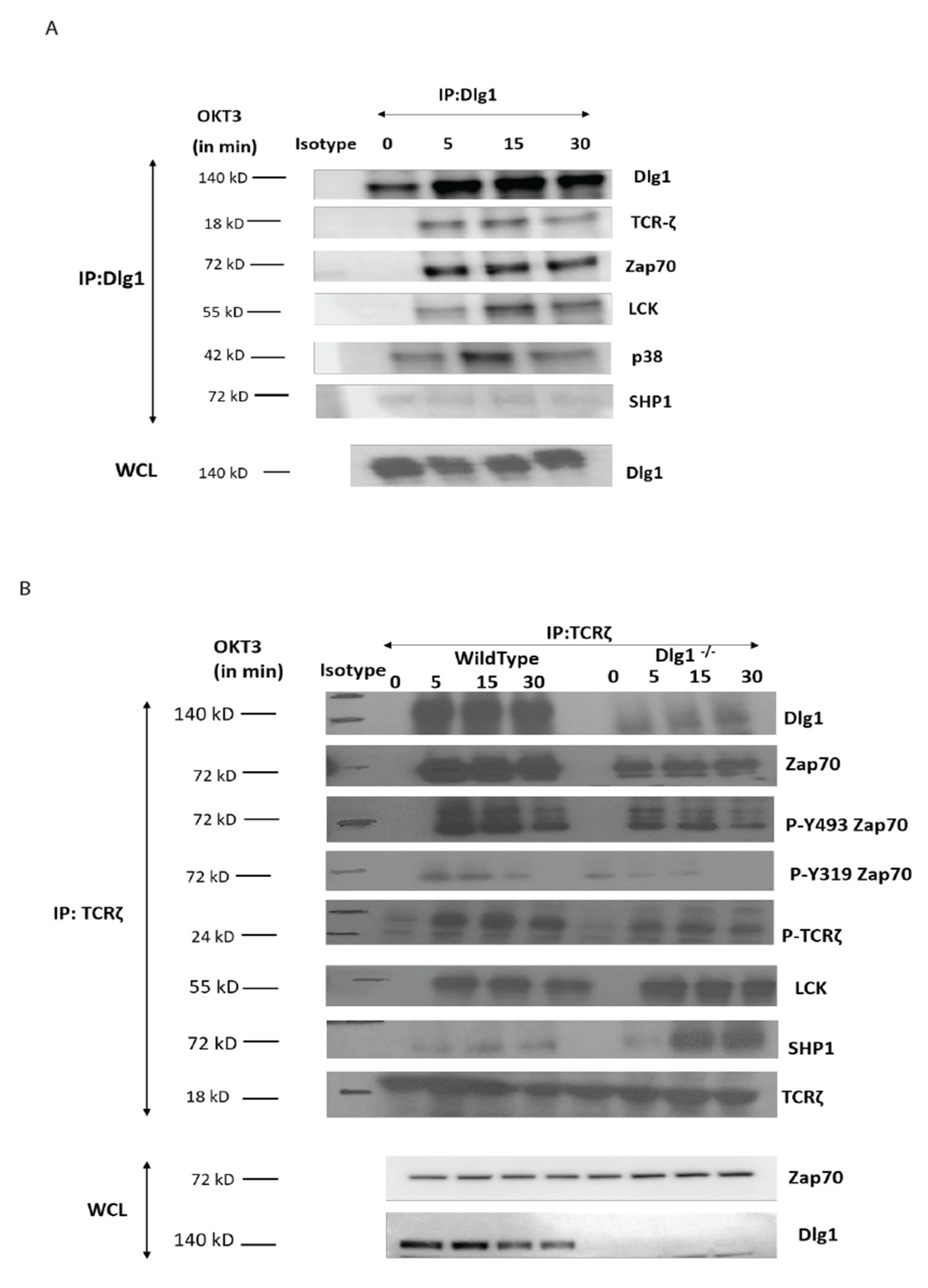
2.4. Co-Immunoprecipitation

T cells were lysed in ice-cold lysis buffer (150 mM NaCl, 20 mM Tris pH 7.5, 1% Triton X-100) supplemented with 1× protease inhibitor and 1× phosphatase inhibitor Roche, Basel, Switzerland). Lysates were cleared by centrifugation at 10,000 r.p.m. for 10 min and subjected to immunoprecipitation using agarose-conjugated TCR-ζ or an IgG1 control antibody overnight at 4 °C. The beads were washed in ice-cold lysis buffer, and the proteins were eluted by boiling in SDS sample buffer. Finally, the protein was resolved by SDS–PAGE and immunoblotted to analyze the interacting partners.

2.5. Confocal Microscopy and Image Analysis

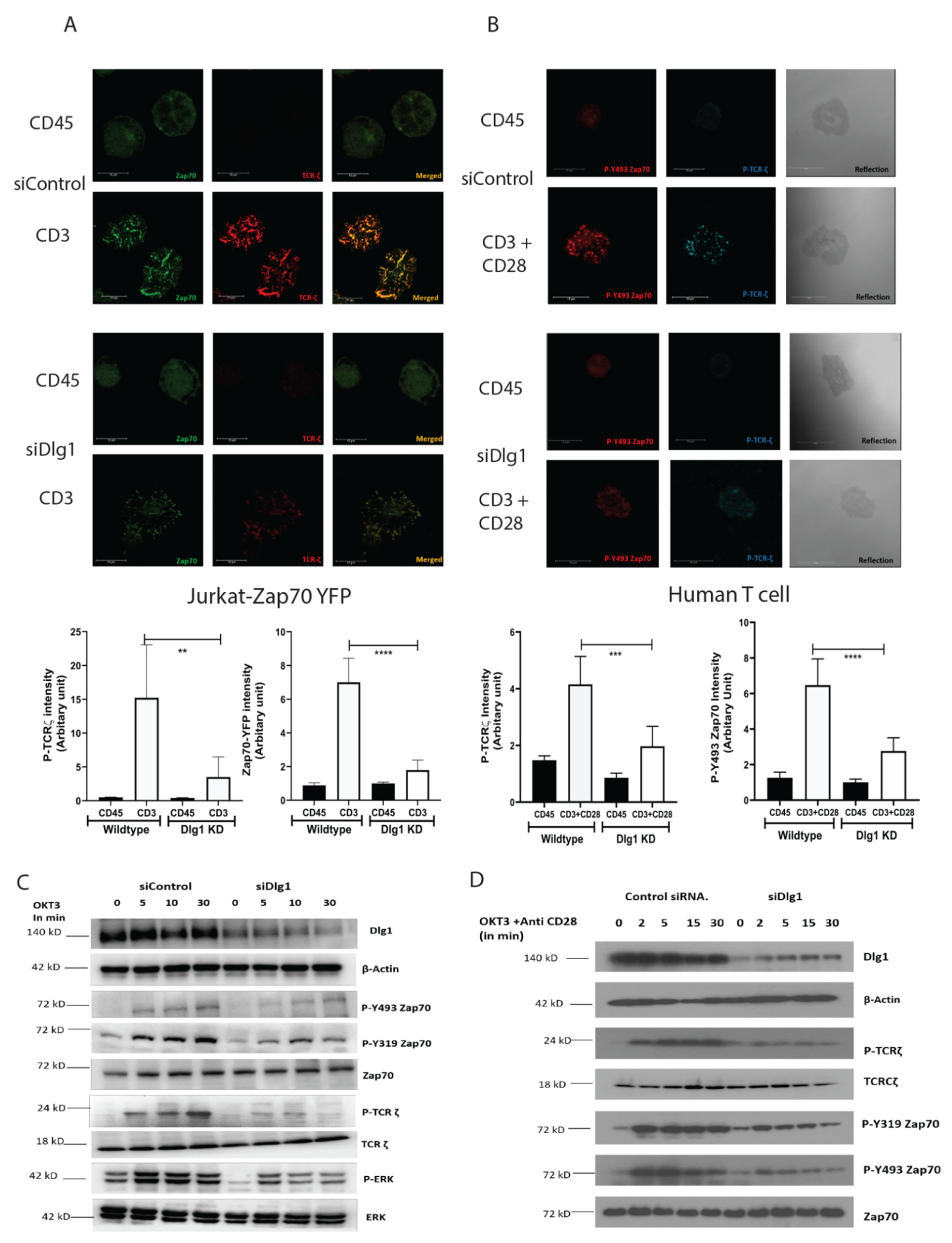
Cells were allowed to spread on coverslips, as described in [5]. In brief, cells were dropped onto polylysine-treated, four-chambered glass coverslips coated with stimulatory anti-CD3 (UCHT-1/HIT3a) or non-stimulatory anti-CD45 (H130) antibodies at 10 μg/mL. Cells were resuspended in a warm medium without phenol red, which was supplemented with 25 mM HEPES, pH 7, and dropped at the bottom of the chamber followed by incubation at 37 °C for 5 min. The cells were fixed with 2.4% paraformaldehyde for 30 min and permeabilized with Triton X-100. The slides were blocked in blocking buffer (2% goat serum) for 30 min and incubated with primary antibodies for 60 min at the appropriate dilution, followed by secondary antibodies (Alexa-Fluor-568-labeled IgG (1:1000) or Alexa-Fluor-647-labeledIgG (1:500)) for 45 min. Cells were washed and resuspended in PBS and images were captured using a Leica SP8 laser-scanning confocal microscope using a 63 × 1.4 numerical aperture (NA) objective (Leica Microsystems Inc., Buffalo Grove, IL, USA). Then, 2- to 3-μm z stacks with a spacing of 0.3 μm were taken of the area contacting the coverslip. Images were processed in Leica AF software and then exported to Adobe Photoshop and Illustrator (Adobe Systems Inc, San Jose, CA, USA) to prepare the composite figures. For quantitative analysis, cluster intensity was analyzed using Image J software. Single cells were analyzed independently in eight different fields from each sample and done in three replicate experiments.

2.6. siRNA-Mediated Knockdown

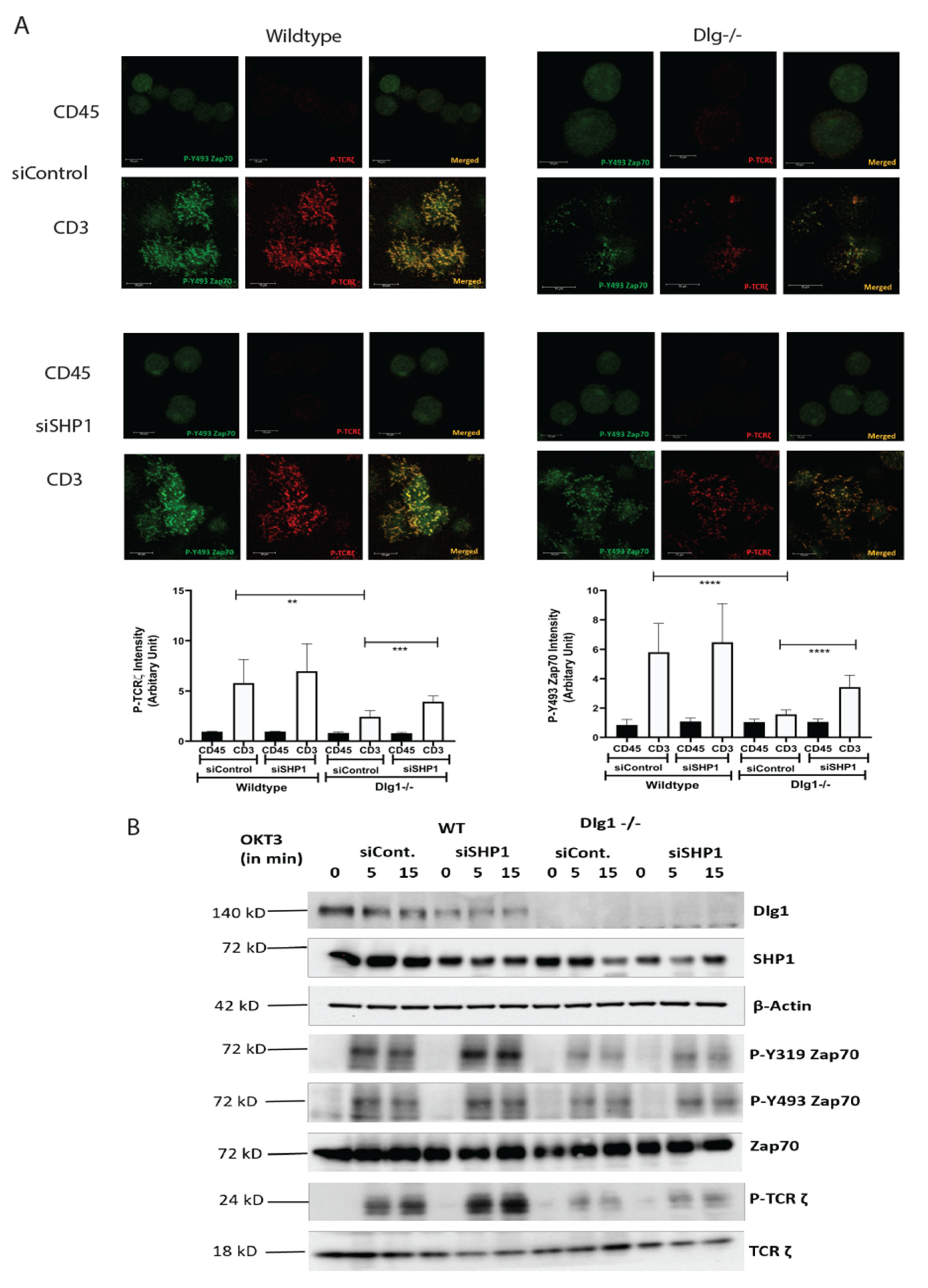
Jurkat or human CD4+ T cells were resuspended in 500 μL of antibiotic-free RPMI medium with 10 μL (100 picomoles) each of the validated siRNAs or the control siRNA-A. They were incubated at room temperature for 5–10 min and they were placed inside a 4-mm cuvette (Bio-Rad). The cuvettes were pulsed using the BTX electroporator (300 V, 10 ms, 960 μF), and the cells were transferred into RPMI medium containing 20% FCS. After 24 h, the live cells were isolated by Ficoll gradient and resuspended in fresh medium containing 10% FCS. Cells were analyzed for knockdown of DLG1 or SHP1 expression after 48 h by immunoblot analysis.

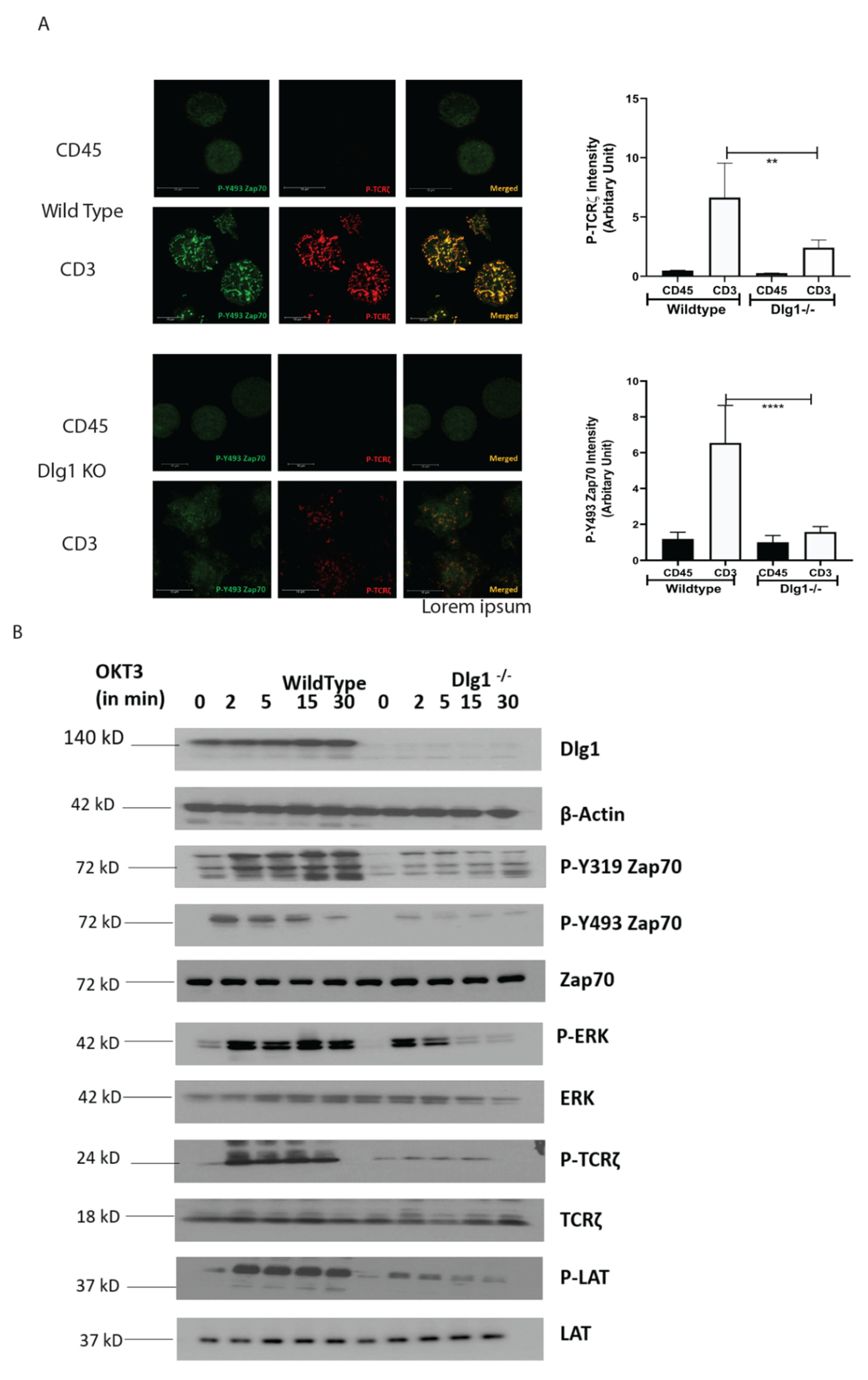
2.7. CRISPR-Cas9 Mediated DLG1 KO and Single Cell Cloning

Guide RNA target sites corresponding to DLG1 were identified using the sgRNA Scorer 2.0 tool (PMID: 28146356). Oligonucleotides corresponding to spacer sequences, TCTGGAGAAAAATGCCGGTC and CCAAAAGGTGCAATGCTCTC, were annealed and ligated into the PX458 plasmid backbone using the Enzymatics Rapid T4 ligase. The pSpCas9(BB)- 2A-GFP PX458) was a gift from Feng Zhang (Addgene plasmid #183 48138 http://n2t.net/addgene:48138;RRID:Addgene_48138) (29 November 2021). Then, 500 ng of cloned plasmid of each guide RNA (1 mg total) were nucleofected into 250,000 Jurkat cells using the Lonza Nucleofector 4D and the SE nucleofection kit. Electroporation settings used were the manufacturer-suggested settings for the Jurkat-E6. After 72 h, GFP-expressing cells (transient expression) were sorted and editing efficiency was checked. Single cell sorting was performed on these GFP-positive cells by BD FACSAria Fusion and single cell clones were generated for 2 weeks. Genomic DNA was extracted from single cell clones and NGS sequencing was done to confirm the generation of knockout clones. Putative Dlg1 KO clones were screened for lack of Dlg1 expression by immunoblotting. Further on, these clones were checked for CD3 expression and only high-CD3-expressing clones were used for experiments. Oligonucleotide sequences used for guide RNA cloning are listed below:
Guide1-F
CAC CGT CTG GAG AAA AAT GCC GGT C
Guide1-R
AAA CGA CCG GCA TTT TTC TCC AGA C
Guide2-F
CAC CGC CAA AAG GTG CAA TGC TCT C
Guide2-R
AAA CGA GAG CAT TGC ACC TTT TGG C

2.8. Flow Cytometry

CD3 expression was checked using OKT3-APC in BD Calibre. Cells were stained with OKT3-APC in FACS buffer for 30 min at 4 °C and analyzed. Intracellular IL-2 was analyzed in primary T cells. Briefly, primary T cells were stimulated with anti CD3+anti-CD28 for 18 h. After that, cells were harvested and washed with PBS. Cells were blocked with Fc receptor blocker for 30 min. Surface staining was done for 30 min at 4 °C. Cells were permeabilized using Fixation and Permeabilization buffer (BD, Cat. No. 554714). Cells were stained for intracellular IL-2 for 30 min at 4 °C and analyzed using BD LSR II Cell Analyzer.

2.9. Statistical Analysis

Statistical analysis was performed with GraphPad Prism 6 software San Diego, CA 92108, USA. Error bars indicated SEM, unless otherwise specified. Statistical significance was determined with the paired t-test, and p < 0.05 was considered to be statistically significant.

3. Results

3.1. DLG1 Is Critical for TCR MC Formation and TCR Proximal Signaling

While the association of DLG1 with the T cell receptor complex has been established, the exact molecular basis and functional significance of this association is still not well understood. In order to interrogate the cellular functionality of DLG1 in T cell synapse formation, we characterized the dynamics of TCR-MC in Jurkat cells. Using small RNA interference, we demonstrated that DLG1 influences microcluster formation. YFP-tagged, ZAP70-expressing Jurkat T cells were dropped onto coverslips coated with stimulatory anti-CD3 or non-stimulatory anti-CD45 specific antibodies. Cells were fixed after 5 min and imaged at the junction of the glass chamber. No distinct ZAP70 clustering could be seen in the cells dropped on the anti-CD45-coated glass chambers. However, when dropped onto anti-CD3-coated glass chambers, distinct ZAP70 puncta appeared. Staining with a mAb specific to the TCR zeta chain showed proper colocalization of ZAP70 to TCR-ζ. However, after treatment with siRNA targeting DLG1, puncta intensity was significantly reduced (Figure 1A). Quantitative analysis of micro cluster intensity from multiple fields across different replicate experiments showed a drastic reduction of cluster intensity in Dlg1 knockdown cells (Figure 1A, graph). DLG1 is also critical for activation of the T cell alternative p38 pathway [12]. The p38 is known to be colocalized with ZAP70 at the immune synapse. [15]. Consistent with ZAP70, phosphorylation of p38 on Y323 was also inhibited under DLG1 knockdown conditions (Supplementary Figure S1A–D).
Next, we analyzed proximal signaling by evaluating the phosphorylation status of ZAP70 and TCR-ζ. Immunoblot analysis showed that phosphorylation of the activating ZAP70 kinase domain residue Tyr 493 was decreased in the DLG1 KD cells. Similarly, the Tyr 319 residue of ZAP70 and Tyr 83 residue of TCR- ζ also showed reduced phosphorylation. Interestingly, phosphorylation of ERK was diminished by siDLG1 treatment (Figure 1C).
Similar results were observed using siRNA treatment in primary human CD4+ T cells. Confocal microscopy of freshly isolated human T cells revealed compromised ZAP70 and TCR-ζ activation in DLG1 KD cells (Figure 1B). Concomitantly, diminished phosphorylation of ZAP70 and TCR-ζ was also observed by Western blot analysis (Figure 1D). Next, we analyzed IL-2 generation from human T cells after DLG1 knockdown. Interestingly, the IL-2 production upon anti-CD3 plus anti-CD28 stimulation was significantly compromised after DLG1 knockdown in both CD4+ and CD8+ T cells (Supplementary Figure S1E).
In order to reinforce our findings from the DLG1 knockdown experiments, we studied the proximal signaling and microcluster dynamics in DLG1 knockout Jurkat cells using CRISPR-Cas9 gene inactivation. DLG1 KO clones were selected by NGS analysis and confirmed by protein expression studies (Supplementary Figures S2A,B and S3A). Jurkat cells sometimes downregulate the TCR-CD3 molecule, which effects its activation status. We, thus, checked the TCR-CD3 expression on the KO clones and found that the KO clone has a comparable TCR-CD3 expression as the wild-type cells, confirming that the dampened signal in the KO clone is not due to TCR-CD3 downregulation (Supplementary Figure S3B). The best clone was examined for DLG1 expression and P-Y493 ZAP70 microcluster formation. Confocal images showed compromised microcluster formation in the absence of DLG1 (Supplementary Figure S3C).
TCR-MC dynamics were studied in a similar way in these cells. Wild-type Jurkat cells dropped onto stimulatory anti-CD3-coated glass surfaces showed distinct puncta for P-Y493 ZAP70 and P-TCR ζ chain, indicative of microcluster formation at 5 min post-activation, as opposed to cells dropped onto non-stimulatory anti-CD45-coated glass surfaces. On the contrary, DLG1 KO cells, when dropped onto stimulatory anti-CD3-coated surfaces, showed much reduced intensity of P- Y493 ZAP70 and TCR-ζ clusters (Figure 2A). Quantitative analysis further validated the finding (Figure 2A, graph).
Downstream signaling was traced by determining the phosphorylation status of the proximal signaling component. Phosphorylation of Y-493 ZAP70 and Y-83 TCR-ζ were significantly reduced, as predicted from the quantitative analysis of cluster intensity. Furthermore, phosphorylation of the Y-319 moiety of ZAP70 was also modestly reduced, indicative of the compromised activation of ZAP70. Additionally, activation of LAT and ERK were markedly diminished in DLG1 KO cells (Figure 2B).
Overall, the combined evidence from knockdown and knockout experiments supports the notion that DLG1 influences proximal TCR signaling events.

3.2. DLG1 Controls the Recruitment of ZAP70 to the TCR Complex

In order to determine the role of DLG1 in proximal TCR signaling, we performed co-immunoprecipitation (Co-IP) experiments. DLG1 immunoprecipitates were analyzed for TCR proximal proteins. Activation-induced association of TCR-ζ, ZAP70, and LCK with DLG1 was evident in wild-type Jurkat cells (Figure 3A). In a reverse Co-IP assay, wild-type and knockout cells were activated with anti-CD3 and cell lysates and, thereafter, were immunoprecipitated with anti-TCR ζ followed by immunoblot analysis. Association of ZAP70, TCR-ζ chain, and LCK was observed as early as 5 min after stimulation in wild-type cells. These associated proteins were activated, as determined from analysis of their phosphorylation status. ZAP70 association with the TCR-ζ chain was drastically compromised in DLG1’s absence. Notably, phosphorylation of the TCR-ζ chain was also reduced (Figure 3B).
ZAP70 is recruited to the TCR complex after the ζ chain is phosphorylated, which opens up a docking site for the protein. As phosphorylation of the ζ chain is compromised in DLG1 KO cells, we examined the status of LCK, the immediate upstream kinase. Surprisingly, LCK recruitment to the complex was unaffected in knockout cells as compared to wild-type cells (Figure 3B).
Phosphorylation status of any protein depends upon the balance between phosphorylation and dephosphorylation. Since LCK recruitment was unaffected by DLG1 KO, we examined the status of SHP1, the critical phosphatase for ζ chain dephosphorylation. Interestingly, SHP1 association with the ζ chain was significantly increased in DLG1-deficient cells (Figure 3B). Thus, the increased association of SHP1 to the TCR complex under conditions of DLG1 deficiency may be a determinant of dysregulated proximal TCR signaling.

3.3. Enhanced Association of SHP1 to the TCR Complex Compromises Proximal TCR Signaling

Next, we aimed to evaluate the role of SHP1 in association with DLG1 in T cell signaling. For this, we re-analyzed the microcluster dynamics in Jurkat cells. Wild-type and DLG1 knockout Jurkat cells were treated with an siRNA against SHP1. After 48 h, the cells were placed onto an anti-CD3- or anti-CD45-coated surface and microclusters were analyzed. After SHP1 siRNA treatment, wild-type cells showed a modest increase in P-ZAP70 and P-TCR ζ signal intensity. On the other hand, the decreased signal intensity in activated DLG1 KO cells was rescued after SHP1 siRNA treatment (Figure 4A).
After TCR activation, DLG1 immunoprecipitates contained LCK, Zap70, p38, and TCR-ζ (Figure 3A). However, significant association with SHP1 was not observed. The downstream effect of this reversal was determined by phospho-protein analysis. Significant increases in P-Y493 ZAP70 and P-Y319 ZAP70 were observed in DLG1 KO cells after siRNA SHP1 treatment as opposed to control siRNA treatment. Increased P-TCR ζ strongly suggested the loss of phosphatase activity in both wild-type and DLG1 KO cells (Figure 4B). The siRNA treatment against SHP2 did not show any significant effects (Supplementary Figure S4).

4. Discussion

DLG1, a homolog of Drosophila Disc large tumor suppressor protein, is a member of the MAGUK (Membrane-associated Guanylate kinase) protein family. It is a scaffolding molecule known to have an important role in the immune synapse, where it organizes signaling components to form synaptic complexes [16]. In epithelial cells, its role is to regulate actin cytoskeleton dynamics and, thus, to maintain cell polarity [17,18]. Apart from its role in neuronal and epithelial cells, DLG1 is also known to have a pivotal function in T cell activation [11].
Several adapter proteins have already been implicated in TCR signal transduction. DLG1 is known to translocate to the TCR complex upon receptor engagement. It orchestrates the inter-molecular interaction between signaling components at the TCR, such as LCK-ZAP70-WASp [11]. Alternative activation of p38 is mediated by LCK-ZAP70 and an association with DLG1 is evident [12]. TCR-MC, which is a platform for signaling cascades, includes activated LCK and ZAP-70. ZAP-70 colocalizes with p38 and activates it alternatively by phosphorylating its Tyr323 moiety [15]. Our data demonstrate that the ZAP-70-p38 association is heavily compromised at TCR-MC when DLG1 is knocked down in Jurkat cells and primary human T cells. This proves that DLG1 is working at the immediate phase of T cell activation, even before the formation of the immunological synapse. Consequently, dampening of downstream signaling is observed by Western Blot analysis under conditions of DLG1 deficiency. The decreased phosphorylation of the TCR-ζ chain explains the subsequent reduction of ZAP-70 phosphorylation at the activating residues Tyr493 and Tyr319. Finally, compromised LAT activation leads to defective LAT signalosome formation, which acts as a key event in T cell activation.
In our study, we showed that DLG1 knockdown in both Jurkat cell and human primary T cell has a direct effect on TCR-MC formation. Visualization of the TCR microcluster revealed loss of ZAP-70 and p-TCR-ζ in the microclusters. DLG1 knockout was performed in Jurkat cells using CRISPR-Cas9 technology and the finding further corroborated the knockdown studies. We also tried to generate DLG1 knockout in human primary T cells using Nucleo-fector 2B (Amaxa Human T cells Nucleofector Kit, VPA-1002) system from Lonza and Invitrogen™ Neon™ Transfection System. However, we found it technically challenging. In the first method, we got very low initial editing efficiency (% GFP positive cells) and cells were non-viable after FACS sorting of the edited population. In the second method, we got a significant proportion of GFP-positive cells and we could successfully sort them. We confirmed DLG1 knockout in one of the Donors by Western blot analysis. However, during the maintenance phase, cells started to become unhappy and eventually died. Due to these technical limitations, reconfirmation of DLG1 knockdown studies in DLG1 knock-out primary T cells remained a caveat of our study. An accurate explanation is still unknown; however, the large size of DLG1 (140 KD) might be a reason behind the failure to generate a proper knockout. As an alternative intervention, IL-2 production was measured, as it is a hallmark of T cell activation and detects the survival cues for T cells. DLG1 deficiency (knockdown) in T cells significantly dampens IL-2 generation and, thus, the survival fate of activated T cells. Overall, the current study could show that this adaptor protein is critical for T cell survival and proliferation, which can be exploited for effective vaccine development.
Molecular interaction between upstream kinases to their substrates moves as a chain reaction as more and more downstream molecules are involved. Interaction between the ζ chain of CD3 to ZAP70 through its SH2 domain is the key event in T cell signal initiation. In the absence of DLG1, we found reduced interaction of ZAP70 to its immediate partner. This may be explained by nonavailability of the docking site for ZAP70 at the ζ chain. The ζ chain is heavily phosphorylated by Lck. However, in the absence of DLG1, a decrease in ζ chain phosphorylation was observed. Interestingly, Lck recruitment to the TCR is intact even in DLG1 deficiency, thus ruling out loss of function of the activating kinase. The alternative mechanism that could be proposed is gain of function of deactivating phosphatases. Surprisingly, the SHP1 association with TCR ζ is significantly enhanced in the absence of DLG1, identifying a role in dampening T cell signal initiation.
Studies in T cell proximal signaling show that the opposing actions of protein tyrosine kinases (PTKs) and protein tyrosine phosphatases (PTPs) are always in constant operation and the balance between the two determines the activation/suppression of T cell signaling. Classical PTPs include SHP1 and SHP2, which are SH2 domain-containing phosphatases implicated in T cell signaling [19]. SHP1 is expressed in all hematopoietic cells; however, its expression is low in epithelial cells [20]. In contrast, SHP2 is ubiquitously expressed [19]. In our study, we found that wild-type Jurkat cells show enhanced phosphorylation of ZAP70, ERK, and TCR ζ when SHP1 is knocked down, identifying a direct effect of SHP1 in suppressing proximal signaling. In Jurkat cells, SHP1 but not SHP2 knockdown partially rescued dampened signaling conferred by DLG1 deficiency. This is in agreement with SHP2 having positive effects or no effect on T cell signaling, although it is found in complex with ITAMs [19]. In this context, it is important to note that SHP1 knockdown has only marginal effect in rescuing the phenotypic defect of DLG1 deficiency. A possible explanation is there can be some counterbalancing effect from any other signaling component (kinase/phosphatase/adaptor protein), which acts as another key player in this orchestra of events and can dictate the phosphorylation–dephosphorylation equilibrium of TCR proximal proteins. Further studies are required to identify these unknown factor(s).
ZAP70 is known to directly bind to SHP1 [21]; however, the precise molecular mechanism of SHP1 recruitment to the TCR/CD3 complex remains unclear. SHP1 is also known to crosstalk with CEACAM [22] and Themis [23] in order to control the outcome of T cell signaling. Another consideration is the increased phosphorylation status of SHP1. Phosphorylation of Y536 increases SHP1 activity about 4–8-fold, while that of Y564 phosphorylation increases it by 1.6-fold. Whether SHP1 is interacting with other partners more strongly or is selectively phosphorylated to increase its PTP activity is an important question for future research.
Our finding that DLG1 regulates proximal T cell signaling is consistent with the known function of other adaptor proteins in T cell activation. However, the microscopic visualization of the proximal molecules in their activation states provides detailed evidence of molecular interaction at the IS. In a recent publication, the authors showed that SHP1 promotes T cell adhesion by activating the adaptor protein CrkII by dephosphorylating it in the IS [24]. In our study, we saw a reverse picture, where an adaptor molecule, DLG1, prevented the recruitment of a phosphatase into the IS, thereby promoting sustained signaling. In the absence of DLG1, this checkpoint is lost and, thus, the T cell fails to form an effective MC. Another docking protein, Gab2, is known to be phosphorylated by ZAP70 and to negatively regulate T cell receptor signaling by recruitment of inhibitory molecules [25]. DLG1 works in an opposing manner to inhibit SHP1 interaction with the TCR. In conclusion, DLG1 is a positive regulator of T cell activation and, thus, it may be important for immune activation in health and disease.

Supplementary Materials

The following are available online at https://www.mdpi.com/article/10.3390/vaccines9121446/s1. Figure S1: Quantitative analysis of 1 TCR-MC intensity. Figure S2: Sequence analysis of DLG1 KO clones. Figure S3: Characteristic analysis of DLG1 KO cells. Figure S4: SHP2 knockdown fails to rescue the inhibition of T cell signaling conferred by DLG1 deficiency.

Author Contributions

Conceptualization, experiment designing, data interpretation, writing, and editing by J.G. Knockout Jurkat cell line generation and validation by R.C. All authors have read and agreed to the published version of the manuscript.

Funding

This work was supported by the Intramural Research Programs of the National Cancer Institute and National Institute of Allergy and Infectious Diseases, National Institutes of Health.

Institutional Review Board Statement

Buffy coats were obtained from healthy U.S. volunteers by counterflow centrifugal elutriation at the National Institutes of Health (NIH) Blood Bank under Institutional Review Board-approved protocols of both the National Institute of Allergy and Infectious Diseases and the Department of Transfusion Medicine.

Informed Consent Statement

Informed consent was obtained from all subjects involved in the study.

Acknowledgments

Jurkat-YFP cells were a kind gift from Lawrence E. Samelson. All the reagents and use of equipment were kindly provided by Jonathan D. Ashwell. Thanks to Philip M. Murphy for carefully editing the manuscript.

Conflicts of Interest

The authors declare no conflict of interest.

References

  1. Brownlie, R.J.; Zamoyska, R. T cell receptor signalling networks: Branched, diversified and bounded. Nat. Rev. Immunol. 2013, 13, 257–269. [Google Scholar] [CrossRef]
  2. Hatada, M.H.; Lu, X.; Laird, E.R.; Green, J.; Morgenstern, J.P.; Lou, M.; Marr, C.S.; Phillips, T.B.; Ram, M.K.; Theriault, K.; et al. Molecular basis for interaction of the protein tyrosine kinase ZAP-70 with the T-cell receptor. Nature 1995, 377, 32–38. [Google Scholar] [CrossRef] [PubMed]
  3. Wange, R.L. LAT, the linker for activation of T cells: A bridge between T cell-specific and general signaling pathways. Sci. STKE 2000, 2000, re1. [Google Scholar] [CrossRef] [PubMed]
  4. Hashimoto-Tane, A.; Saito, T. Dynamic Regulation of TCR-Microclusters and the Microsynapse for T Cell Activation. Front. Immunol. 2016, 7, 255. [Google Scholar] [CrossRef] [PubMed] [Green Version]
  5. Balagopalan, L.; Sherman, E.; Barr, V.A.; Samelson, L.E. Imaging techniques for assaying lymphocyte activation in action. Nat. Rev. Immunol. 2011, 11, 21–33. [Google Scholar] [CrossRef] [PubMed] [Green Version]
  6. Clements, J.L.; Boerth, N.J.; Lee, J.R.; Koretzky, G.A. Integration of T cell receptor-dependent signaling pathways by adapter proteins. Annu. Rev. Immunol. 1999, 17, 89–108. [Google Scholar] [CrossRef] [PubMed]
  7. Zheng, C.Y.; Seabold, G.K.; Horak, M.; Petralia, R.S. MAGUKs, synaptic development, and synaptic plasticity. Neuroscientist 2011, 17, 493–512. [Google Scholar] [CrossRef] [PubMed] [Green Version]
  8. Ludford-Menting, M.J.; Oliaro, J.; Sacirbegovic, F.; Cheah, E.T.; Pedersen, N.; Thomas, S.J.; Pasam, A.; Iazzolino, R.; Dow, L.E.; Waterhouse, N.J.; et al. A network of PDZ-containing proteins regulates T cell polarity and morphology during migration and immunological synapse formation. Immunity 2005, 22, 737–748. [Google Scholar] [CrossRef] [PubMed] [Green Version]
  9. Rebeaud, F.; Hailfinger, S.; Thome, M. Dlgh1 and Carma1 MAGUK proteins contribute to signal specificity downstream of TCR activation. Trends Immunol. 2007, 28, 196–200. [Google Scholar] [CrossRef] [PubMed]
  10. Xavier, R.; Rabizadeh, S.; Ishiguro, K.; Andre, N.; Ortiz, J.B.; Wachtel, H.; Morris, D.G.; Lopez-Ilasaca, M.; Shaw, A.C.; Swat, W.; et al. Discs large (Dlg1) complexes in lymphocyte activation. J. Cell Biol. 2004, 166, 173–178. [Google Scholar] [CrossRef] [PubMed] [Green Version]
  11. Round, J.L.; Tomassian, T.; Zhang, M.; Patel, V.; Schoenberger, S.P.; Miceli, M.C. Dlgh1 coordinates actin polymerization, synaptic T cell receptor and lipid raft aggregation, and effector function in T cells. J. Exp. Med. 2005, 201, 419–430. [Google Scholar] [CrossRef] [Green Version]
  12. Round, J.L.; Humphries, L.A.; Tomassian, T.; Mittelstadt, P.; Zhang, M.; Miceli, M.C. Scaffold protein Dlgh1 coordinates alternative p38 kinase activation, directing T cell receptor signals toward NFAT but not NF-kappaB transcription factors. Nat. Immunol. 2007, 8, 154–161. [Google Scholar] [CrossRef] [PubMed]
  13. Zanin-Zhorov, A.; Lin, J.; Scher, J.; Kumari, S.; Blair, D.; Hippen, K.L.; Blazar, B.R.; Abramson, S.B.; Lafaille, J.J.; Dustin, M.L. Scaffold protein Disc large homolog 1 is required for T-cell receptor-induced activation of regulatory T-cell function. Proc. Natl. Acad. Sci. USA 2012, 109, 1625–1630. [Google Scholar] [CrossRef] [PubMed] [Green Version]
  14. Humphries, L.A.; Shaffer, M.H.; Sacirbegovic, F.; Tomassian, T.; McMahon, K.A.; Humbert, P.O.; Silva, O.; Round, J.L.; Takamiya, K.; Hugamir, R.L.; et al. Characterization of in vivo Dlg1 deletion on T cell development and function. PLoS ONE 2012, 7, e45276. [Google Scholar] [CrossRef] [PubMed]
  15. Giardino Torchia, M.L.; Dutta, D.; Mittelstadt, P.R.; Guha, J.; Gaida, M.M.; Fish, K.; Barr, V.A.; Akpan, I.O.; Samelson, L.E.; Tagad, H.D.; et al. Intensity and duration of TCR signaling is limited by p38 phosphorylation of ZAP-70(T293) and destabilization of the signalosome. Proc. Natl. Acad. Sci. USA 2018, 115, 2174–2179. [Google Scholar] [CrossRef] [PubMed] [Green Version]
  16. Roy, N.H.; Burkhardt, J.K. The Actin Cytoskeleton: A Mechanical Intermediate for Signal Integration at the Immunological Synapse. Front. Cell Dev. Biol. 2018, 6, 116. [Google Scholar] [CrossRef] [PubMed] [Green Version]
  17. Dimitratos, S.D.; Woods, D.F.; Stathakis, D.G.; Bryant, P.J. Signaling pathways are focused at specialized regions of the plasma membrane by scaffolding proteins of the MAGUK family. Bioessays 1999, 21, 912–921. [Google Scholar] [CrossRef]
  18. Bilder, D. PDZ proteins and polarity: Functions from the fly. Trends Genet. 2001, 17, 511–519. [Google Scholar] [CrossRef]
  19. Lorenz, U. SHP-1 and SHP-2 in T cells: Two phosphatases functioning at many levels. Immunol. Rev. 2009, 228, 342–359. [Google Scholar] [CrossRef] [Green Version]
  20. Matthews, R.J.; Bowne, D.B.; Flores, E.; Thomas, M.L. Characterization of hematopoietic intracellular protein tyrosine phosphatases: Description of a phosphatase containing an SH2 domain and another enriched in proline-, glutamic acid-, serine-, and threonine-rich sequences. Mol. Cell. Biol. 1992, 12, 2396–2405. [Google Scholar] [CrossRef] [Green Version]
  21. Brockdorff, J.; Williams, S.; Couture, C.; Mustelin, T. Dephosphorylation of ZAP-70 and inhibition of T cell activation by activated SHP1. Eur. J. Immunol. 1999, 29, 2539–2550. [Google Scholar] [CrossRef]
  22. Nagaishi, T.; Pao, L.; Lin, S.H.; Iijima, H.; Kaser, A.; Qiao, S.W.; Chen, Z.; Glickman, J.; Najjar, S.M.; Nakajima, A.; et al. SHP1 phosphatase-dependent T cell inhibition by CEACAM1 adhesion molecule isoforms. Immunity 2006, 25, 769–781. [Google Scholar] [CrossRef] [PubMed] [Green Version]
  23. Choi, S.; Warzecha, C.; Zvezdova, E.; Lee, J.; Argenty, J.; Lesourne, R.; Aravind, L.; Love, P.E. THEMIS enhances TCR signaling and enables positive selection by selective inhibition of the phosphatase SHP-1. Nat. Immunol. 2017, 18, 433–441. [Google Scholar] [CrossRef] [PubMed]
  24. Azoulay-Alfaguter, I.; Strazza, M.; Peled, M.; Novak, H.K.; Muller, J.; Dustin, M.L.; Mor, A. The tyrosine phosphatase SHP-1 promotes T cell adhesion by activating the adaptor protein CrkII in the immunological synapse. Sci. Signal. 2017, 10, eaal2880. [Google Scholar] [CrossRef] [PubMed] [Green Version]
  25. Yamasaki, S.; Nishida, K.; Hibi, M.; Sakuma, M.; Shiina, R.; Takeuchi, A.; Ohnishi, H.; Hirano, T.; Saito, T. Docking protein Gab2 is phosphorylated by ZAP-70 and negatively regulates T cell receptor signaling by recruitment of inhibitory molecules. J. Biol. Chem. 2001, 276, 45175–45183. [Google Scholar] [CrossRef] [PubMed] [Green Version]
Figure 1. DLG1 knockdown inhibits TCR-MC formation and subsequent proximal signaling. (A,B). Microcluster analysis. Scale bars, 10 μm. (A) Jurkat cell analysis. Microclusters of ZAP-70–YFP (green) and phosphorylated TCR-ζ (red) were visualized in immunostained Jurkat cells stably expressing ZAP-70–YFP, pretreated with control or DLG1-specific siRNA. Cells were dropped onto coverslips coated with anti-CD3 (stimulated) or anti-CD45 (unstimulated) and visualized after 5 min. Quantitative data for specific signals’ intensities (using Image J; collected from eight fields) are graphically represented. (B) Primary human CD4+ T cells. Microclusters of phosphorylated Y493 ZAP-70 (green) and phosphorylated TCR-ζ (red) were visualized in immunostained human T cells, pretreated with control or DLG1-specific siRNA. Cells were dropped onto coverslips coated with anti-CD3+anti-CD28 (stimulated) or anti-CD45 (unstimulated) and visualized after 5 min. Quantitative data for specific signals’ intensities (using Image J; collected from eight fields) are graphically represented. (C,D). Immunoblot analysis of lysates from Jurkat cells (C) and human T cells (D) pretreated with control or DLG1-specific siRNA and then activated with soluble anti-CD3 (monoclonal antibody OKT3) and antibody to mouse immunoglobulin G (IgG), followed by probing for phosphorylated and total proteins. Data are representative of three experiments. Student’s unpaired t tests were used for (A,B). ** p < 0.01, *** p < 0.001, **** p < 0.0001.
Vaccines 09 01446 g001
Figure 2. DLG1 knockout inhibits TCR-MC formation and T cell signaling (A). Microclusters of phosphorylated Y493 ZAP-70 (green) and phosphorylated TCR-ζ (red) in immunostained wild-type or DLG1 KO Jurkat cells were visualized 5 min after the cells were dropped onto coverslips coated with anti-CD3 (stimulated) or anti-CD45 (unstimulated). Scale bars, 10 μm. Quantitative data for specific signals’ intensities (using Image J; collected from eight fields) are graphically represented. (B). Immunoblot analysis of lysates from wild-type and DLG1 KO Jurkat cells activated with soluble anti-CD3 (monoclonal antibody OKT3) and antibody to mouse immunoglobulin G (IgG). Blots were probed for the indicated phosphorylated and total proteins. Data are representative of three experiments. Student’s unpaired t tests were used for (A). ** p < 0.01, **** p < 0.0001.
Vaccines 09 01446 g002
Figure 3. DLG1 is critical for ZAP70 recruitment to the TCR complex. (A). Immunoblot analysis (IB) of the indicated co-immunoprecipitated partners after immunoprecipitation (IP) of DLG1 from activated Jurkat cells. (B). Immunoblot analysis (IB) of the indicated co-immunoprecipitated partners after immunoprecipitation (IP) of TCR-ζ from activated wild-type and DLG1 KO Jurkat cells. Data are representative of three experiments.
Vaccines 09 01446 g003
Figure 4. SHP1 knockdown partially rescues the inhibition of TCR-MC formation and proximal signaling conferred by DLG1 deficiency (A). Microclusters of phosphorylated Y493 ZAP-70 (green) and phosphorylated TCR- ζ (red) were visualized in immunostained wild-type or DLG1 KO Jurkat cells that were pretreated with control or SHP1-specific siRNA. Cells were dropped onto coverslips coated with anti-CD3 (stimulated) or anti-CD45 (unstimulated) and visualized after 5 min. Scale bars, 10 μm. Quantitative data for specific signals’ intensities (using Image J; collected from eight fields) are graphically represented (B). Immunoblot analysis of lysates from wild-type and DLG1 KO Jurkat cells. Cells were pretreated with control or SHP1-specific siRNA, then activated with soluble anti-CD3 (monoclonal antibody OKT3) and antibody to mouse immunoglobulin G (IgG), followed by probing for phosphorylated and total proteins. Data are representative of three experiments. Student’s unpaired t tests were used for (A). ** p < 0.01, *** p < 0.001, **** p < 0.0001.
Vaccines 09 01446 g004
Publisher’s Note: MDPI stays neutral with regard to jurisdictional claims in published maps and institutional affiliations.

