Review

Progress in L2-Based Prophylactic Vaccine Development for Protection against Diverse Human Papillomavirus Genotypes and Associated Diseases

by
Pola Olczak
1 and
Richard B.S. Roden
1,2,3,*
1
Department of Pathology, The Johns Hopkins School of Medicine, Baltimore, MD 21287, USA
2
Department of Oncology, The Johns Hopkins School of Medicine, Baltimore, MD 21287, USA
3
Department of Gynecology and Obstetrics, The Johns Hopkins School of Medicine, Baltimore, MD 21287, USA
*
Author to whom correspondence should be addressed.
Vaccines 2020, 8(4), 568; https://doi.org/10.3390/vaccines8040568
Submission received: 2 September 2020 / Revised: 17 September 2020 / Accepted: 21 September 2020 / Published: 1 October 2020
(This article belongs to the Special Issue Development of Cross-Protective Vaccines)

Abstract

:
The human papillomaviruses (HPVs) are a family of small DNA tumor viruses including over 200 genotypes classified by phylogeny into several genera. Different genera of HPVs cause ano-genital and oropharyngeal cancers, skin cancers, as well as benign diseases including skin and genital warts. Licensed vaccines composed of L1 virus-like particles (VLPs) confer protection generally restricted to the ≤9 HPV types targeted. Here, we examine approaches aimed at broadening the protection against diverse HPV types by targeting conserved epitopes of the minor capsid protein, L2. Compared to L1 VLP, L2 is less immunogenic. However, with appropriate presentation to the immune system, L2 can elicit durable, broadly cross-neutralizing antibody responses and protection against skin and genital challenge with diverse HPV types. Such approaches to enhance the strength and breadth of the humoral response include the display of L2 peptides on VLPs or viral capsids, bacteria, thioredoxin and other platforms for multimerization. Neither L2 nor L1 vaccinations elicit a therapeutic response. However, fusion of L2 with early viral antigens has the potential to elicit both prophylactic and therapeutic immunity. This review of cross-protective HPV vaccines based on L2 is timely as several candidates have recently entered early-phase clinical trials.

Graphical Abstract

1. Introduction

1.1. The Diversity of Human Papillomaviruses and the Diseases They Cause

Papillomaviruses are small DNA tumor viruses with a range of malignant potential; most infections are benign and self-limiting but a ‘high risk’ group of HPVs are the etiologic agents of 4.5% of cancers worldwide [1]. All papillomaviruses are 60 nm diameter non-enveloped T = 7d icosahedral viruses with a double-stranded, covalently closed and histone-bound circular DNA genome [2]. The papillomaviridae encompasses a very broad family of viruses that evolved to propagate in many species, including mammals and birds, but with a highly restricted host range, e.g., human papillomaviruses (HPV) only replicate in humans. Papillomaviruses also demonstrate a strict tissue tropism in that they only propagate in epithelia, and generally show a preference for either mucosa or cutaneous skin. Currently there are 226 known HPV genotypes catalogued in the Papillomavirus Episteme [3] with many more waiting to be recognized (https://pave.niaid.nih.gov/) [4]. Based upon their nucleotide sequences, the known HPVs have been classified into 5 genera termed alpha, beta, gamma, mu and nu. Species within the same genus are 60–70% similar, while novel HPV types have <90% similarity to any other HPV type [5].
The HPV genotypes of genus alpha predominantly infect the genital tract, but other mucosa including the anal and oral cavity are also susceptible. These HPVs are highly transmissible via sexual contact and as many as 60% of people who had sexual contact with an HPV infected person will acquire the virus [6]. These alpha HPVs demonstrate a spectrum of malignant potential. The ‘low risk’ types in this genus, e.g., HPV6 and HPV11, induce benign skin warts as well as laryngeal papillomas that very rarely progress to cancer, as in the slow growing Buschke Lowenstein tumors or lung cancers, respectively. However, even these benign papillomas are associated with significant morbidity and healthcare costs. The International Agency for Research on Cancer (IARC) has identified a baker’s dozen of ‘high risk’ types, HPV16, 18, 31, 33, 35, 39, 45, 51, 52, 56, 58, 59 and 68, as carcinogenic or probably carcinogenic (Groups 1 and 2A, Figure 1A–C) detected in ~97% of cervical cancers [7]. Of these, HPV16 and HPV18 are the most impactful as they contribute to ~50% and 20% each of cases of cervical cancers worldwide [8], whereas HPV16 causes a greater fraction (>80%) in cancer types such as other anogenital and head and neck cancers [9,10]. An additional intermediate risk group (Group 2B, possibly carcinogenic, Figure 1B) appear to drive at least 2% of cases of cervical cancer comprising HPV26, 30, 34, 53, 66, 67, 69, 70, 73 and 82 [11]. Cervical cancer is the fourth most common cancer occurring in women in the world [12], of which 99% is HPV-driven [13]. The global impact of HPV-associated cancers at other sites is lower because they are less common and the attributable fraction is lower and they are primarily driven by HPV16 [14].
HPVs within the beta, gamma, mu and nu genera are mainly associated with benign infections of the squamous epithelia of cutaneous skin [15]. The gamma, mu and nu HPVs cause common warts, plantar warts and pigmented warts which typically resolve within a few months or are subclinical, but can be recalcitrant and disfiguring and require repeated destructive treatments, especially in immunocompromised patients [16].
Beta HPVs typically produce only subclinical infections or flat warts. However, they are the subject of considerable interest as mounting evidence suggests the role of a subset of types as a co-carcinogen with UV light in the development of cutaneous squamous cell carcinoma (CSCC) [17,18]. This is important because of the potential to vaccinate against the relevant beta HPV to prevent CSCC, although the currently licensed HPV vaccines do not target these types [19,20]. A causal association between beta HPV and CSCC remains controversial, but vaccination against beta HPV remains perhaps the best opportunity to directly test this hypothesis [21].
The first evidence supporting beta types as causative factors in CSCC emerged from the identification of the genodermatosis epidermodysplasia verruciformis (EV). Patients with EV frequently develop persistent flat wart-like cutaneous lesions in childhood, and they often develop CSCC later in life in sun-exposed areas. Auto/hetero-inoculation experiments linked these lesions to beta HPV infection [22], and recent genetic studies identify the role of the host’s CIB1-EVER1-EVER2 complex in intrinsic resistance to beta HPV infection [23]. While the most commonly detected beta HPVs in benign EV lesions are HPV5, 8, 9, 12, 14, 15, 17 and 19–25, it is HPV5 and 8 that are detected in 90% of CSCC of EV patients [24]. This link between beta HPV and CSCC also appears to operate in immunocompromised patients including organ transplant recipients and AIDS patients, but is controversial for the general population [25]. HPV beta types accumulate in the aging general population but to a lower extent. It is estimated that in 90% of cases, the cutaneous infections clear within 0.5–2 years [26,27]. Multiple factors including smoking or being immunocompromised contribute to slower clearance [28].

1.2. The Papillomavirus Genome and Proteins

The genome structure of the papillomaviridae is well-conserved and the 8 genes are expressed in a spatially and temporally controlled manner within squamous epithelia. There are six early genes E1, E2, E4, E5, E6 and E7 and two late genes L1 and L2, as well as a long control region (LCR) that serves as the origin of replication and to control transcription [3]. E1 and E2 have DNA-binding capability and support viral replication. E5, E6 and E7 help drive the host cell to proliferate and E4 promotes the release of virus particles from the keratinocytes by collapsing keratin filaments [2]. Interestingly, E5 is not expressed in the HPV Beta types [23].
L1 and L2 encode the major and minor capsid proteins, respectively. Both L1 and L2 are expressed in the granular layer of epithelium, but they are not detectable in the basal epithelia that harbor the infection. Each HPV capsid is composed of 72 pentamers of L1, also known as capsomer(e)s. The capsomers associate to form a virion via disulfide bridges, i.e., 360 L1 proteins per virion. Additionally, there are estimated to be 12–72 L2 proteins per virion [29], but the exact location and topology of L2 remain to be resolved. Recombinant expression of L1 in diverse systems results in its self-assembly into empty capsids, called virus-like particles (VLPs).

1.3. Current L1-Based HPV Vaccines and Remaining Challenges

Animal challenge studies demonstrated that vaccination with L1 VLPs provided robust, but type-restricted protection. However, L1 VLP vaccination did not impact pre-existing infection, likely because L1 is not expressed by infected basal epithelial cells. Currently, there are three licensed vaccines against Human Papillomavirus, all composed of L1-only VLPs derived from the most medically significant genotypes (Cervarix® (GlaxoSmithKline Biologicals Rixensart, Belgium) and Gardasil® and Gardasil 9 (Merck and Co. Inc, Whitehouse Station, NJ, USA)). Cervarix® is composed of HPV16 and 18 L1 VLPs produced in insect cells and formulated with alum and the TLR4 agonist monophosphoryl lipid A (ASO4 adjuvant system). In Gardasil, the aluminum hydroxyphosphate sulfate-adjuvanted VLP are expressed in yeast and derived from L1 of HPV 6, 11, 16 and 18 for the quadrivalent vaccine, and for the predominant nonavalent vaccine HPV31, 33, 45, 52, 58, as well. They induce high and durable titers of neutralizing antibodies as the structure of the particles allows for repetitive epitope display and favors hyperactivation of B cell receptors, and they are likely protective after only a single dose. Passive transfer studies in animal models suggest that these neutralizing antibodies are central to protection, likely acting in exudates at microlesions in the epithelium that are necessary for the viral inoculum to reach and infect the basal epithelia.
Although highly efficacious and safe, these L1 VLP vaccines are limited to protection (i.e., not therapeutic) and target a limited set of mucosal HPV types, albeit the most common in cervical cancer (and genital warts for Gardasil). Therapeutic vaccines aimed at treating pre-existing infections and disease generally target early antigens, primarily E6 and E7, as they are obligately expressed in infected cells and cancer, and are being developed to help those suffering HPV-related disease. It is clear that prophylaxis against a broader range of medically relevant HPV types is desirable for: (1) protection against all carcinogenic and probably carcinogenic alpha HPV that together cause >99% of cervical cancer and eliminate the need and costs for cervical screening, (2) protection against the morbidity and treatment costs associated with benign HPV diseases beyond genital warts, (3) protection against CSCC associated with beta HPV, especially in immunocompromised patients. Unfortunately, the number of different HPV genotypes that cause these diseases is large, rendering production of a sufficiently highly multivalent L1 VLP vaccine impractical, so development of a single cross-protective antigen is of interest as an alternative. For this, the field has generally focused on the minor capsid antigen, L2 (Graphic Abstract).

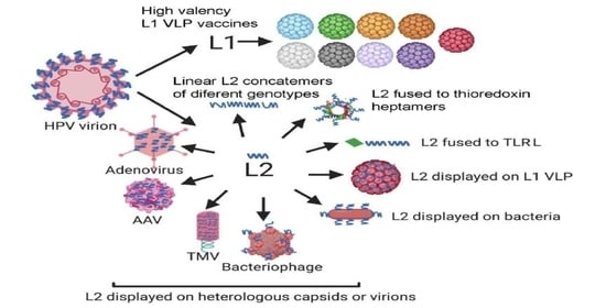
1.4. L2 as a Broadly Protective Vaccine Antigen

The L2 protein is composed of ~500 amino acids and it is required for infection. L2, by itself, does not form the VLPs, but when co-expressed with L1, it co-assembles into capsids and facilitates the encapsidation of the viral genome [30]. Most of the L2 protein is buried within the capsid, but portions are exposed on the capsid surface [31]. L2 was first recognized as a potential antigen for use in prophylactic immunization in the early 1990s. Foundational studies in cattle (bovine papillomavirus, BPV, types 1, 2 and 4) and rabbit (cottontail rabbit papillomavirus, CRPV) challenge models demonstrated that vaccination with L2 produced recombinantly in bacteria can provide durable protection, but it does not impact pre-existing infection [32,33,34,35,36]. Passive transfer studies suggest that neutralizing antibodies confer immunity [37,38]. However, the serum titers of neutralizing antibodies measured in vitro induced by L2 vaccination are substantially lower than achieved with L1 VLP. Perhaps as a result, the development of L2 vaccines languished.
Interest in L2-based vaccination was re-kindled by two observations. First, vaccination with N terminus of the L2 protein elicits both neutralizing antibodies and protective immunity [32]. Second, because the protective epitopes at the N terminus of L2 demonstrated robust sequence conservation (Figure 1), vaccination with this region induced broadly neutralizing antibodies against both mucosal and cutaneous types [39,40] and cross-protection against highly divergent papillomaviruses [41]. These observations were not restricted to animal studies. Vaccination of patients with full-length HPV16 L2 fusion protein even without adjuvant elicited cross-neutralizing antibodies [42]. This suggested the hypothesis that a single L2-based immunogen might provide broad protection and overcome or complement the type-restricted immunity elicited by L1 VLP.
This hypothesis triggered renewed focus on defining the cross-protective epitopes displayed by L2 and how best to present them to the immune system to trigger a robust and durable immunity against the plethora of HPV types with significant medical impact (summarized in Graphic Abstract). These epitopes within L2 were defined by generating cross-neutralizing antibodies or by peptide vaccination. Examples include the identification of the RG1 cross-neutralizing monoclonal antibody which recognizes amino acids 17–36 of L2 [37,43,44] (Figure 2), or Mab5 and Mab13 which recognize residues 69–81 and 108–120 respectively [45,46]. Interestingly, while natural infection or vaccination with virions or L1 + L2 VLP induces significant L1-specific antibody responses, they result in little or no L2-specific neutralizing antibody responses, as L2-neutralizing epitopes are buried and exposed only transiently during infection [47]. The remainder of the review concerns efforts to enhance the presentation of L2 within an immunogen to induce durable and broad immunity elicited by robust neutralizing antibody levels (Table 1).

1.5. How You Measure Neutralizing Antibodies Matters

Neutralizing antibody titers are an important correlate of protection for prophylactic vaccines [88]. Assay of the serum antibody titer after L2 vaccination using ELISA suggests a robust response. Likewise, passive transfer of immune sera confers strong protection. However, measurement of in vitro neutralization titer using native virions or pseudovirions (infectious L1/L2 particles carrying a reporter construct) suggests a weak antibody response despite complete protection to challenge [38]. Day et al. recently showed that the typical in vitro neutralization assay format poorly detects L2-specific neutralizing antibodies as compared to L1-specific antibodies. This likely reflects differences in the in vitro infectious process (e.g., of 293TT target cells) and that in vivo on the basement membrane at the site of epithelial damage. A conformational change is induced by L1-mediated capsid binding to the heparan sulfate proteoglycans of the basement membrane which exposes the amino acid terminal of L2 of the capsid surface, where it can be cleaved by furin, a pro-convertase enzyme critical to infection [89,90,91]. Cleavage of L2′s amino terminus by furin promotes infection of the keratinocyte but renders the virion susceptible to L2-specific neutralizing antibodies [92,93,94]. Because this process is rapid in tissue culture but slow in vivo, the sensitivity for measurement of L2-specific neutralizing antibodies can be substantially improved by using furin pre-cleaved virus for in vitro assays [89,95,96]. Unfortunately, this is a recent observation, and most published studies utilize the first generation neutralization assays that sensitivity to L2-specific neutralizing antibodies, an important caveat in interpreting their findings.

1.6. L2 Vaccines Based on (Poly)peptides

Since vaccination of animals and patients with full-length L2 induced low titers of cross-neutralizing epitopes [42], one possibility was that the antibody response was focused on non-neutralizing regions of L2 that were never exposed on the capsid surface. This might be addressed by focusing the response on the relevant regions by vaccinating with only the protective epitopes. Kawana et al. identified HPV16 L2 residues 108–120 as a cross-protective epitope and showed it was immunogenic in mice via intranasal administration in the absence of adjuvant [50]. When it was similarly administered to patients the HPV16 L2 108–120 peptide induced cross-neutralizing antibodies in 4/5 of patients in the high dose (0.5 mg at weeks 0, 4 and 12) group, but 0/5 at 0.1 mg [51]. This important study suggested the need for an adjuvant and potentially inclusion of T helper epitopes and/or using a longer sequence, which might also confer the advantage of increasing cross-reactivity to more HPV genotypes in addition to providing T helper epitope(s). The immunogenicity of peptide vaccines has been increased by chemical coupling of protective L2 epitopes to the surface amino acids of a protein carrier, typically keyhole limpet hemocyanogen (KLH) which provides a source of T helper epitopes and potentially irregular multimerization tethered at one end only [35,37,56,57]. These vaccines are able to elicit a protective immunity when administered to animal models with adjuvant.
Longer polypeptides can contain multiple protective epitopes and T helper epitopes. Vaccination with full-length L2 protein recombinantly expressed and purified from bacteria induces low levels of neutralizing antibodies and is protective. Cattle vaccinated with the N-terminal 11–200 polypeptide of the L2 of bovine papillomavirus type 4 (BPV4) showed stronger protection against BPV4 challenge and higher serum neutralizing antibody titers than animals vaccinated with the remainder or full-length L2 protein [52]. Vaccination of rabbits with residues 1–88 of bovine papillomavirus type 1 (BPV1) L2 elicited significantly higher titers than full-length protein, and the antisera very broadly neutralized diverse papillomavirus types [40]. Rabbits immunized with L2 polypeptides comprising amino acids 11–88 or 11–200 of cotton rabbit papillomavirus (CRPV), BPV1 and HPV16L2 showed protection against skin challenge with CRPV but also against rabbit oral papillomavirus (ROPV) [41]. L2 polypeptide vaccination did not protect against CRPV DNA challenge, confirming a lack of therapeutic activity. These studies supported the use of a longer L2 polypeptide, but the titers of neutralizing antibodies were much lower than for L1 VLP.
One reason for the high immunogenicity of L1 VLP is repetitive display of the protective epitopes. Jagu et al. explored whether creating concatemers of the L2 protective epitopes, from 11–200, 11–88 and 17–36 would likely increase immunogenicity, and whether the number of repeats mattered by testing 3, 5, 8, 15 and up to 22 repeats [53]. To increase the breadth of the cross-protective immune response, they used an L2 derived from a different HPV type in each repeat. A fusion of L2 11–88 of five or eight different HPV types proved the most effective, and vaccination with this approach using only alum adjuvant provided robust protection against all HPV genotypes tested in the mouse and rabbit challenge models [53,54]. Passive transfer studies indicate that protection was mediated by neutralizing antibodies [54]. This approach is currently being advanced toward the clinic by Bravovax.
Another approach to increase the immunogenicity of multimers of protective L2 peptides is by insertion into a heterologous protein scaffold that itself activates T cell proliferation [58], such as the small redox protein, thioredoxin (Trx) [97]. L2 peptide fusion to thioredoxin showed increased immunogenicity when compared to L2 linear peptides and showed that multimers of the epitopes are more immunogenic than the monomers [58]. Peptide composed of amino acids 20–38 of HPV16 L2 protein fused to Trx produced antibodies that only elicited an immune response against the HPV16 L2 antigen and did not cross neutralize other tested HPV types. Inclusion of L2 sequences from additional HPV types (HPV31 and 51) fused to a single Trx molecule elicited high response to all three types that were covered in the vaccine. The response to this immunization in both mice and guinea pigs was high and robust [59]. This approach has been improved by generating a fusion of residues 20–38 of 8 mucosal HPV types and conjugating it to a thermostable thioredoxin derived from a thermophile archaebacterium [60,61]. The immunogenicity of this vaccine was further improved by the addition of the OVX313 heptamerization domain. The addition of the heptamerization domain also enhanced the T-helper response [62]. Martin Muller (DKFZ) is advancing their product for clinical testing.

1.7. L2 Epitopes Fused to TLR Ligands

The above vaccines are formulated by mixing with a conventional adjuvant prior to administration, but improved immunogenicity might be achieved by direct conjugation of L2 to a defined adjuvant, such as a toll-like receptor (TLR) agonist. HPV16 L2 RG1 peptide fused with a broadly recognized T helper epitope (P25) and toll-like receptor 2 (TLR2) ligand dipalmitoyl-S glycerin cysteine (P2C) generated a potent response far superior to the linear epitope. Vaccinated mice were protected against challenge with HPV16 and HPV45. Sera from vaccinated mice neutralized other papillomavirus types, including HPV5 and BPV1 [63]. This vaccine was generated by direct chemical synthesis, but it requires inclusion of T helper epitopes recognized by the broad and HLA diverse human population. To this end, Zhang et al. generated a lipidated triple RG1 epitope multimer fusion protein fused with a single-chain antibody fragment targeting human FcγRI. Vaccination with this protein purified from bacteria elicited cross-neutralizing antibodies against a broad spectrum of HPV types, but surprisingly still needed additional adjuvant despite carrying a TLR2 ligand [64].
Flagellin is a TLR5 ligand that was tested as a direct fusion to multimer of L2 protective epitopes from various types (HPV6, 18, 31, 39, 52) and was potently immunogenic at low doses without adjuvant [65]. Importantly, it conferred broad protection against diverse HPV genotypes in rabbits and immunity was retained over a one-year period post vaccination [55]. Likewise, concatemers build out of RG1 epitopes of HPV18, 33, 58, 59 and amino acids 11–88 of HPV16 L2 fused to flagellin induced cross-neutralizing antibodies in mucosal secretions and protection from vaginal challenge when administered subcutaneously or intranasally to mice [66].

1.8. L2 Displayed on Bacteria

Bacteria express several TLR ligands that could adjuvant an L2 vaccine response, and the use of lactobacillus as a vector for mucosal vaccines can promote strong responses [98]. Lactobacilli-based recombinant vaccines are considered safe as compared to most other live-vaccine vehicles and can be produced at a low cost. When Lactobacillus casei (L. casei)-expressing residues 1–224 of HPV16 L2 fused to the poly-γ-glutamic acid synthetase A (pgsA) surface protein were administered to Balb/c mice, they elicited neutralizing antibodies against HPV16 and other mucosal types including HPV18, 45, 58 and BPV1, but no protection studies were performed [67].
As an alternative approach to provide surface display for L2 epitopes in the presence of TLR ligands, the outer membrane proteins (OMP) of a gram-negative bacterium that span membranes multiple times can be utilized, e.g., the trimeric OmpF in E. coli form a pore in outer membranes. The L2 antigen inserted in OmpF was expressed in the E. coli and stably presented on the outer membrane, but the immunogenicity was not tested [68].

1.9. L2 Epitopes Displayed on Papillomavirus L1 VLP

The L1 VLP vaccines induce high-titer antibodies even without adjuvant, and these responses are durable. This is believed to reflect the high density and close spacing of neutralizing epitopes that each particle displays (i.e., 360 copies on a 60 nm diameter particle) because this efficiently cross-links and activates B cell receptors, as well as promoting opsonization and uptake by antigen-presenting cells [99]. By contrast, L2 is much less immunogenic because it does not form a particle and, when expressed alone, is a monomeric antigen. Therefore, many groups have attempted to increase the immunogenicity of L2 by displaying it in the immunodominant neutralizing epitope(s) of a VLP. This can be challenging because the assembly and structure of VLP often are compromised by insertion of new sequences, and this is generally more problematic with larger insertions. Therefore, typically only small conserved neutralizing epitopes of L2 are used for insertion, e.g., residues 17–36, 65–81 or 108–120. Selection of the insertion site can be guided by known VLP structures, if available, to identify the immunodominant surface epitopes in the carrier capsid antigen. However, this nevertheless remains quite empiric because most such epitopes are conformational rather than linear so it is hard to predict the impact of insertion on the structure and immunogenicity of the recombinant.
HPV L1 is a logical carrier for the L2 epitope. Unfortunately, the cross-protective L2 epitopes are mostly buried in native virions or L1/L2 VLP and therefore do not effectively trigger an L2 response, which is why only L1 is used in the licensed VLP vaccines. Several groups began to identify potential insertion sites in L1 for conserved L2 protective epitopes, including residues 17–36, 65–81 or 108–120, by focusing on the surface loops [100,101,102]
The DE loop of the L1-based VLP is particularly useful for such display, and it allows for the presentation of an antigen on the surface of the particles. One promising technology is a chimeric VLP that has an RG1 epitope derived from HPV16 displayed on the HPV16 L1 VLP surface [103]. Vaccination with this HPV16L1-16RG1 VLP chimeric vaccine elicits serum antibody responses that protect against a wide range of high-risk HPV types via passive transfer [70]. Importantly, this insertion does not appear to compromise L1-specific immunity [71], and insertions of L2 epitopes from other HPV types is also possible [69,72], or even consensus-derived sequences for alpha and beta HPV-derived L2 ‘RG1 epitope’ which are slightly different as shown in Figure 2. The HPV16L1-16RG1 VLP vaccine is being prepared for clinical studies with support from the NCI PREVENT and SPORE programs, and commercially by Pathovax LLC.
It is possible to use other L1 for display, including HPV18 [104] or BPV1 [73]. For example, a similar vaccine was developed in the HPV18 L1 backbone displaying an HPV45-derived RG1 epitope [105]. While the HPV16-derived RG1 epitope is well-conserved among the hrHPV (alpha) genotypes, the homology and cross-reactivity of antisera is lower for the cutaneous beta HPV types. One approach is to display an RG1 epitope derived from such a representative beta HPV, e.g., the HPV17 RG1 sequence was displayed on the HPV16L1 DE surface loop. Immunization of rabbits with these chimeric VLP produced robust antibody titers against HPV16 L1 VLP, as well as cross neutralizing responses against a variety of HPV beta types, including 5, 8, 20, 24, 36, 23, 80, 49, 92, 96. When in vivo cross-protection was evaluated by passive serum transfer in a murine PsV challenge model, the immune sera to HPV16L1–17RG1 VLP (cross-) protected against beta HPV5/20/24/38/96/16 (but not type 76) [74]. While most studies were performed using baculovirus-based expression, other recombinant systems such as plants [101], yeast [106,107] and possibly even bacteria [108] can also be used.
However, while displaying a conserved L2 epitope on L1 VLPs elicits broad protection via induction of antibodies neutralizing for multiple HPV types that are not included in the vaccine, challenges remain. First, the L2-reactive titers, although associated with cross-protection, are low compared to the L1 VLP-specific responses. This raises concerns about the longevity of cross-protection induced by these HPV16L1–16RG1 VLP, although recent studies in rabbits show broad protection is maintained over 1 year using alum-formulated HPV16L1–16RG1 VLP [109]. Secondly, the conservation of the RG1 epitope is imperfect, and particularly for the beta HPV, suggesting that use of a consensus epitope (Figure 2), or adding a construct with a second different epitope inserted at a different site, and use of a stronger adjuvant like AS04, might be used to cover these types more comprehensively [104].
The HPV16L1–16RG1 VLP (or similar such L2 chimeric construct) might be combined with a conventional multimeric L1 VLP vaccine to help fill in the gaps in coverage of hrHPV genotypes not directly targeted [71,104]. Likewise, no interference with L1 responses was seen when mixing L1 VLP or capsomers with L2 multimer [110], or when fusing to the C-terminus of L1 in a capsomer vaccine [111].

1.10. HPV L2 Epitopes Displayed on Other Eukaryotic Viruses and Their VLP

VLPs generated from the major capsid antigens of many other viruses have also been tested as carriers for surface presentation of L2 epitopes, including hepatitis B core virus-like particles displaying HPV16 L2 14–122 which induced HPV16 neutralizing antibodies [75], as well as potyvirus-like particles [76] and grapevine fanleaf virus (GFLV) VLPs [77] which were not characterized for immunogenicity.
Adeno-associated viruses (AAV) are small, single-stranded DNA paroviruses used in many human gene therapy studies. Their Rep genes code for proteins involved in viral replication and packaging while Cap genes code for structural proteins [112]. When one of the AAV structural proteins, VP3, is expressed along with assembly activating protein (AAP), VLPs can be formed (AAVLP). Recombinant AAVLPs with insertions of both HPV16 and HPV31 L2 residues 17–36 were prepared, as their sequences differ slightly between the two types. Incorporation of both in the AAVLP did not interfere with the capsid assembly and vaccination with the AAVLP (HPV16/31L2) induced broadly neutralizing antibodies against various types, including HPV18, 45, 52, 58 [78]. The use of adjuvant was necessary in mice both for the induction of L2-specific neutralizing antibody and protection against vaginal challenge with HPV16. Co-formulation with alum elicited protection >3 months, but antibody titers were increased by addition of MPL adjuvants. To determine the breadth of immunity, rabbits were immunized three times with AAVLP (HPV16/31L2) either alone, formulated with alum ± MPL, or RIBI adjuvants, and the animals were concurrently challenged with quasivirions with the seven most common oncogenic HPV types or cottontail rabbit papillomavirus (CRPV). Strong protection against all HPV types was observed at both 6 and 12 months post immunization, including robust protection in rabbits receiving the vaccine without adjuvant. Based upon these findings, 2A Pharma has performed a placebo-controlled phase I study assessing safety, tolerability and immunogenicity of the AAVLP (HPV16/31L2) vaccine in healthy adult male and female subjects (NCT03929172). This is the most advanced effort to develop an L2-based preventive HPV vaccine, and the results of this trial are eagerly awaited.
Replication-incompetent adenovirus vectors have been widely tested as vaccines in humans, as they have proven safe and potent. Adenovirus type 5 (Ad5) hexon’s immunodominant neutralizing epitopes have been mapped to particular hypervariable regions (HVR). An L2 epitope comprising of residues 12–41 was inserted or substituted into HVR1 or HVR5 within the context of the virus by homologous recombination. The inclusion of the HPV L2 fragments did not affect the ability of the virus to replicate in 293 cells. Vaccination of mice, in which Adenovirus 5 cannot replicate, induced low neutralizing antibody titers against the L2 epitope as compared to the native virus, although the L2-specific antibody titers could be increased by an addition of adjuvants such as alum and monophosphoryl lipid A [79]. This suggests that not all insertion sites work well, and that predicting the best location to display L2 within a heterologous virus capsid is hard to predict and must be test empirically. Interestingly, these L2-displaying adenovirus 5 constructs would be replication-competent in humans, which would likely enhance the L2-specific antibody responses, although the impact of pre-existing Adenovirus 5 immunity might blunt them.
Vujadinovic et al. chose to display concatemers of L2 aa 17–36 epitopes from multiple HPV types at the C-terminus of protein IX of a rare human adenovirus type 35 (HAdV35) to allow incorporation of larger regions and to avoid issues of pre-existing immunity, respectively. This approach also leaves one end of the L2 unconstrained, perhaps better mimicking furin-cleaved L2 in the infectious intermediate [113]. Insertion did not compromise replication in an E1 complementing cell line, and the recombinants were stable. Upon vaccinating mice without the use of adjuvant, robust antibody responses were detected by ELISA, as well as neutralization of diverse hrHPV types, although protection from experimental challenge was not examined [80].
Instead of genetic insertion, another approach to coupling is the biotinylation of particles [114], in this case Tobacco Mosaic Virus (TMV), and coating them with L2 expressed as a fusion with streptavidin [87]. This latter approach allows for the conjugation of larger polypeptides, in this case residues 61–171 of L2. However, chemical coupling is more complex to control during clinical grade manufacture and therefore most groups have focused on genetic insertion. Indeed, the same group inserted L2 residues 94–122 of cottontail rabbit papillomavirus (CRPV) or rabbit oral papillomavirus (ROPV) into position 155 of TMV’s U1 coat protein, and showed that vaccination of rabbits with the CRPV L2-recombinant TMV provided robust protection against experimental CRPV challenge, but not with ROPV L2-recombinant TMV, reflecting quite divergent sequences in this region [81].

1.11. L2 Epitopes Displayed on Bacteriophage and Their VLP

Antigen display can also be accomplished either by genetic insertion or chemical conjugation of L2 peptides into the capsid surface of bacteriophage like Qβ, an ssRNA bacteriophage which grows in E. coli, or VLPs derived from the phage PP7 [82], an ssRNA bacteriophage of Pseudomonas aeruginosa. In this study, the breadth of cross-reactivity of the L2 residues 65–85 was broadened by using a consensus sequence derived from hrHPV types to couple to the particles.
The insertion of HPV16 L2 17–36 epitope into a surface loop (AB) displayed on a PP7 bacteriophage coat protein dimer allowed for proper VLP assembly without discernable changes to the PP7 morphology. Immunization of mice with these VLPs induced robust L2-specific antibody responses that neutralized and protected mice from vaginal challenge with either HPV16 or HPV45 pseudovirus [115]. Intravaginal vaccination with PP7-L2 VLP was also protective, but not as effective as intramuscular vaccination [83]. However, although some cross-protection occurred, for example against HPV45, it was not strong [115]. PP7 VLPs were able to display the amino acids 17–31 (or equivalent) of L2 of multiple HPV types, and vaccination of mice with a mix of 8 of these PPV-L2 VLP provided robust protection against vaginal challenge with diverse HPV pseudovirion types [84]. Vaccinated mice were strongly protected over one year post immunization, and exogenous and endogenous adjuvant (encapsidated single-stranded RNA) had minor effects on antibody titers which also lasted > 18 months [85].
Since the goal is a single cross-protective construct, an alternative approach to unconstrained display of L2 at the N-terminus of the RNA bacteriophage MS2’s coat protein, to contrast with the presumably more rigid display of L2 within the AB loop of PPV7 coat protein, was tested. Amino acids 20–29, 17–31, 14–40 and 14–65 of HPV16 L2 were each genetically inserted at the N-terminus of the single-chain dimer of MS2 coat protein, and all assembled except for the 14–65 construct, suggesting a potential insertion size limitation. The MS2 VLP displaying HPV16 L2 17–31 elicited the broadest and most robust protection against vaginal challenge with diverse HPV genotypes [86]. These particles are amenable to spray drying, which can potentially stabilize the product to facilitate global delivery without the need to maintain a cold chain [116,117,118].
Breadth of cross-protection could also be further enhanced by incorporation of two epitopes into a single hybrid particle [119], or by the display of a consensus L2 protective epitope [82], or both to achieve similar levels of protection of mice against vaginal HPV pseudovirus challenge with a single MS2–31/16L2 VLP as achieved with Gardasil 9 [120].

1.12. Combination of L2-Based Prophylaxis and Therapeutic HPV Vaccination

The utility of L2 and L1 VLP vaccines is restricted to prophylaxis presumably because the basal keratinocytes that harbor the viral infection do not detectably express either capsid antigen, and therefore are not targeted, whereas the infected cells in the upper epithelial layers that do express them are already dying. Therefore, therapeutic ones targeting HPVs are being developed, and are frequently designed to target the viral oncogenes, E6 and E7, since they are obligately expressed in all HPV-infected cells [121]. Other early viral antigens are sometimes targeted, including E1 and E2, but they are not typically expressed in HPV-related cancers. There have been efforts to combine both types of vaccine, and several such constructs have reached the clinic. Unfortunately, none of the trials address their ability to prevent new infections, but rather focus on triggering regression of existing HPV-related anogenital intraepithelial neoplasia (AGIN) or warts, as summarized in Table 2. However, TA-CIN, a fusion protein composed of HPV16 L2, E7 and E6, was studied in healthy volunteers. It was well-tolerated and induced a significant antibody response even without the use of adjuvant, likely reflecting its amorphous particulate nature [122]. The sera from the vaccinated patients neutralized HPV16, cross-neutralized other hrHPV [42], and conferred protection via passive transfer to naïve mice against vaginal challenge [96]. However, an adjuvant would likely be necessary to achieve durable and robust immunity [123]. The TA-GW vaccine is a fusion protein that consists of the L2 and E7 proteins of HPV6 expressed in E. coli, and is administered as an amorphous antigen on alum. Phase I studies in healthy volunteers [124] and a phase IIa clinical trial in 25 genital wart patients have shown immunogenicity in the form of T cell responses and serum antibodies [125]. In the phase IIa study, five patients completely cleared warts within eight weeks. Those whose warts were not cleared by eight weeks were offered conventional therapy. Recurrence of warts was not seen in any of the 13 persons whose warts cleared by vaccine alone or with conventional therapy, and the relative contributions of the T cell and antibody immunity to these observations remains to be determined. A larger trial using AS02A adjuvant failed to show benefit for HPV6 L2E7 vaccination to enhance the efficacy of treatment of genital warts, but prophylaxis was not examined [126]. Both the L2-specific antibodies and the cellular immune responses against early antigen could potentially be protective against both new infection and re-infection, although the T cell response is unlikely to be cross-type reactive beyond the very closely related HPV11.

2. Conclusions

Currently licensed HPV vaccines offer prophylaxis against a limited range of HPV types, albeit the most common types in anogenital cancer, with low potential of cross-reactivity. It is not feasible to generate sufficiently multivalent L1 VLP vaccine formulations to cover all the oncogenic HPV types, let alone the many other benign types that cause considerable morbidity for patients. Therefore, there remains considerable interest in using an L2-based technology to vaccinate against most, if not all disease-relevant HPV types. This breadth of protection elicited by the well-conserved amino acid sequence of the protective epitopes of L2 may be especially important in immunocompromised patients such as organ transplant recipients and HIV co-infected patients who develop disease associated with a plethora of unusual HPV types.
Although the L1 VLP vaccines produce high antibody titers, durable response and long-term protection, they do not have therapeutic potential. L2-based vaccines alone do not have therapeutic potential, but when fused with early proteins, notably the viral oncoproteins E6 and E7, they might induce both prophylactic and therapeutic immunity. This might be particularly advantageous for catch-up vaccination in older populations already exposed to HPV and thereby reduce cervical cancer rates sooner.
There are many hurdles to the development of a broadly protective L2-based vaccine, including the success of the L1 VLP vaccines. Currently, the most challenging one is ensuring the longevity of L2-based immunity given the relatively lower neutralizing antibody titers than seen for the licensed vaccines, and the likely need for more than a single vaccination. However, it is important to appreciate that the neutralization assays detect protective L2 antibodies poorly, and that passive transfer assays are more informative [43].
We have described the numerous L2-based strategies tested with a goal of achieving durable immunity, and virus-like display appears the most promising. However, immunity lasting over a year in animals has also been seen with repetitive display via L2 multimers formulated in alum or as a flagellin fusion. Furthermore, mice immunized with a single dose of MS2–16L2 VLPs were partially protected from challenge with a high dose of HPV16, one year after immunization even without the use of adjuvant [116]. Single dose immunization may not be such an advantage if the L2-based vaccine is incorporated in combination with other vaccines, e.g., hepatitis B, which have a multidose regimen. In addition, L2 vaccines can target common skin warts that afflict children, providing a rationale to incorporate them into a pediatric vaccination regimen that does not exist for the currently licensed L1 VLP vaccines.
A particularly interesting emerging area of opportunity for L2 vaccination is in prevention of cutaneous squamous cell cancer (CSCC). Despite the use of sunscreen and costly screening and intervention by dermatologists [129], there are 4000–8000 deaths from CSCC in the US every year [130]. CSCC in immunocompromised patients is associated with a plethora of beta HPVs, and this relationship was first described in EV [131,132,133]. While beta HPV infections are clinically inapparent in normal patients, in EV patients they produce disfiguring warts covering which progress to CSCC in sun-exposed areas [134]. The beta HPV are associated with CSCC in organ transplant recipients (OTR) and HIV+ patients, but the link to CSCC in aging but healthy individuals is less clear [135]. While UV exposure is the mutagen, beta HPV infection appears to act by compromising repair of UV-induced DNA damage in the skin. Beta HPV episomes and early gene expression can readily be detected in the precursor lesions, actinic keratosis, and in well-differentiated keratinizing SCC. However, unlike oncogenic alpha HPV, continued expression of beta HPV may not be required, i.e., a hit-and-run mechanism, as seen in poorly differentiated non-keratinizing SCC. No commercial vaccine, including Gardasil9, targets the beta HPVs that cause CSCC. The 50 known cutaneous beta HPVs are too many to practically target with L1 VLP vaccination. However, an L2-based vaccine that broadly protects against all oncogenic βHPV has the potential to reduce the burden of cutaneous squamous cell cancers [136].

Author Contributions

P.O. and R.B.S.R. both contributed fully in the research and writing of the review article. All authors have read and agreed to the published version of the manuscript.

Funding

Funding support from the National Cancer Institute of the National Institutes of Health under awards P30CA06973, P50CA098252, R01CA233486, R01CA237067.

Acknowledgments

The authors apologize to those whose work could not be fully cited due to the constraints of the format. The content is solely the responsibility of the authors and does not necessarily represent the official views of the National Institutes of Health.

Conflicts of Interest

Roden is a co-founder of and has an equity ownership interest in Papivax LLC. Additionally, he owns Papivax Biotech Inc. stock options and is a member of Papivax Biotech Inc.’s Scientific Advisory Board. Roden’s research laboratory has received research funding under a contract between JHU and 2A Pharma. Under a licensing agreement between Papivax Biotech, Inc. and the Johns Hopkins University, the University and Roden are entitled to royalties on an invention described in this article. This arrangement has been reviewed and approved by the Johns Hopkins University in accordance with its conflict of interest policies.

References

  1. de Martel, C.; Plummer, M.; Vignat, J.; Franceschi, S. Worldwide burden of cancer attributable to HPV by site, country and HPV type. Int. J. Cancer 2017, 141, 664–670. [Google Scholar] [CrossRef] [Green Version]
  2. zur Hausen, H. Papillomaviruses and cancer: From basic studies to clinical application. Nat. Rev. Cancer 2002, 2, 342–350. [Google Scholar] [CrossRef]
  3. Van Doorslaer, K.; Li, Z.; Xirasagar, S.; Maes, P.; Kaminsky, D.; Liou, D.; Sun, Q.; Kaur, R.; Huyen, Y.; McBride, A.A. The Papillomavirus Episteme: A major update to the papillomavirus sequence database. Nucleic Acids Res. 2017, 45, D499–D506. [Google Scholar] [CrossRef]
  4. Disease, N.I.o.A.a.I. Papillomavirus Episteme. Available online: https://pave.niaid.nih.gov/-home (accessed on 31 July 2020).
  5. de Villiers, E.M.; Fauquet, C.; Broker, T.R.; Bernard, H.U.; zur Hausen, H. Classification of papillomaviruses. Virology 2004, 324, 17–27. [Google Scholar] [CrossRef] [PubMed] [Green Version]
  6. Longworth, M.S.; Laimins, L.A. Pathogenesis of human papillomaviruses in differentiating epithelia. Microbiol. Mol. Biol. Rev. 2004, 68, 362–372. [Google Scholar] [CrossRef] [Green Version]
  7. Bouvard, V.; Baan, R.; Straif, K.; Grosse, Y.; Secretan, B.; El Ghissassi, F.; Benbrahim-Tallaa, L.; Guha, N.; Freeman, C.; Galichet, L.; et al. A review of human carcinogens--Part B: Biological agents. Lancet Oncol. 2009, 10, 321–322. [Google Scholar] [CrossRef]
  8. de Sanjose, S.; Quint, W.G.; Alemany, L.; Geraets, D.T.; Klaustermeier, J.E.; Lloveras, B.; Tous, S.; Felix, A.; Bravo, L.E.; Shin, H.R.; et al. Human papillomavirus genotype attribution in invasive cervical cancer: A retrospective cross-sectional worldwide study. Lancet Oncol. 2010, 11, 1048–1056. [Google Scholar] [CrossRef]
  9. Alemany, L.; Saunier, M.; Alvarado-Cabrero, I.; Quiros, B.; Salmeron, J.; Shin, H.R.; Pirog, E.C.; Guimera, N.; Hernandez-Suarez, G.; Felix, A.; et al. Human papillomavirus DNA prevalence and type distribution in anal carcinomas worldwide. Int. J. Cancer 2015, 136, 98–107. [Google Scholar] [CrossRef] [PubMed] [Green Version]
  10. Gillison, M.L.; Chaturvedi, A.K.; Anderson, W.F.; Fakhry, C. Epidemiology of Human Papillomavirus-Positive Head and Neck Squamous Cell Carcinoma. J. Clin. Oncol. 2015, 33, 3235–3242. [Google Scholar] [CrossRef] [Green Version]
  11. Geraets, D.; Alemany, L.; Guimera, N.; de Sanjose, S.; de Koning, M.; Molijn, A.; Jenkins, D.; Bosch, X.; Quint, W. Detection of rare and possibly carcinogenic human papillomavirus genotypes as single infections in invasive cervical cancer. J. Pathol. 2012, 228, 534–543. [Google Scholar] [CrossRef]
  12. Arbyn, M.; Weiderpass, E.; Bruni, L.; de Sanjose, S.; Saraiya, M.; Ferlay, J.; Bray, F. Estimates of incidence and mortality of cervical cancer in 2018: A worldwide analysis. Lancet Glob. Health 2020, 8, e191–e203. [Google Scholar] [CrossRef] [Green Version]
  13. Walboomers, J.M.; Jacobs, M.V.; Manos, M.M.; Bosch, F.X.; Kummer, J.A.; Shah, K.V.; Snijders, P.J.; Peto, J.; Meijer, C.J.; Munoz, N. Human papillomavirus is a necessary cause of invasive cervical cancer worldwide. J. Pathol. 1999, 189, 12–19. [Google Scholar] [CrossRef]
  14. Parkin, D.M.; Bray, F. Chapter 2: The burden of HPV-related cancers. Vaccine 2006, 24. [Google Scholar] [CrossRef] [PubMed]
  15. Egawa, N.; Doorbar, J. The low-risk papillomaviruses. Virus Res. 2017, 231, 119–127. [Google Scholar] [CrossRef] [PubMed]
  16. Loo, S.K.; Tang, W.Y. Warts (non-genital). BMJ Clin. Evid. 2014, 2014, 1710. [Google Scholar] [PubMed]
  17. Viarisio, D.; Muller-Decker, K.; Accardi, R.; Robitaille, A.; Durst, M.; Beer, K.; Jansen, L.; Flechtenmacher, C.; Bozza, M.; Harbottle, R.; et al. Beta HPV38 oncoproteins act with a hit-and-run mechanism in ultraviolet radiation-induced skin carcinogenesis in mice. PLoS Pathog 2018, 14, e1006783. [Google Scholar] [CrossRef]
  18. Meyers, J.M.; Uberoi, A.; Grace, M.; Lambert, P.F.; Munger, K. Cutaneous HPV8 and MmuPV1 E6 Proteins Target the NOTCH and TGF-beta Tumor Suppressors to Inhibit Differentiation and Sustain Keratinocyte Proliferation. PLoS Pathog 2017, 13, e1006171. [Google Scholar] [CrossRef] [Green Version]
  19. Vinzon, S.E.; Rosl, F. HPV vaccination for prevention of skin cancer. Hum. Vaccines Immunother. 2015, 11, 353–357. [Google Scholar] [CrossRef] [Green Version]
  20. Vinzon, S.E.; Braspenning-Wesch, I.; Muller, M.; Geissler, E.K.; Nindl, I.; Grone, H.J.; Schafer, K.; Rosl, F. Protective vaccination against papillomavirus-induced skin tumors under immunocompetent and immunosuppressive conditions: A preclinical study using a natural outbred animal model. PLoS Pathog 2014, 10, e1003924. [Google Scholar] [CrossRef]
  21. Zinevich, V.P. Nikolai Nikolaevich Samarin (on the centenary of his birth). Vestn. Khir. Im I I Grek. 1988, 141, 147. [Google Scholar]
  22. Jablonska, S.; Milewski, B. Information on epidermodysplasia verruciformis Lewandowsky-Lutz; positive results of auto- and heteroinoculation. Dermatologica 1957, 115, 1–22. [Google Scholar] [CrossRef] [PubMed]
  23. de Jong, S.J.; Crequer, A.; Matos, I.; Hum, D.; Gunasekharan, V.; Lorenzo, L.; Jabot-Hanin, F.; Imahorn, E.; Arias, A.A.; Vahidnezhad, H.; et al. The human CIB1-EVER1-EVER2 complex governs keratinocyte-intrinsic immunity to beta-papillomaviruses. J. Exp. Med. 2018, 215, 2289–2310. [Google Scholar] [CrossRef] [Green Version]
  24. Majewski, S.; Jablonska, S. Human papillomavirus-associated tumors of the skin and mucosa. J. Am. Acad. Dermatol. 1997, 36, 659–685. [Google Scholar] [CrossRef]
  25. Rollison, D.E.; Viarisio, D.; Amorrortu, R.P.; Gheit, T.; Tommasino, M. An Emerging Issue in Oncogenic Virology: The Role of Beta Human Papillomavirus Types in the Development of Cutaneous Squamous Cell Carcinoma. J. Virol. 2019, 93. [Google Scholar] [CrossRef] [PubMed] [Green Version]
  26. Forcier, M.; Musacchio, N. An overview of human papillomavirus infection for the dermatologist: Disease, diagnosis, management, and prevention. Dermatol. Ther. 2010, 23, 458–476. [Google Scholar] [CrossRef] [PubMed]
  27. Akaaboune, M.; Kenfack, B.; Viviano, M.; Temogne, L.; Catarino, R.; Tincho, E.; Mbobda, J.; Tran, P.L.; Camail, R.; Vassilakos, P.; et al. Clearance and persistence of the human papillomavirus infection among Cameroonian women. Womens Health (Lond) 2018, 14. [Google Scholar] [CrossRef] [PubMed]
  28. Giuliano, A.R.; Sedjo, R.L.; Roe, D.J.; Harri, R.; Baldwi, S.; Papenfuss, M.R.; Abrahamsen, M.; Inserra, P. Clearance of oncogenic human papillomavirus (HPV) infection: Effect of smoking (United States). Cancer Causes Control. 2002, 13, 839–846. [Google Scholar] [CrossRef]
  29. Buck, C.B.; Trus, B.L. The papillomavirus virion: A machine built to hide molecular Achilles’ heels. Adv. Exp. Med. Biol. 2012, 726, 403–422. [Google Scholar] [CrossRef]
  30. Kirnbauer, R.; Taub, J.; Greenstone, H.; Roden, R.; Durst, M.; Gissmann, L.; Lowy, D.R.; Schiller, J.T. Efficient self-assembly of human papillomavirus type 16 L1 and L1-L2 into virus-like particles. J. Virol. 1993, 67, 6929–6936. [Google Scholar] [CrossRef] [Green Version]
  31. Heino, P.; Skyldberg, B.; Lehtinen, M.; Rantala, I.; Hagmar, B.; Kreider, J.W.; Kirnbauer, R.; Dillner, J. Human papillomavirus type 16 capsids expose multiple type-restricted and type-common antigenic epitopes. J. Gen. Virol. 1995, 76 (Pt. 5), 1141–1153. [Google Scholar] [CrossRef]
  32. Campo, M.S.; Grindlay, G.J.; O’Neil, B.W.; Chandrachud, L.M.; McGarvie, G.M.; Jarrett, W.F. Prophylactic and therapeutic vaccination against a mucosal papillomavirus. J. Gen. Virol. 1993, 74, 945–953. [Google Scholar] [CrossRef] [PubMed]
  33. Gaukroger, J.M.; Chandrachud, L.M.; O’Neil, B.W.; Grindlay, G.J.; Knowles, G.; Campo, M.S. Vaccination of cattle with bovine papillomavirus type 4 L2 elicits the production of virus-neutralizing antibodies. J. Gen. Virol. 1996, 77 (Pt. 7), 1577–1583. [Google Scholar] [CrossRef]
  34. Christensen, N.D.; Kreider, J.W.; Kan, N.C.; DiAngelo, S.L. The open reading frame L2 of cottontail rabbit papillomavirus contains antibody-inducing neutralizing epitopes. Virology 1991, 181, 572–579. [Google Scholar] [CrossRef]
  35. Embers, M.E.; Budgeon, L.R.; Pickel, M.; Christensen, N.D. Protective immunity to rabbit oral and cutaneous papillomaviruses by immunization with short peptides of L2, the minor capsid protein. J. Virol. 2002, 76, 9798–9805. [Google Scholar] [CrossRef] [PubMed] [Green Version]
  36. Lin, Y.L.; Borenstein, L.A.; Selvakumar, R.; Ahmed, R.; Wettstein, F.O. Effective vaccination against papilloma development by immunization with L1 or L2 structural protein of cottontail rabbit papillomavirus. Virology 1992, 187, 612–619. [Google Scholar] [CrossRef]
  37. Gambhira, R.; Karanam, B.; Jagu, S.; Roberts, J.N.; Buck, C.B.; Bossis, I.; Alphs, H.; Culp, T.; Christensen, N.D.; Roden, R.B. A protective and broadly cross-neutralizing epitope of human papillomavirus L2. J. Virol. 2007, 81, 13927–13931. [Google Scholar] [CrossRef] [Green Version]
  38. Day, P.M.; Kines, R.C.; Thompson, C.D.; Jagu, S.; Roden, R.B.; Lowy, D.R.; Schiller, J.T. In vivo mechanisms of vaccine-induced protection against HPV infection. Cell Host Microbe 2010, 8, 260–270. [Google Scholar] [CrossRef] [Green Version]
  39. Roden, R.B.; Yutzy, W.H.t.; Fallon, R.; Inglis, S.; Lowy, D.R.; Schiller, J.T. Minor capsid protein of human genital papillomaviruses contains subdominant, cross-neutralizing epitopes. Virology 2000, 270, 254–257. [Google Scholar] [CrossRef] [Green Version]
  40. Pastrana, D.V.; Gambhira, R.; Buck, C.B.; Pang, Y.Y.; Thompson, C.D.; Culp, T.D.; Christensen, N.D.; Lowy, D.R.; Schiller, J.T.; Roden, R.B. Cross-neutralization of cutaneous and mucosal Papillomavirus types with anti-sera to the amino terminus of L2. Virology 2005, 337, 365–372. [Google Scholar] [CrossRef] [PubMed] [Green Version]
  41. Gambhira, R.; Jagu, S.; Karanam, B.; Gravitt, P.E.; Culp, T.D.; Christensen, N.D.; Roden, R.B. Protection of rabbits against challenge with rabbit papillomaviruses by immunization with the N terminus of human papillomavirus type 16 minor capsid antigen L2. J. Virol. 2007, 81, 11585–11592. [Google Scholar] [CrossRef] [PubMed] [Green Version]
  42. Gambhira, R.; Gravitt, P.E.; Bossis, I.; Stern, P.L.; Viscidi, R.P.; Roden, R.B. Vaccination of healthy volunteers with human papillomavirus type 16 L2E7E6 fusion protein induces serum antibody that neutralizes across papillomavirus species. Cancer Res. 2006, 66, 11120–11124. [Google Scholar] [CrossRef] [PubMed] [Green Version]
  43. Wang, J.W.; Wu, W.H.; Huang, T.C.; Wong, M.; Kwak, K.; Ozato, K.; Hung, C.F.; Roden, R.B.S. Roles of Fc Domain and Exudation in L2 Antibody-Mediated Protection against Human Papillomavirus. J. Virol. 2018, 92. [Google Scholar] [CrossRef] [Green Version]
  44. Bywaters, S.M.; Brendle, S.A.; Biryukov, J.; Wang, J.W.; Walston, J.; Milici, J.; Roden, R.B.; Meyers, C.; Christensen, N.D. Production and characterization of a novel HPV anti-L2 monoclonal antibody panel. Virology 2018, 524, 106–113. [Google Scholar] [CrossRef] [PubMed]
  45. Kawana, K.; Yoshikawa, H.; Taketani, Y.; Yoshiike, K.; Kanda, T. Common neutralization epitope in minor capsid protein L2 of human papillomavirus types 16 and 6. J. Virol. 1999, 73, 6188–6190. [Google Scholar] [CrossRef] [PubMed] [Green Version]
  46. Rubio, I.; Seitz, H.; Canali, E.; Sehr, P.; Bolchi, A.; Tommasino, M.; Ottonello, S.; Muller, M. The N-terminal region of the human papillomavirus L2 protein contains overlapping binding sites for neutralizing, cross-neutralizing and non-neutralizing antibodies. Virology 2011, 409, 348–359. [Google Scholar] [CrossRef] [PubMed] [Green Version]
  47. Wang, J.W.; Jagu, S.; Wu, W.H.; Viscidi, R.P.; Macgregor-Das, A.; Fogel, J.M.; Kwak, K.; Daayana, S.; Kitchener, H.; Stern, P.L.; et al. Seroepidemiology of Human Papillomavirus 16 (HPV16) L2 and Generation of L2-Specific Human Chimeric Monoclonal Antibodies. Clin. Vaccine Immunol. 2015, 22, 806–816. [Google Scholar] [CrossRef] [PubMed]
  48. Jarrett, W.F.; Smith, K.T.; O’Neil, B.W.; Gaukroger, J.M.; Chandrachud, L.M.; Grindlay, G.J.; McGarvie, G.M.; Campo, M.S. Studies on vaccination against papillomaviruses: Prophylactic and therapeutic vaccination with recombinant structural proteins. Virology 1991, 184, 33–42. [Google Scholar] [CrossRef]
  49. Pilacinski, W.P.; Glassman, D.L.; Glassman, K.F.; Reed, D.E.; Lum, M.A.; Marshall, R.F.; Muscoplat, C.C.; Faras, A.J. Immunization against bovine papillomavirus infection. Ciba Found. Symp. 1986, 120, 136–156. [Google Scholar] [CrossRef] [PubMed]
  50. Kawana, K.; Kawana, Y.; Yoshikawa, H.; Taketani, Y.; Yoshiike, K.; Kanda, T. Nasal immunization of mice with peptide having a cross-neutralization epitope on minor capsid protein L2 of human papillomavirus type 16 elicit systemic and mucosal antibodies. Vaccine 2001, 19, 1496–1502. [Google Scholar] [CrossRef]
  51. Kawana, K.; Yasugi, T.; Kanda, T.; Kino, N.; Oda, K.; Okada, S.; Kawana, Y.; Nei, T.; Takada, T.; Toyoshima, S.; et al. Safety and immunogenicity of a peptide containing the cross-neutralization epitope of HPV16 L2 administered nasally in healthy volunteers. Vaccine 2003, 21, 4256–4260. [Google Scholar] [CrossRef]
  52. Chandrachud, L.M.; Grindlay, G.J.; McGarvie, G.M.; O’Neil, B.W.; Wagner, E.R.; Jarrett, W.F.; Campo, M.S. Vaccination of cattle with the N-terminus of L2 is necessary and sufficient for preventing infection by bovine papillomavirus-4. Virology 1995, 211, 204–208. [Google Scholar] [CrossRef] [PubMed] [Green Version]
  53. Jagu, S.; Karanam, B.; Gambhira, R.; Chivukula, S.V.; Chaganti, R.J.; Lowy, D.R.; Schiller, J.T.; Roden, R.B. Concatenated multitype L2 fusion proteins as candidate prophylactic pan-human papillomavirus vaccines. J. Natl. Cancer Inst. 2009, 101, 782–792. [Google Scholar] [CrossRef] [PubMed]
  54. Jagu, S.; Kwak, K.; Schiller, J.T.; Lowy, D.R.; Kleanthous, H.; Kalnin, K.; Wang, C.; Wang, H.K.; Chow, L.T.; Huh, W.K.; et al. Phylogenetic considerations in designing a broadly protective multimeric L2 vaccine. J. Virol. 2013, 87, 6127–6136. [Google Scholar] [CrossRef] [PubMed] [Green Version]
  55. Kalnin, K.; Chivukula, S.; Tibbitts, T.; Yan, Y.; Stegalkina, S.; Shen, L.; Cieszynski, J.; Costa, V.; Sabharwal, R.; Anderson, S.F.; et al. Incorporation of RG1 epitope concatemers into a self-adjuvanting Flagellin-L2 vaccine broaden durable protection against cutaneous challenge with diverse human papillomavirus genotypes. Vaccine 2017, 35, 4942–4951. [Google Scholar] [CrossRef] [PubMed]
  56. Kondo, K.; Ishii, Y.; Ochi, H.; Matsumoto, T.; Yoshikawa, H.; Kanda, T. Neutralization of HPV16, 18, 31, and 58 pseudovirions with antisera induced by immunizing rabbits with synthetic peptides representing segments of the HPV16 minor capsid protein L2 surface region. Virology 2007, 358, 266–272. [Google Scholar] [CrossRef] [PubMed] [Green Version]
  57. Campo, M.S.; O’Neil, B.W.; Grindlay, G.J.; Curtis, F.; Knowles, G.; Chandrachud, L. A peptide encoding a B-cell epitope from the N-terminus of the capsid protein L2 of bovine papillomavirus-4 prevents disease. Virology 1997, 234, 261–266. [Google Scholar] [CrossRef] [PubMed]
  58. Rubio, I.; Bolchi, A.; Moretto, N.; Canali, E.; Gissmann, L.; Tommasino, M.; Muller, M.; Ottonello, S. Potent anti-HPV immune responses induced by tandem repeats of the HPV16 L2 (20 -- 38) peptide displayed on bacterial thioredoxin. Vaccine 2009, 27, 1949–1956. [Google Scholar] [CrossRef] [PubMed]
  59. Seitz, H.; Canali, E.; Ribeiro-Muller, L.; Palfi, A.; Bolchi, A.; Tommasino, M.; Ottonello, S.; Muller, M. A three component mix of thioredoxin-L2 antigens elicits broadly neutralizing responses against oncogenic human papillomaviruses. Vaccine 2014, 32, 2610–2617. [Google Scholar] [CrossRef] [PubMed]
  60. Spagnoli, G.; Pouyanfard, S.; Cavazzini, D.; Canali, E.; Maggi, S.; Tommasino, M.; Bolchi, A.; Muller, M.; Ottonello, S. Broadly neutralizing antiviral responses induced by a single-molecule HPV vaccine based on thermostable thioredoxin-L2 multiepitope nanoparticles. Sci. Rep. 2017, 7, 18000. [Google Scholar] [CrossRef] [PubMed] [Green Version]
  61. Spagnoli, G.; Bolchi, A.; Cavazzini, D.; Pouyanfard, S.; Muller, M.; Ottonello, S. Secretory production of designed multipeptides displayed on a thermostable bacterial thioredoxin scaffold in Pichia pastoris. Protein Expr. Purif. 2017, 129, 150–157. [Google Scholar] [CrossRef] [PubMed]
  62. Pouyanfard, S.; Spagnoli, G.; Bulli, L.; Balz, K.; Yang, F.; Odenwald, C.; Seitz, H.; Mariz, F.C.; Bolchi, A.; Ottonello, S.; et al. Minor Capsid Protein L2 Polytope Induces Broad Protection against Oncogenic and Mucosal Human Papillomaviruses. J. Virol. 2018, 92. [Google Scholar] [CrossRef] [PubMed] [Green Version]
  63. Alphs, H.H.; Gambhira, R.; Karanam, B.; Roberts, J.N.; Jagu, S.; Schiller, J.T.; Zeng, W.; Jackson, D.C.; Roden, R.B. Protection against heterologous human papillomavirus challenge by a synthetic lipopeptide vaccine containing a broadly cross-neutralizing epitope of L2. Proc. Natl. Acad. Sci. USA 2008, 105, 5850–5855. [Google Scholar] [CrossRef] [PubMed] [Green Version]
  64. Zhang, T.; Liu, H.; Chen, X.; Wang, Z.; Wang, S.; Qu, C.; Zhang, J.; Xu, X. Lipidated L2 epitope repeats fused with a single-chain antibody fragment targeting human FcgammaRI elicited cross-neutralizing antibodies against a broad spectrum of human papillomavirus types. Vaccine 2016, 34, 5531–5539. [Google Scholar] [CrossRef] [PubMed]
  65. Kalnin, K.; Tibbitts, T.; Yan, Y.; Stegalkina, S.; Shen, L.; Costa, V.; Sabharwal, R.; Anderson, S.F.; Day, P.M.; Christensen, N.; et al. Low doses of flagellin-L2 multimer vaccines protect against challenge with diverse papillomavirus genotypes. Vaccine 2014, 32, 3540–3547. [Google Scholar] [CrossRef] [PubMed] [Green Version]
  66. Zhang, T.; Chen, X.; Liu, H.; Bao, Q.; Wang, Z.; Liao, G.; Xu, X. A rationally designed flagellin-L2 fusion protein induced serum and mucosal neutralizing antibodies against multiple HPV types. Vaccine 2019, 37, 4022–4030. [Google Scholar] [CrossRef]
  67. Yoon, S.W.; Lee, T.Y.; Kim, S.J.; Lee, I.H.; Sung, M.H.; Park, J.S.; Poo, H. Oral administration of HPV-16 L2 displayed on Lactobacillus casei induces systematic and mucosal cross-neutralizing effects in Balb/c mice. Vaccine 2012, 30, 3286–3294. [Google Scholar] [CrossRef]
  68. Chen, H. Production of Viral Vectors with Suicide Genes by Utilizing the Intron-Splicing Mechanism of Insect Cells. Methods Mol. Biol. 2019, 1895, 97–109. [Google Scholar] [CrossRef]
  69. Chen, X.; Zhang, T.; Liu, H.; Hao, Y.; Liao, G.; Xu, X. Displaying 31RG-1 peptide on the surface of HPV16 L1 by use of a human papillomavirus chimeric virus-like particle induces cross-neutralizing antibody responses in mice. Hum. Vaccines Immunother. 2018, 14, 2025–2033. [Google Scholar] [CrossRef]
  70. Schellenbacher, C.; Kwak, K.; Fink, D.; Shafti-Keramat, S.; Huber, B.; Jindra, C.; Faust, H.; Dillner, J.; Roden, R.B.S.; Kirnbauer, R. Efficacy of RG1-VLP vaccination against infections with genital and cutaneous human papillomaviruses. J. Investig Dermatol. 2013, 133, 2706–2713. [Google Scholar] [CrossRef] [PubMed] [Green Version]
  71. Schellenbacher, C.; Huber, B.; Skoll, M.; Shafti-Keramat, S.; Roden, R.B.S.; Kirnbauer, R. Incorporation of RG1 epitope into HPV16L1-VLP does not compromise L1-specific immunity. Vaccine 2019, 37, 3529–3534. [Google Scholar] [CrossRef]
  72. Chen, X.; Liu, H.; Wang, Z.; Wang, S.; Zhang, T.; Hu, M.; Qiao, L.; Xu, X. Human papillomavirus 16L1-58L2 chimeric virus-like particles elicit durable neutralizing antibody responses against a broad-spectrum of human papillomavirus types. Oncotarget 2017, 8, 63333–63344. [Google Scholar] [CrossRef] [PubMed] [Green Version]
  73. Slupetzky, K.; Gambhira, R.; Culp, T.D.; Shafti-Keramat, S.; Schellenbacher, C.; Christensen, N.D.; Roden, R.B.; Kirnbauer, R. A papillomavirus-like particle (VLP) vaccine displaying HPV16 L2 epitopes induces cross-neutralizing antibodies to HPV11. Vaccine 2007, 25, 2001–2010. [Google Scholar] [CrossRef] [PubMed] [Green Version]
  74. Huber, B.; Schellenbacher, C.; Shafti-Keramat, S.; Jindra, C.; Christensen, N.; Kirnbauer, R. Chimeric L2-Based Virus-Like Particle (VLP) Vaccines Targeting Cutaneous Human Papillomaviruses (HPV). PLoS ONE 2017, 12, e0169533. [Google Scholar] [CrossRef] [PubMed]
  75. Diamos, A.G.; Larios, D.; Brown, L.; Kilbourne, J.; Kim, H.S.; Saxena, D.; Palmer, K.E.; Mason, H.S. Vaccine synergy with virus-like particle and immune complex platforms for delivery of human papillomavirus L2 antigen. Vaccine 2019, 37, 137–144. [Google Scholar] [CrossRef] [PubMed]
  76. Cerovska, N.; Hoffmeisterova, H.; Pecenkova, T.; Moravec, T.; Synkova, H.; Plchova, H.; Veleminsky, J. Transient expression of HPV16 E7 peptide (aa 44-60) and HPV16 L2 peptide (aa 108-120) on chimeric potyvirus-like particles using Potato virus X-based vector. Protein Expr. Purif. 2008, 58, 154–161. [Google Scholar] [CrossRef]
  77. Yazdani, R.; Shams-Bakhsh, M.; Hassani-Mehraban, A.; Arab, S.S.; Thelen, N.; Thiry, M.; Crommen, J.; Fillet, M.; Jacobs, N.; Brans, A.; et al. Production and characterization of virus-like particles of grapevine fanleaf virus presenting L2 epitope of human papillomavirus minor capsid protein. BMC Biotechnol. 2019, 19, 81. [Google Scholar] [CrossRef] [Green Version]
  78. Nieto, K.; Stahl-Hennig, C.; Leuchs, B.; Muller, M.; Gissmann, L.; Kleinschmidt, J.A. Intranasal vaccination with AAV5 and 9 vectors against human papillomavirus type 16 in rhesus macaques. Hum. Gene. Ther. 2012, 23, 733–741. [Google Scholar] [CrossRef] [Green Version]
  79. Wu, W.H.; Alkutkar, T.; Karanam, B.; Roden, R.B.; Ketner, G.; Ibeanu, O.A. Capsid display of a conserved human papillomavirus L2 peptide in the adenovirus 5 hexon protein: A candidate prophylactic hpv vaccine approach. Virol. J. 2015, 12, 140. [Google Scholar] [CrossRef] [Green Version]
  80. Vujadinovic, M.; Khan, S.; Oosterhuis, K.; Uil, T.G.; Wunderlich, K.; Damman, S.; Boedhoe, S.; Verwilligen, A.; Knibbe, J.; Serroyen, J.; et al. Adenovirus based HPV L2 vaccine induces broad cross-reactive humoral immune responses. Vaccine 2018, 36, 4462–4470. [Google Scholar] [CrossRef]
  81. Palmer, K.E.; Benko, A.; Doucette, S.A.; Cameron, T.I.; Foster, T.; Hanley, K.M.; McCormick, A.A.; McCulloch, M.; Pogue, G.P.; Smith, M.L.; et al. Protection of rabbits against cutaneous papillomavirus infection using recombinant tobacco mosaic virus containing L2 capsid epitopes. Vaccine 2006, 24, 5516–5525. [Google Scholar] [CrossRef] [PubMed]
  82. Tyler, M.; Tumban, E.; Dziduszko, A.; Ozbun, M.A.; Peabody, D.S.; Chackerian, B. Immunization with a consensus epitope from human papillomavirus L2 induces antibodies that are broadly neutralizing. Vaccine 2014, 32, 4267–4274. [Google Scholar] [CrossRef] [Green Version]
  83. Hunter, Z.; Tumban, E.; Dziduszko, A.; Chackerian, B. Aerosol delivery of virus-like particles to the genital tract induces local and systemic antibody responses. Vaccine 2011, 29, 4584–4592. [Google Scholar] [CrossRef] [PubMed] [Green Version]
  84. Tumban, E.; Peabody, J.; Peabody, D.S.; Chackerian, B. A pan-HPV vaccine based on bacteriophage PP7 VLPs displaying broadly cross-neutralizing epitopes from the HPV minor capsid protein, L2. PLoS ONE 2011, 6, e23310. [Google Scholar] [CrossRef] [PubMed]
  85. Tumban, E.; Peabody, J.; Peabody, D.S.; Chackerian, B. A universal virus-like particle-based vaccine for human papillomavirus: Longevity of protection and role of endogenous and exogenous adjuvants. Vaccine 2013, 31, 4647–4654. [Google Scholar] [CrossRef] [PubMed] [Green Version]
  86. Tumban, E.; Peabody, J.; Tyler, M.; Peabody, D.S.; Chackerian, B. VLPs displaying a single L2 epitope induce broadly cross-neutralizing antibodies against human papillomavirus. PLoS ONE 2012, 7, e49751. [Google Scholar] [CrossRef] [Green Version]
  87. Smith, M.L.; Lindbo, J.A.; Dillard-Telm, S.; Brosio, P.M.; Lasnik, A.B.; McCormick, A.A.; Nguyen, L.V.; Palmer, K.E. Modified tobacco mosaic virus particles as scaffolds for display of protein antigens for vaccine applications. Virology 2006, 348, 475–488. [Google Scholar] [CrossRef] [Green Version]
  88. Robbins, J.B.; Schneerson, R.; Szu, S.C. Perspective: Hypothesis: Serum IgG antibody is sufficient to confer protection against infectious diseases by inactivating the inoculum. J. Infect. Dis. 1995, 171, 1387–1398. [Google Scholar] [CrossRef]
  89. Day, P.M.; Lowy, D.R.; Schiller, J.T. Heparan sulfate-independent cell binding and infection with furin-precleaved papillomavirus capsids. J. Virol. 2008, 82, 12565–12568. [Google Scholar] [CrossRef] [Green Version]
  90. Kines, R.C.; Thompson, C.D.; Lowy, D.R.; Schiller, J.T.; Day, P.M. The initial steps leading to papillomavirus infection occur on the basement membrane prior to cell surface binding. Proc. Natl. Acad. Sci. USA 2009, 106, 20458–20463. [Google Scholar] [CrossRef] [Green Version]
  91. Richards, R.M.; Lowy, D.R.; Schiller, J.T.; Day, P.M. Cleavage of the papillomavirus minor capsid protein, L2, at a furin consensus site is necessary for infection. Proc. Natl. Acad. Sci. USA 2006, 103, 1522–1527. [Google Scholar] [CrossRef] [Green Version]
  92. DiGiuseppe, S.; Bienkowska-Haba, M.; Guion, L.G.; Sapp, M. Cruising the cellular highways: How human papillomavirus travels from the surface to the nucleus. Virus Res. 2017, 231, 1–9. [Google Scholar] [CrossRef] [Green Version]
  93. Day, P.M.; Gambhira, R.; Roden, R.B.; Lowy, D.R.; Schiller, J.T. Mechanisms of human papillomavirus type 16 neutralization by l2 cross-neutralizing and l1 type-specific antibodies. J. Virol. 2008, 82, 4638–4646. [Google Scholar] [CrossRef] [PubMed] [Green Version]
  94. Zhang, P.; Monteiro da Silva, G.; Deatherage, C.; Burd, C.; DiMaio, D. Cell-Penetrating Peptide Mediates Intracellular Membrane Passage of Human Papillomavirus L2 Protein to Trigger Retrograde Trafficking. Cell 2018, 174, 1465–1476 e1413. [Google Scholar] [CrossRef] [PubMed] [Green Version]
  95. Day, P.M.; Pang, Y.Y.; Kines, R.C.; Thompson, C.D.; Lowy, D.R.; Schiller, J.T. A human papillomavirus (HPV) in vitro neutralization assay that recapitulates the in vitro process of infection provides a sensitive measure of HPV L2 infection-inhibiting antibodies. Clin. Vaccine Immunol. 2012, 19, 1075–1082. [Google Scholar] [CrossRef] [PubMed] [Green Version]
  96. Wang, J.W.; Jagu, S.; Wang, C.; Kitchener, H.C.; Daayana, S.; Stern, P.L.; Pang, S.; Day, P.M.; Huh, W.K.; Roden, R.B. Measurement of neutralizing serum antibodies of patients vaccinated with human papillomavirus L1 or L2-based immunogens using furin-cleaved HPV Pseudovirions. PLoS ONE 2014, 9, e101576. [Google Scholar] [CrossRef] [Green Version]
  97. Blum, H.; Rollinghoff, M.; Gessner, A. Expression and co-cytokine function of murine thioredoxin/adult T cell leukaemia-derived factor (ADF). Cytokine 1996, 8, 6–13. [Google Scholar] [CrossRef]
  98. LeCureux, J.S.; Dean, G.A. Lactobacillus Mucosal Vaccine Vectors: Immune Responses against Bacterial and Viral Antigens. mSphere 2018, 3. [Google Scholar] [CrossRef] [Green Version]
  99. Mohsen, M.O.; Zha, L.; Cabral-Miranda, G.; Bachmann, M.F. Major findings and recent advances in virus-like particle (VLP)-based vaccines. Semin. Immunol. 2017, 34, 123–132. [Google Scholar] [CrossRef]
  100. Varsani, A.; Williamson, A.L.; de Villiers, D.; Becker, I.; Christensen, N.D.; Rybicki, E.P. Chimeric human papillomavirus type 16 (HPV-16) L1 particles presenting the common neutralizing epitope for the L2 minor capsid protein of HPV-6 and HPV-16. J. Virol. 2003, 77, 8386–8393. [Google Scholar] [CrossRef] [Green Version]
  101. Chabeda, A.; van Zyl, A.R.; Rybicki, E.P.; Hitzeroth, I. Substitution of Human Papillomavirus Type 16 L2 Neutralizing Epitopes Into L1 Surface Loops: The Effect on Virus-Like Particle Assembly and Immunogenicity. Front. Plant. Sci. 2019, 10, 779. [Google Scholar] [CrossRef]
  102. McGrath, M.; de Villiers, G.K.; Shephard, E.; Hitzeroth, I.; Rybicki, E.P. Development of human papillomavirus chimaeric L1/L2 candidate vaccines. Arch. Virol. 2013, 158, 2079–2088. [Google Scholar] [CrossRef] [PubMed]
  103. Schellenbacher, C.; Roden, R.; Kirnbauer, R. Chimeric L1-L2 virus-like particles as potential broad-spectrum human papillomavirus vaccines. J. Virol. 2009, 83, 10085–10095. [Google Scholar] [CrossRef] [PubMed] [Green Version]
  104. Boxus, M.; Fochesato, M.; Miseur, A.; Mertens, E.; Dendouga, N.; Brendle, S.; Balogh, K.K.; Christensen, N.D.; Giannini, S.L. Broad Cross-Protection Is Induced in Preclinical Models by a Human Papillomavirus Vaccine Composed of L1/L2 Chimeric Virus-Like Particles. J. Virol. 2016, 90, 6314–6325. [Google Scholar] [CrossRef] [PubMed] [Green Version]
  105. Huber, B.; Schellenbacher, C.; Jindra, C.; Fink, D.; Shafti-Keramat, S.; Kirnbauer, R. A chimeric 18L1-45RG1 virus-like particle vaccine cross-protects against oncogenic alpha-7 human papillomavirus types. PLoS ONE 2015, 10, e0120152. [Google Scholar] [CrossRef] [Green Version]
  106. Bredell, H.; Smith, J.J.; Gorgens, J.F.; van Zyl, W.H. Expression of unique chimeric human papilloma virus type 16 (HPV-16) L1-L2 proteins in Pichia pastoris and Hansenula polymorpha. Yeast 2018, 35, 519–529. [Google Scholar] [CrossRef] [Green Version]
  107. Sanchooli, A.; Aghaiypour, K.; Kiasari, B.A.; Samarbaf-Zadeh, A.; Ghadiri, A.; Makvandi, M. VLP Production from Recombinant L1/L2 HPV-16 Protein Expressed in Pichia Pastoris. Protein Pept. Lett. 2018, 25, 783–790. [Google Scholar] [CrossRef]
  108. Chen, Y.; Liu, Y.; Zhang, G.; Wang, A.; Dong, Z.; Qi, Y.; Wang, J.; Zhao, B.; Li, N.; Jiang, M. Human papillomavirus L1 protein expressed in Escherichia coli self-assembles into virus-like particles that are highly immunogenic. Virus Res. 2016, 220, 97–103. [Google Scholar] [CrossRef]
  109. Hu, J.; Balog, K.; Matsui, K.; Tan, H.; Olczak, P.; Buchman, G.; Howard, B.; White, J.; Kennedy, M.; Boring, D.; et al. Towards a CGMP-grade chimeric papillomavirus vaccine: Protection over one year of HPV16RG1- VLP compared to a Gardasil-9 vaccination in pre-clinical papillomavirus animal models. In Proceedings of the international Papillomavirus Conference, Virtual, Barcelona, Spain, 20–24 July 2020. [Google Scholar]
  110. Jagu, S.; Kwak, K.; Garcea, R.L.; Roden, R.B. Vaccination with multimeric L2 fusion protein and L1 VLP or capsomeres to broaden protection against HPV infection. Vaccine 2010, 28, 4478–4486. [Google Scholar] [CrossRef] [Green Version]
  111. Wu, W.H.; Gersch, E.; Kwak, K.; Jagu, S.; Karanam, B.; Huh, W.K.; Garcea, R.L.; Roden, R.B. Capsomer vaccines protect mice from vaginal challenge with human papillomavirus. PLoS ONE 2011, 6, e27141. [Google Scholar] [CrossRef] [Green Version]
  112. Santiago-Ortiz, J.L.; Schaffer, D.V. Adeno-associated virus (AAV) vectors in cancer gene therapy. J. Control. Release 2016, 240, 287–301. [Google Scholar] [CrossRef] [Green Version]
  113. Wang, J.W.; Jagu, S.; Kwak, K.; Wang, C.; Peng, S.; Kirnbauer, R.; Roden, R.B. Preparation and properties of a papillomavirus infectious intermediate and its utility for neutralization studies. Virology 2014, 449, 304–316. [Google Scholar] [CrossRef] [PubMed] [Green Version]
  114. Chackerian, B.; Lenz, P.; Lowy, D.R.; Schiller, J.T. Determinants of autoantibody induction by conjugated papillomavirus virus-like particles. J. Immunol. 2002, 169, 6120–6126. [Google Scholar] [CrossRef] [PubMed] [Green Version]
  115. Caldeira Jdo, C.; Medford, A.; Kines, R.C.; Lino, C.A.; Schiller, J.T.; Chackerian, B.; Peabody, D.S. Immunogenic display of diverse peptides, including a broadly cross-type neutralizing human papillomavirus L2 epitope, on virus-like particles of the RNA bacteriophage PP7. Vaccine 2010, 28, 4384–4393. [Google Scholar] [CrossRef] [PubMed] [Green Version]
  116. Peabody, J.; Muttil, P.; Chackerian, B.; Tumban, E. Characterization of a spray-dried candidate HPV L2-VLP vaccine stored for multiple years at room temperature. Papillomavirus Res. 2017, 3, 116–120. [Google Scholar] [CrossRef]
  117. Tumban, E.; Muttil, P.; Escobar, C.A.; Peabody, J.; Wafula, D.; Peabody, D.S.; Chackerian, B. Preclinical refinements of a broadly protective VLP-based HPV vaccine targeting the minor capsid protein, L2. Vaccine 2015, 33, 3346–3353. [Google Scholar] [CrossRef] [Green Version]
  118. Saboo, S.; Tumban, E.; Peabody, J.; Wafula, D.; Peabody, D.S.; Chackerian, B.; Muttil, P. Optimized Formulation of a Thermostable Spray-Dried Virus-Like Particle Vaccine against Human Papillomavirus. Mol. Pharm. 2016, 13, 1646–1655. [Google Scholar] [CrossRef] [Green Version]
  119. Tyler, M.; Tumban, E.; Peabody, D.S.; Chackerian, B. The use of hybrid virus-like particles to enhance the immunogenicity of a broadly protective HPV vaccine. Biotechnol. Bioeng. 2014, 111, 2398–2406. [Google Scholar] [CrossRef] [Green Version]
  120. Zhai, L.; Peabody, J.; Pang, Y.S.; Schiller, J.; Chackerian, B.; Tumban, E. A novel candidate HPV vaccine: MS2 phage VLP displaying a tandem HPV L2 peptide offers similar protection in mice to Gardasil-9. Antivir. Res. 2017, 147, 116–123. [Google Scholar] [CrossRef]
  121. Gilboa, E. The makings of a tumor rejection antigen. Immunity 1999, 11, 263–270. [Google Scholar] [CrossRef] [Green Version]
  122. de Jong, A.; O’Neill, T.; Khan, A.Y.; Kwappenberg, K.M.; Chisholm, S.E.; Whittle, N.R.; Dobson, J.A.; Jack, L.C.; St Clair Roberts, J.A.; Offringa, R.; et al. Enhancement of human papillomavirus (HPV) type 16 E6 and E7-specific T-cell immunity in healthy volunteers through vaccination with TA-CIN, an HPV16 L2E7E6 fusion protein vaccine. Vaccine 2002, 20, 3456–3464. [Google Scholar] [CrossRef]
  123. Karanam, B.; Gambhira, R.; Peng, S.; Jagu, S.; Kim, D.J.; Ketner, G.W.; Stern, P.L.; Adams, R.J.; Roden, R.B. Vaccination with HPV16 L2E6E7 fusion protein in GPI-0100 adjuvant elicits protective humoral and cell-mediated immunity. Vaccine 2009, 27, 1040–1049. [Google Scholar] [CrossRef] [PubMed] [Green Version]
  124. Thompson, H.S.; Davies, M.L.; Holding, F.P.; Fallon, R.E.; Mann, A.E.; O’Neill, T.; Roberts, J.S. Phase I safety and antigenicity of TA-GW: A recombinant HPV6 L2E7 vaccine for the treatment of genital warts. Vaccine 1999, 17, 40–49. [Google Scholar] [CrossRef]
  125. Lacey, C.J.; Thompson, H.S.; Monteiro, E.F.; O’Neill, T.; Davies, M.L.; Holding, F.P.; Fallon, R.E.; Roberts, J.S. Phase IIa safety and immunogenicity of a therapeutic vaccine, TA-GW, in persons with genital warts. J. Infect. Dis. 1999, 179, 612–618. [Google Scholar] [CrossRef]
  126. Vandepapeliere, P.; Barrasso, R.; Meijer, C.J.; Walboomers, J.M.; Wettendorff, M.; Stanberry, L.R.; Lacey, C.J. Randomized controlled trial of an adjuvanted human papillomavirus (HPV) type 6 L2E7 vaccine: Infection of external anogenital warts with multiple HPV types and failure of therapeutic vaccination. J. Infect. Dis. 2005, 192, 2099–2107. [Google Scholar] [CrossRef]
  127. Smyth, L.J.; Van Poelgeest, M.I.; Davidson, E.J.; Kwappenberg, K.M.; Burt, D.; Sehr, P.; Pawlita, M.; Man, S.; Hickling, J.K.; Fiander, A.N.; et al. Immunological responses in women with human papillomavirus type 16 (HPV-16)-associated anogenital intraepithelial neoplasia induced by heterologous prime-boost HPV-16 oncogene vaccination. Clin. Cancer Res. 2004, 10, 2954–2961. [Google Scholar] [CrossRef] [PubMed] [Green Version]
  128. Davidson, E.J.; Faulkner, R.L.; Sehr, P.; Pawlita, M.; Smyth, L.J.; Burt, D.J.; Tomlinson, A.E.; Hickling, J.; Kitchener, H.C.; Stern, P.L. Effect of TA-CIN (HPV 16 L2E6E7) booster immunisation in vulval intraepithelial neoplasia patients previously vaccinated with TA-HPV (vaccinia virus encoding HPV 16/18 E6E7). Vaccine 2004, 22, 2722–2729. [Google Scholar] [CrossRef]
  129. Guy, G.P., Jr.; Machlin, S.R.; Ekwueme, D.U.; Yabroff, K.R. Prevalence and costs of skin cancer treatment in the U.S., 2002–2006 and 2007–2011. Am. J. Prev. Med. 2015, 48, 183–187. [Google Scholar] [CrossRef] [PubMed]
  130. Rogers, H.W.; Weinstock, M.A.; Feldman, S.R.; Coldiron, B.M. Incidence Estimate of Nonmelanoma Skin Cancer (Keratinocyte Carcinomas) in the U.S. Population, 2012. JAMA Dermatol. 2015, 151, 1081–1086. [Google Scholar] [CrossRef] [PubMed]
  131. Nindl, I.; Gottschling, M.; Stockfleth, E. Human papillomaviruses and non-melanoma skin cancer: Basic virology and clinical manifestations. Dis. Markers 2007, 23, 247–259. [Google Scholar] [CrossRef] [Green Version]
  132. McLaughlin-Drubin, M.E. Human papillomaviruses and non-melanoma skin cancer. Semin. Oncol. 2015, 42, 284–290. [Google Scholar] [CrossRef]
  133. Tommasino, M. The biology of beta human papillomaviruses. Virus Res. 2017, 231, 128–138. [Google Scholar] [CrossRef] [PubMed]
  134. Przybyszewska, J.; Zlotogorski, A.; Ramot, Y. Re-evaluation of epidermodysplasia verruciformis: Reconciling more than 90 years of debate. J. Am. Acad. Dermatol. 2017, 76, 1161–1175. [Google Scholar] [CrossRef] [PubMed]
  135. Bouwes Bavinck, J.N.; Feltkamp, M.C.W.; Green, A.C.; Fiocco, M.; Euvrard, S.; Harwood, C.A.; Nasir, S.; Thomson, J.; Proby, C.M.; Naldi, L.; et al. Human papillomavirus and posttransplantation cutaneous squamous cell carcinoma: A multicenter, prospective cohort study. Am. J. Transpl. 2018, 18, 1220–1230. [Google Scholar] [CrossRef]
  136. Hasche, D.; Vinzon, S.E.; Rosl, F. Cutaneous Papillomaviruses and Non-melanoma Skin Cancer: Causal Agents or Innocent Bystanders? Front. Microbiol. 2018, 9, 874. [Google Scholar] [CrossRef] [PubMed] [Green Version]
Figure 1. ClustalX sequence comparison of the highly conserved protective 11–88 epitope of HPV L2 minor capsid protein within groups associated with differing diseases and oncogenicity. The alignment was generated using Jalview 2.11.1.0. (http://www.jalview.org/). The default color scheme for ClustalX was used. Each of the residues is assigned a color if the threshold percentage of particular amino acids in the column was met. The following colors are assigned to the categories of amino acids: blue (hydrophobic), red (positive charge), magenta (negative charge), green (polar), pink (cysteines), orange (glycines), yellow (prolines), cyan (aromatic), white (unconserved). The aligned sequences start at the furin cleavage site conserved at the amino terminus of all L2 proteins including (A) High-risk alpha HPV types, (B) Intermediate-risk alpha HPV, (C) EV beta types, (D) Genital wart-relevant alpha types, (E) Benign skin wart types.
Vaccines 08 00568 g001
Figure 2. Designing consensus sequences within a conserved protective epitope of L2 of alpha and beta HPV genotypes. Plot of the probability of listed amino acids at given position within the RG-1 sequence (i.e., defined by HPV16 L2 17–36) of L2 of medically significant HPV types. (A) Oncogenic or probably oncogenic alpha types (HPV16, 18, 26, 30, 31, 33, 34, 35, 39, 45, 51, 52, 53, 56, 58, 59, 66, 67, 68, 69, 70, 73, 82 and 85). (B) EV-associated HPV beta types (HPV 5, 8, 9, 12, 14, 15, 17, 20). The figures were generated using ggseqlogo package in R studio.
Vaccines 08 00568 g002
Table 1. Summary of published L2 vaccine technologies
Technology for DisplayDescriptionRegion of L2Citations
Linear monomer, and multimers of L2 (poly)peptidesL2 proteinFull length L2 protein[34,36,42,48,49]
Synthetic HPV16 L2 peptideHPV16 L2 AA 108–120 peptide[50,51]
BPV4 L2 polypeptideBPV4 L2 AA 11-200 polypeptide[52]
BPV1 L2 polypeptideBPV1 L2 AA 11–88 polypeptide[40]
L2 multimersHPV (6, 16, 18, 31, 39, 51, 56, 73) L2 AA 11–88 or 11–200[41,53,54,55]
L2 peptides linked to KLHL2 peptidesVarious HPV types and BPV4, L2 short peptides[35,56,57]
Thioredoxin fused peptidesThioredoxin conjugated concatemersHPV16 L2 AA 1–120 (20–38; 28–42; 56–75; 64–81; 96–115; 108–120) fused to thioredoxin
HPV 16, 31 and 51, AA 20–38 fused to thioredoxin
[58,59]
HPV16 AA 20–38 × 3 fused to PfTrx
HPV16 AA 20–38 × 3 fused to PfTrx (+heptamerization domain)
[60,61,62]
L2 epitopes fused to TLR ligandsL2 peptide fused to Th and P2CHPV 16 L2 AA 17–36 fused with T helper epitope (P25) and dipalmitoyl-S glycerin cysteine (P2C)[63]
RG1 epitopes fused with human Fc and lipidatedHPV (multiple types) AA 17–36 fused with antibody fragment targeting human FcγRI[64]
L2 linear multimers fused to FlagellinHPV-16 L2 11(AA 11–200)/L2 fusion of 5 or 8 HPV types, AA 11–88 peptides[65]
HPV 8, 33, 58, 59, AA 17–36 of L2 and HPV16 AA 11–88 of L2[66]
L2 displayed on bacterial surfaceHPV16 L2 displayed on Lactobacillus caseiHPV16 AA 1–224 of L2 fused to poly-γ-glutamic acid synthetase A (pgsA)[67]
L2 antigen inserted in OmpF was expressed in Escherichia coliAA 17–33[68]
Display of L2 on papillomavirus L1 VLPChimeric HPV16 L1-RG1 cVLPHPV16, 18, 31, 58 AA 17–36 of L2[69,70,71,72]
Chimeric various HPV L1-RG1 cVLPHPV4, 5, 17, 45, AA 17–36 or 53–72 of L2[73,74]
HPV L2 displayed on eukaryotic viruses and their VLPHPV16 L2 displayed on hepatitis B core virus-like particles HPV16 L2, AA14–122 of L2[75]
HPV16 L2 displayed on potyvirus-like particlesHPV 16 L2 AA 108–120[76]
HPV16 L2 displayed on grapevine fanleaf virus (GFLV) VLPsL2 17–31[77]
HPV16/31 L2 epitopes displayed on AAV2 particlesHPV16, 31 AA 17–36 of L2[78]
L2 displayed on human Adenovirus 5HPV16 L2 AA 12–41[79]
L2 displayed on protein IX of human Adenovirus 35Various concatamers of different HPV types, AA17–36 of L2[80]
CRPV or ROPV, L2 AA 94–122CRPV or ROPV, AA 94–122 of L2[81]
L2 epitopes displayed on bacteriophage and their VLPsHPV L2 peptide VLP displayed on PP7HPV1,5,6,11,16,18, 45, or HPV58, AA 65–85 of L2
HPV16 AA 17–36 of L2
17–31 (or equivalent) of L2 of multiple HPV types
[82,83,84,85]
HPV L2 displayed on MS2 coat proteinHPV16 L2 17–31[86]
CRPV/ROPV L2 display on U1 of TMVCOPV L2 61-171
CRPV/ROPV L2 94-122
[87]
[81]
Table 2. Summary of clinical studies testing vaccines that include the minor capsid protein L2
VaccineSpecific ConditionPhaseTechnology UsedCitations
Vaccines with Prophylactic and Therapeutic Potential
TA-CIN Healthy volunteers
High-grade AGIN
HPV16+ VIN/High-grade AGIN
HPV 16+ Cervical Cancer
Phase I
Phase II
Phase I
Fusion protein HPV16 L2-E7-E6[122,127,128]
Cantab Pharmaceuticals/Xenova
NCT02405221
TA-GW Healthy volunteers/Genital wartsPhase I
Phase IIa
Fusion protein HPV6 L2E7[124]
Cantab Pharmaceuticals/Xenova
pNGVL4a-CRTE6E7L2 DNAHPV16+ CIN2/3Phase IpNGVL4aCRTE6E7L2 HPV DNA Vaccine + electroporationNCT04131413
PVX-6HPV16+ ASC-US, ASC-H,
LSIL
Phase IpNGVL4aCRTE6E7L2 DNA i.m. vaccination twice and single IM TA-CINNCT03913117
PVX-2HPV16+ ASC-US, ASC-H, LSILPhase IIpNGVL4aSig/E7(detox)/HSP70 DNA i.m. vaccination twice and single IM TA-CIN NCT03911076
Vaccines with Only Prophylactic Potential
HPV16 L2 AA 108–120 peptideHealthy volunteersCompletedSynthetic peptide consisting of the AA 108–120 of HPV16 L2[51]
αHPV L2 multimersOncogenic and cutaneous papillomavirus infectionsIn preparationL2 11–88 of five or eight different αHPVBravovax
Thioredoxin- conjugated L2Oncogenic and cutaneous papillomavirus infectionsIn preparationHeptamerized L2 8-mer thioredoxin single-peptide antigenDKFZ
HPV16L1–16RG1 VLPOncogenic and cutaneous papillomavirus infectionsIn preparation RG1 display of on L1 VLPNCI PREVENT and SPORE
Pathovax LLC
AAVLP-HPVPapillomavirus infectionsPhase IHPV16 and 31 RG1 insertion on AAVLP2A Pharma AB
NCT03929172

