Article

Recognition of Dimeric Lewis X by Anti-Dimeric Lex Antibody SH2

1
Department of Chemistry, University of Guelph, Guelph, ON N1G 2W1, Canada
2
Immunology Department, University of Toronto, 1 King’s College Circle, Toronto, ON M5S-1A8, Canada
3
Department of Chemistry, Universitas Jenderal Soedirman, Purwokerto, Jawa Tengah 53123, Indonesia
4
Department of Chemistry, University of Toronto, 80 St. George Street, Toronto, ON M5S-3H6, Canada
5
Research and Development, Ludger Ltd., Culham Science Centre, Abingdon, Oxfordshire OX14-3EB, UK
6
Quality Control, SteriMax Inc., 2770 Portland Dr, Oakville, ON L6H-6R4, Canada
7
Department of Chemistry, Simon Fraser University, Burnaby, BC V5A1S6, Canada
8
SGS-CSTC Standards Technical Services Co., Ltd. 4/F, 4th Building, 889 Yishan Road, Xuhui District, Shanghai 200233, China
9
IQVIA, QuintilesIMS, Clinical Research, 10188 Telesis Ct #400, San Diego, CA 92121, USA
*
Author to whom correspondence should be addressed.
Vaccines 2020, 8(3), 538; https://doi.org/10.3390/vaccines8030538
Submission received: 6 August 2020 / Revised: 11 September 2020 / Accepted: 14 September 2020 / Published: 17 September 2020
(This article belongs to the Special Issue Carbohydrate Immunogens in Vaccines)

Abstract

:
The carbohydrate antigen dimeric Lewis X (DimLex), which accumulates in colonic and liver adenocarcinomas, is a valuable target to develop anti-cancer therapeutics. Using the native DimLex antigen as a vaccine would elicit an autoimmune response against the Lex antigen found on normal, healthy cells. Thus, we aim to study the immunogenic potential of DimLex and search internal epitopes displayed by DimLex that remain to be recognized by anti-DimLex monoclonal antibodies (mAbs) but no longer possess epitopes recognized by anti-Lex mAbs. In this context, we attempted to map the epitope recognized by anti-DimLex mAb SH2 by titrations and competitive inhibition experiments using oligosaccharide fragments of DimLex as well as Lex analogues. We compare our results with that reported for anti-Lex mAb SH1 and anti-polymeric Lex mAbs 1G5F6 and 291-2G3-A. While SH1 recognizes an epitope localized to the non-reducing end Lex trisaccharide, SH2, 1G5F6, and 291-2G3-A have greater affinity for DimLex conjugates than for Lex conjugates. We show, however, that the Lex trisaccharide is still an important recognition element for SH2, which (like 1G5F6 and 291-2G3-A) makes contacts with all three sugar units of Lex. In contrast to mAb SH1, anti-polymeric Lex mAbs make contact with the GlcNAc acetamido group, suggesting that epitopes extend further from the non-reducing end Lex. Results with SH2 show that this epitope is only recognized when DimLex is presented by glycoconjugates. We have reported that DimLex adopts two conformations around the β-d-GlcNAc-(1→3)-d-Gal bond connecting the Lex trisaccharides. We propose that only one of these conformations is recognized by SH2 and that this conformation is favored when the hexasaccharide is presented as part of a glycoconjugate such as DimLex-bovine serum albumin (DimLex-BSA). Proper presentation of the oligosaccharide candidate via conjugation to a protein or lipid is essential for the design of an anti-cancer vaccine or immunotherapeutic based on DimLex.

Graphical Abstract

1. Introduction

One molecular hallmark of cancer is aberrant glycosylation that results from abnormally expressed glycosyltransferases and glycosidases in tumor cells [1]. Cancer cells display glycans at different levels and profiles than normal cells [2,3]. The overexpression of these tumor associated carbohydrate antigens (TACAs) is frequently correlated with poor prognosis, allowing TACAs to be diagnostic markers [4]. In addition, there is mounting evidence that the overexpression of TACAs correlates with various stages of cancer, and that they play an important role in cancer proliferation, tumor cell metastasis, and invasiveness [5,6]. Thus, TACAs are of considerable interest in the search for anti-cancer immunotherapeutics, particularly since they may allow the differentiation between tumor and normal cells [1].
Amongst the many TACAs that have been characterized, several papers have reported the accumulation of fucose-containing glycosphingolipids in adenocarcinomas [7,8,9]. Of particular interest is the glycolipid displaying the dimeric Lewis X (DimLex) hexasaccharide, which is reported to accumulate in colonic and liver adenocarcinomas and is associated with the progression of colorectal cancer [3,8,10]. In normal tissues, most of the type 2 chains (i.e., Gal-β-(1→4)-GlcNAc linkage) are branched by β-(1→6)-GlcNAc transferase [10,11,12]. However, tumor tissues undergo blocked synthesis of the branched lactosamine, synthesizing unbranched type 2 chains. These unbranched structures, upon straight chain elongation, undergo increased fucosylation and/or sialylation. The accumulation of DimLex on tumor tissues is a result of the enhanced activity of the β-(1→3)-GlcNAc transferase and increased fucosylation [10,11,12]. Numerous monoclonal antibodies (mAbs) directed against various cancers (gastric cancer, colonic and small cell adenocarcinomas, lung squamous carcinoma) and cancer cell lines (leukemia HL-60, SCLC) were shown to react with the Lewis X (Lex) determinant similarly to a mAb directed against the stage-specific embryonic antigen-1 (SSEA-1) [11,13,14,15,16,17,18,19,20,21,22,23,24,25,26]. Antibodies directed against the Lex determinant (anti-SSEA-1, FH3, etc.) were also shown to react within a number of normal tissues and cells (granulocytes, erythrocytes, colon mucosa, liver, etc.) [11,17,19,23,24,26]. In contrast, mAbs directed against di- and trimeric Lex structures, such as FH4 and ACFH18, were found to react more specifically with certain tumor cell lines [25] and cancer tissues [27]. These results suggest that mAbs, such as ACH18 and FH4, are selective for internal epitopes presented by polymeric Lex structures, while anti-monomeric Lex mAbs (anti-SSEA-1, FH3 etc.) react with the terminal Lex trisaccharide presented by mono-, di- and trimeric Lex glycolipids on cancer cells and tissues [11,25]. Of particular interest to us are mAbs SH1 and SH2, which were raised upon immunization of mice with the Lex glycolipid (III3FucnLc4) and DimLex glycolipid (III3V3Fuc2nLc6), respectively [3]. Much like mAbs anti-SSEA-1 and FH3, SH1 was shown to react with all fucosylated type 2 glycolipids displaying the Lex trisaccharide at their non-reducing end. In contrast, much like mAb FH4, mAb SH2 shows a strong preference for di- and tri- fucosylated type 2 chains (III3V3Fuc2nLc6 and III3V3VII3Fuc3nLc8) displaying DimLex and TrimLex, while it does not react with the monofucosylated Lex glycolipid (III3FucnLc4) [3]. SH1 and SH2 were used to demonstrate increased levels of Lex and DimLex glycolipids and glycoproteins in the sera of patients suffering from various types of adenocarcinomas [3].
Anti-monomeric Lex and anti-multimeric Lex mAbs have also been isolated upon infection of mice with Schistosoma mansoni. Indeed, it has been shown that mono- and polymeric Lex glycolipids are expressed at various life stages of the parasite. Studies have illustrated that different oligomeric presentations of the Lex antigen gave rise to the production of different groups of anti-Lex mAbs in mice [28,29,30,31]. Depending on their binding profile to human serum albumin (HSA) glycoconjugates, Van Roon et al. proposed a classification of anti-Lex mAbs into three groups: group I binds mono-, di-, and trimeric Lex conjugates, group II binds di- and trimeric Lex conjugates, and group III specifically binds the trimeric Lex-HSA conjugate [28].
Another anti-Lex mAb (1G5F6) was cloned upon immunization of mice with the Gram-negative bacterial pathogen Helicobacter pylori O:3 cells [32]. The cell envelope of H. pylori O-3 lipopolysaccharide (LPS) O-specific antigen (O-chain) were shown to be abundant in both Lex and Ley blood group epitopes [33,34]. The mAb 1G5F6 (IgG3) was found to recognize polymeric Lex structures with greater affinity than monomeric Lex, suggesting that it recognizes epitopes that are either extended from the terminal non-reducing end Lex or internal to DimLex [32]. Indeed, titration experiments that we recently reported [35] support that mAb 1G5F6 has greater affinity for a DimLex glycoconjugate than for a Lex glycoconjugate. However, since mAb 1G5F6 retains binding to monomeric Lex, we propose to add a new group to Van Roon’s classification: [28] group IIB includes those mAbs that have greater affinity towards polymeric Lex structures but still retain some binding to monomeric Lex.
In the past, we have extensively studied the group I anti-Lex mAb SH1 [36,37]. In this paper we focus our studies on the murine IgG3 mAb SH2, which, as mentioned above, was raised in mice immunized with the purified DimLex glycolipid coated on Salmonella Minnesota [3]. SH2 was shown to react strongly with di- and trimeric Lex glycolipids, while it does not bind to the monomeric Lex ceramide pentasaccharide (LNFPIII) [3]. Thus, these preliminary studies suggest that SH2 is a group II anti-Lex mAb as per the classification introduced earlier. For this reason, it is of interest to characterize the mAb, as this will provide insight into the internal epitopes displayed by DimLex on cancer cells.

2. Materials and Methods

2.1. Ascites Containing mAb SH2

Ascites containing mAb SH2 aliquots were a generous gift from S.-I. Hakomori from the Pacific Northwest Research Institute. In brief, immunization of BALB/c mice with Lex pentasaccharide and DimLex glycolipids coated on Salmonella Minnesota was followed by the fusion of spleen cells with mouse Sp2 myeloma cells and the screening of antibody-secreting hybridomas by automated fluorescence immunoassay using mono- and dimeric Lex glycolipids. Clone SH2 was selected and analyzed to be an IgG3 [3].

2.2. Preparation of the GDimLex-BSA (5) Glycoconjugate

The synthesis of the GDimLex cysteamine derivatives was previously reported [38]. The hexasaccharide was desalted on Dowex OH. A solution (39 µL of 10 μL/mL, 1 equiv.) of 3,4-diethoxy-3-cyclobutene-1,2-dione (diethyl squarate) (Sigma Aldrich) in freshly distilled MeOH was added to a solution of the desalted hexasaccharide (2.9 mg, 2.5 μmol), in freshly distilled MeOH (300 μL). The reaction mixture was left at room temperature (RT) (4–6 h), and thin layer chromatography (TLC) (5:3:1 iPrOH-NH4OH-H2O) showed that the carbohydrate was quantitatively converted to the desired squarate adduct. Following concentration to dryness, the squarate adduct was solubilized in pH 10 carbonate buffer (100 µL, 0.1 M). The solution was transferred to a tube containing bovine serum albumin (BSA, 5.8 mg). The flask that contained the squarate solution was washed with more buffer, which was added to the reaction mixture (final volume of 300 μL). The reaction was left to proceed for 9 days at RT. The glycoconjugate was filtered against Milli-Q (MQ) H2O (7 × 8 mL) using an Amicon ultrafiltration cell equipped with a Diaflo membrane (Millipore, 25 mm, 30 kDa cut-off). The conjugate was then lyophilized to give the pure glycoconjugate: GDimLex-BSA 5 (7.2 mg). The level of incorporation of the hexasaccharide to BSA was evaluated by MALDI-TOF (positive mode, matrix: sinapic acid) [39], which gave a hapten loading (n) of 16 GDimLex hexasaccharide per BSA (m/z: 86835).

2.3. Indirect Titration ELISA Procedures

MaxiSorp NUNC 96-well enzyme-linked immunosorbent assay (ELISA) microtiter plate (Thermo Fisher Scientific) was coated with a dilution of glycoconjugates 15 and BSA (100 μL per well, 10 μg/mL or 5 µg/mL as indicated in Figure 2) in a 10 mM phosphate-buffered saline (PBS) solution at pH 7.1. The plate was covered with sealing tape and incubated at 4 °C overnight. The antigen solution was discarded, and the plate was washed (using ELx405 auto plate washer, 5 × 15 s) with a 10 mM PBS buffer at pH 7.3 containing 0.05% Tween 20. The plate was blocked with 0.05% skim milk in 10 mM PBS (300 μL per well) and incubated for 1 h at 37 °C. The plate was then washed with 10 mM PBS-0.05% Tween 20. A 1:100 dilution of SH2 ascites was prepared and 146 μL of the dilution was distributed in the wells corresponding to the primary dilution. All other wells received 100 μL of the 10 mM PBS-0.05% Tween 20 pH 7.3 buffer. In-plate serial dilutions were performed in which 46 μL of the primary dilution was pipetted downward along the rows. The well contents were mixed by rinsing the pipette tips (7×). Lastly, 46 μL of the mAb solution was removed and discarded from the wells, which received the final solution of mAb (final volume in all wells, 100 μL). The last row of the 96-well plate was used as the blank control. MAb SH2 ascites were not pipetted into these wells, rather 100 μL of 10 mM PBS-0.05% Tween 20 was added. The plate was incubated for 2 h at 23 °C (in the dark). The plate was subsequently washed using the plate washer with PBS-0.05% Tween 20. A dilution of commercially available horseradish peroxidase labeled goat anti-mouse antibody (Mandel Scientific) (1:5000 in 10 mM PBS-0.05% Tween 20, 100 μL per well) was added to each well. After 1 h of incubation at 23 °C (in the dark), the plate was washed with PBS-0.05% Tween 20. A solution of the chromogenic substrate 3,3′,5,5′-tetramethylbenzidine (TMB) (Mandel Scientific) (100 μL per well) was added. After a 10 min incubation period at 23 °C (in the dark), 100 μL of 5% Phosphoric Acid stop solution was added to the wells to quench the reaction. The absorbance values were read at 450 nm employing a PowerWave XS plate reader. All samples were prepared in triplicate. The absorbance values were plotted against an increasing serum dilution. The values were fitted to a 4-parameter logistics sigmoidal equation: y = y0 + a/[1 + (x/x0)b] using Sigma Plot®® 10.0.

2.4. Competitive ELISA Procedures

A MaxiSorp NUNC 96-well ELISA microtiter plate was coated with a dilution of the (DimLex)16-BSA conjugate 1 (100 μL per well, 5 μg/mL) in a 10 mM PBS solution at pH 7.1. The plate was covered with sealing tape and incubated at 4 °C overnight. The antigen solution was discarded, and the plate was washed (using ELx405 auto plate washer, 5 × 15 s) with a 10 mM PBS buffer at pH 7.3 containing 0.05% Tween 20. In 1.5 mL Eppendorf tubes, 175 μL dilutions of competitors (concentration ranging from 0.632 mg/mL to 2 mg/mL in 10 mM PBS-0.05% Tween 20) were incubated for 1 h at 23 °C (in the dark) with 175 μL of a solution of SH2 ascites (1:500 dilution in 10 mM PBS-0.05% Tween 20). At the same time, the plate was blocked with 0.05% skim milk in PBS (300 μL per well). After 1 h of incubation at 37 °C, the plate was washed with 10 mM PBS-0.05% Tween 20. Each well then received 100 μL of the competitor-mAb mixture. Three wells were used as the blank control in which the competitor-mAb mixture was not added, rather received 100 μL of 10 mM PBS-0.05% Tween 20. The plate was incubated for 3 h at 23 °C (in the dark). The plate was subsequently washed using the plate washer with PBS-0.05% Tween 20. A dilution of commercially available horseradish peroxidase labeled goat anti-mouse antibody (1:5000 in 10 mM PBS-0.05% Tween 20, 100 μL per well) was added to each well. After 1 h of incubation at 23 °C (in the dark), the plate was washed with 10 mM PBS-0.05% Tween 20. A solution of the chromogenic substrate TMB (100 μL per well) was added. After a 10 min incubation period at 23 °C (in the dark), 100 μL of 5% Phosphoric Acid stop solution was added to the wells to quench the reaction. The absorbance values were read at 450 nm employing a PowerWave XS plate reader. All samples were prepared in triplicate. The absorbance values were plotted as the percentage inhibition against an increasing concentration of competitor, calculated using wells containing no competitor as the reference point. The values were fitted to a 4-parameter logistics sigmoidal equation: y = y0 + a/[1 + (x/x0)b] using Sigma Plot 10.0. The concentration of each analogue required for 50% inhibition (IC50) of Lex-SH2 binding and the corresponding changes in free energy Δ(ΔG) of binding (kcal·mol−1) using DimLexOPr 6 (Table 1) or LexOMe 7 (Table 2) as the reference were calculated.

3. Results

3.1. Titration Experiments

The ascites of SH2 were titrated against five glycoconjugates shown in Figure 1: (DimLex)16-BSA 1, (DimLex)6-TT 2, (Lex)10-BSA 3, (Lex)35-BSA 4, and (GDimLex)16-BSA 5 as well as BSA.
The preparations of glycoconjugates 14 have been described previously, Refs. [35,36] and the preparation of glycoconjugate 5 is described above. All antigens were coated at a concentration of 10 μg/mL, with the exception of (DimLex)16-BSA (1), which was coated at both 10 μg/mL and 5 µg/mL (Figure 2). Titration curves were generated by plotting the absorbance measured at 450 nm against varying dilutions of SH2, and the curves were fitted to a four-parameter logistics sigmoidal equation (Figure 2).
The titration against BSA confirmed that mAb SH2 had no affinity for the carrier protein and that the results obtained with the glycoconjugates resulted from the ability for SH2 to bind to the oligosaccharide. As expected from the initial study by Singhal et al. [3], the mAb SH2 showed high specificity for the (DimLex)16-BSA glycoconjugate 1 whether the plate was coated with a 10 or 5 µg/mL solution of the conjugate (EC50 of ~ 1:600 for both concentrations). However, coating with a 3 µg/mL concentration of the DimLex)16-BSA conjugate (see Figure S1) gave a titration curve that did not reach maximum optical density (OD). In contrast, the mAb displayed only very little binding to (DimLex)6-TT 2 even when using higher coating concentration of 20 µg/mL (not shown). This lack of binding suggests that either the hexasaccharide is sterically hindered and not accessible to the mAb when on tetanous toxoid (TT), or that the presentation of the antigen by TT is inadequate for binding by SH2 [40,41,42]. In contrast to anti-Lex mAbs SH1 and 1G5F6 [35,36], SH2 displayed no binding to conjugate (Lex)10-BSA 3 (Figure 2, green). We tested whether the lack of recognition to glycoconjugate 3 was the result of avidity, by immobilizing the (Lex)35-BSA glycoconjugate 4. From Figure 2, it can be seen that SH2 only weakly binds to glycoconjugate 4 (blue), portraying a binding curve similar to (DimLex)6-TT 2.
Our previous study of the anti-Lex mAb SH1 showed that replacing the galactose residue in Lex by a glucose unit (GlcLex) resulted in a total loss of binding by SH1 [36]. Thus, we had postulated that a DimLex-based vaccine candidate that may elicit group II and III antibodies but not elicit the production of group I anti-Lex antibodies could be an analogue of DimLex in which the non-reducing end galactose unit was replaced by glucose (GDimLex). To test the hypothesis that such an analogue would be recognized by group II/III type antibodies, we titrated SH2 against immobilized the conjugate (GDimLex)16-BSA 5. Unfortunately, SH2 displayed no binding to (GDimLex)16-BSA even with a higher coating concentration (20 μg/mL, not shown) of the glycoconjugate. The weak recognition of glycoconjugates Lex-BSA 3 and 4 suggests that SH2 binds an internal epitope of DimLex that involves the non-reducing end galactosyl residue. Hence, we attempted to map this internal epitope using competitive inhibition experiments with various soluble fragments of the hexasaccharide.

3.2. Competitive Inhibition Studies with DimLex Fragments and Glycoconjugates

We have previously reported the chemical synthesis of the DimLex propyl hexasaccharide 6 as well as that of the Lex methyl glycoside 7 [38,43]. In addition, we have also described the synthesis of tri-, tetra-, and pentasaccharide fragments of DimLex 8, 1014 [44,45,46,47] shown in Figure 3. Tetrasaccharide fragment 9 (Lex[1,3]Gal, Figure 3) was a generous gift of Samain and co. [48].
The affinity of mAb SH2 for DimLex 6 was compared to its affinity for this panel of DimLex fragments 614. We performed competitive ELISA experiments using DimLex-BSA 1 (coated at 5 μg/mL) as an immobilized antigen, DimLex fragments 614 as soluble competitors, and SH2 ascites dilutions of 1:500. Figure 4 shows the inhibition curves only for those compounds 6, 7, 9, 10 that showed some inhibition, and Table 1 lists the corresponding IC50 values. Table 1 also shows the changes in free energy of binding [Δ(ΔG)] for fragments 7, 9, 10 that were calculated using DimLex 6 as a reference.
In addition to DimLex (6), only three fragments: Lex (7), LexGal (9) and GlcNAc[1,3″]Lex (10), albeit weakly, were shown to inhibit the DimLex-SH2 binding. The best inhibitors: DimLex (6) Lex (7) and Lex[1,3]Gal (10) all contain the terminal non-reducing end Lex trisaccharide, which is absent in all other fragments. Indeed, both fucosyl and galactosyl residues in the non-reducing end Lex trisaccharide are required for inhibition, as is shown by the lack of competitive binding of the LacNAc[1,3″]Lex (13) and Fuc[1,3,]GlcNAc[1,3″]Lex (14) pentasaccharide fragments. Most surprisingly, our inhibition results showed that all compounds, including the native DimLex antigen 6 were weaker inhibitors than the Lex trisaccharide 7 (Table 1). In fact, the DimLex hexasaccharide 6 and the Lex[1,3]Gal tetrasaccharide 9 gave similar values of IC50 (~500 µM, Table 1, entries 1 and 3), while the Lex trisaccharide 7 had greater binding affinity with an IC50 of 47 µM, resulting in a greater binding to SH2 by a Δ(ΔG) of -1.4 kcal.mol−1 when compared to DimLex 6 (Table 1, entry 2). These results are surprising since both hexasaccharide 6 and tetrasaccharide 9 display the reducing end Lex trisaccharide, which we would expect binding to SH2 with the same extent than the Lex trisaccharide 7. These results also come in sharp contrast with the very little to no binding of SH2 to the Lex-BSA conjugates 3 and 4 in our initial titration experiments (Figure 2). Thus, it appears that presentation of the Lex trisaccharide and DimLex hexasaccharide has an impact on their ability to be recognized by SH2.
To further confirm the results of our titration experiments, we carried out competitive inhibition experiments using the (DimLex)16-BSA 1 as the immobilized antigen and the conjugates (DimLex)16-BSA (1), (Lex)10-BSA (3), and (Lex)35-BSA (4) as soluble inhibitors. Indeed, as expected, only the DimLex conjugate 1 was able to inhibit binding, while the Lex conjugates did not (Figure 5).

3.3. Competitive Inhibition Binding Studies with Lex Analogues
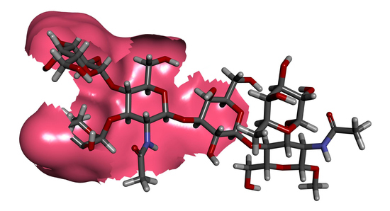
Given the surprising ability of the Lex trisaccharide 7 to inhibit the binding of SH2 to (DimLex)16-BSA 1, we also investigated the inhibition of this binding by our previously reported [43,49] Lex analogues 1518 (Figure 6). While D-GlcNAc and D-Gal are replaced by a D-glucose unit in analogue 15 (LacLex) and 16 (GlcLex), respectively, the L-fucose residue is replaced by an L-rhamnose unit in analogue 17 (RhaLex). In analogue 18, both the D-GlcNAc and L-Fuc are replaced by D-glucose and L-rhamnose units, respectively. Our previous studies [36] have shown that such analogues maintain the typical stacked conformation of the Lex trisaccharide [36,50,51,52,53,54]. We performed competitive inhibitions experiments using the (DimLex)16-BSA conjugate 1 (5 μg/mL) as the immobilized ligand and analogues 1518 as soluble competitors. Figure 6 shows the corresponding inhibition curves, while Table 2, entries 2–5, gives the IC50 for each analogue and the corresponding changes in free energy of binding Δ(ΔG) taking Lex 7 as the reference inhibitor (entry 1).
As can be seen, LacLex 15, GlcLex 16, and RhaLac 18 showed no inhibition of binding. The lack of inhibition of analogues 15 (LacLex) and 18 (RhaLac) indicates the importance of the N-acetylglucosamine residue, suggesting that the amide group participates in an essential antibody–carbohydrate interaction. Moreover, since replacing the galactose unit by a glucose residue also resulted in a complete loss in binding to GlcLex 16, we identified the galactose unit as a crucial element in epitope recognition by the mAb. This later result was in agreement with the titrations experiments performed with (GDimLex)16-BSA that showed no binding of SH2 (Figure 2). In contrast, substitution of the fucose unit by a rhamnose residue in RhaLex (17) resulted only in a 1.7 kcal.mol−1 decrease in free energy of binding compared to the Lex antigen 7 (Table 2, entry 4). This loss of binding correlates with the loss of a key polar interaction or H-bond, occurring between either Fuc 2-OH or 4-OH and an amino acid side chain in the SH2 binding site [55,56].
Given the lack of inhibition observed for GlcLex 16 and the lack of binding of the (GDimLex)16-BSA conjugate in our titration experiments (Figure 2), we investigated the importance of the galactosyl 4-OH group for recognition by SH2 using the previously described [57] analogues 1922 in competitive binding experiments. In these analogues, the galactosyl 4-OH is either methylated (4″-MeOLex, 19), deoxygenated (4″-HLex, 20), or replaced by a halogen in the 4″-ClLex (21) and 4″-FLex (22) (Figure 7).
Accordingly, we carried out competitive inhibitions experiments using the (DimLex)16-BSA conjugate 1 (5 μg/mL) as the immobilized ligand and analogues 1922 as soluble competitors. Figure 7 shows the corresponding inhibition curves, while Table 2, entries 6–9, gives the IC50 values for each analogue and the corresponding changes in free energy of binding Δ(ΔG) taking Lex 7 as the reference inhibitor. As can be seen in Figure 7, all analogues modified at O-4″ were able to inhibit the (DimLex)16-BSA binding to SH2 almost as well as or better than the Lex trisaccharide 7. Compared to Lex 7, the 4″-MeOLex analogue 19 resulted only in a small 0.7 kcal.mol−1 decrease in free energy of binding, suggesting that the galactosyl 4-OH is partially solvent exposed (Table 2, entry 6) [56]. Since the 4″-deoxy analogue 20 (4″HLex) only resulted in a loss of 0.3 kcal.mol−1 binding energy (Table 2, entry 7), it appears that the galactose 4-OH is not involved in any strong polar interaction or H-bond within the SH2 binding site [56]. Finally, the slight increases in binding of −0.5 and −0.6 kcal.mol−1 for the 4″-halogenated analogues 21 (4″-ClLex) and 22 (4″-FLex) suggest that the 4″-OH galactose is involved in weak van der Waals interactions. Indeed, this enhanced binding reflects the ability of the halogens (Cl and F) to produce stronger van der Waals forces than the hydroxyl group [58].

4. Discussion

MAb SH2 is shown here to bind (DimLex)16-BSA (1) but not (DimLex)6-TT (2) nor to the (Lex)10-BSA conjugate (3). Thus, these results seem to indicate that SH2 is binding an internal epitope on DimLex, which is not displayed by the Lex-BSA conjugate and not accessible on the DimLex-TT conjugate possibly as a result of the bulky protein carrier or inadequate presentation by the protein [40,41,42]. However, we also report here that the binding of SH2 to DimLex-BSA is inhibited better by Lex trisaccharide (7) than by DimLex hexasaccharide (6). This latter result contrasts with the hypothesis that SH2 binds an internal epitope on DimLex. One might propose that the SH2 mAb binding site is a deep pocket, which does not allow proper binding to the Lex trisaccharide when displayed on BSA due to steric hindrance, while it would be more accessible on the (DimLex)16-BSA conjugate. If this was so, one would then expect equivalent inhibition by all soluble inhibitors that display the reducing end Lex, such as DimLex (6), Lex (7) and Lex[1,3]Gal (9). This was not the case, since DimLex (6) and Lex[1,3]Gal (9) were much weaker inhibitors than the Lex trisaccharide (7). Thus, we propose that recognition by mAb SH2 of the epitope displayed by the DimLex antigen involves the reducing end Lex trisaccharide extending further to part of the reducing end Lex trisaccharide. However, binding of this epitope by SH2 is subject to correct presentation by the carrier molecule. Indeed, it has been shown that the recognition of oligosaccharides by lectins, enzymes, and antibodies was influenced by the different geometries of presentation of the carbohydrate within the glycan-carrier system (i.e., glycoconjugates, glycoproteins, glycolipids) [40,41,42,59]. Extended epitope presentation from the non-reducing end Lex trisaccharide in DimLex to the mAb SH2 binding site should also be considered in the context of the β-d-GlcNAc-(1→3)-d-Gal glycosidic bond conformation. Indeed, this glycosidic bond, that links the non-reducing end Lex trisaccharide to the reducing end Lex moiety in DimLex (6) or to galactose in Lex[1,3]Gal (9), has been shown to be highly flexible in various oligosaccharides [60,61,62,63,64,65,66]. Conformations around glycosidic bonds are defined by two dihedral angles: Φ (O5-C1-O1-Cx) and Ψ (C1-O1-Cx + 1). While it has been well established that the Lex trisaccharide adopts a rigid “stacked” conformation [36,50,51,52,53,54], we have shown [66] that the DimLex hexasaccharide could adopt two distinct conformations (I and II) around the Ψ dihedral angle for the β-d-GlcNAc-(1→3)-d-Gal glycosidic bond (Figure 8). These conformations were shown by NMR to exist in fast exchange for the hexasaccharide in solution [66].
In conformation I (Figure 8A), the non-reducing end β-GlcNAc (GlcNAc labelled in blue) unit is in the same plane as the reducing end galactosyl unit (Gal labelled in blue), while in conformation II, these two sugar units are perpendicular to one another (Figure 8B). Thus, these two conformations result in a much different environment around the N-acetyl group of the non-reducing end β-GlcNAc, which ends up in much closer proximity to the reducing end fucose unit (Fuc labelled in red) in conformation II than in conformation I. Taking together the importance of epitope presentation for binding of SH2 to Lex and DimLex discussed above and the occurrence of two distinct conformations of the DimLex hexasaccharide (and analogues) in solution, we propose that one of these conformations impedes the binding of SH2 to the non-reducing end Lex trisaccharide. This explains the greater affinity of SH2 for the Lex trisaccharide 7 than for the DimLex hexasaccharide 6 and the Lex[1,3]Gal fragment 9. We propose that immobilization on BSA in the (DimLex)16-BSA conjugate 1 favors one conformation of the DimLex oligosaccharide that presents the correct epitope accessible for binding with SH2.
Our results with the analogues and fragments suggest that, pending proper presentation, the non-reducing end Lex trisaccharide is an essential binding element for recognition by mAb SH2, despite the fact that SH2 does not react with the Lex-BSA conjugate nor with the Lex ceramide pentasaccharide (LNFPIII) [3]. These results are interesting when compared to the results that we have already described for mAbs SH1 and 1G5F6. As mentioned before, murine mAb SH1 (IgG3) was raised against the monomeric Lex ceramide pentasaccharide (LNFPIII) coated on acid-treated Salmonella Minnesota. It was shown to exhibit high affinity for monomeric and polymeric Lex structures regardless of chain length and is therefore classified as an anti-Lex group I mAb that binds the terminal non reducing end Lex trisaccharide [3,28]. In contrast, the murine mAb 1G5F6 (IgG3) raised against Helicobacter pylori O:3, much like mAb SH2, was shown to recognized polymeric Lex structures with greater affinity than monomeric Lex [32,35]. However, since 1G5F6 still retains binding to the Lex trisaccharide, we propose to classify it as a group IIB anti-Lex mAb. We have previously studied the recognition of monomeric Lex by anti-Lex mAb SH1 and anti-polymeric Lex mAb 1G5F6 [35,36,37]. For comparison with SH2, the difference in changes of free energy reported for competitive inhibition experiments with SH1 and 1G5F6 are reproduced in Table 2 taking the Lex (7) trisaccharide as a reference. As can be seen, substitution of the β-D-GlcNAc unit by the β-D-Glc greatly affected binding to SH2 and 1G5F6, (Table 2, entry 2) but not to SH1 [35,36]. Thus, in contrast to group I mAb SH1, the N-acetyl group of the GlcNAc residue is an essential binding element for recognition by anti-polymeric Lex mAbs SH2 and 1G5F6. Indeed, we have proposed—much like we do here for SH2—that 1G5G6 recognizes an epitope that, including the Lex non-reducing end trisaccharide, extends towards the galactosyl ring of the reducing end Lex trisaccharide. Replacing the galactose unit by a glucose residue led to total loss of recognition by both SH1 and SH2 and a loss of binding by 2.7 kcal.mol−1 for 1G5F6 [35,36]. Such reduced binding cannot solely be explained by the loss of favorable interactions between the binding sites of mAb SH1, SH2 or 1G5F6, and the axial galactose 4-OH group. Therefore, it is likely that the equatorial orientation of the 4″-OH group in GlcLex 16 disturbs the hydrophobic patch normally present in the β-galactosyl α face and leads to these results. Indeed, the β-galactosyl α face, which is defined by H-1, H-3, H-4, and H-5 of the galactose ring [67,68], is known to constitute an important recognition element due to its interaction with aromatic amino acid residues present in anti-carbohydrate antibodies and lectin binding sites [29,37,55,69]. Thus, as for mAbs SH1 and 1G5F6, we propose that the hydrophobic α-face of the galactose residue is probably involved in stacking interactions with aromatic side chains within the binding site [35,36,37]. Results with the RhaLex analogue 17 (Table 2, entry 4) showed that all three mAbs are involved in polar interactions or hydrogen bonds (H-bonds) involving the Fuc 2-OH and/or 4-OH with amino acid side chains in the binding sites. Noticeably, mAbs SH2 and 1G5F6 led to greater changes in free energy of binding [Δ(ΔG) = 1.6–1.7 kcal.mol−1] than mAb SH1 [Δ(ΔG) = 1.1 kcal.mol−1] suggesting that for these mAbs, both 2-OH and 4-OH of the fucosyl residue are involved in binding when only one of these is involved in binding to mAb SH1. Furthermore, for all three mAbs a cumulative effect was observed when both N-acetylglucosamine and fucose units were substituted by glucose and rhamnose residues (18, Table 2, entry 5), respectively (Table 2, entry 5). Taken together, these results illustrate that, while all three sugar residues are involved in the recognition of Lex by mAbs SH2 and 1G5F6, mAb SH1 makes contacts with the galactosyl and fucosyl residues, while no interaction with the GlcNAc acetamido group is detected.
Studies in the past have used methylated analogues of carbohydrates to distinguish between hydroxyl groups located at the periphery of mAb binding sites from those that are solvent exposed. It has been shown that partially solvent exposed hydroxyl groups can be replaced by methoxy groups with only a minor change in binding energy [70,71]. Thus, results with the 4″-MeOLex analogue 19 (Table 2, entry 6) indicate that, while the galactosyl 4-OH is likely positioned at the periphery or within the biding sites of mAbs SH1 and 1G5F6, it is partially exposed to bulk solvent in the mAb SH2 binding site. Results with the 4″-deoxy Lex 20 (4″-HLex, entry 7) show that, while the galactosyl 4-OH is likely involved in hydrogen bonding within the SH1 binding site [Δ(ΔG) = 1.3 kcal.mol−1], it does not contribute to the to the binding of Lex to mAbs SH2 and 1G5F6. Similarly, while replacing the galactosyl 4-OH by a chlorine or fluorine (entries 8 and 9) results in large drops in free energy of binding (1.6 and 2.1 kcal.mol−1) with mAb SH1, these substitutions have relatively little impact on the binding to mAbs SH2 and 1G5F6. Thus, while this hydroxyl group acts as an H-bond donor within the SH1 binding site [37], it is located at the periphery of the binding site within the 1G5F6 binding site [35] and partially exposed to bulk solvent in the SH2 binding site only contributing weak polar contacts in the latter two cases. Collectively, these results emphasize the different roles of galactose in the recognition of Lex by the three mAbs. In mAb SH1, the galactose participates in both hydrogen bond formation and hydrophobic interactions with the galactosyl α-face. In contrast, in mAbs SH2 and 1G5F6, the galactose predominantly participates through hydrophobic interactions with the galactosyl α-face.
Our results should be compared to the work of Van Roon et al. who studied anti-Lex antibodies generated in mice infected with Schistosoma mansoni cercariae [28,29,72]. In their study, Van Roon et al. cloned mAb 291-2G3A (IgG3), which, similarly to SH2 and 1G5F6, was shown by surface plasmon resonance (SPR) studies to recognize a DimLex-HSA conjugate with greater affinity than Lacto-N-fucopentaose-HSA (LNFPIII-HSA) conjugate [29,72]. To understand the specificity of the antibody for Lex, Van Roon et al. performed X-ray crystallographic analysis of the Fab fragment of mAb 291-2G3-A in complex with the Lex trisaccharide. The binding site of mAb 291-2G3-A is described as a shallow pocket that, much like SH2 and 1G5F6, makes contact with all three sugar units of the Lex trisaccharide. Again, similarly to SH2 and 1G5F6, the 4-OH of galactose does not participate in an H-bond with the mAb 291-2G3-A binding site, but a tryptophan (W33) residue forms favorable aromatic stacking interactions with the hydrophobic patch of the galactosyl α face [29]. Finally, the authors also report an H-bond between the glucosamine acetamido nitrogen and an asparagine residue (Asn L91) in the binding site. Unfortunately, the authors were not able to obtain a crystal structure with DimLex and thus could not conclude if binding to 291-2G3-A to DimLex would extend further from the reducing end GlcNAc residue.

5. Conclusions

These results taken collectively clearly demonstrate that different groups of antibodies are produced depending on the presentation of Lex to the immune system. Since SH1 was raised against the Lex ceramide pentasaccharide (LNFPIII), it recognizes an epitope localized to the non-reducing end Lex trisaccharide in all Lex-displaying analogues and conjugates. In contrast, those mAbs raised against polymeric Lex structures, such as SH2, 1G5F6, and 291-2G3-A, have greater affinity for DimLex conjugates than for conjugates only displaying Lex. However, the results presented in this work for SH2, as well as those reported for 1G5F6 and 291-2G3-A, indicate that the non-reducing end Lex trisaccharide is still an important recognition element for these mAbs [29,36,37]. Indeed, all three mAbs make similar contacts with all three sugar units of the Lex trisaccharide including the galactosyl hydrophobic α face. Since all three mAbs (SH2, 1G5F6, 291-2G3-A) make contact with the GlcNAc acetamido group and, given their higher affinity towards DimLex conjugates than for Lex conjugates, we also conclude that they recognise an epitope that extends further from the non-reducing end Lex trisaccharide. However, results with SH2 indicate that this extended epitope is only recognized when DimLex is presented by conjugates such as BSA glycoconjugates or glycolipids [3]. Indeed, while SH2 recognizes the DimLex-BSA conjugate 1 but does not bind (or binds poorly) to the Lex-BSA conjugates 3 or 4 (Figure 1), it displays weaker binding to the DimLex propyl glycoside 6 than for the Lex methyl glycoside 7. Interestingly, the known flexibility of the β-d-GlcNAc-(1→3)-d-Gal glycosidic bond that connects the two Lex trisaccharides in DimLex results in the hexasaccharide adopting two conformations in fast exchange when in solution (Figure 8). Thus, it is reasonable to assume that only one of these conformations is recognized by SH2, and we propose that this conformation is favored when the hexasaccharide is presented as part of a glycolipid such as the DimLex glycolipid (III3V3Fuc2nLc6) or as glycoprotein such as DimLex-BSA 1. Given the steric hindrance observed around the GlcNAc acetamido group in conformation II (Figure 8B), it is reasonable to suggest that conformation I (Figure 8B) is the conformation recognized by SH2.
Additional studies with mAb 1G5F6 and the analogues and fragments used in the present work will establish if presentation of DimLex also has an impact on epitope recognition by mAb 1G5F6. Based on the results reported here, we propose in Figure 9 a schematic representation of the epitope recognized by SH2 in conformation I of DimLex.
While the DimLex antigen is almost exclusively expressed at the surface of tumor cells, this hexasaccharide displays at its reducing end the Lex trisaccharide that is expressed on a number of normal tissues and cells. Thus, the development of anti-cancer vaccines able to target the DimLex antigen safely, while avoiding an autoimmune response against the Lex antigen, requires that we identify and target epitopes that are presented by the DimLex hexasaccharide but not displayed by the Lex trisaccharide. Such epitopes are recognized by mAbs such as SH2 and 1G5F6. Indeed, these two mAbs bind strongly to DimLex conjugates, while they do not, or very poorly, bind glycoconjugates displaying only the Lex trisaccharide. Therefore, the development of anti-cancer vaccines able to target the DimLex antigen rests on the accurate mapping of epitopes recognized by mAbs such as SH2 and 1G5F6. Most important to the field of TACA-based anticancer therapies, the results reported here indicate that proper presentation of the DimLex-based antigen target via conjugation to a protein or lipid is vital to the design of a successful vaccine or immunotherapeutic. Further mapping of this epitope will be explored using oligosaccharide analogues and fragments conjugated to BSA to ensure appropriate presentation of the epitope.

Supplementary Materials

The following are available online at https://www.mdpi.com/2076-393X/8/3/538/s1, Figure S1: Titration curves for SH2 ascites with various coating concentrations of conjugate (DimLex)16-BSA 1.

Author Contributions

Conceptualization, F.-I.A. and S.J.; methodology, S.J.; investigation, S.J.; resources: syntheses of 1–4 and 6–8 and 10–22, A.A., A.F., J.L.H., C.J.M., A.N., A.W. and J.-W.W.; writing, S.J and F.-I.A.; supervision and project administration, F.-I.A.; funding acquisition, F.-I.A. All authors have read and agreed to the published version of the manuscript.

Funding

This work was supported by the Natural Sciences and Engineering Research Council of Canada (RG03820).

Acknowledgments

The authors thank S.-I. Hakomori from the Pacific Northwest Research Institute (Seattle, WA, USA) for the generous gift of mAb SH2 and E. Samain from the Centre de recherches sur les macromolécules végétales (CERMAV, Grenoble, France) for the generous gift of tetrasaccharide 9.

Conflicts of Interest

The authors declare no conflict of interest.

References

  1. Soliman, C.; Yuriev, E.; Ramsland, P.A. Antibody recognition of aberrant glycosylation on the surface of cancer cells. Curr. Opin. Struct. Biol. 2007, 44, 1–8. [Google Scholar] [CrossRef] [PubMed]
  2. Hakomori, S.-I. Glycosylation defining cancer malignancy: New wine in an old bottle. Proc. Natl. Acad. Sci. USA 2002, 99, 10231–10233. [Google Scholar] [CrossRef] [PubMed] [Green Version]
  3. Singhal, A.K.; Ørntoft, T.F.; Nudelman, E.; Nance, S.; Schibig, L.; Stroud, M.R.; Clausen, H.; Hakomori, S. Profiles of Lewisx-containing Glycoproteins and Glycolipids in Sera of Patients with Adenocarcinoma. Cancer Res. 1990, 50, 1375–1380. [Google Scholar] [PubMed]
  4. Hakomori, S.; Kannagi, R. Glycosphingolipids as Tumor-Associated and Differentiation Markers. J. Natl. Cancer Inst. 1983, 71, 231–251. [Google Scholar]
  5. Hakomori, S. Tumor malignancy defined by aberrant glycosylation and sphingo(glyco)lipid metabolism. Cancer Res. 1996, 56, 5309–5318. [Google Scholar]
  6. Yanagisawa, M.; Yu, R.K. The expression and functions of glycoconjugates in neural stem cells. Glycobiology 2007, 17, 57–74. [Google Scholar] [CrossRef] [Green Version]
  7. Hakomori, S.; Jeanloz, R.W. Isolation of a Glycolipid Containing Fucose, Galactose, Glucose, and Glucosamine from Human Cancerous Tissue. J. Biol. Chem. 1964, 239, 3606–3607. [Google Scholar]
  8. Hakomori, S.; Nudelman, E.; Levery, S.B.; Kannagi, R. Novel fucolipids accumulating in human adenocarcinoma. I. Glycolipids with di- or trifucosylated type 2 chain. J. Biol. Chem. 1984, 259, 4672–4680. [Google Scholar]
  9. Feng, D.; Shaikh, A.S.; Wang, F. Recent Advance in Tumor-associated Carbohydrate Antigens (TACAs)-based Antitumor Vaccines. ACS Chem. Biol. 2016, 11, 850–863. [Google Scholar] [CrossRef]
  10. Hakomori, S.-I. Tumor-associated glycolipid antigens, their metabolism and organization. Chem. Phys. Lipids 1986, 42, 209–233. [Google Scholar] [CrossRef]
  11. Hakomori, S. Tumor-Associated Carbohydrate Antigens. Annu Rev. Immunol. 1984, 2, 103–126. [Google Scholar] [CrossRef] [PubMed]
  12. Singhal, A.; Hakomori, S.-I. Molecular changes in carbohydrate antigens associated with cancer. Bioessays 1990, 12, 223–230. [Google Scholar] [CrossRef]
  13. Brockhaus, M.; Magnani, J.L.; Herlyn, M.; Blaszczyk, M.; Steplewski, Z.; Koprowski, H.; Ginsburg, V. Monoclonal antibodies directed against the sugar sequence of lacto-N-fucopentaose III are obtained from mice immunized with human tumors. Arch. Biochem. Biophys. 1982, 217, 647–651. [Google Scholar] [CrossRef]
  14. Cuttitta, F.; Rosen, S.; Gazdar, A.F.; Minna, J.D. Monoclonal-Antibodies That Demonstrate Specificity for Several Types of Human-Lung Cancer. Proc. Natl Acad Sci. Biol. USA 1981, 78, 4591–4595. [Google Scholar] [CrossRef] [PubMed] [Green Version]
  15. Huang, L.C.; Brockhaus, M.; Magnani, J.L.; Cuttitta, F.; Rosen, S.; Minna, J.D.; Ginsburg, V. Many monoclonal antibodies with an apparent specificity for certain lung cancers are directed against a sugar sequence found in lacto-N-fucopentaose III. Arch. Biochem. Biophys. 1983, 220, 318–320. [Google Scholar] [CrossRef]
  16. Huang, L.C.; Civin, C.I.; Magnani, J.L.; Shaper, J.H.; Ginsburg, V. My-1, the Human Myeloid-Specific Antigen Detected by Mouse Monoclonal-Antibodies, Is a Sugar Sequence Found in Lacto-N-Fucopentaose-Iii. Blood 1983, 61, 1020–1023. [Google Scholar] [CrossRef] [Green Version]
  17. Gooi, H.C.; Thorpe, S.J.; Hounsell, E.F.; Rumpold, H.; Kraft, D.; Forster, O.; Feizi, T. Marker of Peripheral-Blood Granulocytes and Monocytes of Man Recognized by 2 Monoclonal-Antibodies Vep8 and Vep9 Involves the Trisaccharide 3-Fucosyl-N-Acetyllactosamine. Eur. J. Immunol. 1983, 13, 306–312. [Google Scholar] [CrossRef]
  18. Urdal, D.L.; Brentnall, T.A.; Bernstein, I.D.; Hakomori, S.I. A granulocyte reactive monoclonal antibody, 1G10, identifies the Gal beta 1-4 (Fuc alpha 1-3) GlcNAc (X determinant) expressed in HL-60 cells on both glycolipid and glycoprotein molecules. Blood 1983, 62, 1022–1026. [Google Scholar] [CrossRef] [Green Version]
  19. Combs, S.G.; Marder, R.J.; Minna, J.D.; Mulshine, J.L.; Polovina, M.R.; Rosen, S.T. Immunohistochemical Localization of the Immunodominant Differentiation Antigen Lacto-N-Fucopentaose-Iii in Normal Adult and Fetal Tissues. J. Histochem. Cytochem. 1984, 32, 982–988. [Google Scholar] [CrossRef] [Green Version]
  20. Solter, D.; Knowles, B.B. Monoclonal Antibody Defining a Stage-Specific Mouse Embryonic Antigen (Ssea-1). Proc. Natl. Acad. Sci. USA 1978, 75, 5565–5569. [Google Scholar] [CrossRef] [Green Version]
  21. Hakomori, S.; Nudelman, E.; Levery, S.; Solter, D.; Knowles, B.B. The Hapten Structure of a Developmentally Regulated Glycolipid Antigen (Ssea-1) Isolated from Human-Erythrocytes and Adenocarcinoma—A Preliminary Note. Biochem. Biophys. Res. Commun. 1981, 100, 1578–1586. [Google Scholar] [CrossRef]
  22. Gooi, H.C.; Feizi, T.; Kapadia, A.; Knowles, B.B.; Solter, D.; Evans, M.J. Stage-Specific Embryonic Antigen Involves Alpha-1-]3 Fucosylated Type-2 Blood-Group Chains. Nature 1981, 292, 156–158. [Google Scholar] [CrossRef] [PubMed]
  23. Kannagi, R.; Nudelman, E.; Levery, S.B.; Hakomori, S. A series of human erythrocyte glycosphingolipids reacting to the monoclonal antibody directed to a developmentally regulated antigen SSEA-1. J. Biol. Chem. 1982, 257, 14865–14874. [Google Scholar]
  24. Hakomori, S.; Nudelman, E.; Kannagi, R.; Levery, S.B. The Common Structure in Fucosyllactosaminolipids Accumulating in Human Adenocarcinomas, and Its Possible Absence in Normal Tissue. Biochem. Biophys. Res. Commun. 1982, 109, 36–44. [Google Scholar] [CrossRef]
  25. Fukushi, Y.; Hakomori, S.; Nudelman, E.; Cochran, N. Novel fucolipids accumulating in human adenocarcinoma. II. Selective isolation of hybridoma antibodies that differentially recognize mono-, di-, and trifucosylated type 2 chain. J. Biol. Chem. 1984, 259, 4681–4685. [Google Scholar]
  26. Fox, N.; Damjanov, I.; Knowles, B.B.; Solter, D. Immunohistochemical Localization of the Mouse Stage-specific Embryonic Antigen 1 in Human Tissues and Tumors. Cancer Res. 1983, 43, 669–678. [Google Scholar]
  27. Fukushi, Y.; Hakomori, S.; Shepard, T. Localization and alteration of mono-, di-, and trifucosyl alpha 1----3 type 2 chain structures during human embryogenesis and in human cancer. J. Exp. Med. 1984, 160, 506–520. [Google Scholar] [CrossRef] [Green Version]
  28. Van Roon, A.-M.M.; Van de Vijver, K.K.; Jacobs, W.; Van Marck, E.A.; Van Dam, G.J.; Hokke, C.H.; Deelder, A.M. Discrimination between the anti-monomeric and the anti-multimeric Lewis X response in murine schistosomiasis. Microbes Infect. 2004, 6, 1125–1132. [Google Scholar] [CrossRef]
  29. van Roon, A.-M.M.; Pannu, N.S.; de Vrind, J.P.M.; van der Marel, G.A.; van Boom, J.H.; Hokke, C.H.; Deelder, A.M.; Abrahams, J.P. Structure of an Anti-Lewis X Fab Fragment in Complex with Its Lewis X Antigen. Structure 2004, 12, 1227–1236. [Google Scholar] [CrossRef] [PubMed] [Green Version]
  30. van Remoortere, A.; Vermeer, H.J.; van Roon, A.M.; Langermans, J.A.; Thomas, A.W.; Wilson, R.A.; van Die, I.; van den Eijnden, D.H.; Agoston, K.; Kerekgyarto, J.; et al. Dominant antibody responses to Fuc alpha 1-3GalNAc and Fuc alpha 1-2Fuc alpha 1-3GlcNAc containing carbohydrate epitopes in Pan troglodytes vaccinated and infected with Schistosoma mansoni. Exp. Parasitol. 2003, 105, 219–225. [Google Scholar] [CrossRef] [PubMed] [Green Version]
  31. de Geus, D.C.; van Roon, A.-M.M.; Thomassen, E.A.J.; Hokke, C.H.; Deelder, A.M.; Abrahams, J.P. Characterization of a diagnostic Fab fragment binding trimeric Lewis, X. Proteins Struct. Funct. Bioinform. 2009, 76, 439–447. [Google Scholar] [CrossRef]
  32. Altman, E.; Harrison, B.A.; Hirama, T.; Chandan, V.; To, R.; MacKenzie, R. Characterization of murine monoclonal antibodies against Helicobacter pylori lipopolysaccharide specific for Lex and Ley blood group determinants. Biochem. Cell Biol. 2005, 83, 589–596. [Google Scholar] [CrossRef]
  33. Appelmelk, B.J.; Simoons-Smit, I.; Negrini, R.; Moran, A.P.; Aspinall, G.O.; Forte, J.G.; De Vries, T.; Quan, H.; Verboom, T.; Maaskant, J.J.; et al. Potential role of molecular mimicry between Helicobacter pylori lipopolysaccharide and host Lewis blood group antigens in autoimmunity. Infect. Immun. 1996, 64, 2031–2040. [Google Scholar] [CrossRef] [Green Version]
  34. Aspinall, G.O.; Monteiro, M.A.; Shaver, R.T.; Kurjanczyk, L.A.; Penner, J.L. Lipopolysaccharides of Helicobacter Pylori Serogroups O:3 and O:6 Structures of a Class of Lipopolysaccharides with Reference to the Location of Oligomeric Units of D-Glycero-α-D-Manno-Heptose Residues. Eur. J. Biochem. 1997, 248, 592–601. [Google Scholar] [CrossRef]
  35. Jegatheeswaran, S.; Auzanneau, F.I. Recognition of Lewis X by Anti-Le(x) Monoclonal Antibody IG5F6. J. Immunol. 2019, 203, 3037–3044. [Google Scholar] [CrossRef] [PubMed]
  36. Wang, J.-W.; Asnani, A.; Auzanneau, F.-I. Synthesis of a BSA-Lex glycoconjugate and recognition of Lex analogues by the anti-Lex monoclonal antibody SH1, The identification of a non-cross reactive analogue. Bioorg. Med. Chem. 2010, 18, 7174–7185. [Google Scholar] [CrossRef]
  37. Moore, C.J.; Auzanneau, F.I. Understanding the Recognition of Lewis X by Anti-Le Monoclonal Antibodies. J. Med. Chem. 2013, 56, 8183–8190. [Google Scholar] [CrossRef] [Green Version]
  38. Hendel, J.L.; Auzanneau, F.-I. Convergent Preparation of DimLex Hexasaccharide Analogues. Eur. J. Org. Chem. 2011, 2011, 6864–6876. [Google Scholar] [CrossRef]
  39. Kamath, V.P.; Diedrich, P.; Hindsgaul, O. Use of diethyl squarate for the coupling of oligosaccharide amines to carrier proteins and characterization of the resulting neoglycoproteins by MALDI-TOF mass spectrometry. Glycoconj. J. 1996, 13, 315–319. [Google Scholar] [CrossRef]
  40. Leteux, C.; Stoll, M.S.; Childs, R.A.; Chai, W.; Vorozhaikina, M.; Feizi, T. Influence of oligosaccharide presentation on the interactions of carbohydrate sequence-specific antibodies and the selectins—Observations with biotinylated oligosaccharides. J. Immunol. Methods 1999, 227, 109–119. [Google Scholar] [CrossRef]
  41. Solis, D.; Bruix, M.; Gonzalez, L.; Diaz-Maurino, T.; Rico, M.; Jimenez-Barbero, J.; Feizi, T. Carrier protein-modulated presentation and recognition of an N-glycan: Observations on the interactions of Man(8) glycoform of ribonuclease B with conglutinin. Glycobiology 2001, 11, 31–36. [Google Scholar] [CrossRef] [PubMed] [Green Version]
  42. Solis, D.; Feizi, T.; Yuen, C.T.; Lawson, A.M.; Harrison, R.A.; Loveless, R.W. Differential Recognition by Conglutinin and Mannan-Binding Protein of N-Glycans Presented on Neoglycolipids and Glycoproteins with Special Reference to Complement Glycoprotein C3 and Ribonuclease-B. J. Biol. Chem. 1994, 269, 11555–11562. [Google Scholar] [PubMed]
  43. Asnani, A.; Auzanneau, F.-I. Synthesis of Lewis X and three Lewis X trisaccharide analogues in which glucose and rhamnose replace N-acetylglucosamine and fucose, respectively. Carbohydr. Res. 2008, 343, 1653–1664. [Google Scholar] [CrossRef]
  44. Wang, A.; Auzanneau, F.-I. Selective protection of 2-azido-lactose and in situ ferrier rearrangement during glycosylation: Synthesis of a dimeric lewis X fragment. J. Org. Chem. 2007, 72, 3585–3588. [Google Scholar] [CrossRef]
  45. Wang, A.; Auzanneau, F.-I. Synthesis of LeaLex oligosaccharide fragments and efficient one-step deprotection. Carbohydr. Res. 2010, 345, 1216–1221. [Google Scholar] [CrossRef] [PubMed]
  46. Nejatie, A.; Jegatheeswaran, S.; Auzanneau, F.I. Synthesis of LacNAcLex- and DimLex-BSA Conjugates and Binding to Anti-Polymeric Lex mAbs. Eur. J. Org. Chem. 2019, 2019, 6631–6645. [Google Scholar] [CrossRef]
  47. Forman, A.; Hendel, J.; Auzanneau, F.I. Convergent synthesis of tetra- and penta- saccharide fragments of dimeric Lewis, X. Carbohydr. Res. 2019, 482, 107730. [Google Scholar] [CrossRef]
  48. Dumon, C.; Bosso, C.; Utille, J.P.; Heyraud, A.; Samain, E. Production of Lewis x Tetrasaccharides by Metabolically Engineered Escherichia coli. ChemBioChem 2006, 7, 359–365. [Google Scholar] [CrossRef]
  49. Hendel, J.L.; Cheng, A.; Auzanneau, F.-I. Application and limitations of the methyl imidate protection strategy of N-acetylglucosamine for glycosylations at O-4, Synthesis of Lewis A and Lewis X trisaccharide analogues. Carbohydr. Res. 2008, 343, 2914–2923. [Google Scholar] [CrossRef]
  50. Imberty, A.; Mikros, E.; Koca, J.; Mollicone, R.; Oriol, R.; Pérez, S. Computer simulation of histo-blood group oligosaccharides: Energy maps of all constituting disaccharides and potential energy surfaces of 14 ABH and Lewis carbohydrate antigens. Glycoconj. J. 1995, 12, 331–349. [Google Scholar] [CrossRef]
  51. Perez, S.; Mouhous-Riou, N.; Nifant’ev, N.E.; Tsvetkov, Y.E.; Bachet, B.; Imberty, A. Crystal and molecular structure of a histo-blood group antigen involved in cell adhesion: The Lewis x trisaccharide. Glycobiology 1996, 6, 537–542. [Google Scholar]
  52. Reynolds, M.; Fuchs, A.; Lindhorst, T.K.; Perez, S. The hydration features of carbohydrate determinants of Lewis antigens. Mol. Simul. 2008, 34, 447–460. [Google Scholar]
  53. Miller, K.E.; Mukhopadhyay, C.; Cagas, P.; Bush, C.A. Solution structure of the Lewis x oligosaccharide determined by NMR spectroscopy and molecular dynamics simulations. Biochemistry 1992, 31, 6703–6709. [Google Scholar]
  54. Haselhorst, T.; Weimar, T.; Peters, T. Molecular Recognition of Sialyl Lewisx and Related Saccharides by Two Lectins. J. Am. Chem. Soc. 2001, 123, 10705–10714. [Google Scholar]
  55. Bundle, D.R. Antibody Combining Sites and Oligosaccharide Determinants Studied by Competitive-Binding, Sequencing and X-Ray Crystallography. Pure Appl. Chem. 1989, 61, 1171–1180. [Google Scholar]
  56. Bundle, D.R. Recognition of Carbohydrate Antigens by Antibody Binding Sites. In Biorganic Chemistry: Carbohydrates; Hecht, S.M., Ed.; Oxford University Press: New York, NY, USA, 1999. [Google Scholar]
  57. Moore, C.J.; Auzanneau, F.-I. Synthesis of 4 manipulated Lewis X trisaccharide analogues. Beilstein J. Org. Chem. 2012, 8, 1134–1143. [Google Scholar]
  58. Bundle, D.R.; Eichler, E.; Gidney, M.A.J.; Meldal, M.; Ragauskas, A.; Sigurskjold, B.W.; Sinnott, B.; Watson, D.C.; Yaguchi, M.; Young, N.M. Molecular Recognition of a Salmonella Trisaccharide Epitope by Monoclonal-Antibody Se155-4. Biochemistry 1994, 33, 5172–5182. [Google Scholar]
  59. Yamasaki, R.; Schneider, H.; Griffiss, J.M.; Mandrell, R. Epitope expression of Gonococcal lipooligosaccharide (LOS). Importance of the lipoidal moiety for expression of an epitope that exists in the oligosaccharide moiety of LOS. Mol. Immunol. 1988, 25, 799–809. [Google Scholar]
  60. Sawen, E.; Hinterholzinger, F.; Landersjo, C.; Widmalm, G. Conformational flexibility of the pentasaccharide LNF-2 deduced from NMR spectroscopy and molecular dynamics simulations. Org. Biomol. Chem. 2012, 10, 4577–4585. [Google Scholar]
  61. Zaccheus, M.; Pendrill, R.; Jackson, T.A.; Wang, A.; Auzanneau, F.I.; Widmalm, G. Conformational Dynamics of a Central Trisaccharide Fragment of the LeaLex Tumor Associated Antigen Studied by NMR Spectroscopy and Molecular Dynamics Simulations. Eur. J. Org. Chem. 2012, 2012, 4705–4715. [Google Scholar]
  62. Xia, J.C.; Daly, R.P.; Chuang, F.C.; Parker, L.; Jensen, J.H.; Margulis, C.J. Sugar folding: A novel structural prediction tool for oligosaccharides and polysaccharides. J. Chem. Theory Comput. 2007, 3, 1629–1643. [Google Scholar] [CrossRef] [PubMed]
  63. Xia, J.C.; Margulis, C. A tool for the prediction of structures of complex sugars. J. Biomol. NMR 2008, 42, 241–256. [Google Scholar] [CrossRef] [PubMed]
  64. Sawen, E.; Stevensson, B.; Ostervall, J.; Maliniak, A.; Widmalm, G. Molecular Conformations in the Pentasaccharide LNF-1 Derived from NMR Spectroscopy and Molecular Dynamics Simulations. J. Phys. Chem. B 2011, 115, 7109–7121. [Google Scholar] [CrossRef]
  65. Jackson, T.A.; Robertson, V.; Imberty, A.; Auzanneau, F.-I. The flexibility of the Le(a)Le(x) Tumor Associated Antigen central fragment studied by systematic and stochastic searches as well as dynamic simulations. Bioorg. Med. Chem. 2009, 17, 1514–1526. [Google Scholar] [CrossRef]
  66. Jackson, T.A.; Robertson, V.; Auzanneau, F.I. Evidence for Two Populated Conformations for the Dimeric Le(X) and Le(A)Le(X) Tumor-Associated Carbohydrate Antigens. J. Med. Chem. 2014, 57, 817–827. [Google Scholar] [CrossRef] [Green Version]
  67. Quiocho, F.A. Protein-carbohydrate interactions: Basic molecular features. Pure Appl. Chem. 1989, 61, 1293–1306. [Google Scholar] [CrossRef]
  68. Lemieux, R.U. How Water Provides the Impetus for Molecular Recognition in Aqueous Solution. Acc. Chem. Res. 1996, 29, 373–380. [Google Scholar] [CrossRef]
  69. Asensio, J.L.; Arda, A.; Canada, F.J.; Jimenez-Barbero, J. Carbohydrate-Aromatic Interactions. Acc. Chem. Res. 2013, 46, 946–954. [Google Scholar] [CrossRef] [Green Version]
  70. Bundle, D.R. Bioorganic Chemistry: Carbohydrates; Hecht, S.M., Ed.; Oxford University Press: New York, NY, USA, 1999. [Google Scholar]
  71. Nikrad, P.V.; Beierbeck, H.; Lemieux, R.U. Molecular Recognition 10. A Novel Procedure for the Detection of the Intermolecular Hydrogen-Bonds Present in a Protein-Bullet-Oligosaccharide Complex. Can. J. Chem. 1992, 70, 241–253. [Google Scholar] [CrossRef]
  72. Van Roon, A.M.M.; Pannu, N.S.; Hokke, C.H.; Deelder, A.M.; JAbrahams, P. Crystallization and preliminary X-ray analysis of an anti-LewisX Fab fragment with and without its LewisX antigen. Acta Crystallogr. D 2003, 59, 1306–1309. [Google Scholar] [CrossRef]
Figure 1. Glycoconjugates used in the titrations.
Vaccines 08 00538 g001
Figure 2. Titration curves for SH2 ascites with conjugates 15 and BSA.
Vaccines 08 00538 g002
Figure 3. Structure of inhibitors 614 used in this study.
Vaccines 08 00538 g003
Figure 4. Competitive inhibitions curves for those compounds 6, 7, 9, 10 that showed some inhibition. Coated inhibitor (DimLex)16-BSA (1) and SH2 serum dilution 1:500.
Vaccines 08 00538 g004
Figure 5. Competitive inhibition with glycoconjugates 1, 3, and 4 as soluble inhibitors. Coated inhibitor (DimLex)16-BSA (1). SH2 serum dilution, 1:500.
Vaccines 08 00538 g005
Figure 6. Structures of analogue 1518 and competitive inhibition with 7 and 1518 as soluble inhibitors. Coated inhibitor (DimLex)16-BSA (1). SH2 serum dilution, 1:500.
Vaccines 08 00538 g006
Figure 7. Structures of analogue 1922 and competitive inhibition with 7 and 1922 as soluble inhibitors. Coated inhibitor (DimLex)16-BSA (1). SH2 serum dilution, 1:500.
Vaccines 08 00538 g007
Figure 8. Known [66] conformations of the DimLex hexasaccharide in fast exchange: (A) Conformation I; (B) Conformation II.
Vaccines 08 00538 g008
Figure 9. Proposed extended epitope recognized by SH2 in conformation I of the DimLex hexasaccharide.
Vaccines 08 00538 g009
Table 1. Inhibition data for mAb SH2 and those compounds 6, 7, 9, 10 that showed some inhibition.
EntryInhibitorIC50 a (μM)∆(∆G) b (kcal·mol−1)
1DimLex (6)5030
2Lex (7)47−1.4
3Lex[1,3]Gal (9)5290.03
4GlcNAc[1,3″]Lex (10)186372.1
a Concentration of inhibitor required for 50% inhibition using DimLex-BSA conjugate 1 as immobilized antigen. b Values determined from the expression Δ(ΔG) = RT ln([I1]/[I2] where [I2] is the IC50 measured for the reference inhibitor DimLex 6, and [I1] is the IC50 measured for each fragment with R = 1.98 cal K−1and T = 296 K.
Table 2. Inhibition data for mAb SH2, SH1, and 1G5F6 with Lex analogues 7, 1522.
SH2SH1 d1G5F6 e
EntryInhibitorIC50 a (μM)∆(∆G) b (kcal·mol−1)∆(∆G) d (kcal·mol−1)∆(∆G) e (kcal·mol−1)
1Lex (7)47000
2LacLex (15)>>1800---c0.22.5
3GlcLex (16)>>1800---c---c2.7
4RhaLex (17)8271.71.11.6
5RhaLac (18)>>1800---c1.53.2
64″-MeOLex (19)1480.7---c---c
74″-HLex (20)740.31.30.3
84″-ClLex (21)19-0.51.60.3
94″-FLex (22)17-0.62.1−0.1
a Concentration of inhibitor required for 50% inhibition using DimLex-BSA conjugate 1 as immobilized antigen. b Values determined from the expression Δ(ΔG) = RT ln([I1]/[I2] where [I2] is the IC50 measured for the reference inhibitor Lex 7 (shaded in yellow), and [I1] is the IC50 measured for each analogue 15–22 with R = 1.98 cal K−1and T = 296 K. c No inhibition. d Inhibition data published [36,37] for SH1 using Lex-BSA conjugate 4 as immobilized antigen and Lex trisaccharide 7 as reference. e Inhibition data published [35] for 1G5F6 using DimLex-BSA conjugate 1 as immobilized antigen and Lex trisaccharide 7 as reference.

