Article

Immunomodulation of Patients with Canine Visceral Leishmaniasis at Different Stages: A 12-Month Follow-Up Study Using LaSap

by
Kelvinson Fernandes Viana
1,*,
Adrieli Barboza de Souza
1,
Sara Torres
1,
Maria Camila Escobar Garcia
1,
Açucena Veleh Rivas
1,
Alex Sander Rodrigues Cangussu
2,
Francisca Hildemagna Guedes da Silva
3 and
Rodolfo Cordeiro Giunchetti
4
1
Vaccine Development Technology Laboratory, Federal University for Latin American Integration (UNILA), Foz do Iguaçu 85870-650, PR, Brazil
2
Vaccine Laboratory, Federal University of Tocantins (UFT), Gurupi 77410-530, TO, Brazil
3
Leopoldo de Meis Institute of Medical Biochemistry, National Institute of Science and Technology for Structural Biology and Bioimaging, National Center of Nuclear Magnetic Resonance Jiri Jonas, Federal University of Rio de Janeiro, Rio de Janeiro 21941-599, RJ, Brazil
4
Cellular Interaction Biology Laboratory, Department of Morphology, Federal University of Minas Gerais (UFMG), Belo Horizonte 31270-901, MG, Brazil
*
Author to whom correspondence should be addressed.
Vaccines 2025, 13(9), 933; https://doi.org/10.3390/vaccines13090933
Submission received: 23 July 2025 / Revised: 29 August 2025 / Accepted: 29 August 2025 / Published: 1 September 2025
(This article belongs to the Special Issue Parasitic Infections: Therapy for Host Immunity and Vaccination)

Abstract

Background/Objectives: Canine visceral leishmaniasis (CVL) is one of the main neglected protozoan diseases in the world. Dogs play a fundamental role in the maintenance of Leishmania infantum in the Americas, and we have already encountered resistance problems with drugs currently used in these animals. Methods: In view of this, two new immunotherapeutic protocols were tested in 48 dogs, using L. amazonensis antigens plus saponin (LaSap) and only L. amazonensis antigens (La) as a control group. Dogs naturally infected with L. infantum were divided into four groups, according to clinical staging. A total of 24 dogs (stages 1 and 2) received a four-dose protocol, and another 24 dogs (stages 3 to 5) received six doses. All animals received a booster dose every three months until they were one year old. Results: Our results showed that dogs in the early stages of the disease respond better and are able to remain stable for longer, maintaining baseline laboratory biomarkers, in addition to having a lower parasite load. Conclusions: On the other hand, dogs in more advanced stages have a poor response, with stage 3 being a key point in clinical progression or regression.

1. Introduction

According to the World Health Organization (WHO), leishmaniasis is a complex of neglected diseases caused by protozoa of the genus Leishmania sp. [1]. Insects of the genus Lutzomyia and Phlebotomus are the transmitters of the parasite in the New and Old World, respectively [2]. Widely distributed throughout the world, leishmaniasis is mainly related to areas with worse socioeconomic conditions [3]. In 2023, the WHO announced that Bangladesh had eliminated visceral leishmaniasis (VL) as a public health problem, the first country in the world to do so. This status was validated after Bangladesh reported less than 1 case per 10,000 people for the previous 3 consecutive years. This result was achieved through a series of factors associated with disease control [4].
VL is one of the most prevalent human diseases, with more than 20 Leishmania species reported worldwide [3]. The protozoan disease can be classified as anthroponotic (AVL) or zoonotic (ZVL), depending on the different vulnerable species involved [5]. In the context of ZVL, canine visceral leishmaniasis (CVL) is strongly linked to human cases. Furthermore, not only symptomatic dogs but also asymptomatic ones can act as a source of infection for the main phlebotomine vector, Lutzomyia longipalpis, in Latin America [6,7].
In both Brazil and Europe, dogs infected with L. infantum must be treated or euthanized. However, there are marked differences in the vector’s seasonality and infection pressure between the two regions. The monthly distribution and abundance of sand flies are influenced by biotic and abiotic factors. In addition, temperature, humidity, and precipitation influence the populations of these vectors [8,9]. These factors directly interfere with the treatment of dogs, and it is common for these animals to undergo new protocols with miltefosine at increasingly shorter intervals, directly impacting parasite resistance.
Immunotherapy as a treatment proposal for leishmaniasis has been a promising alternative for decades. Different researchers have used this treatment approach in humans and dogs, obtaining very encouraging results [10,11,12]. Our research group has been working for some years with total antigens of L. amazonensis associated with saponin as an alternative in the clinical management of canine patients with VL. However, the challenging complexity of this multifaceted disease makes us understand that dogs in different clinical stages may require different immunotherapeutic protocols. This particularity may be necessary because the canine patient requires more or less antigenic stimulation to remain clinically stable [13,14]. Therefore, in the present study, we present an analysis using two protocols in dogs with different forms of VL.

2. Materials and Methods

2.1. Animals

This study involved 48 dogs (24 males and 24 females) of different breeds, most of which were mixed breeds. Their ages ranged from 8 months to 10 years. All animals were naturally infected with L. infantum (diagnosed by serology and PCR) and staged according to the Brazilian and Leishvet guidelines. All animals underwent a battery of clinical, hematological (blood count), biochemical (AST, ALT, urea, and creatinine), and molecular (bone marrow qPCR) tests to establish the clinical staging of each patient. All dogs were from owners in the city of Foz do Iguaçu, Paraná, Brazil. The study was approved by the Ethics Committee for the Use of Animals (CEUA-UNILA), protocol 003/2022, on 8 April 2022.

2.2. Inclusion and Exclusion Criteria and Randomization

Only dogs that tested positive for L. infantum—based on laboratory diagnostics using ELISA (Biogene®, Recife, Pernambucio, Brazil), indirect immunofluorescence (Immunodot®, Jaboticabal, São Paulo, Brazil), and qPCR of bone marrow—were included in this clinical study. In addition, all owners agreed to sign a free and informed consent form. All blood samples from the dogs underwent serological and molecular tests to detect hemoparasitosis such as Babesia, Ehrlichia and Anaplasma. Animals that tested positive in any of the tests were treated before starting the study.
Dogs that presented any serious adverse reaction (e.g., seizure or anaphylaxis) after the application of immunotherapy would be monitored by the responsible clinical veterinarian and excluded from the study. Dogs that presented any other clinical pathological manifestation throughout the study, such that the therapeutic conduct could interfere with the results, would be excluded from the experiment. The animals were randomized using “https://www.random.org/”.

2.3. Experimental Design

All dogs were clinically staged between grades 1 and 5 and randomized into four groups, each containing 12 dogs. The study was divided into two treatment protocols: dogs in stages 1 and 2 received the four-dose immunotherapy protocol containing 100 micrograms of total L. amazonensis antigens (MHOM/BR/1977/LTB0016) plus 100 micrograms of saponin subcutaneously at 21-day intervals (LaSap). The control group also consisted of 12 dogs that received only 100 micrograms of total L. amazonensis antigens (La) subcutaneously at 21-day intervals. After the protocol ended, all dogs in both groups received a booster dose of their respective formulations every three months, until they completed 12 months.
The second protocol consisted of dogs in clinical stages between grades 3 and 5. There were two groups (LaSap and La), with 12 dogs per group. In the LaSap group, all dogs received six doses of immunotherapy, with the first two doses 7 days apart, the third dose 15 days after the second, and the last three doses 30 days apart. The control group (La) received the same protocol, but without saponin in the formulation (Figure 1). After the protocol ended, the animals received a booster dose every 3 months for 12 months. Blood samples were collected from the animals at a specific time point (15 days before the start of the experiments) and on the day of each immunotherapy booster. The samples were used for hematological, biochemical, serological, and molecular analyses (zero time).

2.4. Hematology and Biochemistry

The hematological profile was analyzed by blood cell counts using an electronic hematology particle counter (BC2800Vet, Mindray, Hamburg, Germany). Differential leukocyte counting was performed on Giemsa-stained blood smears, and a total of 100 cells were counted. The biochemical evaluations consisted of the following tests: renal function (urea and creatinine) and hepatic function tests (alanine aminotransferase (ALT) and aspartate aminotransferase (AST)). For this analysis, the automated biochemical system (CELM SBA-200, Barueri, SP, Brazil) and Labtest® commercial kits (Labtest Diagnóstica S.A., Lagoa Santa, MG, Brazil) were used, following the method described by the manufacturer.

2.5. ELISA

Two ELISA diagnostic kits were used to detect antibodies stimulated by immunotherapy (HSP70—Biogene Company, Brazil). For IgG Total analysis, sera were added at a 1:100 dilution. Peroxidase-conjugated rabbit anti-dog IgG antibodies were added at a 1:10,000 dilution for 30 min. The reaction was developed using TMB, and absorbance was measured on a Multiskan® MCC 340 (Labsystems, Helsinki, Finland) automatic microplate reader at 450 nm. Cut-off was calculated according to each manufacturer’s determinations.

2.6. Isolation of Peripheral Blood Mononuclear Cells

All blood samples (20 mL) were collected in heparinized tubes from the 48 dogs. Peripheral blood mononuclear cells (PBMCs) were isolated as previously described [13]. Cells were counted in a Neubauer hemocytometer chamber to determine the number of monocytes or lymphocytes per milliliter. Isolated PBMC suspensions adjusted to 1 × 10 monocytes/well were added to 24-well plates (NUNC, Thermo Fisher Scientific Inc., Waltham, MA, USA). Cells were stimulated with 10 micrograms of soluble antigen from L. infantum and L. amazonensis (SLiAg and SLaAg) and maintained in culture for 72 h. Subsequently, the supernatant of each culture was collected and stored at −80 °C for cytokine measurement.

2.7. ELISA Cytokine Assay

Briefly, the plates were coated with anti-IFN-γ and anti-IL-10 mAb in PBS, pH 7.4, and incubated at 4 °C overnight. After blocking the wells using buffer containing PBS plus 0.05% (v/v) Tween 20 and 0.1% (w/v) BSA, supernatants were added to each well. Biotin-labeled mAb in incubation buffer was added to each well, and streptavidin–HRP was used as an enzyme. The reaction was developed using 3,3′,5,5′-tetramethylbenzidine substrate and stopped with 2.5 M H2SO4 solution. The plates were washed after each step of incubation using PBS plus 0.05% (v/v) Tween 20. Minimum sensitivity levels were 68 pg/mL for IFN-γ and 16 pg/mL for IL-10. All experiments were performed using 96-well plates (COSTAR, Corning Inc., Glendale, AZ, USA) and according to the instructions of R&D Systems. The reading was performed using a microplate automatic reader (LoccusTM, Cotia, São Paulo, Brazil) at a wavelength of 450 nm [15].

2.8. Parasite Load

The parasite loads were calculated by real-time PCR according to a method described elsewhere [16]. Skin biopsy was performed in the middle region of the left ear. A fragment was removed with the aid of sterile biopsy punch 5 mm in diameter (Punch for Biopsy®, Kolplast LTDA, São Paulo, São Paulo, Brazil) and stored in a freezer at −80 °C for further analysis of the parasite load. This material was collected before the start of immunotherapy at T0 and at all times after the end of the initial protocol. DNA was extracted from the skin using Nucleo Spin® Tissue (Macherey-Nagel, Oerdt, France) according to the manufacturer’s instructions. The parasite loads were estimated using the following primers: forward, 5′TGTCGCTTGCAGACCAGATG 3′ and reverse, 5′GCATCGCAGGTGTGAGCAC 3′. These primers amplified a 90 bp fragment of a single-copy-DNA polymerase gene (DNA polα) from L. infantum (Gen-Bank accession number AF009147). PCR was carried out in a final volume of 10 μL containing 2.0 pmol of each primer, 2X SYBR® Green PCR Master Mix (Applied Biosystems, Waitham, MA, USA), 4.0 μL of DNA with a final concentration of approximately 20 ng/mL, and ultrapure water. Reactions were processed and analyzed in an ABI Prism 7500 Sequence Detection System (SDS Applied Biosystems, Foster City, CA, USA). The following steps were programmed: an initial denaturation at 95 °C for 10 min followed by 40 cycles of denaturation at 95 °C for 15 s and annealing/extension at 60 °C for 1 min. Parasite quantification for each bone marrow and skin sample was calculated by interpolation of the standard curve included in the same run, performed in duplicate, and expressed as the number of L. infantum organisms per 20 ng of total DNA.

2.9. Statistical Analysis

The statistical analysis was performed with GraphPad Prism 9.0 software (Prism Software, Irvine, CA, USA). Data normality was assessed using the Kolmogorov–Smirnov test. The analyses were performed using a repeated-measures ANOVA. Differences were considered significant at p < 0.05.

3. Results

3.1. Immunotherapy Improves the Clinical Profile of Patients with CVL

Dogs in the initial stages that received the four-dose LaSap protocol showed improvement in clinical manifestations, especially in relation to exfoliative dermatitis and lymphadenopathy. The same was observed in the control group (La). In both groups, there was no recurrence of these clinical signs over the course of 12 months. In the group of patients in stages between 3 and 5, who received six doses, the LaSap group showed a more marked improvement compared to the La group. However, over the course of 12 months, some dogs returned to presenting the same clinical signs they had before starting treatment (Table 1 and Table 2).

3.2. LaSap Immunotherapy Stabilizes Most Staged Patients

The hematologic profile of patients in stages 1 and 2 remained stable throughout the treatment protocol, as well as throughout the four booster doses over 12 months. This profile was observed in both treatment groups, all remaining within the reference values (Table 3). On the other hand, in animals in more advanced stages (3 to 5), who received six doses of LaSap or La, it was observed that dogs in the La group presented a reduction in platelets (Table 4). In the LaSap group, most patients were able to maintain leukocyte levels within reference values. Of the seven dogs in stage 3, one progressed to stage 4 and six improved to stage 2. Of a total of four dogs that began the study in stage 4, two dogs progressed to level 5 in the last experimental trimester. One patient in stage 5 showed no improvement in his clinical condition (Table 3 and Table 4). Biochemical analyses indicated that only dogs in more advanced stages showed increased urea and creatinine levels in both treated groups.

3.3. Dogs May Exhibit Variable Antibody Levels Throughout Treatment with LaSap

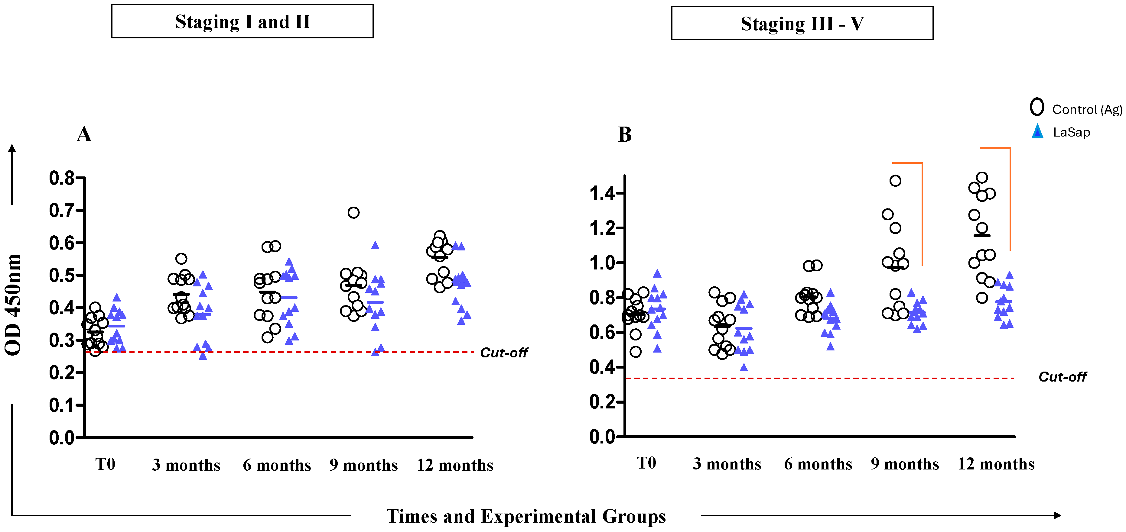
Serological analyses performed using the HSP70 protein (commercial kit Biogene®), present in all Leishmania species, as a target, indicated that dogs in stages 1 and 2 were able to maintain antibody production at lower levels throughout the treatment. Dogs in stages between 3 and 5 already presented higher absorbances at time zero due to clinical conditions. However, the LaSap group was able to maintain production at lower values compared to the La group, especially at 9 and 12 months (Figure 2).

3.4. LaSap Reduces and Stabilizes Parasite Load in Most Treated Dogs

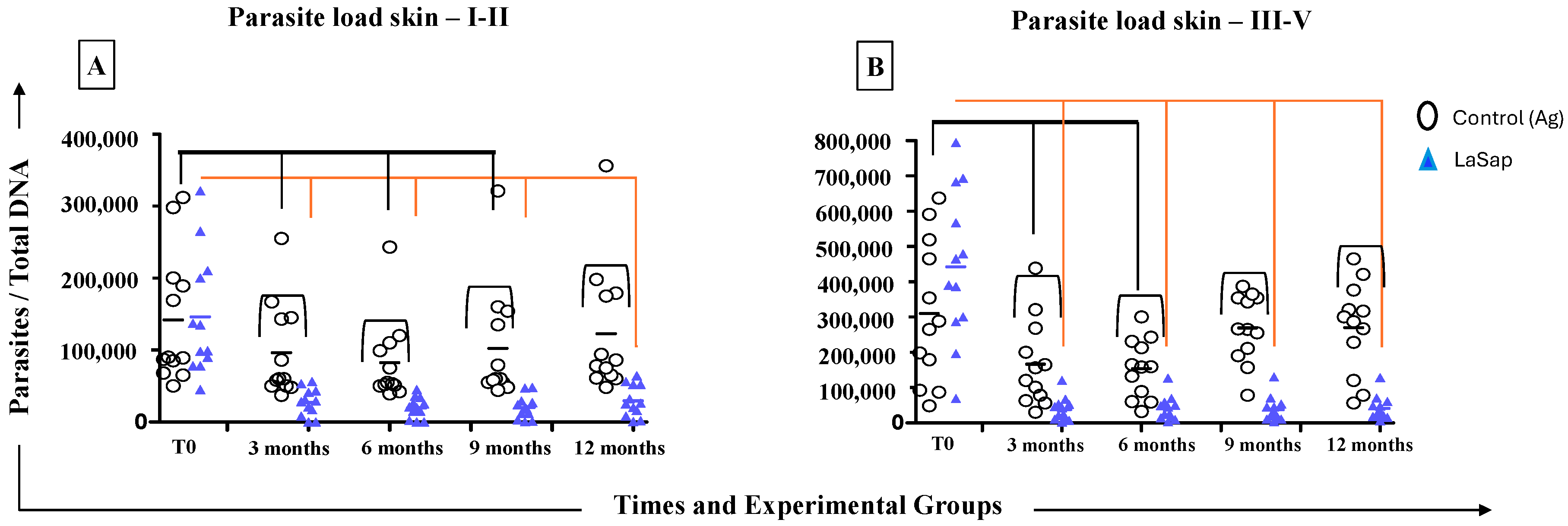
Dogs in stages 1 and 2 of the La group showed a significant reduction in parasite load on the skin up to 9 months after the end of the initial protocol (p = 0.04, 91,050 ± 57,520). On the other hand, dogs in the LaSap group remained with a lower parasite load up to 12 months (p = 0.0008, 84,800 ± 21,880). In addition, the parasite load of the LaSap group at 12 months was lower than that of the La group (p = 0.005, 90,000 ± 21,880) (Figure 3). In dogs in stages 3 and 5, the La group managed to reduce the parasite load up to 6 months after the end of the initial protocol (p = 0.023, 203,100 ± 83,260). The LaSap group maintained a lower load up to 12 months (p = 0.0001, 40,830 ± 33,620) compared to time zero (p = 0.0001, 442,400 ± 215,800). Similarly, there was a lower load compared to the La group (p = 0.0001, 269,700 ± 129,700) at 12 months.
In the bone marrow, dogs in the LaSap group in stages 1 and 2 maintained a lower parasite load at all times after the initial protocol, compared to time zero (T0 = 122,000; 3 months = 14,460; 6 months = 11,120; 9 months = 9745 and 12 months = 8726). At the last time, they also maintained a lower load than the La group (p = 0.006, 63,230 ± 8726). In the groups of dogs in stages between 3 and 5, there was a significant reduction in the load at times 9 (p = 0.043, 414,700 ± 139,500) and 12 months (p = 0.039, 536,200 ± 201,100).

3.5. LaSap Stimulates Higher Levels of IFN-γ in Dogs Naturally Infected with L. infantum

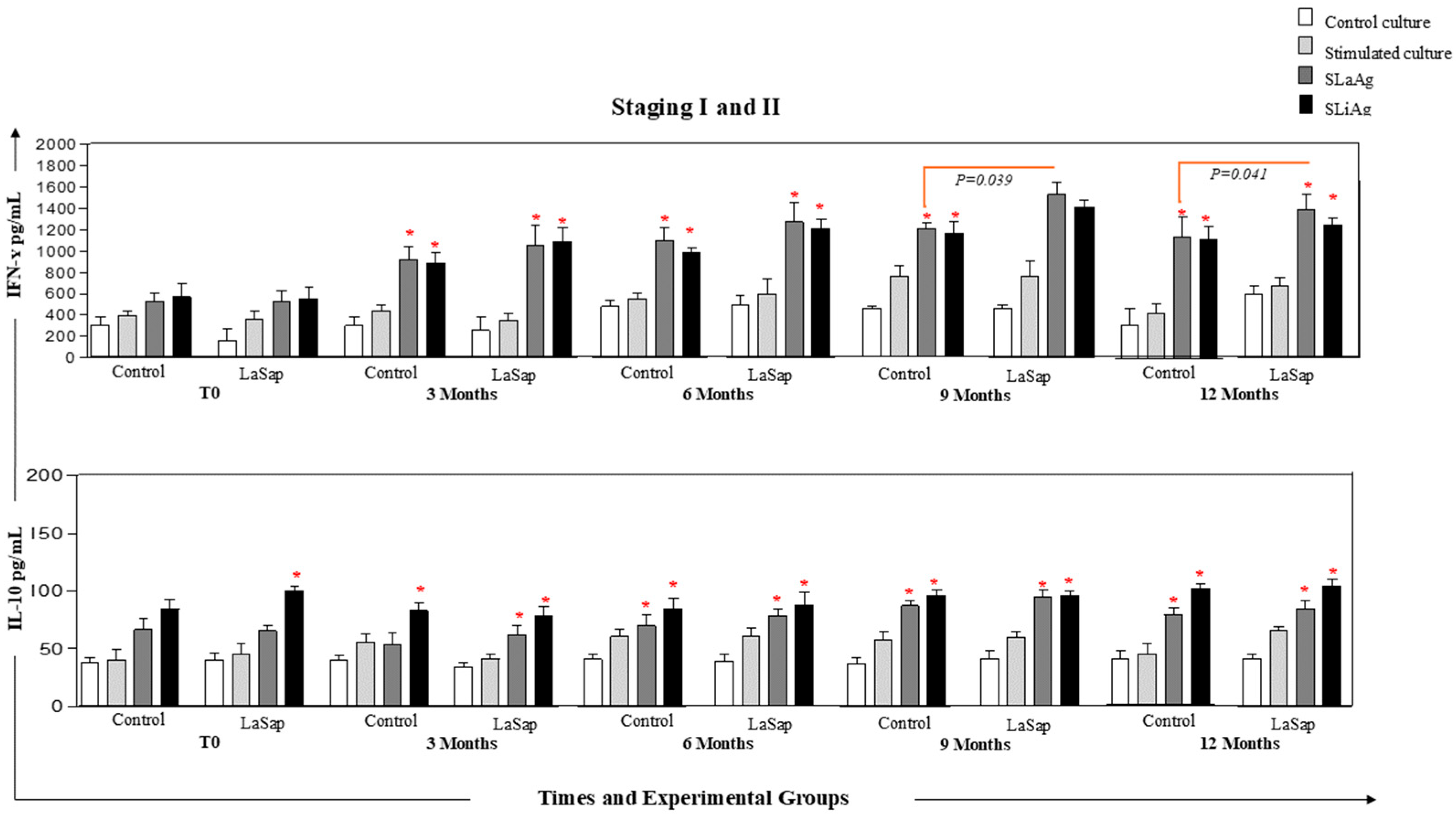
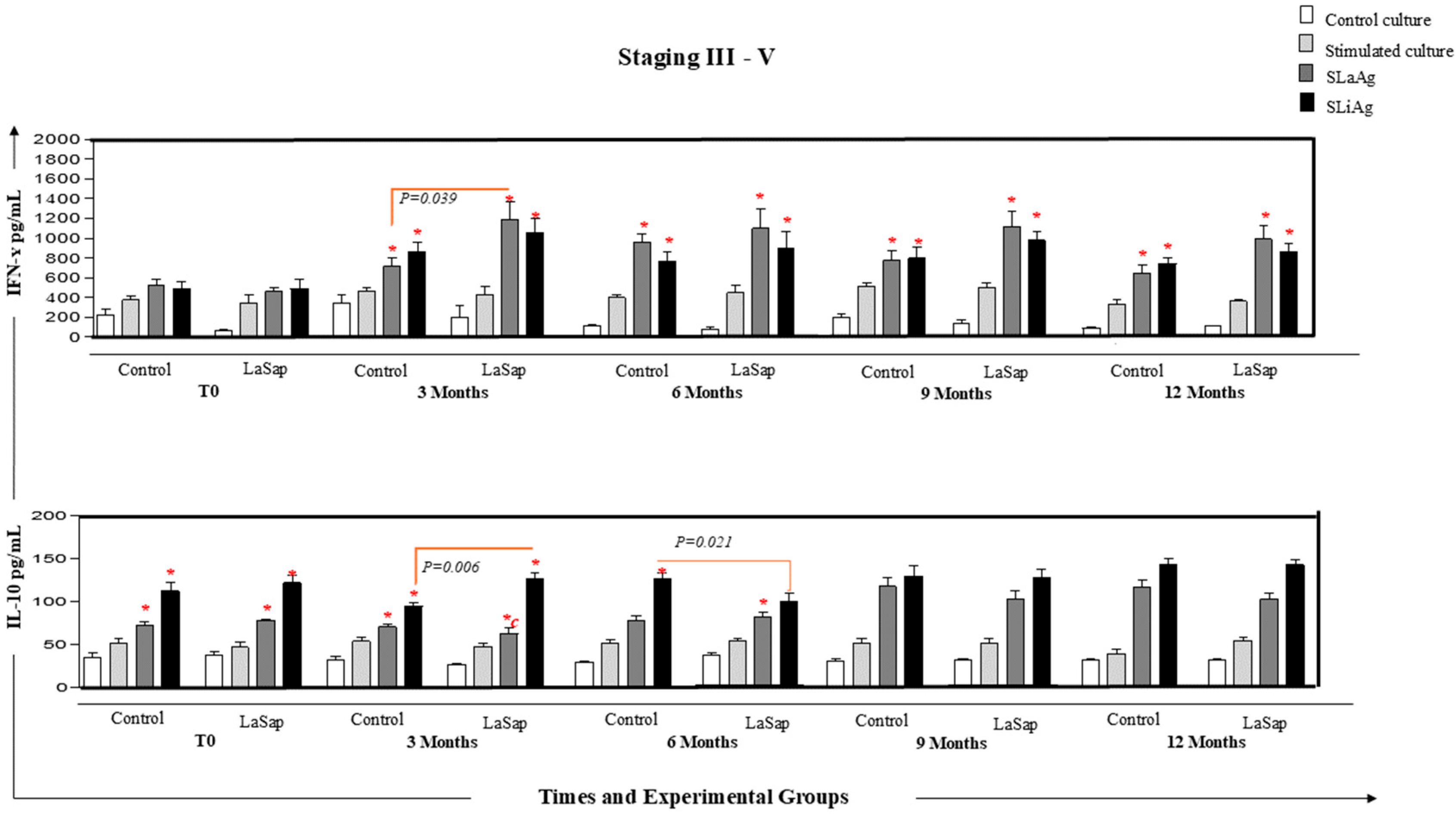
In dogs in stages 1 and 2, at all times analyzed, and in both the La and LaSap groups, there was greater production of IFN-γ in cultures stimulated with SLaAg and SLiAg compared to control cultures. However, at the 9-month time point (p = 0.041, 1.210, 1.533), there was greater production of the cytokine in the culture stimulated with SLaAg of the LaSap group compared to the La group. Regarding the production of IL-10, at all times, there was greater production of this cytokine in the stimulated cultures compared to the control cultures (Figure 4). A similar pattern was observed in dogs with stages between 3 and 5 (Figure 5). However, the levels of IL-10 were extremely low (p < 0.05) compared to IFN-g in the overall average (Figure 6).

4. Discussion

When it comes to CVL, there are different factors that directly impact the ability to control the transmission of L. infantum. Dogs are the main domestic reservoirs of the parasite in urban areas [17]. Euthanasia has not proven effective in most countries where this procedure has been used, in addition to the fact that dogs today have a completely different profile than in past decades, being treated as true members of the family [18]. Another factor that has emerged as even more aggravating is climate change, favoring the emergence of autochthonous cases in regions where there were no recorded cases, with the expansion of the vector [19,20,21] among many other factors.
Assuming that there are no drugs capable of achieving a sterilizing cure in dogs, CVL treatment aims to improve the patient’s clinical condition by reducing the parasite load; normalizing renal, hepatic, and hematologic profiles; preventing or delaying relapses; and reducing the transmission rate to the vector [22,23]. Until recently, the only drug licensed for the treatment of CVL in Brazil was miltefosine. However, after a few months, the treated dog experiences relapses, requiring further treatment with the same drug, generating parasite resistance [24]. The use of marbofloxacin as a leishmanicidal drug for the treatment of CVL was recently approved in Brazil. However, this is a concern among researchers due to the obvious issue of antibiotic resistance, especially because it is a protozoan, in addition to the transmission pressure in the country. In this context, this research group has been working for some years on an immunotherapy proposal containing L. amazonensis antigens plus saponin (LaSap) [14,16]. Previous results guided the present study, so that it was possible to verify the maintenance of dogs naturally infected with L. infantum for 12 months after the end of the initial protocol.
Dogs in stages 1 and 2 (Brasileish and Leishvet) [25,26] remained clinically stable throughout the study, with remission of the clinical signs presented before the start of treatment. Furthermore, this was observed in both treated groups, indicating that in these cases, the L. amazonensis antigen alone is sufficient. This is important because in clinical routine, there are patients who are sensitive to saponin [27] and who cannot receive the complete formulation. Dogs in stages 3 to 5 had greater difficulty in remaining stable over 12 months, especially patients in stage 4. Most dogs in stage 3 improved their scores, returning to stage 2. However, it is important to emphasize that animals in these stages already have kidney problems, directly impacting the patient’s clinical condition [28,29,30]. Another very important factor in the clinical management of the patient is the immunological condition itself, since there are dogs that are unable or partially capable of responding satisfactorily to the parasite [16,31,32].
Dogs in early stages treated with LaSap maintained lower IgG levels compared to animals in more advanced stages (Figure 2). There are several points to consider in this regard: (1) increased antibody levels are directly linked to clinical disease progression [33,34,35]; (2) immunotherapy also stimulates antibodies, but the ELISA kit used in this study did not allow us to distinguish between antibodies stimulated by infection and immunotherapy, as the HSP70 protein in the kit is present throughout the Leishmania genus. However, dogs that better control the infection produce fewer antibodies [24,36]. Over the course of 12 months after the initial treatment, both in the skin and in the bone marrow, dogs treated with LaSap presented lower parasite loads compared to the La group in all stages analyzed, indicating the importance of saponin in immunotherapy. This was also verified in previous studies [13,14]; however, with different treatment protocols than the current study. Improvement in parasitological indices was also obtained in other studies with different formulations, including nutraceuticals [37,38,39]. In this context, it was evident that immunotherapy with LaSap stimulated higher levels of IFN-γ in relation to IL-10 over a year after the initial treatment, maintaining a booster dose every three months. IFN-γ plays an important role in the activation of macrophages and consequent reduction in the parasite load [40,41,42,43].
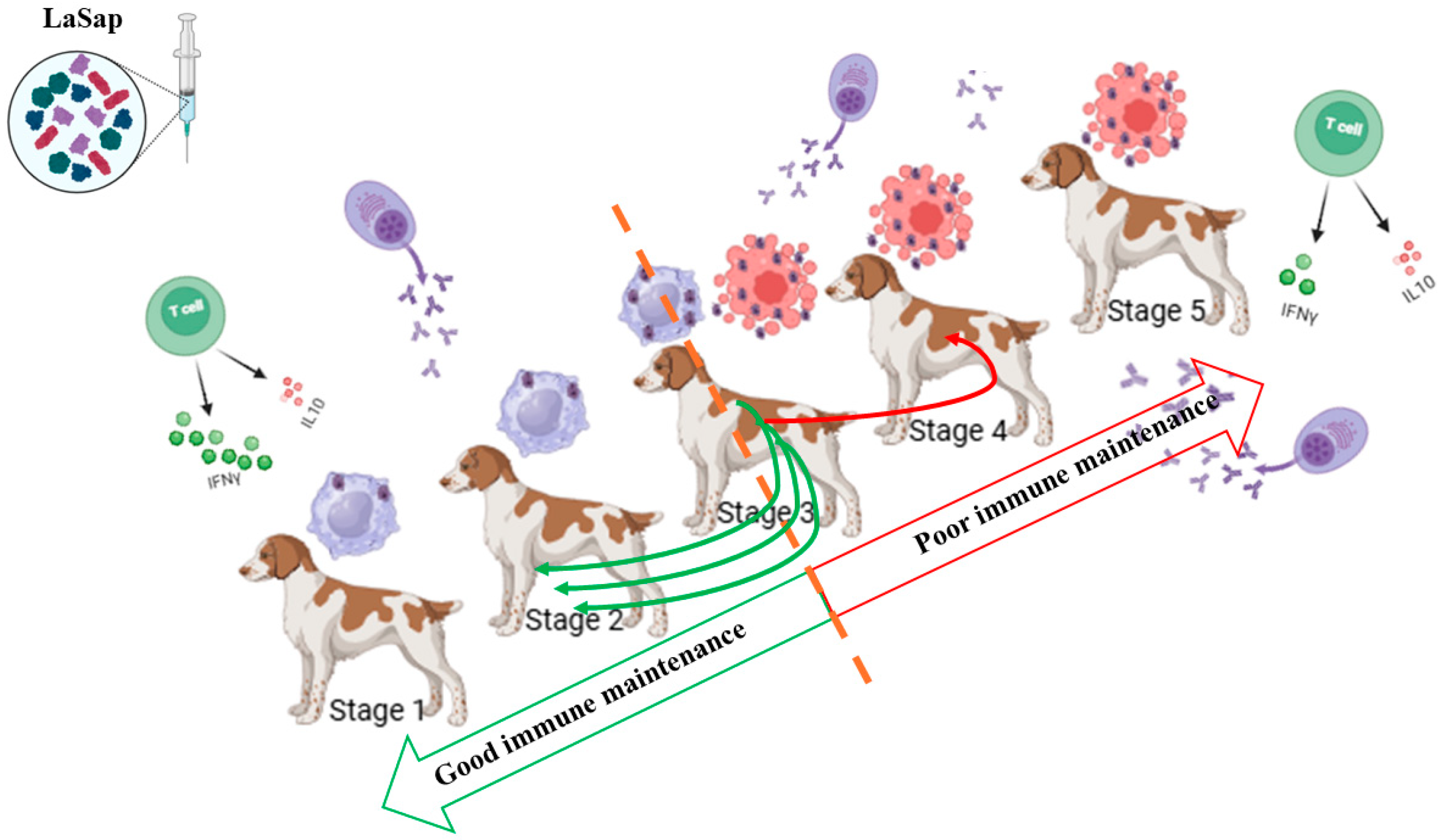
Considering that CVL is a multifaceted and complex disease in which each patient responds very uniquely to the same treatment, it is reasonable to infer that dogs in different stages should receive different immunotherapeutic treatment protocols with LaSap. This was most evident when we found that stage 3 of the disease is the threshold between keeping the patient stable and returning to stage 2 or progressing to stage 4 (Figure 7). Dogs in more critical conditions received an initial protocol of six doses, the first two administered seven days apart, as a way of generating a greater stimulus in a shorter time. Thereafter, the doses were applied at longer intervals (Figure 1). In contrast, dogs in earlier stages can receive fewer doses at the same interval, since they are in better clinical condition. Furthermore, it is clear that dogs in critical conditions, especially those with advanced renal disease, cannot be maintained with immunotherapy alone. However, based on the results of this study, dogs in stages 2 and 3, depending on the response conditions, can be maintained stabilized with LaSap alone for 12 months (Figure 7).

5. Conclusions

In addition to demonstrating safety, the strategy of using different immunotherapeutic protocols with LaSap according to the patient’s clinical staging appears to be appropriate, considering the results obtained in this clinical study.

Author Contributions

Conceptualization, K.F.V.; methodology, K.F.V.; validation, A.V.R., A.B.d.S. and M.C.E.G.; formal analysis, R.C.G.; investigation, A.S.R.C. and A.V.R.; resources, K.F.V.; data curation, M.C.E.G., S.T., F.H.G.d.S. and R.C.G.; writing—original draft preparation, K.F.V.; writing—review and editing, A.V.R., R.C.G., S.T., A.B.d.S. and A.S.R.C.; funding acquisition, K.F.V. All authors have read and agreed to the published version of the manuscript.

Funding

This research was financed with resources from Notice 105/2020/PRPPG, Priority Latin-America and the Caribbean, from UNILA.

Institutional Review Board Statement

The animal study protocol herein was approved by the Unila Animal Use Ethics Committee (CEUA-UNILA), protocol no. 003/2022, on 8 April 2022.

Informed Consent Statement

Not applicable.

Data Availability Statement

The data presented in this study are available on request from the corresponding author.

Acknowledgments

This study was funded by the Araucaria Foundation and the Federal University for Latin-American Integration.

Conflicts of Interest

The author K.F.V. is an equity holder in the company Devax Biotech, which develops personalized therapies. The remaining authors declare no conflicts of interest.

Abbreviations

The following abbreviations are used in this manuscript:
CVLCanine visceral leishmaniasis
LaSapL. amazonensis antigens plus saponin
WHOWorld Health Organization
VLVisceral leishmaniasis
LaL. amazonensis
ALTAlanine aminotransferase
ASTAspartate aminotransferase
PBMCPeripheral blood mononuclear cells
SLiAgMicrograms of soluble L. infantum antigen
SLaAgMicrograms of soluble L amazonensis antigen
DNA polαDNA polymerase

References

  1. World Health Organization. Leishmaniasis 2023. Available online: https://www.who.int/news-room/fact-sheets/detail/leishmaniasis (accessed on 31 January 2025).
  2. Akhoundi, M.; Kuhls, K.; Cannet, A.; Votýpka, J.; Marty, P.; Delaunay, P.; Sereno, D. A Historical Overview of the Classification, Evolution, and Dispersion of Leishmania Parasites and Sandflies. PLoS Neglected Trop. Dis. 2016, 10, e0004349. [Google Scholar] [CrossRef]
  3. Oryan, A.; Akbari, M. Worldwide risk factors in leishmaniasis. Asian Pac. J. Trop. Med. 2016, 9, 925–932. [Google Scholar] [CrossRef]
  4. Nagi, N. Bangladesh eliminates visceral leishmaniasis. Lancet. Microbe 2024, 5, e420. [Google Scholar] [CrossRef]
  5. Postigo, J.A. Leishmaniasis in the World Health Organization Eastern Mediterranean Region. Int. J. Antimicrob. Agents 2010, 36 (Suppl. 1), S62–S65. [Google Scholar] [CrossRef]
  6. Lainson, R.; Rangel, E.F. Lutzomyia longipalpis and the eco-epidemiology of American visceral leishmaniasis, with particular reference to Brazil: A review. Mem. Inst. Oswaldo Cruz. 2005, 100, 811–827. [Google Scholar] [CrossRef] [PubMed]
  7. Laurenti, M.D.; Rossi, C.N.; da Matta, V.L.; Tomokane, T.Y.; Corbett, C.E.; Secundino, N.F.; Pimenta, P.F.; Marcondes, M. Asymptomatic dogs are highly competent to transmit Leishmania (Leishmania) infantum chagasi to the natural vector. Vet. Parasitol. 2013, 196, 296–300. [Google Scholar] [CrossRef] [PubMed]
  8. Grimaldi, G., Jr.; Teva, A.; Dos-Santos, C.B.; Santos, F.N.; Pinto, I.D.; Fux, B.; Leite, G.R.; Falqueto, A. Field trial of efficacy of the Leish-tec® vaccine against canine leishmaniasis caused by Leishmania infantum in an endemic area with high transmission rates. PLoS ONE 2017, 12, e0185438. [Google Scholar] [CrossRef]
  9. de Oliveira, E.F.; dos Santos Fernandes, C.E.; Araújo e Silva, E.; Brazil, R.P.; de Oliveira, A.G. Climatic factors and population density of Lutzomyia longipalpis (Lutz & Neiva, 1912) in an urban endemic area of visceral leishmaniasis in midwest Brazil. J. Vector Ecol. J. Soc. Vector Ecol. 2013, 38, 224–228. [Google Scholar] [CrossRef]
  10. Convit, J.; Ulrich, M.; Fernández, C.T.; Tapia, F.J.; Cáceres-Dittmar, G.; Castés, M.; Rondón, A.J. The clinical and immunological spectrum of American cutaneous leishmaniasis. Trans. R. Soc. Trop. Med. Hyg. 1993, 87, 444–448. [Google Scholar] [CrossRef]
  11. Mayrink, W.; Magalhaes, P.A.; Michalick, M.S.; da Costa, C.A.; Lima, A.d.O.; Melo, M.N.; Toledo, V.P.; Nascimento, E.; Dias, M.; Genaro, O. Immunotherapy as a treatment of American cutaneous leishmaniasis: Preliminary studies in Brazil. Parassitologia 1992, 34, 159–165. [Google Scholar] [CrossRef] [PubMed]
  12. Ferreira, S.deA.; Ituassu, L.T.; de Melo, M.N.; de Andrade, A.S. Evaluation of the conjunctival swab for canine visceral leishmaniasis diagnosis by PCR-hybridization in Minas Gerais State, Brazil. Vet. Parasitol. 2008, 152, 257–263. [Google Scholar] [CrossRef]
  13. Viana, K.F.; Lacerda, G.; Teixeira, N.S.; Rodrigues Cangussu, A.S.; Sousa Aguiar, R.W.; Giunchetti, R.C. Therapeutic vaccine of killed Leishmania amazonensis plus saponin reduced parasite burden in dogs naturally infected with Leishmania infantum. Vet. Parasitol. 2018, 254, 98–104. [Google Scholar] [CrossRef] [PubMed]
  14. Clasta, R.B.; Rivas, A.V.; Souza, A.B.; Dos Santos, A.G.V.; Le Quesne, A.H.M.; Gonçalves, A.A.M.; Cangussu, A.S.R.; Giunchetti, R.C.; Viana, K.F. LaSap vaccine: Immunotherapy and immunochemotherapy associated with allopurinol in dogs naturally infected with Leishmania infantum. Parasite Immunol. 2024, 46, e13028. [Google Scholar] [CrossRef]
  15. Viana, K.F.; Fiuza, J.A.; Gannavaram, S.; Dey, R.; Selvapandiyan, A.; Bartholomeu, D.C.; da Silveira-Lemos, D.; Bueno, L.L.; Dutra, W.O.; Fujiwara, R.T.; et al. Application of rapid in vitro co-culture system of macrophages and T-cell subsets to assess the immunogenicity of dogs vaccinated with live attenuated Leishmania donovani centrin deleted parasites (LdCen-/-). Parasit. Vectors 2016, 9, 250. [Google Scholar] [CrossRef] [PubMed] [PubMed Central]
  16. Reis, A.B.; Martins-Filho, O.A.; Teixeira-Carvalho, A.; Giunchetti, R.C.; Carneiro, C.M.; Mayrink, W.; Tafuri, W.L.; Corrêa-Oliveira, R. Systemic and compartmentalized immune response in canine visceral leishmaniasis. Vet. Immunol. Immunopathol. 2009, 128, 87–95. [Google Scholar] [CrossRef]
  17. Moreno, J. Assessment of Vaccine-Induced Immunity Against Canine Visceral Leishmaniasis. Front. Vet. Sci. 2019, 6, 168. [Google Scholar] [CrossRef] [PubMed]
  18. Costa, C.H. How effective is dog culling in controlling zoonotic visceral leishmaniasis? A critical evaluation of the science, politics and ethics behind this public health policy. Rev. Da Soc. Bras. Med. Trop. 2011, 44, 232–242. [Google Scholar] [CrossRef]
  19. Ready, P.D. Leishmaniasis emergence and climate change. Rev. Sci. Tech. (Int. Off. Epizoot.) 2008, 27, 399–412. [Google Scholar] [CrossRef]
  20. Travi, B.L.; Adler, G.H.; Lozano, M.; Cadena, H.; Montoya-Lerma, J. Impact of habitat degradation on phlebotominae (Diptera: Psychodidae) of tropical dry forests in Northern Colombia. J. Med. Entomol. 2002, 39, 451–456. [Google Scholar] [CrossRef]
  21. Tazerji, S.S.; Nardini, R.; Safdar, M.; Shehata, A.A.; Duarte, P.M. An Overview of Anthropogenic Actions as Drivers for Emerging and Re-Emerging Zoonotic Diseases. Pathogens 2022, 11, 1376. [Google Scholar] [CrossRef] [PubMed]
  22. Noli, C.; Saridomichelakis, M.N. An update on the diagnosis and treatment of canine leishmaniosis caused by Leishmania infantum (syn. L. chagasi). Vet. J. 2014, 202, 425–435. [Google Scholar] [CrossRef]
  23. Morales-Yuste, M.; Martín-Sánchez, J.; Corpas-Lopez, V. Canine Leishmaniasis: Update on Epidemiology, Diagnosis, Treatment, and Prevention. Vet. Sci. 2022, 9, 387. [Google Scholar] [CrossRef] [PubMed]
  24. Gonçalves, A.A.M.; Leite, J.C.; Resende, L.A.; Mariano, R.M.D.S.; Silveira, P.; Melo-Júnior, O.A.O.; Ribeiro, H.S.; de Oliveira, D.S.; Soares, D.F.; Santos, T.A.P.; et al. An Overview of Immunotherapeutic Approaches Against Canine Visceral Leishmaniasis: What Has Been Tested on Dogs and a New Perspective on Improving Treatment Efficacy. Front. Cell. Infect. Microbiol. 2019, 9, 427. [Google Scholar] [CrossRef]
  25. Solano-Gallego, L.; Di Filippo, L.; Ordeix, L.; Planellas, M.; Roura, X.; Altet, L.; Martínez-Orellana, P.; Montserrat, S. Early reduction of Leishmania infantum-specific antibodies and blood parasitemia during treatment in dogs with moderate or severe disease. Parasites Vectors 2016, 9, 235. [Google Scholar] [CrossRef] [PubMed]
  26. Ribeiro, V.M.; da Silva, S.M.; Menz, I.; Tabanez, P.; Nogueira, F.d.S.; Werkhaüser, M.; da Fonseca, A.L.; Dantas-Torres, F. Brasileish—A Study Group about Animal Leishmaniasis Control of visceral leishmaniasis in Brazil: Recommendations from Brasileish. Parasites Vectors 2013, 6, 8. [Google Scholar] [CrossRef]
  27. Sun, H.X.; Xie, Y.; Ye, Y.P. Advances in saponin-based adjuvants. Vaccine 2009, 27, 1787–1796. [Google Scholar] [CrossRef]
  28. Araújo Verçosa, B.L.; Muniz-Junqueira, M.I.; Menezes-Souza, D.; Mourão Dias Magalhães, L.; Fujiwara, R.T.; Melo, M.N.; Vasconcelos, A.C. Enhanced apoptotic index, chemokines and inflammatory recruitment in renal tissues shows relationship with the clinical signs in Leishmania-infected dogs. Vet. Parasitol. 2021, 300, 109611. [Google Scholar] [CrossRef]
  29. Alves, A.F.; Pereira, R.A.; Rodrigues, M.A.; Campos, L.S.; do Carmo, D.D.; de Abreu Teles, P.P.; Andrade, H.M.; de Araújo, S.A.; Gomes, D.A.; Tafuri, W.L. Leishmania (L.) infantum BH401 strain induces classic renal lesions in dogs: Histological and confocal microscopy study. Exp. Parasitol. 2022, 242, 108342. [Google Scholar] [CrossRef] [PubMed]
  30. Roura, X.; Cortadellas, O.; Day, M.J.; Benali, S.L.; Zatelli, A.; Canine Leishmaniosis Working Group. Canine leishmaniosis and kidney disease: Q&A for an overall management in clinical practice. J. Small Anim. Pract. 2021, 62, 3. [Google Scholar] [CrossRef]
  31. Vasconcelos, T.C.; Bruno, S.F.; de Miranda, L.H.M.; Conceição-Silva, F.; Belo, V.S.; Figueiredo, F.B. Parasite load, iNOS and cytokine profiles, and histopathological aspects of Leishmania infantum infection in dogs with different clinical presentations. Scielo Braz. 2019, 49, e20180984. [Google Scholar] [CrossRef]
  32. Batista, L.F.S.; Torrecilha, R.B.P.; Silva, R.B.; Utsunomiya, Y.T.; Silva, T.B.F.; Tomokane, T.Y.; Pacheco, A.D.; Bosco, A.M.; Paulan, S.C.; Rossi, C.N.; et al. Chromosomal segments may explain the antibody response cooperation for canine leishmaniasis pathogenesis. Vet. Parasitol. 2020, 288, 109276. [Google Scholar] [CrossRef]
  33. Boggiatto, P.M.; Ramer-Tait, A.E.; Metz, K.; Kramer, E.E.; Gibson-Corley, K.; Mullin, K.; Hostetter, J.M.; Gallup, J.M.; Jones, D.E.; Petersen, C.A. Immunologic indicators of clinical progression during canine Leishmania infantum infection. Clin. Vaccine Immunol. CVI 2010, 17, 267–273. [Google Scholar] [CrossRef]
  34. Proverbio, D.; Spada, E.; Bagnagatti de Giorgi, G.; Perego, R.; Valena, E. Relationship between Leishmania IFAT titer and clinicopathological manifestations (clinical score) in dogs. BioMed Res. Int. 2014, 2014, 412808. [Google Scholar] [CrossRef] [PubMed]
  35. Solano-Gallego, L.; Miró, G.; Koutinas, A.; Cardoso, L.; Pennisi, M.G.; Ferrer, L.; Bourdeau, P.; Oliva, G.; Baneth, G.; The LeishVet Group. LeishVet guidelines for the practical management of canine leishmaniosis. Parasites Vectors 2011, 4, 86. [Google Scholar] [CrossRef] [PubMed]
  36. Reis, A.B.; Giunchetti, R.C.; Carrillo, E.; Martins-Filho, O.A.; Moreno, J. Immunity to Leishmania and the rational search for vaccines against canine leishmaniasis. Trends Parasitol. 2010, 26, 341–349. [Google Scholar] [CrossRef]
  37. Toepp, A.; Larson, M.; Wilson, G.; Grinnage-Pulley, T.; Bennett, C.; Leal-Lima, A.; Anderson, B.; Parrish, M.; Anderson, M.; Fowler, H.; et al. Randomized, controlled, double-blinded field trial to assess Leishmania vaccine effectiveness as immunotherapy for canine leishmaniosis. Vaccine 2018, 36, 6433–6441. [Google Scholar] [CrossRef]
  38. Roatt, B.M.; Aguiar-Soares, R.D.; Reis, L.E.; Cardoso, J.M.; Mathias, F.A.; de Brito, R.C.; da Silva, S.M.; Gontijo, N.F.; Ferreira, S.A.; Valenzuela, J.G.; et al. A Vaccine Therapy for Canine Visceral Leishmaniasis Promoted Significant Improvement of Clinical and Immune Status with Reduction in Parasite Burden. Front. Immunol. 2017, 8, 217. [Google Scholar] [CrossRef]
  39. Miró, G.; Segarra, S.; Cerón, J.J.; Ferrer, L.; Solano-Gallego, L.; Montell, L.; Costa, E.; Teichenne, J.; Mariné-Casadó, R.; GALILEI trial Group; et al. New immunomodulatory treatment protocol for canine leishmaniosis reduces parasitemia and proteinuria. PLoS Neglected Trop. Dis. 2024, 18, e0012712. [Google Scholar] [CrossRef]
  40. Barbiéri, C.L. Immunology of canine leishmaniasis. Parasite Immunol. 2006, 28, 329–337. [Google Scholar] [CrossRef]
  41. Carrillo, E.; Moreno, J. Cytokine profiles in canine visceral leishmaniasis. Vet. Immunol. Immunopathol. 2009, 128, 67–70. [Google Scholar] [CrossRef]
  42. Abbehusen, M.M.C.; Almeida, V.D.A.; Solcà, M.D.S.; Pereira, L.D.S.; Costa, D.J.; Gil-Santana, L.; Bozza, P.T.; Fraga, D.B.M.; Veras, P.S.T.; Dos-Santos, W.L.C.; et al. Clinical and immunopathological findings during long term follow-up in Leishmania infantum experimentally infected dogs. Sci. Rep. 2017, 7, 15914. [Google Scholar] [CrossRef] [PubMed]
  43. Samant, M.; Sahu, U.; Pandey, S.C.; Khare, P. Role of Cytokines in Experimental and Human Visceral Leishmaniasis. Front. Cell. Infect. Microbiol. 2021, 11, 624009. [Google Scholar] [CrossRef] [PubMed]
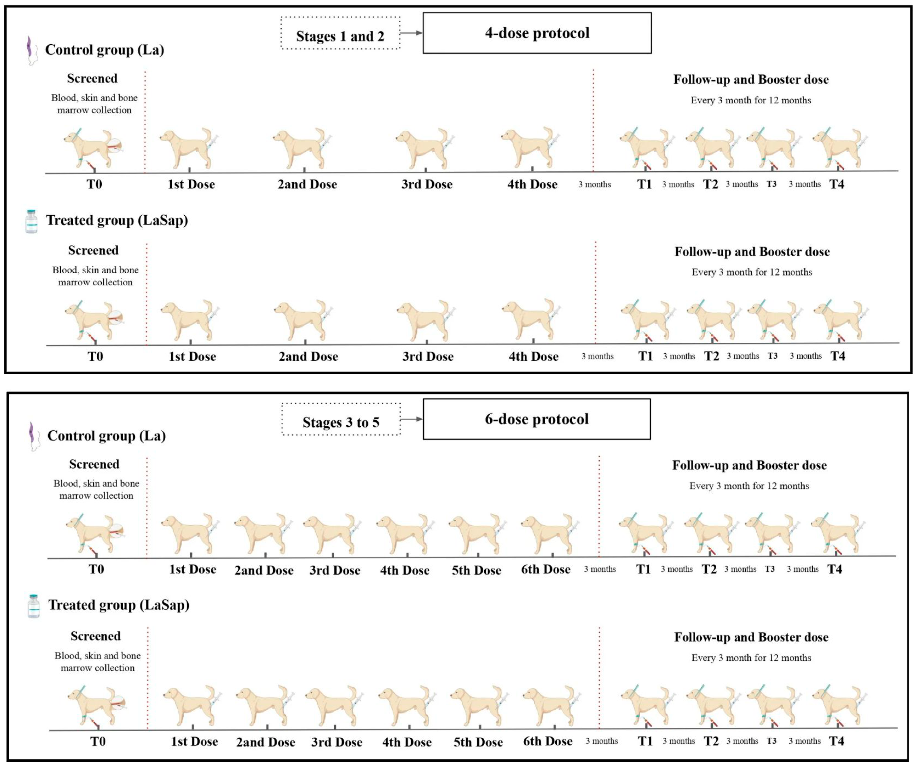
Figure 1. Experimental design using LaSap: Dogs in stages 1 and 2 received four doses at 21-day intervals via the subcutaneous route. Dogs in stages 3 to 5 received two initial doses at 7-day intervals; the third dose 15 days after the second dose; and the last three doses at 30-day intervals each. After this, all dogs received a booster dose every three months for one year. Time zero was 15 days before starting the experiment.
Vaccines 13 00933 g001
Figure 2. Enzyme-linked immunosorbent assay (ELISA) using recombinant HSP70 protein with sera from dogs naturally infected with L. infantum and treated with LaSap for 12 months after the initial protocol. (A) indicates the results from dogs in stages 1 and 2. (B) indicates the results from dogs in stages 3 to 5. The dashed lines indicate the cut-off; the connecting lines indicate statistical significance (p < 0.05).
Vaccines 13 00933 g002
Figure 3. Analyses of parasite loads on the skin of 48 dogs naturally infected with L. infantum and treated with LaSap. (A) indicates the results in patients in stages 1 and 2. (B) indicates the results in patients in stages 3 to 5. The connecting lines indicate significant results (p < 0.05).
Vaccines 13 00933 g003
Figure 4. Analyses of parasite loads on the bone marrow of 48 dogs naturally infected with L. infantum and treated with LaSap. (A) indicates the results in patients in stages 1 and 2. (B) indicates the results in patients in stages 3 to 5. The connecting lines indicate significant results (p < 0.05).
Vaccines 13 00933 g004
Figure 5. Analyses of IFN-γ and IL-10 cytokine levels from PBMC cultures of 24 dogs naturally infected with L. infantum and treated with LaSap over 12 months after the initial four-dose protocol (stages 1 and 2). Control culture Vaccines 13 00933 i001, culture stimulated with LPS Vaccines 13 00933 i002, SLaAg Vaccines 13 00933 i003 and SLiAg Vaccines 13 00933 i004. * and connecting lines indicate statistical differences within and outside the groups.
Vaccines 13 00933 g005
Figure 6. Analyses of IFN-γ and IL-10 cytokine levels from PBMC cultures of 24 dogs naturally infected with L. infantum and treated with LaSap over 12 months after the initial six-dose protocol (stages 3 to 5). Control culture Vaccines 13 00933 i001, culture stimulated with LPS Vaccines 13 00933 i002, SLaAg Vaccines 13 00933 i003 and SLiAg Vaccines 13 00933 i004. * and connecting lines indicate statistical differences within and outside the groups.
Vaccines 13 00933 g006
Figure 7. Overview of dogs in different clinical stages of visceral leishmaniasis (Brasileish and Leishvet) treated with LaSap. Dogs in stages 1 and 2 present better results with monotherapy, including clinical maintenance of the patient, with lower production of IgG antibodies, greater cellular response, and more incisive reduction of the parasite load (skin and bone marrow). On the other hand, dogs in more advanced stages, especially those in stage 4, present a poor response profile using LaSap alone. Dogs in stage 3 play a key role in the progression to level 4 or regression to level 2 (dashed orange line), and this seems to be more related to the individual immune response profile of the patient. However, most dogs in this stage can return to stage 2 if there is clinical monitoring by the veterinarian, especially with monitoring of the renal profile.
Vaccines 13 00933 g007
Table 1. Relationship between dogs naturally infected with L. infantum (stages 1 and 2) and treated with LaSap, with clinical signs present at different analyses times over 12 months after the initial 4-dose protocol. Time zero (before starting the protocol), T1 (3 months after the protocol), T2 (6 months after the protocol), T3 (9 months after the protocol), and T4 (12 months after the protocol).
Clinical Signs
Staging 1 and 2
0 Time3 Months6 Months9 Months12 Months
LaLaSapLaLaSapLaLaSapLaLaSapLaLaSap
Alopecia/exfoliative dermatitis10/129/1200000000
Lymphadenopathy8/127/1200000000
Lethargy
Cachexia
Epistaxis
Vasculitis
Arthritis
Table 2. Relationship between dogs naturally infected with L. infantum (stages 3 to 5) and treated with LaSap, with clinical signs present at different analyses times over 12 months after the initial six-dose protocol. Time zero (before starting the protocol), T1 (3 months after the protocol), T2 (6 months after the protocol), T3 (9 months after the protocol) and T4 (12 months after the protocol).
Clinical Signs
Staging 3–5
0 Time3 Months6 Months9 Months12 Months
LaLaSapLaLaSapLaLaSapLaLaSapLaLaSap
Alopecia/exfoliative dermatitis8/1210/120/120/122/1204/121/125/121/12
Lymphadenopathy10/1212/124/120/124/121/125/121/125/122/12
Lethargy4/125/120/120/121/120/121/120/122/121/12
Cachexia3/124/120/120/121/120/121/120/121/120/12
Epistaxis2/122/120/120/121/120/122/120/122/122/12
Vasculitis6/126/123/120/123/120/124/121/125/122/12
Arthritis1/122/120/120/121/120/121/121/121/121/12
Table 3. Hematological and biochemical renal and hepatic results of the La and LaSap groups in stages 1 and 2 of CVL at different analysis times over 12 months after the initial four-dose protocol. Time zero (before starting the protocol), T1 (3 months after the protocol), T2 (6 months after the protocol), T3 (9 months after the protocol) and T4 (12 months after the protocol).
Clinical Signs
Staging 1–2
0 Time3 Months6 Months9 Months12 MonthsReference
LaLaSapLaLaSapLaLaSapLaLaSapLaLaSap
Total leukocytes12,401806812,8918914881910,46310,25010,604823492146000–17,000 mm3
Rods00000000000–300
Segmented78754228789354954704558049845833468050223000–11,500
Lymphocytes35702579389128583072390039213682295432401000–4800
Monocytes7673178663718008431024810498672150–1350
Eosinophils222186239188243140321279102280100–1250
Platelets346,000315,000345,000329,000322,000354,000319,000377,000320,000350,000200,000–500,000 mm3
AST4552374058443931644810–88 U/L
ALT5042332857396146523710–88 U/L
Urea2237292338423039433820–50 mg/dL
Creatinine0.91.01.21.10.891.01.11.21.00.80.5–1.4 mg/dL
Table 4. Hematological and biochemical renal and hepatic results of the La and LaSap groups in stages 3 to 5 of CVL at different analyses times over 12 months after the initial six-dose protocol. Time zero (before starting the protocol), T1 (3 months after the protocol), T2 (6 months after the protocol), T3 (9 months after the protocol) and T4 (12 months after the protocol).
Clinical Signs
Staging 3–5
0 Time3 Months6 Months9 Months12 MonthsReference
LaLaSapLaLaSapLaLaSapLaLaSapLaLaSap
Total leukocytes74207968937010,31172509230782110,590629487606000–17,000 mm3
Rods00000000000–300
Segmented48235557646573204640639848497200440556063000–11,500
Lymphocytes19461788201024101620189019722287120024681000–4800
Monocytes319388621300660785840847422398150–1350
Eosinophils332238274281330157160256267288100–1250
Platelets240,000276,000350,000392,000228,000312,000196,000260,000182,000220,000200,000–500,000 mm3
AST6574616569577660856310–88 U/L
ALT7269595666557567736510–88 U/L
Urea5052363840354744555020–50 mg/dL
Creatinine1.61.71.31.11.41.21.41.11.51.40.5–1.4 mg/dL
Disclaimer/Publisher’s Note: The statements, opinions and data contained in all publications are solely those of the individual author(s) and contributor(s) and not of MDPI and/or the editor(s). MDPI and/or the editor(s) disclaim responsibility for any injury to people or property resulting from any ideas, methods, instructions or products referred to in the content.