Review

Stopping Tuberculosis at the Gate: The Role of M. tuberculosis Adhesins in Infection and Intervention

1
Research and Development Centre, Beijing Zhifei Lvzhu Biopharmaceutical Co., Ltd., Beijing 100176, China
2
Beijing Bacterial Vaccine Engineering Research Centre, Beijing 100176, China
3
School of Life Science and Biopharmaceutics, Shenyang Pharmaceutical University, Shenyang 110016, China
*
Authors to whom correspondence should be addressed.
These authors contributed equally to this work.
Vaccines 2025, 13(7), 676; https://doi.org/10.3390/vaccines13070676
Submission received: 19 May 2025 / Revised: 21 June 2025 / Accepted: 23 June 2025 / Published: 24 June 2025

Abstract

The global burden of tuberculosis (TB), exacerbated by the rise of drug-resistant Mycobacterium tuberculosis (M. tuberculosis), underscores the need for alternative intervention strategies. One promising approach is to block the infection at its earliest stage—bacterial adhesion to host cells—thereby preventing colonization and transmission without exerting selective pressure. Adhesins, surface-exposed molecules mediating this critical interaction, have therefore emerged as attractive targets for early prevention. This review outlines the infection process driven by bacterial adhesion and describes the architecture of the M. tuberculosis outer envelope, emphasizing components that contribute to host interaction. We comprehensively summarize both non-protein and protein adhesins, detailing their host receptors, biological roles, and experimental evidence. Recent progress in the computational prediction of adhesins, particularly neural network-based tools like SPAAN, is also discussed, highlighting its potential to accelerate adhesin discovery. Additionally, we present a detailed, generalized workflow for predicting M. tuberculosis adhesins, which synthesizes current approaches and provides a comprehensive framework for future studies. Targeting bacterial adhesion presents a therapeutic strategy that interferes with the early stages of infection while minimizing the risk of developing drug resistance. Consequently, anti-adhesion strategies may serve as valuable complements to conventional therapies and support the development of next-generation TB vaccines and treatments.

1. Introduction

TB remains one of the most significant infectious diseases worldwide. In 2023, TB was expected to once again become the leading cause of death from a single infectious agent, after being temporarily overtaken by COVID-19. Over 10 million people contract TB annually, and the rise of drug-resistant Mycobacterium tuberculosis strains complicates treatment efforts. To eliminate TB by 2030, the United Nations (UN) and the World Health Organization (WHO) have endorsed a global strategy that requires improved screening, treatment, and novel therapeutic approaches [1,2].
One promising strategy focuses on the initial stages of infection, particularly the adhesion of M. tuberculosis to host cells. Adhesion plays a key role in pathogenesis. It allows bacteria to colonize host tissues, form biofilms, and resist stress [3,4,5,6]. M. tuberculosis adhesins bind to specific host receptors. This promotes bacterial attachment and colonization, both of which are critical for infection persistence [7,8]. The surface properties of the bacterium, such as surface free energy, charge, and hydrophobicity, also influence these interactions [9].
Targeting bacterial adhesion offers a novel approach for TB treatment. Anti-adhesion therapies can prevent bacterial attachment, reducing colonization, enhancing treatment effectiveness, and minimizing drug resistance [10,11]. For instance, in other pathogens such as Helicobacter pylori and uropathogenic Escherichia coli (UPEC), anti-adhesion agents like bovine lactoferrin (BLF) and FimH inhibitors have shown promise in improving infection outcomes [12,13,14,15,16]. These findings suggest that similar strategies could be effective in combating M. tuberculosis. Supporting this, serum from tuberculosis patients containing IgM antibodies against the adhesin HBHA has been shown to neutralize the bacterium’s entry into epithelial cells, implying that adhesin-specific immunity may offer protection against extrapulmonary dissemination. This evidence highlights the therapeutic potential of targeting adhesins, even in intracellular pathogens such as M. tuberculosis [17].
Despite advances in understanding M. tuberculosis adhesins, significant gaps remain. This review focuses on the mechanisms by which M. tuberculosis adheres to host cells, the role of important adhesins in infection, and their protective effects. While the prediction of adhesins in M. tuberculosis is still an emerging area of research, we will briefly outline the principles behind current prediction methods and highlight relevant findings from existing studies. By consolidating this knowledge, we aim to support the development of targeted therapies that could improve the treatment and prevention of TB.

2. Spatiotemporal Dynamics of Mycobacterial Adhesion During Infection

The adhesion of M. tuberculosis plays a critical role throughout its lifecycle, beginning with the inhalation of aerosol droplets containing the bacteria, which are expelled through the cough of an infected individual. Due to their small size, these droplets bypass the upper respiratory tract’s microbial flora and immune defenses, allowing M. tuberculosis to reach the alveolar spaces of the lungs [18,19]. Once in the alveoli, the bacteria are engulfed by alveolar macrophages (AM), initiating a complex immune response that leads to the formation of granulomas, structures that contain the bacteria in a latent state [20].
The initial adhesion of M. tuberculosis is mediated by surface-exposed lipids and glycoconjugates that interact with specific receptors on host cell surfaces, primarily on macrophages and alveolar epithelial cells (AECs). This process facilitates the bacteria’s attachment and entry into the host cells [21,22]. However, more recent studies have shown that M. tuberculosis may also utilize specialized adhesins for tissue dissemination. For example, Mycobacterium bovis Bacillus Calmette-Guérin (M. bovis BCG), as well as M. tuberculosis, uses heparin-binding hemagglutinin adhesin (HBHA), which binds to heparan sulfate-containing receptors on epithelial cells, aiding in the bacteria’s spread beyond the lungs [23]. This suggests that adhesins may help M. tuberculosis cross the alveolar epithelium, a process that may also occur via the transmigration of infected macrophages [24].
After the bacteria adhere to macrophages and AECs, the immune response can lead to the formation of granulomas, which contain the bacteria in a latent phase. In immunocompromised individuals, or children, the bacteria can undergo intense intracellular multiplication, which may lead to the breakdown of granulomas or the reactivation of latent infections. This can result in the dissemination of bacteria to distant sites, including the nervous system, bones, genitourinary system, and skin [25].
During hematogenous spread, M. tuberculosis likely adheres to extracellular matrix (ECM) components to overcome mechanical shear forces and promote colonization in new tissues. For example, during hematogenous dissemination, the bacteria may bind plasma fibronectin and subsequently ECM proteins to invade new tissues (Figure 1) [26].
Thus, the adhesins of M. tuberculosis play an essential role not only in the initial stages of infection in the lungs but also in the subsequent spread and persistence of infection in distant organs. These adhesins enable the bacteria to cross epithelial barriers, invade new tissues, and evade immune clearance, making them key determinants of pathogenicity and transmission. Furthermore, adhesion is also a prerequisite for the formation of biofilms, which have been observed within granulomatous lesions. In this context, adhesins contribute to the establishment and stability of biofilm-like structures, which provide a protective microenvironment that enhances bacterial survival, limits antibiotic penetration, and promotes persistence during chronic infection [5]. This link between adhesion and biofilm formation underscores the broader role of adhesins in M. tuberculosis pathogenesis and highlights their potential as targets for disrupting both colonization and long-term persistence.

3. The M. tuberculosis Capsule: A Reservoir for Adhesins

The bacterial cell envelope provides mechanical rigidity, facilitates solute and protein transport, and protects against environmental stresses. It consists of several layers, including the cytoplasmic membrane, cell wall, surface lipids, and capsule (Figure 2). The capsule, which is the outermost structure, contains key components such as lipids, glycoconjugates, and proteins. Many of these components function as adhesins, playing a crucial role in the bacterium’s ability to adhere to host receptors and establish infection [27].

3.1. Cytoplasmic Membrane

The mycobacterial cytoplasmic membranes contain glycerophospholipids, along with phosphatidylinositol mannosides (PIMs), lipomannans (LMs), and lipoarabinomannans (LAMs), which are derivatives of PIMs. While primarily contributing to cell envelope permeability and antibiotic resistance, PIMs, LMs, and LAMs also play a role in adhesion [28]. These molecules interact with host cells and influence immune responses, potentially affecting bacterial attachment and immune evasion [29].

3.2. Peptidoglycan (PG)

PG is a fundamental component of the mycobacterial cell wall, providing structural integrity and resistance to osmotic pressure. It consists of linear chains of alternating N-acetylmuramic acid and N-acetylglucosamine (GlcNAc), which are crosslinked by peptides to form a layered, honeycomb-like structure [30]. Beyond its structural role, PG is actively involved in host–pathogen interactions. Mycobacteria produce a unique form of peptidoglycan-derived muramyl dipeptide (MDP), known as N-glycolyl MDP, which enhances immune recognition by activating the pattern recognition receptor NOD2 [31]. Additionally, PG levels remain stable across different growth phases, suggesting that it may contribute to bacterial persistence within the host [32]. By engaging immune receptors and influencing granuloma formation, PG may play a role in bacterial adhesion and long-term survival during infection.

3.3. Arabinogalactan (AG)

The middle layer of the mycobacterial cell wall core consists of arabinogalactan sugars, which are linked to peptidoglycan via a rhamnose-GlcNAc disaccharide [32]. Beyond its structural role, AG has been identified as a virulence factor in M. tuberculosis, contributing to infection severity. Recent findings indicate that AG interacts with host receptors, including Galectin-9, a β-galactoside-binding protein, which activates the TAK1-ERK-MMP signaling pathway and leads to pathological changes in the lung [33,34]. This interaction suggests that AG may also play a role in bacterial adhesion, potentially influencing host–pathogen interactions during infection.

3.4. Mycolic Acids (MAs), Trehalose Monomycolate (TMM), and Trehalose 6,6′-Dimycolate (TDM)

Mycolic acids (MAs) are long-chain fatty acids (C60-C90) that form a major component of the mycobacterial cell envelope. They serve as the structural backbone of the mycomembrane by covalently linking to the arabinogalactan–peptidoglycan (AG-PG) matrix. MAs also exist on the bacterial surface in free or glycosylated forms, such as TMM and TDM [35]. Beyond their structural role, MAs and their derivatives may contribute to mycobacterial adhesion. As key components of the outermost layer of the bacterial envelope, they provide a direct interface with the host. While the precise mechanisms remain unclear, MAs have been implicated in interactions with host immune receptors such as Mincle and TREM2, which may modulate immune recognition and bacterial attachment [36]. Given their surface localization and involvement in host–pathogen interactions, MAs and their derivatives could play a role in facilitating bacterial colonization and dissemination during infection.

3.5. Phthiocerol Dimycocerosate (PDIM)

PDIM is a key surface lipid of M. tuberculosis, playing an essential role in both immune evasion and bacterial adhesion. PDIM helps the bacteria evade the innate immune system by masking pathogen-associated molecular patterns (PAMPs) on the cell wall, thus preventing the activation of immune responses. This immune evasion enables the recruitment of permissive monocytes, which are more favorable for bacterial survival, rather than microbicidal macrophages [37]. In addition to immune modulation, PDIM may also facilitate adhesion. Its methyl-branched fatty acids increase lipid mobility, potentially enhancing bacterial attachment to host cells such as epithelial cells and macrophages. While PDIM primarily functions to hide bacterial PAMPs, its role in promoting initial bacterial attachment suggests a dual function in immune evasion and colonization [37,38].

4. M. tuberculosis Adhesins

Adhesins play a crucial role in M. tuberculosis attachment to and entry into host cells. They can be broadly classified into non-protein and protein adhesins, both of which interact with host receptors to mediate adhesion. M. tuberculosis adhesion is a multifactorial process that involves the simultaneous action of multiple adhesins, rather than a single dominant factor. Major surface adhesins of M. tuberculosis and their host receptors are listed in Table 1. The following sections provide a detailed overview of these adhesins and their corresponding host receptors.

4.1. Non-Protein Adhesins in the Mycobacterial Cell Wall and Their Host Interactions

4.1.1. Mannose-Capped Lipoarabinomannan (ManLAM) and PIMs

ManLAM and PIMs, two key mannose-capped components of the M. tuberculosis cell wall, act as adhesins by binding to the dendritic cell-specific ICAM-3-grabbing non-integrin (DC-SIGN) and Dectin-2 receptors on immature dendritic cells and macrophages [39,41,42]. This interaction has been linked to both immune activation and immune evasion. On the one hand, the binding of manLAM to Dectin-2 on dendritic cells promotes the activation of T cell responses and enhances immunity, as seen in certain experimental models [42]. On the other hand, this interaction can also impair dendritic cell maturation and inhibit the immune response, contributing to the evasion of protective immunity against tuberculosis. Additionally, manLAM and higher-order PIMs prevent the fusion of phagosomes with lysosomes in macrophages, helping the bacteria avoid degradation [81]. However, studies on mycobacterial strains lacking manLAM or PIMs suggest that the manLAM/PIM-DC-SIGN interaction is not essential for cytokine release in vitro or for protective immunity in vivo, indicating that these adhesins might be more important for bacterial survival and immune modulation rather than directly triggering immune responses [41,82].

4.1.2. MAs

MAs, which are α-alkyl-β-hydroxy fatty acids, are the major lipid components of the M. tuberculosis cell wall. These hydrophobic molecules contribute significantly to the bacterial resistance to drugs and oxidative stress by forming a protective barrier. In addition to their structural role, MAs influence host–pathogen interactions. Experimental studies show that MAs can directly interact with macrophages, affecting both cell survival and immune response [83]. Specifically, MAs at lower concentrations promote macrophage survival, while higher concentrations induce apoptosis by triggering the release of pro-apoptotic cytokines [83]. This suggests that MAs not only modulate immune responses but also interact with macrophages to influence their behavior.
Furthermore, MAs interact with immune molecules such as galectin-3 and Clec12A [45,46]. Galectin-3, a lectin involved in immune recognition, specifically binds to the mycolic acid components of the M. tuberculosis cell wall, which may facilitate bacterial adhesion and immune evasion [45]. Mycobacterial MAs also bind to the inhibitory C-type lectin receptor Clec12A, found on immune cells. This interaction suppresses immune responses, as evidenced by enhanced immune reactions in Clec12A-deficient mice after infection, and increased susceptibility in human Clec12A transgenic mice [46]. These findings underscore the diverse role of MAs in modulating the host immune response and suggest that targeting these interactions could offer new therapeutic approaches for combating mycobacterial infections.

4.1.3. TDM

TDM, a key glycolipid in the M. tuberculosis cell wall, is an important adhesin in infection. It is recognized by several pattern recognition receptors (PRRs), including macrophage-inducible C-type lectin (Mincle), the macrophage receptor with a collagenous structure (MARCO), and Dectin-3. Mincle, a C-type lectin receptor, is essential for TDM-induced macrophage activation, promoting the release of pro-inflammatory cytokines and nitric oxide. In Mincle-deficient macrophages, these immune responses are completely suppressed, highlighting Mincle’s crucial role in TDM recognition. TDM also induces granuloma formation in vivo, a hallmark of mycobacterial infection, which is absent in mincle-deficient mice [47,48].
Dectin-3 also recognizes TDM and promotes Mincle expression, amplifying the immune response. In Dectin-3-deficient macrophages, this activation is significantly impaired, emphasizing Dectin-3’s importance in the TDM-induced immune response [49]. Additionally, MARCO, a class A scavenger receptor, binds TDM and activates the TLR2 signaling pathway, enhancing cytokine production. MARCO-deficient macrophages show a reduced immune response to TDM [50,51].
Together, these findings demonstrate that TDM, through its interactions with multiple receptors, plays a vital role in both the bacterial adhesion and modulation of the host immune system. These interactions not only facilitate M. tuberculosis colonization but also shape the host immune response, highlighting TDM as a key player in tuberculosis pathogenesis.

4.1.4. α-Glucan

M. tuberculosis α-glucan functions as an adhesin and a novel ligand for the C-type lectin receptor DC-SIGN, with binding mediated by internal glucosyl residues. This interaction induces IL-10 production in LPS-activated monocyte-derived dendritic cells (DCs), contributing to immune suppression [52]. Additionally, α-glucan interferes with monocyte differentiation, preventing their maturation into fully competent DCs and promoting the development of CD1-negative dendritic cells (CD1⁻ DCs). By blocking class I CD1 molecule expression, α-glucan disrupts CD1-mediated antigen presentation, which is crucial for the activation of CD1-restricted lipid-specific T cells [53]. Since M. tuberculosis has a high content of antigenic lipids, this immune evasion mechanism may impair the host’s ability to mount an effective cell-mediated immune response. Notably, CD1-restricted T cell responses are significantly reduced in patients with active pulmonary tuberculosis, further highlighting the role of α-glucan-DC-SIGN interaction in M. tuberculosis pathogenesis [84].

4.2. Protein Adhesins in the Mycobacterial Cell Wall and Their Host Interactions

4.2.1. Early Secretory Antigenic Target 6 kDa (ESAT-6) and Culture Filtrate Protein 10 kDa (CFP-10)

Although ESAT-6 and CFP-10 are classically characterized as secreted virulence factors, increasing evidence supports their additional role as non-canonical adhesins that mediate direct interaction with host cells [55,75]. ESAT-6 and CFP-10 form a stable 1:1 complex and are coordinately regulated during M. tuberculosis infection [54]. CFP-10 likely functions as a chaperone, stabilizing ESAT-6, which is implicated in disrupting phagosomal membrane integrity through an incompletely understood mechanism [85]. Beyond their roles in membrane disruption, ESAT-6 and CFP-10 also act as adhesins, facilitating bacterial invasion and dissemination within the host [86].
During infection, ESAT-6 is recognized as a PAMP that stimulates Toll-like receptors (TLRs), triggering innate immune responses [87]. In pneumocytes, surface-expressed ESAT-6 mediates bacterial adhesion to the basolateral laminin-expressing surface, contributing to host cell damage and basement membrane degradation. This interaction promotes bacterial dissemination through the alveolar wall [55]. Renshaw et al. demonstrated that the ESAT-6-CFP-10 complex directly mediates macrophage binding, enhancing bacterial uptake and survival [54]. Furthermore, studies in embryonic zebrafish suggest that ESAT-6 enhances the trafficking of infected macrophages within granulomas, facilitating early bacterial dissemination [86].
In vitro studies using an alveolar barrier model, composed of human alveolar epithelial and endothelial cells cultured on opposite sides of a basement membrane, revealed that strains lacking ESAT-6 exhibit reduced migration across this barrier [88]. These findings collectively support a model in which ESAT-6 and CFP-10 contribute to adhesion-mediated steps of infection, consistent with their inclusion in the current review as functionally relevant adhesins, despite their classification as secreted proteins.

4.2.2. Antigen 85 (Ag85) Complex

The Ag85 complex in M. tuberculosis comprises three paralogous proteins—Ag85A, Ag85B, and Ag85C—which are secreted in large amounts and elicit robust humoral and cell-mediated immune responses [89]. Interestingly, despite their high secretion levels, a significant portion of these proteins remains associated with the bacterial surface, suggesting a potential role in host–pathogen interactions [90]. All three Ag85 proteins have been shown to bind fibronectin (Fn), a key extracellular matrix protein, highlighting their importance in mediating bacterial adhesion—a critical step in host invasion [56].
The functional relevance of the Ag85-Fn interaction has been demonstrated through the siRNA-mediated depletion of Fn, which significantly reduces the binding of purified Ag85B to human epithelial cells [91]. This finding is further supported by the identification of Ag85-Fn complexes in clinical samples from tuberculosis patients, underscoring the biological significance of this interaction in infection [92]. Notably, the specific amino acid residues that are critical for Fn binding have been mapped to the sequence 98FEWYYQSGLSV108 in Ag85B from Mycobacterium kansasii. This sequence appears to be unique among known prokaryotic and eukaryotic Fn-binding proteins, suggesting a distinct mechanism of interaction [93]. These findings highlight Ag85 as a key adhesin facilitating M. tuberculosis attachment to host tissues, contributing to its pathogenicity.

4.2.3. HBHA

M. tuberculosis adheres to, invades, and proliferates in both professional phagocytes and epithelial cells. Among its adhesins, HBHA plays a crucial role in mediating adherence to epithelial cells and facilitating extrapulmonary dissemination [58]. HBHA specifically binds to sulfated glycoconjugates on epithelial cells, and the disruption of its synthesis significantly impairs bacterial dissemination from the lungs to other organs, such as the spleen [86].
Experimental studies have demonstrated the mechanistic role of HBHA in receptor-mediated endocytosis. Franco D. Menozzi and colleagues showed that HBHA, when conjugated to colloidal gold particles, rapidly attaches to the membranes of human laryngeal epithelial cells and type II pneumocytes. This process is dependent on the heparin-binding domain of HBHA, as gold particles coated with a truncated HBHA lacking this domain fail to bind. Furthermore, pretreatment of epithelial cells with heparinase III, an enzyme that selectively cleaves heparan sulfate chains, abolishes HBHA-mediated attachment [94]. These findings establish HBHA as a key adhesin that facilitates M. tuberculosis adherence to host epithelial cells through interactions with heparan sulfate-containing proteoglycans.

4.2.4. Mycobacterium Tuberculosis Curli Pili (MTP)

MTP, encoded by the Rv3312A (mtp) gene, is a surface-associated adhesin that plays a critical role in host–pathogen interactions [95]. It facilitates the initial attachment of M. tuberculosis to host cells, a key step in establishing infection. MTP specifically binds to the extracellular matrix protein laminin and contributes to biofilm formation, which provides a reservoir for persistent bacterial cells that are resistant or tolerant to antibiotics [62].
Experimental studies have demonstrated the functional significance of MTP in adhesion and invasion. M. tuberculosis strains lacking mtp exhibit impaired MTP production in vitro and a significantly reduced laminin-binding capacity [63]. In macrophages, an mtp deletion mutant (Δmtp) displayed markedly reduced adhesion and invasion compared to the wild-type strain, while a complemented strain overexpressing mtp showed enhanced macrophage interaction [61]. Similarly, the loss of MTP significantly diminished the ability of M. tuberculosis to adhere to and invade A549 pulmonary epithelial cells. Complementation of the mtp mutant restored its adhesive and invasive properties to wild-type levels [60].
These findings establish MTP as a crucial adhesin that mediates M. tuberculosis interaction with host cells, promoting bacterial colonization and persistence.

4.2.5. 19-kDa Antigen

The 19-kDa antigen of M. tuberculosis is a cell wall-associated lipoprotein that functions as an adhesin, facilitating bacterial interaction with host cells [96,97]. It is glycosylated and acylated, enabling its recognition by host receptors. Studies have demonstrated that the 19-kDa antigen preferentially binds to THP-1 macrophages, with binding inhibited by mannose receptor competitor sugars, calcium chelators, and monoclonal antibodies against the mannose receptor. This suggests that the mannose receptor mediates the adhesion of M. tuberculosis through this antigen. Additionally, screening assays using biotin-labeled culture filtrate proteins identified the 19-kDa antigen as a dominant adhesin, further supporting its role in host–pathogen interactions [65].

4.2.6. Alanine and Proline-Rich Protein (Apa)

The Apa is a secreted adhesin of M. tuberculosis that plays a crucial role in host–pathogen interactions. Apa facilitates bacterial adhesion to host cells by binding to Fn, with key Fn-binding motifs identified within residues 269–280, particularly the conserved 273RWFV276 sequence. Blocking the Apa-Fn interaction significantly reduces bacterial attachment and invasion, highlighting its importance in mycobacterial pathogenesis [68]. Additionally, Apa interacts with pulmonary surfactant protein A (PSP-A), a C-type lectin involved in innate immune recognition. This binding is calcium- and mannose-dependent, as shown by the loss of interaction upon EDTA treatment or deglycosylation. These findings suggest that Apa functions as a multifunctional adhesin, mediating bacterial attachment through both fibronectin and PSP-A interactions [69].

4.2.7. Mycobacterial Mammalian Cell Entry Protein 1A (Mce1A)

Mce1A is a mycobacterial adhesin that facilitates bacterial attachment and invasion into host cells. Studies using Escherichia coli expressing Mce1A have demonstrated enhanced bacterial adhesion and intracellular survival in human monocytes and alveolar epithelial cells, accompanied by increased MCP-1 and IL-8 production [73,74]. In Mycobacterium leprae, Mce1A mediates invasion into human microvascular endothelial cells (HMVEC), which serve as reservoirs before bacterial dissemination to Schwann cells. Blocking specific regions of Mce1A with antibodies significantly reduces bacterial entry, highlighting its essential function in host–pathogen interactions [72].

4.2.8. PE_PGRS33 (Rv1818c)

PE_PGRS33 is a surface antigen of M. tuberculosis that functions as an adhesin, facilitating bacterial entry into macrophages [77]. The PE domain of PE_PGRS33 is essential for its translocation across the mycobacterial cell wall, while the PGRS domain plays a key role in interacting with host receptors [77,78].
In macrophages, this interaction occurs through Toll-like receptor 2 (TLR2) in a calcium-dependent manner, as evidenced by the failure of macrophages from TLR2-deficient mice to internalize the bacteria [77,79,98]. Notably, the PGRS domain, particularly the region spanning amino acids 140–260, is critical for mediating bacterial entry [78]. Supporting this, antibodies against PE_PGRS33 significantly reduce bacterial uptake, while M. tuberculosis strains lacking Rv1818c exhibit impaired entry. Furthermore, supplementing these mutant strains with recombinant PE_PGRS33 restores their ability to invade macrophages. Immunization with native recombinant PE_PGRS33 has also been shown to enhance bacterial restriction in vivo, highlighting its potential role in host–pathogen interactions and immune modulation [78,79].

5. Advances in In Silico Prediction of M. tuberculosis Adhesins

Before the application of computational techniques in predicting M. tuberculosis adhesins, researchers primarily relied on experimental methods to identify putative adhesins. This approach led to the identification of several adhesins, including HBHA, and PE_PGRS proteins [79,99]. However, experimental verification is labor-intensive and costly, limiting its feasibility for the development of novel tuberculosis vaccines. To address this challenge, computational techniques, which have been widely used in biopharmaceutical research, were introduced into this field.
Traditional methods, such as homologous sequence alignment and structural analysis, offer a preliminary means to identify potential adhesins. However, the lack of universally conserved motifs among bacterial adhesins and the limited number of functionally characterized proteins restrict the broader applicability of these methods [100]. With the emergence of reverse vaccinology and the availability of the complete M. tuberculosis genome, computational prediction has become a critical tool for elucidating protein function [101,102,103]. One such method is the Software Program for the Prediction of Adhesins and Adhesin-like Proteins using Neural Networks (SPAAN), which predicts adhesins based on non-homologous sequence features and supports accelerated vaccine research [104].
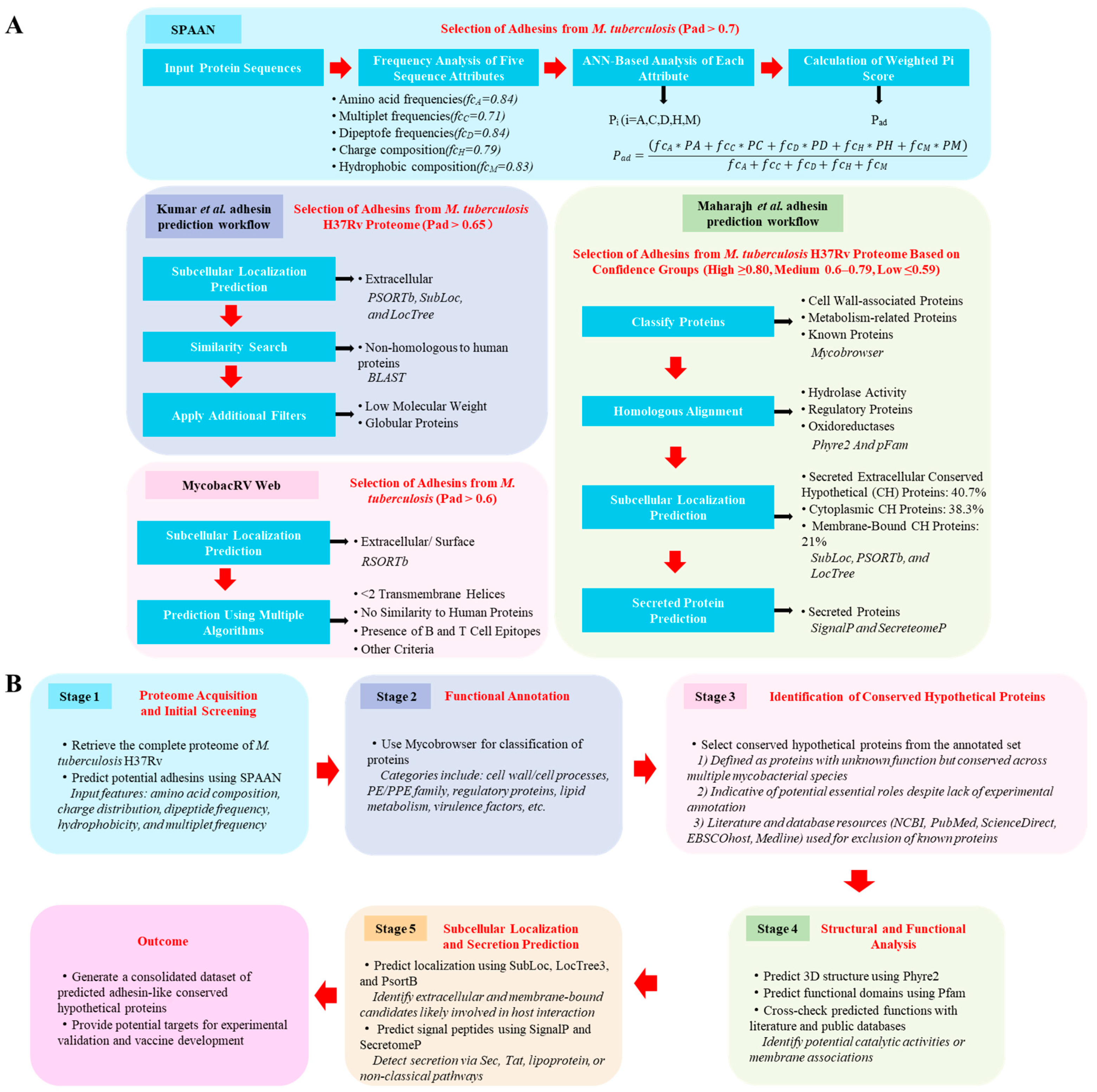
The SPAAN evaluates 105 compositional features of proteins, grouped into amino acid frequencies, multiplet frequencies, dipeptide frequencies, charge composition, and hydrophobic composition. Each group is analyzed by an individual artificial neural network (ANN), and the combined outputs generate a final prediction score (Pad), representing the likelihood of a protein being an adhesin. The SPAAN model was optimized using iterative training with known adhesins and non-adhesins. A threshold of Pad ≥ 0.51 was found to yield high predictive accuracy and specificity. Additionally, the SPAAN showed reliable performance when applied to viral adhesins, supporting its generalizability. For M. tuberculosis, a stricter Pad threshold of >0.7 was suggested to reduce false-positive predictions, considering that HBHA, an experimentally confirmed adhesin, had a Pad score of 0.6763 (Figure 3A).
Despite its strengths, the SPAAN has limitations. Its dependence on full-length amino acid sequences constrains its utility in cases with incomplete sequence data. Furthermore, the presence of false positives necessitates downstream validation using complementary computational or experimental approaches.
Since adhesins are typically located on the bacterial surface or secreted extracellularly to mediate interactions with host proteins, integrating subcellular localization predictions enhances adhesin identification [105,106]. Based on this rationale, Kumar et al. and the MycobacRV web server combined SPAAN outputs with localization tools such as PSORTb, SubLoc, and LocTree [71,107]. Recognizing that some experimentally validated adhesins had Pad scores below 0.7, these studies used relaxed thresholds (Pad ≥ 0.65 or 0.6) to capture a broader range of candidates (Figure 3A).
Kumar et al. applied the SPAAN to the entire M. tuberculosis H37Rv proteome and refined predictions through homology-based functional annotation, structural modeling, and evaluation of extracellular matrix-binding capacity [71]. The MycobacRV web server extended this pipeline to 23 M. tuberculosis strains and selected extracellular or cell wall-associated proteins using PSORTb v3.0 [107]. These predictions were further analyzed using immunoinformatics tools to identify antigenic adhesin epitopes as vaccine candidates.
These integrative approaches expanded the spectrum of predicted M. tuberculosis adhesins. For example, Kumar et al. identified 20 putative adhesins in H37Rv, and experimental validation confirmed the adhesin roles of Rv0309, Rv2599, and Rv3717 [71]. Similarly, MycobacRV identified 233 vaccine candidate sequences, including 37 novel adhesins in H37Rv, contributing to tuberculosis vaccine development [75].
While these studies focused on subsets of proteins, Maharajh et al. conducted a comprehensive evaluation of SPAAN-based predictions and proposed a systematic workflow for classifying uncharacterized H37Rv proteins [108]. Rather than using a fixed Pad threshold, they stratified 167 predicted adhesins into high (≥0.80), medium (0.6–0.79), and low (≤0.59) confidence groups. Additional analyses, including homology modeling, localization, and secretion prediction, improved the robustness of the predictions. The use of a large dataset enhanced the credibility of SPAAN-based workflows (Figure 3A).
Among existing tools, the SPAAN program remains the foundation for adhesin prediction in M. tuberculosis, but its high false-positive rate necessitates further refinement. To improve prediction accuracy, Kumar et al. and the MycobacRV platform incorporated subcellular localization and domain-based information. Maharajh et al. proposed a more systematic SPAAN-based workflow, stratifying candidates into confidence levels and integrating additional bioinformatic evidence such as secretion signals and structural modeling.
Building upon these developments, we developed a stepwise prediction pipeline (Figure 3B) that offers a more detailed and user-friendly workflow. While largely based on the strategy proposed by Maharajh et al., our version expands the evaluation steps to include cross-referencing with public databases (e.g., PubMed, ScienceDirect) and literature mining, which may assist researchers in prioritizing adhesin candidates for experimental validation. This practical framework aims to facilitate more reproducible and transparent adhesin prediction in M. tuberculosis and potentially other pathogens.

6. Challenges and Controversies in Adhesin-Targeted Strategies

6.1. Functional Complexity and Contradictory Evidence

The infection process of M. tuberculosis is inherently complex and often paradoxical. Granuloma formation, while primarily intended to contain infection, also contributes to tissue damage and bacterial persistence [109]. Similarly, cytokines induced during infection can exhibit both protective and pathogenic roles depending on the immune context [110]. These examples reflect the broader challenge of interpreting immune responses to M. tuberculosis, where host-beneficial and pathogen-favoring mechanisms frequently coexist.
This complexity is also evident in the study of adhesins. Although adhesins are recognized as promising targets for TB intervention, their functions are not always straightforward. A well-characterized example is ManLAM, which interacts with receptors such as DC-SIGN and Dectin-2 on dendritic cells and macrophages. These interactions have been reported to either promote antigen presentation and T cell priming or inhibit dendritic cell maturation and facilitate immune evasion [42,81]. Several factors may contribute to these contradictions: the function of an adhesin may vary depending on the stage of infection; the same adhesin–receptor interaction may trigger distinct outcomes in different host cell types; and structural variations in the adhesin itself—such as differences in glycosylation or lipid composition—may influence its immunological effects.
A more balanced interpretation of these findings—acknowledging both the immunostimulatory and immunosuppressive outcomes of adhesin–host interactions—is essential for advancing our understanding of their roles in pathogenesis and for informing rational vaccine and therapeutic design. Future studies should therefore incorporate infection-stage-specific models, single-cell immune profiling, and detailed structure–function analyses to clarify the precise biological functions of adhesins under physiologically relevant conditions.

6.2. Translational and Practical Challenges

Despite the conceptual appeal of targeting M. tuberculosis adhesins as a prophylactic or therapeutic strategy, several translational and practical barriers remain. First, adhesins are not essential for bacterial viability, and their immunogenicity may not suffice to induce sterilizing immunity, particularly in individuals with latent or active tuberculosis. As such, anti-adhesin approaches are most suitable for pre-exposure prophylaxis, aiming to block bacterial colonization at the earliest stage of infection. By interfering with host–pathogen interactions rather than bacterial survival, these strategies exert minimal selective pressure, which could be advantageous in reducing the risk of drug resistance. However, their application in post-infection settings remains limited. In such cases, adhesin-targeted interventions may help restrict dissemination or reinfection, particularly when integrated into combination therapies that also target intracellular survival and persistence mechanisms.
Another major obstacle is the high degree of functional redundancy among M. tuberculosis adhesins. The bacterium expresses a large array of surface-exposed ligands—both proteinaceous and non-proteinaceous—many of which bind overlapping host targets or operate synergistically. Targeting a single adhesin may be insufficient due to compensatory mechanisms, making broad protection difficult to achieve with monovalent formulations. Additionally, the development of effective delivery platforms that ensure antigen stability, accessibility, and proper immune engagement remains a practical challenge.

6.3. Strategic Solutions and Future Directions

Addressing these challenges will require a combination of rational target selection and innovative technological approaches. One critical step is the identification of conserved, immunogenic, and surface-accessible adhesins that are expressed during key stages of infection. Spatiotemporal mapping of adhesin expression may help distinguish constitutively expressed adhesins from those that are only induced in specific niches, guiding more precise targeting strategies. The construction of cross-species adhesin databases could further facilitate the discovery of broadly relevant antigens, while CRISPR-based genome editing and high-throughput interaction screens may support the functional validation and ranking of candidate targets.
To overcome the limitations of individual adhesins, multivalent vaccines incorporating multiple key adhesins—either as fusion proteins, multi-epitope constructs, or mRNA-encoded cocktails—are under consideration. These could be further optimized by co-delivery with antigens expressed during later stages of infection, such as ESAT-6 or Ag85B, thereby enhancing coverage across the entire infection timeline. Such combined approaches may enable adhesin-based strategies to serve both prophylactic and adjunct therapeutic roles. Continued integration of computational prediction, structural modeling, and experimental validation will be essential for translating adhesin research into clinically viable interventions.

7. Conclusions and Future Perspectives

M. tuberculosis infection begins with a well-coordinated adhesion process. Multiple adhesins mediate attachment to host cells and extracellular matrix components. This early step enables the pathogen to evade clearance, establish granulomas, and eventually disseminate to distant tissues. This review outlines the complex architecture of the mycobacterial cell envelope. These structures facilitate host receptor interactions and contribute to immune modulation. We also highlighted the diverse types of M. tuberculosis adhesins—including proteins, glycolipids, and other non-protein components—and summarized their respective roles in host colonization. We also reviewed advances in computational adhesin prediction. SPAAN-based pipelines have enabled the large-scale identification of candidate adhesins in multiple M. tuberculosis strains.
Targeting bacterial adhesion offers a promising strategy for tuberculosis prevention and treatment, especially in light of rising drug resistance. Unlike bactericidal drugs, adhesion inhibitors function by preventing bacterial attachment rather than by killing the pathogen. This approach minimizes selective pressure and may help reduce the emergence of resistant strains. Since adhesion is not essential for bacterial survival, these strategies may preserve commensal microbiota. They may also reduce inflammation caused by bacterial cell lysis. Preclinical studies have demonstrated that antibodies or receptor mimetics targeting major adhesins—such as HBHA, Apa, and the 19-kDa protein—can block M. tuberculosis attachment and infection. However, due to the extensive repertoire of adhesins expressed by M. tuberculosis, effective therapeutic interventions may require a multi-target approach. Identifying and validating a core set of adhesins that contribute most significantly to virulence and host interactions will be critical for advancing this strategy.
Looking ahead, integrating computational prediction, structural biology, and experimental validation will be essential. This will help map the M. tuberculosis adhesome more completely. A deeper understanding of the spatial and temporal expression of adhesins during different stages of infection may reveal new opportunities for targeted intervention. Furthermore, the development of adhesin-based vaccines or adjunct therapies could complement existing tuberculosis treatment regimens, shorten therapy duration, and improve outcomes for drug-resistant TB cases. As the field moves forward, adhesins hold significant potential as both diagnostic markers and novel therapeutic targets in the global fight against tuberculosis.

Author Contributions

H.Y. conceived of and designed the study, created all figures and schematics, conducted the literature review, and drafted the manuscript. Y.M. contributed to the section on the computational prediction of adhesins, created Figure 3A, and assisted in manuscript revision under the guidance of H.Y., X.L., S.C. and S.Z. collected the data and revised the manuscript. L.D., S.L., and G.S. provided critical feedback on the manuscript. All authors have read and agreed to the published version of the manuscript.

Funding

This research received no external funding.

Institutional Review Board Statement

Not applicable.

Informed Consent Statement

Not applicable.

Data Availability Statement

Not applicable.

Conflicts of Interest

Haoyan Yang, Xinkui Lei, Siyu Chai, Sigen Zhang, Guimin Su, and Lin Du were employed by the company Beijing Zhifei Lvzhu Biopharmaceutical Co., Ltd. during the course of this study. The remaining authors declare that the research was conducted in the absence of any commercial or financial relationships that could be construed as a potential conflict of interest.

Abbreviations

The following abbreviations are used in this manuscript:
GlossaryFull Term
AECAlveolar epithelial cells
AGArabinogalactan
AG–PGArabinogalactan–peptidoglycan
AMAlveolar macrophages
Antigen 85Ag85
ANNArtificial neural network
ApaAlanine and proline-rich protein
BLFBovine lactoferrin
CD1⁻ DCsCD1-negative dendritic cells
CFP-10Culture filtrate protein 10 kDa
CHConserved Hypothetica
COVID-19Coronavirus disease
DC-SIGNDendritic cell-specific ICAM-3-grabbing non-integrin
ECMExtracellular matrix
ESAT-6Early secretory antigenic target 6 kDa
FnFibronectin
GAPDHGlyceraldehyde-3-phosphate dehydrogenase
GlcNAcN-acetylglucosamine
GlcBMalate synthase
HBHAHeparin-binding hemagglutinin adhesin
HMVECHuman microvascular endothelial cells
H. pyloriHelicobacter pylori
LAMsLipoarabinomannans
LMsLipomannans
MARCOMacrophage receptor with collagenous structure
MAsMycolic acids
ManLAMMannose-capped lipoarabinomannan
M. bovis BCGMycobacterium bovis Bacillus Calmette-Guérin
Mce1AMycobacterial mammalian cell entry protein 1A
MDPMuramyl dipeptide
MincleMacrophage-inducible C-type lectin
MRMacrophage mannose receptor
MTPMycobacterium tuberculosis curli pili
M. tuberculosisMycobacterium tuberculosis
TBTuberculosis
TDMTrehalose 6,6′-dimycolate
TLR2Toll-like receptor 2
TLRsToll-like receptors
TMMTrehalose monomycolate
UNUnited Nation
UPECUropathogenic Escherichia coli
PadProtein being an adhesin
PAMPsPathogen-associated molecular patterns
PDIMPhenolic glycolipid
PGPeptidoglycan
PIMsPhosphatidylinositol mannosides
PlgPlasminogen
PRRsPattern recognition receptors
PSP-AHuman pulmonary surfactant protein A
SPAANSoftware Program for Prediction of Adhesins and Adhesin-like Proteins using Neural Networks
WHOWorld Health Organization

References

  1. Global Tuberculosis Report 2024; World Health Organization: Geneva, Switzerland, 2024; Licence: CC BY-NC-SA 3.0 IGO.
  2. Global Tuberculosis Report 2023; World Health Organization: Geneva, Switzerland, 2023; Licence: CC BY-NC-SA 3.0 IGO.
  3. Berne, C.; Ellison, C.K.; Ducret, A.; Brun, Y.V. Bacterial adhesion at the single-cell level. Nat. Rev. Microbiol. 2018, 16, 616–627. [Google Scholar] [CrossRef] [PubMed]
  4. Costerton, J.W.; Lewandowski, Z.; Caldwell, D.E.; Korber, D.R.; Lappin-Scott, H.M. Microbial biofilms. Annu. Rev. Microbiol. 1995, 49, 711–745. [Google Scholar] [CrossRef] [PubMed]
  5. Costerton, J.W.; Stewart, P.S.; Greenberg, E.P. Bacterial biofilms: A common cause of persistent infections. Science 1999, 284, 1318–1322. [Google Scholar] [CrossRef] [PubMed]
  6. Berne, C.; Ducret, A.; Hardy, G.G.; Brun, Y.V. Adhesins Involved in Attachment to Abiotic Surfaces by Gram-Negative Bacteria. Microbiol. Spectr. 2015, 3. [Google Scholar] [CrossRef]
  7. Pizarro-Cerdá, J.; Cossart, P. Bacterial adhesion and entry into host cells. Cell 2006, 124, 715–727. [Google Scholar] [CrossRef]
  8. Stones, D.H.; Krachler, A.M. Against the tide: The role of bacterial adhesion in host colonization. Biochem. Soc. Trans. 2016, 44, 1571–1580. [Google Scholar] [CrossRef]
  9. Viljoen, A.; Dufrêne, Y.F.; Nigou, J. Mycobacterial Adhesion: From Hydrophobic to Receptor-Ligand Interactions. Microorganisms 2022, 10, 454. [Google Scholar] [CrossRef]
  10. Pecoraro, C.; Carbone, D.; Parrino, B.; Cascioferro, S.; Diana, P. Recent Developments in the Inhibition of Bacterial Adhesion as Promising Anti-Virulence Strategy. Int. J. Mol. Sci. 2023, 24, 4872. [Google Scholar] [CrossRef]
  11. Asadi, A.; Razavi, S.; Talebi, M.; Gholami, M. A review on anti-adhesion therapies of bacterial diseases. Infection 2019, 47, 13–23. [Google Scholar] [CrossRef]
  12. Wang, N.; Cai, T.; Liu, X.; Zhu, W. Bovine lactoferrin inhibits resistant Helicobacter pylori in vitro and protects gastric mucosal injury in vivo. Int. Dairy. J. 2024, 147, 105770. [Google Scholar] [CrossRef]
  13. Hablass, F.H.; Lashen, S.A.; Alsayed, E.A. Efficacy of Lactoferrin with Standard Triple Therapy or Sequential Therapy for Helicobacter pylori Eradication: A Randomized Controlled Trial. Turk. J. Gastroenterol. 2021, 32, 742–749. [Google Scholar] [CrossRef] [PubMed]
  14. Imoto, I.; Yasuma, T.; D’Alessandro-Gabazza, C.N.; Oka, S.; Misaki, M.; Horiki, N.; Gabazza, E.C. Antimicrobial Effects of Lactoferrin against Helicobacter pylori Infection. Pathogens 2023, 12, 599. [Google Scholar] [CrossRef] [PubMed]
  15. Zou, J.; Dong, J.; Yu, X.F. Meta-analysis: The effect of supplementation with lactoferrin on eradication rates and adverse events during Helicobacter pylori eradication therapy. Helicobacter 2009, 14, 119–127. [Google Scholar] [CrossRef] [PubMed]
  16. Sarshar, M.; Behzadi, P.; Ambrosi, C.; Zagaglia, C.; Palamara, A.T.; Scribano, D. FimH and Anti-Adhesive Therapeutics: A Disarming Strategy Against Uropathogens. Antibiotics 2020, 9, 397. [Google Scholar] [CrossRef]
  17. Shin, A.R.; Lee, K.S.; Lee, J.S.; Kim, S.Y.; Song, C.H.; Jung, S.B.; Yang, C.S.; Jo, E.K.; Park, J.K.; Paik, T.H.; et al. Mycobacterium tuberculosis HBHA protein reacts strongly with the serum immunoglobulin M of tuberculosis patients. Clin. Vaccine Immunol. 2006, 13, 869–875. [Google Scholar] [CrossRef]
  18. Cambier, C.J.; Falkow, S.; Ramakrishnan, L. Host evasion and exploitation schemes of Mycobacterium tuberculosis. Cell 2014, 159, 1497–1509. [Google Scholar] [CrossRef]
  19. Bisht, D.; Meena, L.S. Adhesion molecules facilitate host-pathogen interaction & mediate Mycobacterium tuberculosis pathogenesis. Indian. J. Med. Res. 2019, 150, 23–32. [Google Scholar] [CrossRef]
  20. Russell, D.G. Who puts the tubercle in tuberculosis? Nat. Rev. Microbiol. 2007, 5, 39–47. [Google Scholar] [CrossRef]
  21. Ishikawa, E.; Mori, D.; Yamasaki, S. Recognition of Mycobacterial Lipids by Immune Receptors. Trends Immunol. 2017, 38, 66–76. [Google Scholar] [CrossRef]
  22. Neyrolles, O.; Guilhot, C. Recent advances in deciphering the contribution of Mycobacterium tuberculosis lipids to pathogenesis. Tuberculosis 2011, 91, 187–195. [Google Scholar] [CrossRef]
  23. Pethe, K.; Alonso, S.; Biet, F.; Delogu, G.; Brennan, M.J.; Locht, C.; Menozzi, F.D. The heparin-binding haemagglutinin of M. tuberculosis is required for extrapulmonary dissemination. Nature 2001, 412, 190–194. [Google Scholar] [CrossRef] [PubMed]
  24. Nguyen, L.; Pieters, J. The Trojan horse: Survival tactics of pathogenic mycobacteria in macrophages. Trends Cell Biol. 2005, 15, 269–276. [Google Scholar] [CrossRef] [PubMed]
  25. Yang, H.; Lei, X.; Chai, S.; Su, G.; Du, L. From pathogenesis to antigens: The key to shaping the future of TB vaccines. Front. Immunol. 2024, 15, 1440935. [Google Scholar] [CrossRef] [PubMed]
  26. Vidal Pessolani, M.C.; Marques, M.A.; Reddy, V.M.; Locht, C.; Menozzi, F.D. Systemic dissemination in tuberculosis and leprosy: Do mycobacterial adhesins play a role? Microbes Infect. 2003, 5, 677–684. [Google Scholar] [CrossRef]
  27. Franklin, A.; Salgueiro, V.C.; Layton, A.J.; Sullivan, R.; Mize, T.; Vázquez-Iniesta, L.; Benedict, S.T.; Gurcha, S.S.; Anso, I.; Besra, G.S.; et al. The mycobacterial glycoside hydrolase LamH enables capsular arabinomannan release and stimulates growth. Nat. Commun. 2024, 15, 5740. [Google Scholar] [CrossRef]
  28. Kitzmiller, C.E.; Cheng, T.Y.; Prandi, J.; Sparks, I.L.; Moody, D.B.; Morita, Y.S. Detergent-induced quantitatively limited formation of diacyl phosphatidylinositol dimannoside in Mycobacterium smegmatis. J. Lipid Res. 2024, 65, 100533. [Google Scholar] [CrossRef]
  29. Gonzales-Huerta, L.E.; Williams, T.J.; Aljohani, R.; Robertson, B.; Evans, C.A.; Armstrong-James, D. Mycobacterial lipoarabinomannan negatively interferes with macrophage responses to Aspergillus fumigatus in-vitro. bioRxiv, 2024; preprint. [Google Scholar] [CrossRef]
  30. Chiang, C.Y.; West, N.P. The fall of the mycobacterial cell wall: Interrogating peptidoglycan synthesis for novel anti-TB agents. PeerJ 2024, 12, e18404. [Google Scholar] [CrossRef]
  31. Behr, M.A.; Divangahi, M. Freund’s adjuvant, NOD2 and mycobacteria. Curr. Opin. Microbiol. 2015, 23, 126–132. [Google Scholar] [CrossRef]
  32. Dulberger, C.L.; Rubin, E.J.; Boutte, C.C. The mycobacterial cell envelope—A moving target. Nat. Rev. Microbiol. 2020, 18, 47–59. [Google Scholar] [CrossRef]
  33. Qin, L.; Xu, J.; Chen, J.; Wang, S.; Zheng, R.; Cui, Z.; Liu, Z.; Wu, X.; Wang, J.; Huang, X.; et al. Cell-autonomous targeting of arabinogalactan by host immune factors inhibits mycobacterial growth. eLife 2024, 13, RP92737. [Google Scholar] [CrossRef] [PubMed]
  34. Wu, X.; Wu, Y.; Zheng, R.; Tang, F.; Qin, L.; Lai, D.; Zhang, L.; Chen, L.; Yan, B.; Yang, H.; et al. Sensing of mycobacterial arabinogalactan by galectin-9 exacerbates mycobacterial infection. EMBO Rep. 2021, 22, e51678. [Google Scholar] [CrossRef] [PubMed]
  35. Su, C.C.; Klenotic, P.A.; Cui, M.; Lyu, M.; Morgan, C.E.; Yu, E.W. Structures of the mycobacterial membrane protein MmpL3 reveal its mechanism of lipid transport. PLoS Biol. 2021, 19, e3001370. [Google Scholar] [CrossRef]
  36. Iizasa, E.; Chuma, Y.; Uematsu, T.; Kubota, M.; Kawaguchi, H.; Umemura, M.; Toyonaga, K.; Kiyohara, H.; Yano, I.; Colonna, M.; et al. TREM2 is a receptor for non-glycosylated mycolic acids of mycobacteria that limits anti-mycobacterial macrophage activation. Nat. Commun. 2021, 12, 2299. [Google Scholar] [CrossRef]
  37. Cambier, C.J.; Banik, S.M.; Buonomo, J.A.; Bertozzi, C.R. Spreading of a mycobacterial cell-surface lipid into host epithelial membranes promotes infectivity. eLife 2020, 9, e60648. [Google Scholar] [CrossRef]
  38. Mittal, E.; Roth, A.T.; Seth, A.; Singamaneni, S.; Beatty, W.; Philips, J.A. Single cell preparations of Mycobacterium tuberculosis damage the mycobacterial envelope and disrupt macrophage interactions. eLife 2023, 12, e85416. [Google Scholar] [CrossRef]
  39. Driessen, N.N.; Ummels, R.; Maaskant, J.J.; Gurcha, S.S.; Besra, G.S.; Ainge, G.D.; Larsen, D.S.; Painter, G.F.; Vandenbroucke-Grauls, C.M.; Geurtsen, J.; et al. Role of phosphatidylinositol mannosides in the interaction between mycobacteria and DC-SIGN. Infect. Immun. 2009, 77, 4538–4547. [Google Scholar] [CrossRef]
  40. Beatty, W.L.; Rhoades, E.R.; Hsu, D.K.; Liu, F.T.; Russell, D.G. Association of a macrophage galactoside-binding protein with Mycobacterium-containing phagosomes. Cell Microbiol. 2002, 4, 167–176. [Google Scholar] [CrossRef]
  41. Appelmelk, B.J.; den Dunnen, J.; Driessen, N.N.; Ummels, R.; Pak, M.; Nigou, J.; Larrouy-Maumus, G.; Gurcha, S.S.; Movahedzadeh, F.; Geurtsen, J.; et al. The mannose cap of mycobacterial lipoarabinomannan does not dominate the Mycobacterium-host interaction. Cell Microbiol. 2008, 10, 930–944. [Google Scholar] [CrossRef]
  42. Yonekawa, A.; Saijo, S.; Hoshino, Y.; Miyake, Y.; Ishikawa, E.; Suzukawa, M.; Inoue, H.; Tanaka, M.; Yoneyama, M.; Oh-Hora, M.; et al. Dectin-2 is a direct receptor for mannose-capped lipoarabinomannan of mycobacteria. Immunity 2014, 41, 402–413. [Google Scholar] [CrossRef]
  43. Decout, A.; Silva-Gomes, S.; Drocourt, D.; Blattes, E.; Rivière, M.; Prandi, J.; Larrouy-Maumus, G.; Caminade, A.M.; Hamasur, B.; Källenius, G.; et al. Deciphering the molecular basis of mycobacteria and lipoglycan recognition by the C-type lectin Dectin-2. Sci. Rep. 2018, 8, 16840. [Google Scholar] [CrossRef] [PubMed]
  44. Schlesinger, L.S.; Hull, S.R.; Kaufman, T.M. Binding of the terminal mannosyl units of lipoarabinomannan from a virulent strain of Mycobacterium tuberculosis to human macrophages. J. Immunol. 1994, 152, 4070–4079. [Google Scholar] [CrossRef] [PubMed]
  45. Barboni, E.; Coade, S.; Fiori, A. The binding of mycolic acids to galectin-3: A novel interaction between a host soluble lectin and trafficking mycobacterial lipids? FEBS Lett. 2005, 579, 6749–6755. [Google Scholar] [CrossRef] [PubMed]
  46. Nishimura, N.; Tomiyasu, N.; Torigoe, S.; Mizuno, S.; Fukano, H.; Ishikawa, E.; Katano, H.; Hoshino, Y.; Matsuo, K.; Takahashi, M.; et al. Mycobacterial mycolic acids trigger inhibitory receptor Clec12A to suppress host immune responses. Tuberculosis 2023, 138, 102294. [Google Scholar] [CrossRef]
  47. Ishikawa, E.; Ishikawa, T.; Morita, Y.S.; Toyonaga, K.; Yamada, H.; Takeuchi, O.; Kinoshita, T.; Akira, S.; Yoshikai, Y.; Yamasaki, S. Direct recognition of the mycobacterial glycolipid, trehalose dimycolate, by C-type lectin Mincle. J. Exp. Med. 2009, 206, 2879–2888. [Google Scholar] [CrossRef]
  48. Schoenen, H.; Bodendorfer, B.; Hitchens, K.; Manzanero, S.; Werninghaus, K.; Nimmerjahn, F.; Agger, E.M.; Stenger, S.; Andersen, P.; Ruland, J.; et al. Cutting edge: Mincle is essential for recognition and adjuvanticity of the mycobacterial cord factor and its synthetic analog trehalose-dibehenate. J. Immunol. 2010, 184, 2756–2760. [Google Scholar] [CrossRef]
  49. Zhao, X.Q.; Zhu, L.L.; Chang, Q.; Jiang, C.; You, Y.; Luo, T.; Jia, X.M.; Lin, X. C-type lectin receptor dectin-3 mediates trehalose 6,6'-dimycolate (TDM)-induced Mincle expression through CARD9/Bcl10/MALT1-dependent nuclear factor (NF)-κB activation. J. Biol. Chem. 2014, 289, 30052–30062. [Google Scholar] [CrossRef]
  50. Bowdish, D.M.; Sakamoto, K.; Kim, M.J.; Kroos, M.; Mukhopadhyay, S.; Leifer, C.A.; Tryggvason, K.; Gordon, S.; Russell, D.G. MARCO, TLR2, and CD14 are required for macrophage cytokine responses to mycobacterial trehalose dimycolate and Mycobacterium tuberculosis. PLoS Pathog. 2009, 5, e1000474. [Google Scholar] [CrossRef]
  51. Martinez, N.; Ketheesan, N.; West, K.; Vallerskog, T.; Kornfeld, H. Impaired Recognition of Mycobacterium tuberculosis by Alveolar Macrophages From Diabetic Mice. J. Infect. Dis. 2016, 214, 1629–1637. [Google Scholar] [CrossRef]
  52. Geurtsen, J.; Chedammi, S.; Mesters, J.; Cot, M.; Driessen, N.N.; Sambou, T.; Kakutani, R.; Ummels, R.; Maaskant, J.; Takata, H.; et al. Identification of mycobacterial alpha-glucan as a novel ligand for DC-SIGN: Involvement of mycobacterial capsular polysaccharides in host immune modulation. J. Immunol. 2009, 183, 5221–5231. [Google Scholar] [CrossRef]
  53. Gagliardi, M.C.; Lemassu, A.; Teloni, R.; Mariotti, S.; Sargentini, V.; Pardini, M.; Daffé, M.; Nisini, R. Cell wall-associated alpha-glucan is instrumental for Mycobacterium tuberculosis to block CD1 molecule expression and disable the function of dendritic cell derived from infected monocyte. Cell Microbiol. 2007, 9, 2081–2092. [Google Scholar] [CrossRef] [PubMed]
  54. Renshaw, P.S.; Lightbody, K.L.; Veverka, V.; Muskett, F.W.; Kelly, G.; Frenkiel, T.A.; Gordon, S.V.; Hewinson, R.G.; Burke, B.; Norman, J.; et al. Structure and function of the complex formed by the tuberculosis virulence factors CFP-10 and ESAT-6. Embo J. 2005, 24, 2491–2498. [Google Scholar] [CrossRef] [PubMed]
  55. Kinhikar, A.G.; Verma, I.; Chandra, D.; Singh, K.K.; Weldingh, K.; Andersen, P.; Hsu, T.; Jacobs, W.R., Jr.; Laal, S. Potential role for ESAT6 in dissemination of M. tuberculosis via human lung epithelial cells. Mol. Microbiol. 2010, 75, 92–106. [Google Scholar] [CrossRef]
  56. Viljoen, A.; Alsteens, D.; Dufrêne, Y. Mechanical Forces between Mycobacterial Antigen 85 Complex and Fibronectin. Cells 2020, 9, 716. [Google Scholar] [CrossRef]
  57. Xolalpa, W.; Vallecillo, A.J.; Lara, M.; Mendoza-Hernandez, G.; Comini, M.; Spallek, R.; Singh, M.; Espitia, C. Identification of novel bacterial plasminogen-binding proteins in the human pathogen Mycobacterium tuberculosis. Proteomics 2007, 7, 3332–3341. [Google Scholar] [CrossRef]
  58. Lebrun, P.; Raze, D.; Fritzinger, B.; Wieruszeski, J.M.; Biet, F.; Dose, A.; Carpentier, M.; Schwarzer, D.; Allain, F.; Lippens, G.; et al. Differential contribution of the repeats to heparin binding of HBHA, a major adhesin of Mycobacterium tuberculosis. PLoS ONE 2012, 7, e32421. [Google Scholar] [CrossRef]
  59. Silva, C.A.; Danelishvili, L.; McNamara, M.; Berredo-Pinho, M.; Bildfell, R.; Biet, F.; Rodrigues, L.S.; Oliveira, A.V.; Bermudez, L.E.; Pessolani, M.C. Interaction of Mycobacterium leprae with human airway epithelial cells: Adherence, entry, survival, and identification of potential adhesins by surface proteome analysis. Infect. Immun. 2013, 81, 2645–2659. [Google Scholar] [CrossRef]
  60. Ramsugit, S.; Pillay, B.; Pillay, M. Evaluation of the role of Mycobacterium tuberculosis pili (MTP) as an adhesin, invasin, and cytokine inducer of epithelial cells. Braz. J. Infect. Dis. 2016, 20, 160–165. [Google Scholar] [CrossRef]
  61. Ramsugit, S.; Pillay, M. Mycobacterium tuberculosis Pili promote adhesion to and invasion of THP-1 macrophages. Jpn. J. Infect. Dis. 2014, 67, 476–478. [Google Scholar] [CrossRef]
  62. Ramsugit, S.; Guma, S.; Pillay, B.; Jain, P.; Larsen, M.H.; Danaviah, S.; Pillay, M. Pili contribute to biofilm formation in vitro in Mycobacterium tuberculosis. Antonie Van Leeuwenhoek 2013, 104, 725–735. [Google Scholar] [CrossRef]
  63. Alteri, C.J.; Xicohténcatl-Cortes, J.; Hess, S.; Caballero-Olín, G.; Girón, J.A.; Friedman, R.L. Mycobacterium tuberculosis produces pili during human infection. Proc. Natl. Acad. Sci. USA 2007, 104, 5145–5150. [Google Scholar] [CrossRef] [PubMed]
  64. Pitarque, S.; Herrmann, J.L.; Duteyrat, J.L.; Jackson, M.; Stewart, G.R.; Lecointe, F.; Payre, B.; Schwartz, O.; Young, D.B.; Marchal, G.; et al. Deciphering the molecular bases of Mycobacterium tuberculosis binding to the lectin DC-SIGN reveals an underestimated complexity. Biochem. J. 2005, 392, 615–624. [Google Scholar] [CrossRef] [PubMed]
  65. Diaz-Silvestre, H.; Espinosa-Cueto, P.; Sanchez-Gonzalez, A.; Esparza-Ceron, M.A.; Pereira-Suarez, A.L.; Bernal-Fernandez, G.; Espitia, C.; Mancilla, R. The 19-kDa antigen of Mycobacterium tuberculosis is a major adhesin that binds the mannose receptor of THP-1 monocytic cells and promotes phagocytosis of mycobacteria. Microb. Pathog. 2005, 39, 97–107. [Google Scholar] [CrossRef] [PubMed]
  66. Kinhikar, A.G.; Vargas, D.; Li, H.; Mahaffey, S.B.; Hinds, L.; Belisle, J.T.; Laal, S. Mycobacterium tuberculosis malate synthase is a laminin-binding adhesin. Mol. Microbiol. 2006, 60, 999–1013. [Google Scholar] [CrossRef]
  67. Gani, Z.; Boradia, V.M.; Kumar, A.; Patidar, A.; Talukdar, S.; Choudhary, E.; Singh, R.; Agarwal, N.; Raje, M.; Iyengar Raje, C. Mycobacterium tuberculosis glyceraldehyde-3-phosphate dehydrogenase plays a dual role-As an adhesin and as a receptor for plasmin(ogen). Cell Microbiol. 2021, 23, e13311. [Google Scholar] [CrossRef]
  68. Kuo, C.J.; Gao, J.; Huang, J.W.; Ko, T.P.; Zhai, C.; Ma, L.; Liu, W.; Dai, L.; Chang, Y.F.; Chen, T.H.; et al. Functional and structural investigations of fibronectin-binding protein Apa from Mycobacterium tuberculosis. Biochim. Biophys. Acta Gen. Subj. 2019, 1863, 1351–1359. [Google Scholar] [CrossRef]
  69. Ragas, A.; Roussel, L.; Puzo, G.; Rivière, M. The Mycobacterium tuberculosis cell-surface glycoprotein apa as a potential adhesin to colonize target cells via the innate immune system pulmonary C-type lectin surfactant protein A. J. Biol. Chem. 2007, 282, 5133–5142. [Google Scholar] [CrossRef]
  70. Esparza, M.; Palomares, B.; García, T.; Espinosa, P.; Zenteno, E.; Mancilla, R. PstS-1, the 38-kDa Mycobacterium tuberculosis glycoprotein, is an adhesin, which binds the macrophage mannose receptor and promotes phagocytosis. Scand. J. Immunol. 2015, 81, 46–55. [Google Scholar] [CrossRef]
  71. Kumar, S.; Puniya, B.L.; Parween, S.; Nahar, P.; Ramachandran, S. Identification of novel adhesins of M. tuberculosis H37Rv using integrated approach of multiple computational algorithms and experimental analysis. PLoS ONE 2013, 8, e69790. [Google Scholar] [CrossRef]
  72. Idris, I.; Abdurrahman, A.H.; Fatulrachman; Aftitah, V.B.; Fadlitha, V.B.; Sato, N.; Fujimura, T.; Takimoto, H. Invasion of human microvascular endothelial cells by Mycobacterium leprae through Mce1A protein. J. Dermatol. 2019, 46, 853–858. [Google Scholar] [CrossRef]
  73. Kohwiwattanagun, J.; Kawamura, I.; Fujimura, T.; Mitsuyama, M. Mycobacterial mammalian cell entry protein 1A (Mce1A)-mediated adherence enhances the chemokine production by A549 alveolar epithelial cells. Microbiol. Immunol. 2007, 51, 253–261. [Google Scholar] [CrossRef] [PubMed]
  74. Blake, R.; Jensen, K.; Mabbott, N.; Hope, J.; Stevens, J. The role of Mce proteins in Mycobacterium avium paratuberculosis infection. Sci. Rep. 2024, 14, 14964. [Google Scholar] [CrossRef] [PubMed]
  75. Ramsugit, S.; Pillay, M. Identification of Mycobacterium tuberculosis adherence-mediating components: A review of key methods to confirm adhesin function. Iran. J. Basic. Med. Sci. 2016, 19, 579–584. [Google Scholar]
  76. Stamm, C.E.; Pasko, B.L.; Chaisavaneeyakorn, S.; Franco, L.H.; Nair, V.R.; Weigele, B.A.; Alto, N.M.; Shiloh, M.U. Screening Mycobacterium tuberculosis Secreted Proteins Identifies Mpt64 as a Eukaryotic Membrane-Binding Bacterial Effector. mSphere 2019, 4, e00354-19. [Google Scholar] [CrossRef]
  77. Kramarska, E.; Squeglia, F.; De Maio, F.; Delogu, G.; Berisio, R. PE_PGRS33, an Important Virulence Factor of Mycobacterium tuberculosis and Potential Target of Host Humoral Immune Response. Cells 2021, 10, 161. [Google Scholar] [CrossRef]
  78. Palucci, I.; Camassa, S.; Cascioferro, A.; Sali, M.; Anoosheh, S.; Zumbo, A.; Minerva, M.; Iantomasi, R.; De Maio, F.; Di Sante, G.; et al. PE_PGRS33 Contributes to Mycobacterium tuberculosis Entry in Macrophages through Interaction with TLR2. PLoS ONE 2016, 11, e0150800. [Google Scholar] [CrossRef]
  79. Minerva, M.; De Maio, F.; Camassa, S.; Battah, B.; Ivana, P.; Manganelli, R.; Sanguinetti, M.; Sali, M.; Delogu, G. Evaluation of PE_PGRS33 as a potential surface target for humoral responses against Mycobacterium tuberculosis. Pathog. Dis. 2017, 75, ftx100. [Google Scholar] [CrossRef]
  80. Espitia, C.; Laclette, J.P.; Mondragón-Palomino, M.; Amador, A.; Campuzano, J.; Martens, A.; Singh, M.; Cicero, R.; Zhang, Y.; Moreno, C. The PE-PGRS glycine-rich proteins of Mycobacterium tuberculosis: A new family of fibronectin-binding proteins? Microbiology 1999, 145 Pt 12, 3487–3495. [Google Scholar] [CrossRef]
  81. Torrelles, J.B.; Azad, A.K.; Schlesinger, L.S. Fine discrimination in the recognition of individual species of phosphatidyl-myo-inositol mannosides from Mycobacterium tuberculosis by C-type lectin pattern recognition receptors. J. Immunol. 2006, 177, 1805–1816. [Google Scholar] [CrossRef]
  82. Ehlers, S. DC-SIGN and mannosylated surface structures of Mycobacterium tuberculosis: A deceptive liaison. Eur. J. Cell Biol. 2010, 89, 95–101. [Google Scholar] [CrossRef]
  83. Nuzzo, I.; Galdiero, M.; Bentivoglio, C.; Galdiero, R.; Romano Carratelli, C. Apoptosis modulation by mycolic acid, tuberculostearic acid and trehalose 6,6'-dimycolate. J. Infect. 2002, 44, 229–235. [Google Scholar] [CrossRef] [PubMed]
  84. Shao, M.M.; Yi, F.S.; Huang, Z.Y.; Peng, P.; Wu, F.Y.; Shi, H.Z.; Zhai, K. T Cell Receptor Repertoire Analysis Reveals Signatures of T Cell Responses to Human Mycobacterium tuberculosis. Front. Microbiol. 2022, 13, 829694. [Google Scholar] [CrossRef] [PubMed]
  85. Bates, T.A.; Trank-Greene, M.; Nguyenla, X.; Anastas, A.; Gurmessa, S.K.; Merutka, I.R.; Dixon, S.D.; Shumate, A.; Groncki, A.R.; Parson, M.A.H.; et al. ESAT-6 undergoes self-association at phagosomal pH and an ESAT-6-specific nanobody restricts M. tuberculosis growth in macrophages. eLife 2024, 12, RP91930. [Google Scholar] [CrossRef]
  86. Krishnan, N.; Robertson, B.D.; Thwaites, G. The mechanisms and consequences of the extra-pulmonary dissemination of Mycobacterium tuberculosis. Tuberculosis 2010, 90, 361–366. [Google Scholar] [CrossRef]
  87. Passos, B.B.S.; Araújo-Pereira, M.; Vinhaes, C.L.; Amaral, E.P.; Andrade, B.B. The role of ESAT-6 in tuberculosis immunopathology. Front. Immunol. 2024, 15, 1383098. [Google Scholar] [CrossRef]
  88. Ryndak, M.B.; Chandra, D.; Laal, S. Understanding dissemination of Mycobacterium tuberculosis from the lungs during primary infection. J. Med. Microbiol. 2016, 65, 362–369. [Google Scholar] [CrossRef]
  89. Tengattini, S.; Bavaro, T.; Rinaldi, F.; Temporini, C.; Pollegioni, L.; Terreni, M.; Piubelli, L. Novel tuberculosis vaccines based on TB10.4 and Ag85B: State-of-art and advocacy for good practices. Vaccine 2025, 53, 126932. [Google Scholar] [CrossRef]
  90. Rambukkana, A.; Das, P.K.; Burggraaf, J.D.; Yong, S.; Faber, W.R.; Thole, J.E.; Harboe, M. Heterogeneity of monoclonal antibody-reactive epitopes on mycobacterial 30-kilodalton-region proteins and the secreted antigen 85 complex and demonstration of antigen 85B on the Mycobacterium leprae cell wall surface. Infect. Immun. 1992, 60, 5172–5181. [Google Scholar] [CrossRef]
  91. Kuo, C.J.; Bell, H.; Hsieh, C.L.; Ptak, C.P.; Chang, Y.F. Novel mycobacteria antigen 85 complex binding motif on fibronectin. J. Biol. Chem. 2012, 287, 1892–1902. [Google Scholar] [CrossRef]
  92. Bentley-Hibbert, S.I.; Quan, X.; Newman, T.; Huygen, K.; Godfrey, H.P. Pathophysiology of antigen 85 in patients with active tuberculosis: Antigen 85 circulates as complexes with fibronectin and immunoglobulin G. Infect. Immun. 1999, 67, 581–588. [Google Scholar] [CrossRef]
  93. Naito, M.; Ohara, N.; Matsumoto, S.; Yamada, T. The novel fibronectin-binding motif and key residues of mycobacteria. J. Biol. Chem. 1998, 273, 2905–2909. [Google Scholar] [CrossRef] [PubMed]
  94. Menozzi, F.D.; Reddy, V.M.; Cayet, D.; Raze, D.; Debrie, A.S.; Dehouck, M.P.; Cecchelli, R.; Locht, C. Mycobacterium tuberculosis heparin-binding haemagglutinin adhesin (HBHA) triggers receptor-mediated transcytosis without altering the integrity of tight junctions. Microbes Infect. 2006, 8, 1–9. [Google Scholar] [CrossRef] [PubMed]
  95. Ashokcoomar, S.; Reedoy, K.S.; Loots, D.T.; Beukes, D.; van Reenen, M.; Pillay, B.; Pillay, M.M. tuberculosis curli pili (MTP) facilitates a reduction of microbicidal activity of infected THP-1 macrophages during early stages of infection. Comp. Immunol. Microbiol. Infect. Dis. 2022, 90–91, 101907. [Google Scholar] [CrossRef] [PubMed]
  96. Wang, W.; Ning, Y.; Wang, Y.; Deng, G.; Pace, S.; Barth, S.A.; Menge, C.; Zhang, K.; Dai, Y.; Cai, Y.; et al. Mycobacterium tuberculosis-Induced Upregulation of the COX-2/mPGES-1 Pathway in Human Macrophages Is Abrogated by Sulfasalazine. Front. Immunol. 2022, 13, 849583. [Google Scholar] [CrossRef]
  97. Yeremeev, V.V.; Lyadova, I.V.; Nikonenko, B.V.; Apt, A.S.; Abou-Zeid, C.; Inwald, J.; Young, D.B. The 19-kD antigen and protective immunity in a murine model of tuberculosis. Clin. Exp. Immunol. 2000, 120, 274–279. [Google Scholar] [CrossRef]
  98. Yeruva, V.C.; Kulkarni, A.; Khandelwal, R.; Sharma, Y.; Raghunand, T.R. The PE_PGRS Proteins of Mycobacterium tuberculosis Are Ca2+ Binding Mediators of Host-Pathogen Interaction. Biochemistry 2016, 55, 4675–4687. [Google Scholar] [CrossRef]
  99. Parada, C.; Neri-Badillo, I.C.; Vallecillo, A.J.; Segura, E.; Silva-Miranda, M.; Guzmán-Gutiérrez, S.L.; Ortega, P.A.; Coronado-Aceves, E.W.; Cancino-Villeda, L.; Torres-Larios, A.; et al. New Insights into the Methylation of Mycobacterium tuberculosis Heparin Binding Hemagglutinin Adhesin Expressed in Rhodococcus erythropolis. Pathogens 2021, 10, 1139. [Google Scholar] [CrossRef]
  100. Kulmanov, M.; Hoehndorf, R. DeepGOPlus: Improved protein function prediction from sequence. Bioinformatics 2020, 36, 422–429. [Google Scholar] [CrossRef]
  101. Goodswen, S.J.; Kennedy, P.J.; Ellis, J.T. A guide to current methodology and usage of reverse vaccinology towards in silico vaccine discovery. FEMS Microbiol. Rev. 2023, 47, fuad004. [Google Scholar] [CrossRef]
  102. Cole, S.T.; Brosch, R.; Parkhill, J.; Garnier, T.; Churcher, C.; Harris, D.; Gordon, S.V.; Eiglmeier, K.; Gas, S.; Barry, C.E., 3rd; et al. Deciphering the biology of Mycobacterium tuberculosis from the complete genome sequence. Nature 1998, 393, 537–544. [Google Scholar] [CrossRef]
  103. Kapopoulou, A.; Lew, J.M.; Cole, S.T. The MycoBrowser portal: A comprehensive and manually annotated resource for mycobacterial genomes. Tuberculosis 2011, 91, 8–13. [Google Scholar] [CrossRef] [PubMed]
  104. Sachdeva, G.; Kumar, K.; Jain, P.; Ramachandran, S. SPAAN: A software program for prediction of adhesins and adhesin-like proteins using neural networks. Bioinformatics 2005, 21, 483–491. [Google Scholar] [CrossRef] [PubMed]
  105. Nwoko, E.Q.A.; Okeke, I.N. Bacteria autoaggregation: How and why bacteria stick together. Biochem. Soc. Trans. 2021, 49, 1147–1157. [Google Scholar] [CrossRef] [PubMed]
  106. Lipke, P.N.; Ragonis-Bachar, P. Sticking to the Subject: Multifunctionality in Microbial Adhesins. J. Fungi 2023, 9, 419. [Google Scholar] [CrossRef]
  107. Chaudhuri, R.; Kulshreshtha, D.; Raghunandanan, M.V.; Ramachandran, S. Integrative immunoinformatics for Mycobacterial diseases in R platform. Syst. Synth. Biol. 2014, 8, 27–39. [Google Scholar] [CrossRef]
  108. Maharajh, R.; Pillay, M.; Senzani, S. A computational method for the prediction and functional analysis of potential Mycobacterium tuberculosis adhesin-related proteins. Expert. Rev. Proteomics 2023, 20, 483–493. [Google Scholar] [CrossRef]
  109. Fonseca, K.L.; Lozano, J.J.; Despuig, A.; Habgood-Coote, D.; Sidorova, J.; Aznar, D.; Arias, L.; Del Río-Álvarez, Á.; Carrillo-Reixach, J.; Goff, A.; et al. Unravelling the transcriptome of the human tuberculosis lesion and its clinical implications. Nat. Commun. 2025, 16, 5028. [Google Scholar] [CrossRef]
  110. Hossain, M.M.; Norazmi, M.N. Pattern recognition receptors and cytokines in Mycobacterium tuberculosis infection--the double-edged sword? BioMed Res. Int. 2013, 2013, 179174. [Google Scholar] [CrossRef]
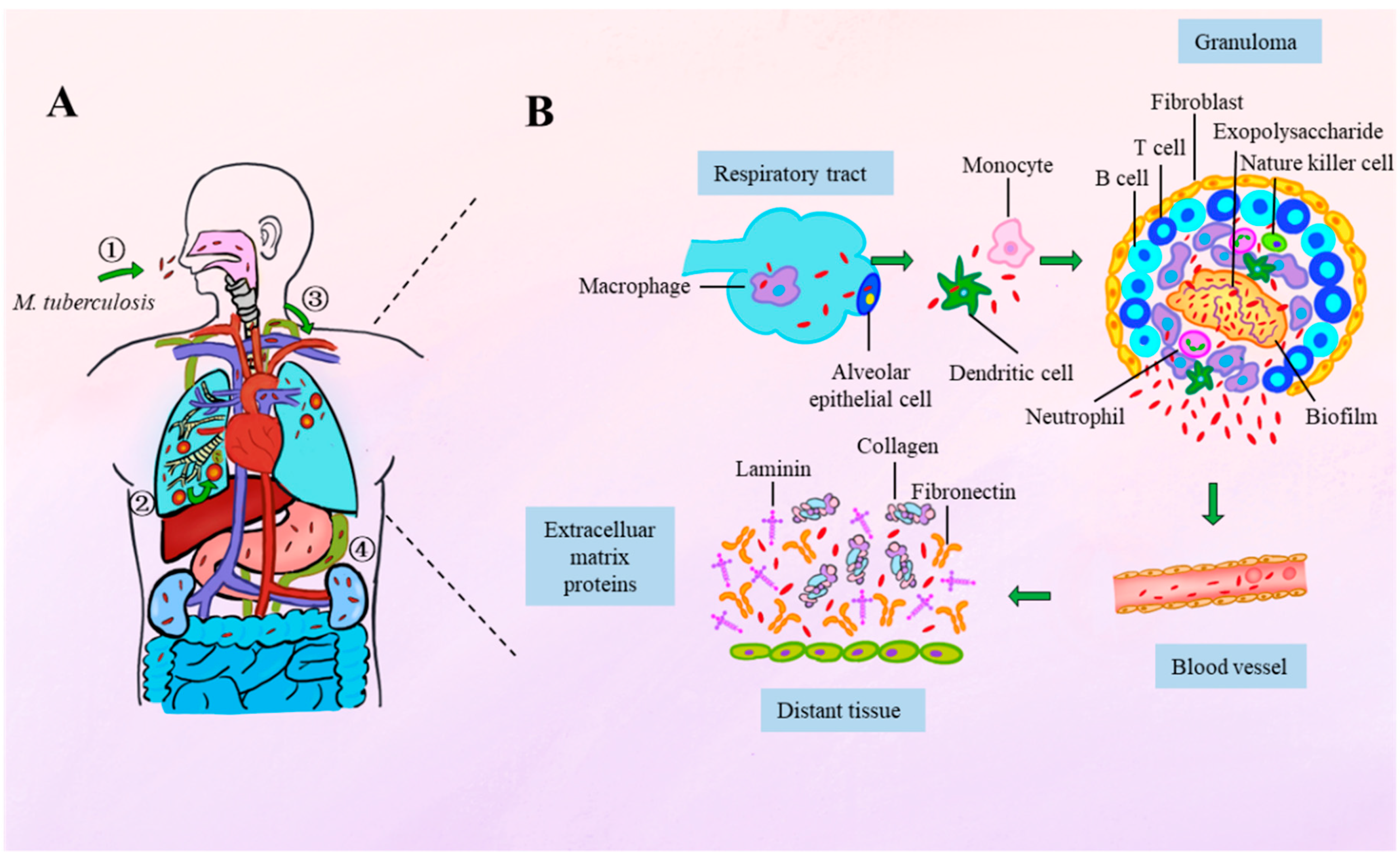
Figure 1. Spatiotemporal dynamics of M. tuberculosis infection progression and bacterial adhesion. (A) M. tuberculosis infection progression. (1) Transmission: TB is primarily contracted through inhalation of air containing M. tuberculosis. (2) Initial pulmonary infection: Inhaled bacilli reach the alveoli and are phagocytosed by alveolar macrophages, initiating a local immune response and leading to the formation of granulomas. These granulomas serve as a containment strategy but may also harbor persistent bacteria. (3) Lymphatic spread: Once the bacteria exit the lungs, they enter the lymphatic system and are likely to reach the circulatory system via the thoracic duct, which drains into the subclavian vein. (4) Extrapulmonary dissemination: Hematogenous spread allows the bacteria to colonize distant organs, leading to extrapulmonary tuberculosis. (B) Adhesion mechanisms throughout infection. Pulmonary infections initiate with adhesion to alveolar macrophages or epithelial cells. Following the migration across the alveolar epithelium, bacilli adhere to monocytes or dendritic cells. The immune response to the infection can sequester the bacteria within granulomas, where recent evidence has identified the presence of M. tuberculosis biofilms. Upon the breakdown of granulomas, the bacteria can attach to extracellular matrix (ECM) components. During the hematogenous spread, tubercle bacilli can bind plasma fibronectin, ultimately adhering to ECM proteins to facilitate invasion into new tissues.
Vaccines 13 00676 g001
Figure 2. Schematic model of the M. tuberculosis cell envelope. (A) General illustration of the M. tuberculosis bacterial cell. (B) Detailed view of the multilayered architecture of the mycobacterial cell envelope. Phosphoinositol mannosides (PIMs), shown in light and dark purple, are short glycolipids consisting of an inositol core and mannose residues. Lipomannans (LMs) are elongated derivatives of PIMs, while lipoarabinomannans (LAMs) further incorporate branched arabinose chains (depicted in pink). Peptidoglycan is represented in blue, arabinogalactan in yellow (galactan) and pink (arabinan). Trehalose molecules, illustrated as paired dark pink hexagons, are conjugated with mycolic acids to form trehalose monomycolate (TMM) and trehalose dimycolate (TDM), key components of the outer lipid barrier. Mycolic acids are indicated in orange and light green, representing structurally distinct forms with different cyclopropane ring modifications or functional groups. Phthiocerol dimycocerosate (PDIM), a virulence-associated lipid, is marked in dark red. The blue-purple spiral represents α-glucan, while the remaining protein-like structures correspond to cell envelope proteins. Other classes of lipids and membrane components are not shown in this model for simplicity.
Vaccines 13 00676 g002
Figure 3. In silico prediction of M. tuberculosis adhesins. (A) Various in silico prediction methods for identifying M. tuberculosis adhesins described in the literature. (B) Bioinformatic pipeline for predicting adhesin-like proteins from M. tuberculosis using latest general prediction methods. This pipeline consists of five sequential stages. In Stage 1, the complete proteome of M. tuberculosis H37Rv is retrieved and screened using the SPAAN to predict potential adhesins based on amino acid composition and related features. In Stage 2, functionally annotated proteins are selected using Mycobrowser, focusing on categories relevant to host–pathogen interaction. Stage 3 refines the candidates by identifying conserved hypothetical proteins, defined as uncharacterized proteins conserved across mycobacterial species, while excluding known proteins using multiple databases. In Stage 4, structure prediction is performed using Phyre2, and functional domains are analyzed using Pfam, complemented by the literature-based functional assessment. Stage 5 involves subcellular localization and secretion prediction using tools such as SubLoc, PsortB, and SignalP, prioritizing extracellular and membrane-bound proteins. Each of the five steps is visually represented as a separate module in the diagram, with arrows indicating the flow of analysis.
Vaccines 13 00676 g003
Table 1. Key mycobacterial adhesins and their host receptors.
No.AdhesinReceptorPhenotype During InfectionRefs.
1Phosphatidylinositol-mannosides (PIMs)Dendritic cell-specific ICAM-3-grabbing non-integrin (DC-SIGN)PIMs contribute to the interaction between mycobacteria and dendritic cells, although other unknown ligands may be more dominant in this process.[39]
Galectin-3Infection of galectin-3-deficient mice with M. tuberculosis reveals a reduced capacity to clear late-stage, but not early-stage, infection.[40]
2Mannose-capped lipoarabinomannan (manLAM)DC-SIGNManLAM’s mannose cap is thought to inhibit phagolysosome fusion and promote IL-10 production, but this effect is less important in interactions with living bacteria.[41]
Dectin-2Dectin-2 deficiency exacerbates lung pathology during mycobacterial infection in mice.[42,43,44]
3Arabinogalactan (AG)Galectin-9Deletion of galectin-9 blocks AG-induced lung pathology, and the AG-galectin-9 axis exacerbates M. tuberculosis infection in mice.[34]
4Mycolic acid (MA)Galectin-3N.A.[45]
Clec12AClec12A-deficient mice show enhanced innate and T cell responses after infection, while human Clec12A transgenic mice are more susceptible to M. tuberculosis infection.[46]
5Trehalose 6,6′-dimycolate (TDM)MincleIn vivo administration of TDM triggers a significant increase in inflammatory cytokines in serum and induces characteristic lung inflammation, including granuloma formation.[47,48]
Dectin-3TDM stimulation of Dectin-3 induces Mincle expression, potentially boosting the host’s innate immune response to Mycobacterium infection.[49]
Macrophage receptor with collagenous structure (MARCO)MARCO-expressing macrophages secrete pro-inflammatory cytokines in response to TDM, while macrophages from MARCO(−/−) mice produce significantly lower cytokine levels upon infection with virulent M. tuberculosis.[50,51]
6α-glucanDC-SIGNInhibits DC maturation and suppresses DC-mediated immune responses; additionally, induces IL-10 secretion.[52,53]
7Early secretory antigenic target 6 kDa (ESAT-6) *, culture filtrate protein 10 kDa (CFP-10) *N.A.The binding of the fluorescently labeled ESAT-6-CFP-10 complex to the surface of macrophages is directly mediated by the protein complex. Moreover, the flexible C-terminal arm of CFP-10 plays a crucial role in this interaction, forming an essential part of the binding site for the cell surface receptor.[54]
8ESAT-6 *LamininESAT-6 induces cytolysis in both type 1 and type 2 pneumocytes.[55]
9Antigen 85 (Ag85) complexFibronectin (Fn)Fn depletion through siRNA significantly impaired the binding of purified Ag85B to human epithelial cells, highlighting the crucial role of the Ag85B-Fn interaction in epithelial adhesion.[56]
Ag85BPlasminogen (Plg)Ag85B binds to human Plg, and promotes its activation to plasmin (Plm), potentially enhancing the extracellular matrix degradation and tissue invasion of M. tuberculosis.[57]
10Heparin-Binding Hemagglutinin Adhesin (HBHA)Heparan sulfate glycosaminoglycansFacilitates bacillary adhesion to host cells and promotes epithelial transcytosis, enabling M. tuberculosis to establish systemic infection.[58,59]
11Mycobacterium tuberculosis curli pili (MTP)N.A.An MTP-deficient strain of Mycobacterium tuberculosis exhibits a significant reduction in its ability to adhere to and invade A549 pulmonary epithelial cells, with decreases of 69.39% (p = 0.047) and 56.20% (p = 0.033), respectively.[60]
N.A.Adhesion to and invasion of macrophages are reduced by 42.16% (p = 0.107) and 69.02% (p = 0.052), respectively, in the pili-deficient Δmtp mutant compared to the wild-type.[61]
LamininMTP binds to the extracellular matrix protein laminin and contributes to biofilm formation. Isogenic mtp mutants lose the ability to produce MTP in vitro and demonstrate decreased laminin-binding capabilities.[62,63]
1219-kDa antigenDC-SIGNN.A.[64]
Mannose receptorThe 19-kDa antigen (Rv3763) binds to THP-1 macrophages through the macrophage mannose receptor (MR), promoting mycobacterial uptake.[65]
13Malate synthase (GlcB)Laminin, FnAntibodies targeting the C-terminal laminin/fibronectin-binding domain inhibit the binding of M. tuberculosis to laminin and fibronectin, significantly reducing its adherence to A549 lung epithelial cells.[66]
14Glyceraldehyde-3-phosphate dehydrogenase (GAPDH)Plg, PlmEnhances bacterial binding to and translocation across lung epithelial cell barriers.[67]
15Alanine and proline-rich protein (Apa)DC-SIGNN.A.[64]
FibronectinPeptides 177-201 and 269-292 of Apa-A specifically bind fibronectin, with peptide 269-292 additionally inhibiting full-length protein interactions.[68]
human pulmonary surfactant protein A (PSP-A)Apa remains stably associated with the cell wall, providing a platform that facilitates the attachment of PSP-A, thereby enhancing bacterial adhesion to host surfaces.[69]
16PstS-1Mannose receptorN.A.[70]
17N-acetylmuramoyl-L-alanine amidase (Rv3717)Fn, lamininN.A.[71]
18Mycobacterial mammalian cell entry protein 1A (Mce1A)N.A.Mce1A protein is crucial for Mycobacterium leprae invasion into human microvascular endothelial cells (HMVECs), and antibodies targeting Mce1A may inhibit this process.[72]
N.A.Mce1A enhances bacterial adhesion and intracellular survival, promoting infection in monocytes and 3D bovine enteroids.[73,74]
19GlnA1Plg, FnGlnA1 binds to human Plg and facilitates its activation to Plm, while also interacting with the extracellular matrix protein fibronectin, potentially enhancing M. tuberculosis tissue invasion.[57]
20Mpt64N.A.N.A.[57,75,76]
21PE_PGRS33 (Rv1818c)Toll-like receptor 2 (TLR2)PE-PGRS33 binds to TLR2 on macrophages in a calcium-dependent manner, facilitating bacterial adhesion and entry.[77,78,79]
22PE_PGRS81 (Rv1759c)FnPE-PGRS81 binds fibronectin via its C-terminal fragment and shows reactivity with sera from TB patients.[80]
* Although ESAT-6 and CFP-10 are primarily characterized as secreted virulence factors, they are included here as non-canonical adhesins based on studies reporting their direct interaction with host cells and facilitation of bacterial entry or dissemination.
Disclaimer/Publisher’s Note: The statements, opinions and data contained in all publications are solely those of the individual author(s) and contributor(s) and not of MDPI and/or the editor(s). MDPI and/or the editor(s) disclaim responsibility for any injury to people or property resulting from any ideas, methods, instructions or products referred to in the content.