Article

Synthetic Cyclic C5-Curcuminoids Increase Antioxidant Defense and Reduce Inflammation in 6-OHDA-Induced Retinoic Acid-Differentiated SH-SY5Y Cells

1
Department of Pharmaceutical Biology, Faculty of Pharmacy, University of Pécs, Rókus str. 4, H-7624 Pécs, Hungary
2
Department of Biochemistry and Medical Chemistry, Medical School, University of Pécs, Szigeti str. 12, H-7624 Pécs, Hungary
3
Institute of Pharmaceutical Chemistry, Faculty of Pharmacy, University of Pécs, Rókus str. 4, H-7624 Pécs, Hungary
*
Author to whom correspondence should be addressed.
In memoriam.
Antioxidants 2025, 14(9), 1057; https://doi.org/10.3390/antiox14091057
Submission received: 24 July 2025 / Revised: 27 August 2025 / Accepted: 27 August 2025 / Published: 28 August 2025

Abstract

Parkinson’s disease (PD) is recognized as one of the most common neurodegenerative disorders globally. The primary factor contributing to this condition is the loss of dopaminergic neurons, which results in both motor and nonmotor symptoms. The etiology of neurodegeneration remains unclear. However, it is characterized by the elevated production of reactive oxygen species, which subsequently leads to oxidative stress, lipid peroxidation, mitochondrial dysfunction, and inflammation. The investigation of the applicability of natural compounds and their derivatives to various diseases is becoming increasingly important. The possible role of curcumin from Curcuma longa L. and its derivatives in the treatment of PD has been partially investigated, but there are no data on the action of synthetic cyclic C5-curcuminoids and chalcones tested in a Parkinson’s model. Two chalcones and five synthetic cyclic C5-curcuminoids with potential antioxidant properties were investigated in an in vitro model of 6-hydroxydopamine (6-OHDA)-induced neurodegeneration in differentiated SH-SY5Y cells. Reactive oxygen species (ROS) production, total antioxidant capacity, antioxidant enzyme activity, thiol and ATP levels, caspase-3 activity, and cytokine release were examined after treatment with the test compounds. Based on these results, one cyclic chalcone (compound 5) and three synthetic cyclic C5-curcuminoids (compounds 9, 12, and 13) decreased oxidative stress and apoptosis in our in vitro model of neurodegeneration. Compounds 5 and 9 were also successful in decreasing the production of pro-inflammatory cytokines (IL-6, IL-8, and TNF-α), while promoting the release of anti-inflammatory cytokines (IL-4 and IL-10). These findings indicate that these two compounds exhibit potential antioxidant, anti-apoptotic, and anti-inflammatory properties, rendering them promising candidates for drug development.

1. Introduction

Neurodegenerative disorders are emerging diseases worldwide. According to the latest estimates, in two decades, the number of people with Parkinson’s disease (PD) will reach 14.2 million [1]. PD is characterized by motor symptoms such as muscle rigidity, tremors at rest, and balance issues, as well as nonmotor symptoms like cognitive challenges, autonomic system issues, and depressive disorders [2].
The etiology of PD remains incompletely elucidated. Factors such as aging, genetic predispositions, environmental influences, and social determinants, including lifestyle and stress, contribute to the pathogenesis of the disease [3]. Approximately 10–15% of individuals with PD, particularly those with early-onset forms, exhibit genetic variations in PD-associated genes, notably GBA1 and LRRK2, which are significant genetic risk factors [4]. The pathology of PD is characterized by the degeneration of dopaminergic neurons, leading to a reduction in dopamine synthesis. Oxidative stress, which contributes to mitochondrial dysfunction leading to energy stress and iron accumulation, initiates ROS production and lipid peroxidation, resulting in ferroptosis [5,6,7,8,9].
Treatments of PD aim to restore dopamine levels using dopamine receptor agonists, inhibitors of dopamine-degrading enzymes, monoamine oxidase (MAO)-B inhibitors, and levodopa, which is metabolized to dopamine [10,11,12,13]. Another possibility is invasive deep brain stimulation and Duopa™ therapy to decrease tremors [14,15].
In the field of pharmaceutical research, natural compounds derived from herbs often serve as essential sources of lead molecules and potential drug candidates [16,17]. Species in the Zingiberaceae (ginger) family, such as Curcuma longa L. and Curcuma domestica L., are the parent plants of turmeric (Curcuma), containing curcumin (I) as the major component (Figure 1). It exhibits a wide range of advantageous biological properties, such as antimicrobial, antifungal, anti-inflammatory, antioxidant, neuroprotective, cytotoxic, and antitumor effects [18]. The antiradical and antioxidant activities of curcumin (I) have been extensively investigated [19]. The antioxidant ability of curcumin is due to both the phenolic aromatic rings and the 1,3-dicarbonyl moiety. Monocarbonyl C5-curcumin (II) is a component of turmeric (Figure 1) and has strong antioxidant properties [19]. Truncated C5-curcumin (II) can be synthesized from curcumin (I) via pyrolysis [20]. Another chemically closely related group of natural compounds with one enone motif in their structure is the large group of chalcones (III, Figure 1). Chalcones are found in many plants and, like curcumin, have demonstrated a number of beneficial biological effects. Synthetic cyclic chalcones also possess advantageous pharmacological properties [21].
Owing to the chemical and biochemical shortcomings of curcumin (I), there is an ongoing interest in the synthesis of more stable and effective cyclic analogs of C5-curcuminoids (V) [22,23]. Their pleiotropic feature may be responsible for the coexisting antioxidant, multiple drug resistance (MDR) revertant, and other beneficial properties of these derivatives [24]. However, studies on the antioxidant abilities of cyclic C5-curcuminoids are limited.
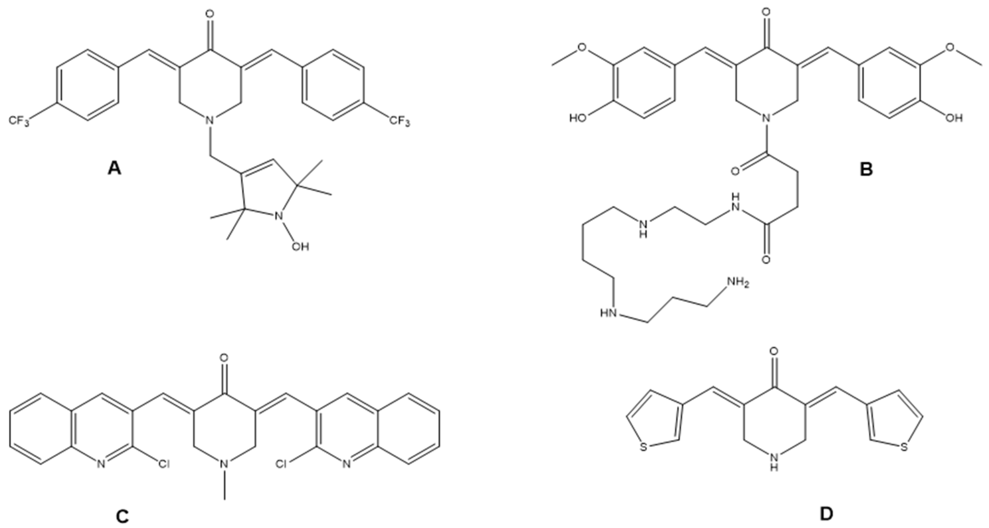
The molecules, including A and its derivatives, feature a unique –NOH function in their structure (Figure 2), which offers antioxidant protection to healthy tissues while still enabling the compounds to retain their toxicity against cancer cells. This special feature provides selective toxicity to heterocyclic C5-curcuminoid compounds [25]. Another study reported that C5-curcuminoids, such as cyclic C5-curcuminoid B conjugated with a polyamine function on the central nitrogen atom (Figure 2), allowing the compounds to cross mitochondrial membranes and develop mitochondria-directed neuroprotective effects [26].
Quinoline-based antifungal cyclic C5-curcuminoid C (Figure 2) and its derivatives prepared in a Claisen-Schmidt condensation reaction showed better radical scavenging activity than butylated hydroxytoluene [27]. Antibacterial (E. coli and S. aureus) derivative D (Figure 2) prepared from piperidin-4-one hydrochloride and thiophene-2-carboxaldehyde proved to be a strong antioxidant because of its high free radical scavenging ability in nine different assays [28].
A review has examined the potential application of curcumin and its derivatives in addressing neurodegenerative disorders, including Alzheimer’s and Parkinson’s diseases, as well as brain malignancies [29]. Another article reviewed three clinical trials, indicating that curcumin may provide modest benefits as an adjunctive treatment for patients with PD. These findings are preliminary and necessitate further trials for validation [30]. We did not identify any publications concerning synthetic cyclic C5-curcuminoids tested in an antiparkinsonian context.
In this study, we investigated two cyclic chalcones (4 and 5) and five synthetic cyclic C5-curcuminoids (8, 9, 11, 12, and 13) for their potential antioxidant properties using an in vitro model of neuronal damage induced by 6-OHDA. SH-SY5Y cells were differentiated with all-trans-retinoic acid and subsequently treated with 6-OHDA to simulate oxidative stress. These cells were then exposed to our sample compounds (4, 5, 8, 9, 11, 12, and 13), and their effects on iron accumulation, oxidative stress defense, thiol, malondialdehyde (MDA) and ATP concentrations, apoptosis, and cytokine release were assessed. The findings indicated that cyclic chalcone (5) and three synthetic cyclic C5-curcuminoids (9, 12, and 13) were effective in mitigating oxidative stress and apoptosis. Notably, only compounds 5 and 9 significantly reduced pro-inflammatory cytokine levels. Additionally, these two compounds exhibited the highest IC50 values in the in vitro model, indicating their potential as promising drug candidates.

2. Materials and Methods

2.1. Chemical Experimental Protocol

A Barnstead-Electrothermal 9100 apparatus (Thermo Fisher Scientific Inc., Waltham, MA, USA) was used to ascertain the uncorrected melting points. Silica gel 60 (0.2–0.5 mm, Merck Life Sciences Kft., Budapest, Hungary) was used for column chromatography and pre-coated silica gel 60 (F-254, Merck Life Sciences Kft., Budapest, Hungary) plates for TLC.
The nuclear magnetic resonance (NMR) 1H- and 13C-spectra were recorded using a Bruker Avance III 500 (500.15/125.77 MHz for 1H/13C) spectrometer (Bruker Corp., Ettlingen, Germany) according to our previous publication [31]. The chemical shifts were calibrated against the residual solvent signals. The measurements were conducted at a probe temperature of 298 K using a dimethyl sulfoxide (DMSO)-d6 solution. All the 1H and 13C NMR spectra were in good agreement with the expected structures.
A Thermo Dionex Ultimate 3000 liquid chromatograph (Dionex, Sunnyvale, CS, USA) coupled with a Thermo Q Exactive Focus Hybrid Quadrupole-Orbitrap mass spectrometer (Thermo Fisher Scientific, Waltham, MA, USA) was utilized for High-Performance Liquid Chromatography-Mass Spectrometry (HPLC-MS) analyses according to our previous publication [31]. For data analysis and evaluation, Q Exactive Focus 2.1 Software and Xcalibur 4.2. Software (Thermo Fisher Scientific Inc., Waltham, MA, USA) were applied. Separation of the compounds was performed using a Thermo Hypersil GOLD C18 analytical column. The mobile phase consisted of a 0.1% formic acid solution (A) and methanol containing 0.1% formic acid (B). Gradient elution was performed as follows: 0 min, 20% B; 6 min, 80% B; 8 min, 80% B; 10 min, 20% B; and 10 min stop. Chromatographic analysis was conducted at 40 °C at a flow rate of 0.3 mL/min. The injection volume was 5 μL [31].
The physical and analytical data of the synthesized compounds 4 [32], 8 and 9 [33], 11 [34], 12 [35] were identical to the original ones. The numbering of the compounds follows that of the schemes outlined in the Results section.

2.2. Cell Culture and Treatment

SH-SY5Y human neuroblastoma cells (ATCC-CRL-2266; LGC Ltd., Teddington, UK) were grown in DMEM/F12 medium (Biowest Ltd., Nuaillé, France) that was enriched with 10% fetal bovine serum (FBS; Biowest Ltd., Nuaillé, France), along with 1% non-essential amino acids (NEAA; Biowest Ltd., Nuaillé, France) and 1% penicillin/streptomycin (P/S; Biowest Ltd., Nuaillé, France). To differentiate SH-SY5Y cells into dopaminergic neurons, 1 µM all-trans retinoic acid (ATRA; Merck Life Sciences Kft., Budapest, Hungary) was added for 5 days in a complete culture medium with reduced serum (1% FBS) [36]. Due to its low iron content, it becomes depleted over the 5-day incubation period. The medium is replaced only after differentiation, with the serum content remaining reduced. To enhance iron transport into cells, the fresh culture medium was supplemented with 50 µM ferric ammonium citrate (FAC; Merck Life Sciences Kft., Budapest, Hungary) during 6-OHDA treatment. This supplement was also administered to the control cells to ensure comparability. Oxidative stress was induced after differentiation by adding 150 µM 6-OHDA (Merck Life Sciences Kft., Budapest, Hungary) for 24 h. The differentiated SH-SY5Y cells were then treated with seven different synthetic cyclic sample compounds for 24 h. Rasagiline (Merck Life Sciences Kft., Budapest, Hungary), a monoamine oxidase B inhibitor, was used as the positive control at a concentration of 1 µM for 24 h [37].

2.3. Viability Measurements

The viability of differentiated SH-SY5Y cells was assessed using a TOX8 kit (Merck Life Sciences Kft., Budapest, Hungary) following the manufacturer’s guidelines. Briefly, SH-SY5Y cells were cultured in 96-well plates at a density of 104 cells. The cells were then differentiated for 5 days with ATRA. Then, the cells were treated with the sample compounds at increasing concentrations of 50 nM–1 µM and 1 µM–20 µM. The compounds were first dissolved in 100% DMSO and subsequently diluted with the suitable culture medium before being applied to the cells. Following a 24 h treatment period, 10 µL of TOX8 reagent was introduced to each well, and the mixture was incubated for an additional 2 h. The optical density was assessed at a wavelength of 600 nm using a MultiSkan GO spectrophotometer (Thermo Fisher Scientific Inc., Waltham, MA, USA) [38]. DMSO-treated cells were used as controls. The optimal 6-OHDA and rasagiline concentrations were determined as previously described [38]. The IC50 values were derived from cell viability data using GraphPad Prism 8 software (GraphPad Software, San Diego, CA, USA).

2.4. Determination of the Reactive Oxygen Species (ROS)

SH-SY5Y cells after differentiation were seeded in a 96-well plate at a concentration of 104 cells/well and exposed to 6-OHDA treatment for 24 h. The cells were then treated with the test compounds according to their IC50 values. Three initial concentrations of each compound were used for ROS measurements (Table 1). The amount of ROS was determined in SH-SY5Y cells using a Fluorometric Intracellular ROS Kit (Merck Life Sciences Kft., Budapest, Hungary). The assay was conducted following the guidelines provided by the manufacturer. Cells were exposed to the detection reagent for 30 min in a humidified environment with 5% CO2 at 37 °C. The cells were incubated for 30 min with the detection reagent in a humidified atmosphere containing 5% CO2 at 37 °C. Fluorescence intensity was measured at an excitation wavelength of 640 nm and emission wavelength of 675 nm using an EnSight Multimode microplate reader (PerkinElmer, Rodgau, Germany). The ROS level was compared to that of DMSO-treated control cells and expressed as a percentage of the control, which was considered 100% [39].

2.5. Total Antioxidant Capacity (TAC) Measurement

The Antioxidant Assay Kit (Merck Life Science Kft., Budapest, Hungary) was utilized to measure the total antioxidant capacity encompassing both small-molecule and enzymatic antioxidant defenses. Differentiated SH-SY5Y cells were cultured and treated in 6-well plates at 5 × 10⁵ cells/well (Sarstedt Inc., Nümbrecht, Germany). The cells were then treated with 6-OHDA for 24 h. The cells were then treated with the test compounds according to their IC50 values, similar to the ROS measurements listed in Table 1. DMSO-treated cells were used as controls. Following incubation, SH-SY5Y cells were harvested by spinning them at 1200 rpm for 15 min. The resulting cell pellets were rinsed once with ice-cold 1× phosphate-buffered saline (PBS, Capricorn Scientific GmbH, Ebsdorfergrund, Germany). The pellets were then lysed in ice-cold PBS. After another round of centrifugation, 100 µL from each sample was taken for analysis. Optical density was measured at 570 nm using a MultiSkan GO spectrophotometer. Antioxidant concentrations were expressed as Trolox equivalents (µM) [40].

2.6. Glutathione Peroxidase (GPx) Activity Measurement

GPx activity was determined using a Glutathione Peroxidase Assay Kit (Merck Life Science Kft., Budapest, Hungary) according to the manufacturer’s protocol. Differentiated SH-SY5Y cells were cultured and treated in 6-well plates at 5 × 10⁵ cells/well. Cells were treated with 6-OHDA, sample compounds, and rasagiline. After incubation, SH-SY5Y cells were collected by centrifugation at 1200 rpm for 15 min. SH-SY5Y cell pellets were washed once with ice-cold 1× PBS and lysed in 200 µL of PBS by sonication. After centrifugation for 10 min at 14,000× g, glutathione peroxidase activity was measured using 10 µL of the supernatant from each sample. The optical density at 340 nm was determined using a Multiskan GO spectrophotometer. The activity of GPx was reported in units per liter (U/L).

2.7. Superoxide Dismutase (SOD) Activity Determination

SOD activity was determined using a Superoxide Dismutase Assay Kit (Merck Life Science Kft., Budapest, Hungary) according to the manufacturer’s protocol. Differentiated SH-SY5Y cells were cultured and treated in 6-well plates at 5 × 10⁵ cells/well. The cells were treated with 6-OHDA and then with the sample compounds and rasagiline. After incubation, SH-SY5Y cells were collected by centrifugation at 1200 rpm for 15 min. The cell pellets were washed once with ice-cold 1× PBS. The cells were disrupted in 500 µL of cold 1× Lysis Buffer on ice for 10 min and then collected through centrifugation. To assess SOD activity, 20 µL of the supernatant was taken from each sample. The optical density was determined at 440 nm using a Multiskan GO spectrophotometer. SOD activity was calculated following the manufacturer’s guidelines and reported in U/mL [39].

2.8. Total Intracellular Iron Content Determination

The intracellular iron content was determined using a colorimetric ferrozine-based assay described by Riemer et al. [41]. Differentiated SH-SY5Y cells were cultured at a density of 5 × 10⁵ cells/well in 6-well plates. The cells were then treated with 6-OHDA for 24 h. After incubation, cells were treated with the sample compounds and rasagiline. After incubation, SH-SY5Y cells were collected by centrifugation at 1200 rpm for 15 min. The SH-SY5Y cell pellets were washed once with ice-cold 1× PBS. The cell pellets were lysed with 200 µL of 50 mM NaOH at room temperature for 2 h. Following incubation, the lysates were neutralized by adding an equal amount of 1N HCl. Subsequently, 100 µL of the sample was combined with 100 µL of an iron-releasing reagent (1.4 M HCl, 4.5% w/v) and incubated for 2 h at 60 °C. Once the incubation was complete, the samples were allowed to cool to room temperature. Then, 30 µL of an iron detection reagent was introduced, and the mixture was left to incubate at room temperature for 30 min. The optical density was measured in 96-well plates at 550 nm using a Multiskan GO spectrophotometer. A standard curve of FeCl3 was used to determine its concentration, which was treated in the same manner as for the samples. The total iron content measurement encompasses both free and protein-bound iron within the cells, assessed after reduction in the form of the Fe2+-ferrozine complex and normalized to the protein content of the cells. The protein concentration for each sample was measured using a DC Protein Assay Kit (Bio-Rad Inc., Hercules, CA, USA). The amount of iron within the cells was quantified as µM of iron/mg of protein [36].

2.9. Thiol Concentration Measurement

The total thiol concentration of the cells was determined using a Fluorometric Thiol Assay Kit (Merck Life Science Kft., Budapest, Hungary). Differentiated SH-SY5Y cells were cultured in 25 cm2 flasks at 106 cells/flask (Sarstedt Inc., Nümbrecht, Germany). The cells were treated with 6-OHDA for 24 h, and then with the test compounds for 24 h. After incubation, the cells were collected by centrifugation at 1200 rpm for 15 min. The cell pellets were washed once with 1 mL ice-cold PBS and lysed in 100 µL assay buffer. The measurements were performed in 96-well plates following the manufacturer’s guidelines. For the analysis, 50 µL of both standards and samples were combined with 50 µL of the Detection Reagent in the wells. The plates were then incubated at RT for 1 h. Fluorescence intensity was recorded at excitation and emission wavelengths of 490/535 nm using the EnSight Multimode Plate Reader. The results were calculated based on the standard curve values of reduced glutathione (GSH) and expressed in µM [39].

2.10. Malondialdehyde (MDA) Detection

The levels of MDA were measured using the MDA Colorimetric Assay Kit (Thermo Fisher Scientific Inc., Waltham, MA, USA). Differentiated SH-SY5Y cells were cultured and treated in 25 cm2 flasks at 106 cells/flask. After incubation, SH-SY5Y cells were collected by centrifugation. The cells were homogenized in 400 μL of PBS, and 200 μL supernatant was used for the MDA determination. The optical density was determined at 532 nm using a Multiskan GO spectrophotometer. The MDA concentration was calculated using a standard curve (0–40 μmol). The results were normalized to the protein content of the supernatant and expressed as μmol/mg protein.

2.11. ATP Concentration Determination

The intracellular ATP concentration of the treated cells was measured using an ATP Determination Kit (Thermo Fisher Scientific Inc., Waltham, MA, USA). Differentiated SH-SY5Y cells were cultured and treated in 6-well plates at 5 × 10⁵ cells/well. After incubation, SH-SY5Y cells were collected by centrifugation at 1200 rpm for 15 min. SH-SY5Y cell pellets were lysed in 100 µL of 1× Reaction Buffer, and 10 µL of the lysate was added to 1× Reaction Buffer in a total volume of 100 µL. The luminescence was measured using an EnSight Multimode microplate reader. Values are expressed as nM [37].

2.12. Cytochrome c Measurement

Cytochrome c levels were determined using a Human Cytochrome c ELISA Kit (Thermo Fisher Scientific Inc., Waltham, MA, USA), according to the manufacturer’s protocol. Differentiated SH-SY5Y cells were cultured at a density of 5 × 10⁵ cells/well in 6-well plates. The cells were exposed to 6-OHDA for 24 h. Following the incubation period, cells were treated with the sample compounds and rasagiline. After incubation, SH-SY5Y cells were harvested, and the pellets were washed once with ice-cold 1× PBS. The cells were disrupted in lysis buffer for 1 h at RT with gentle shaking and then centrifuged at 200× g for 15 min. The supernatants were diluted following the manufacturer’s guidelines, and 100 µL of each was utilized for ELISA. The absorbance was recorded at 450 nm using a Multiskan GO spectrophotometer. Concentrations were calculated using SkanIt software 5.1 (Thermo Fisher Scientific, Waltham, MA, USA). The results were expressed in ng/mL [42].

2.13. Caspase-3 Activity Assay

The activity of caspase-3 was measured using a Caspase-3 Assay Kit (Merck Life Science Kft., Budapest, Hungary), following the instructions provided by the manufacturer. Differentiated SH-SY5Y cells were cultured and treated with 6-OHDA for 24 h, then with the sample compounds or rasagiline for 24 h, in 25 cm2 flasks using 106 cells/flask. DMSO-treated cells were used as controls. After incubation, SH-SY5Y cells were harvested, and then the cell pellets were washed once with ice-cold 1× PBS. The cells were lysed in 300 µL of ice-cold lysis buffer (50 mM HEPES, pH 7.2, 100 mM NaCl, 0.5% (v/v) Triton X-100) with shaking for 30 min on ice. Cell lysates were centrifuged at 2500× g for 10 min at 4 °C. Caspase-3 activity was measured in 50 µL of supernatant. The fluorescent intensity (FI) was determined at λEx 400 nm/λEm 490 nm using an EnSight Multimode Microplate Reader. The FI was normalized to the control values and expressed as ∆FI.

2.14. Enzyme-Linked Immunosorbent Assay (ELISA)

Following the administration of the sample compounds and rasagiline to SH-SY5Y cells, the culture media from both the control and treated groups were collected and kept at −80 °C until the analysis. Cytokine and chemokine secretion of the differentiated SH-SY5Y cells was determined using ELISA kits from Thermo Fisher Scientific Inc.: Human IL-4 ELISA Kit, Human IL-6 ELISA Kit, Human IL-8 ELISA Kit, Human IL-10 ELISA Kit, Human TNF-α ELISA Kit, and Human Fractalkine ELISA Kit. All the ELISA kits were used according to the manufacturer’s instructions. The levels of secreted proteins were expressed in pg/mL [40].

2.15. Statistical Analysis

The experiments were repeated three times. Five technical replicates were used for viability, TAC, and ROS measurements. Three technical replicates were performed for GPx, SOD, iron, thiol, MDA, ATP, cytochrome c, caspase-3 activity, and cytokine ELISA. Data analysis was conducted using SPSS software (version 24.0; IBM Corporation, Armonk, NY, USA). The determination of statistical significance was carried out through one-way ANOVA followed by Tukey’s post hoc test. Results are expressed as mean ± standard deviation (SD), and a p-value of less than 0.05 was considered to indicate statistical significance.

3. Results

3.1. Chemical Synthesis

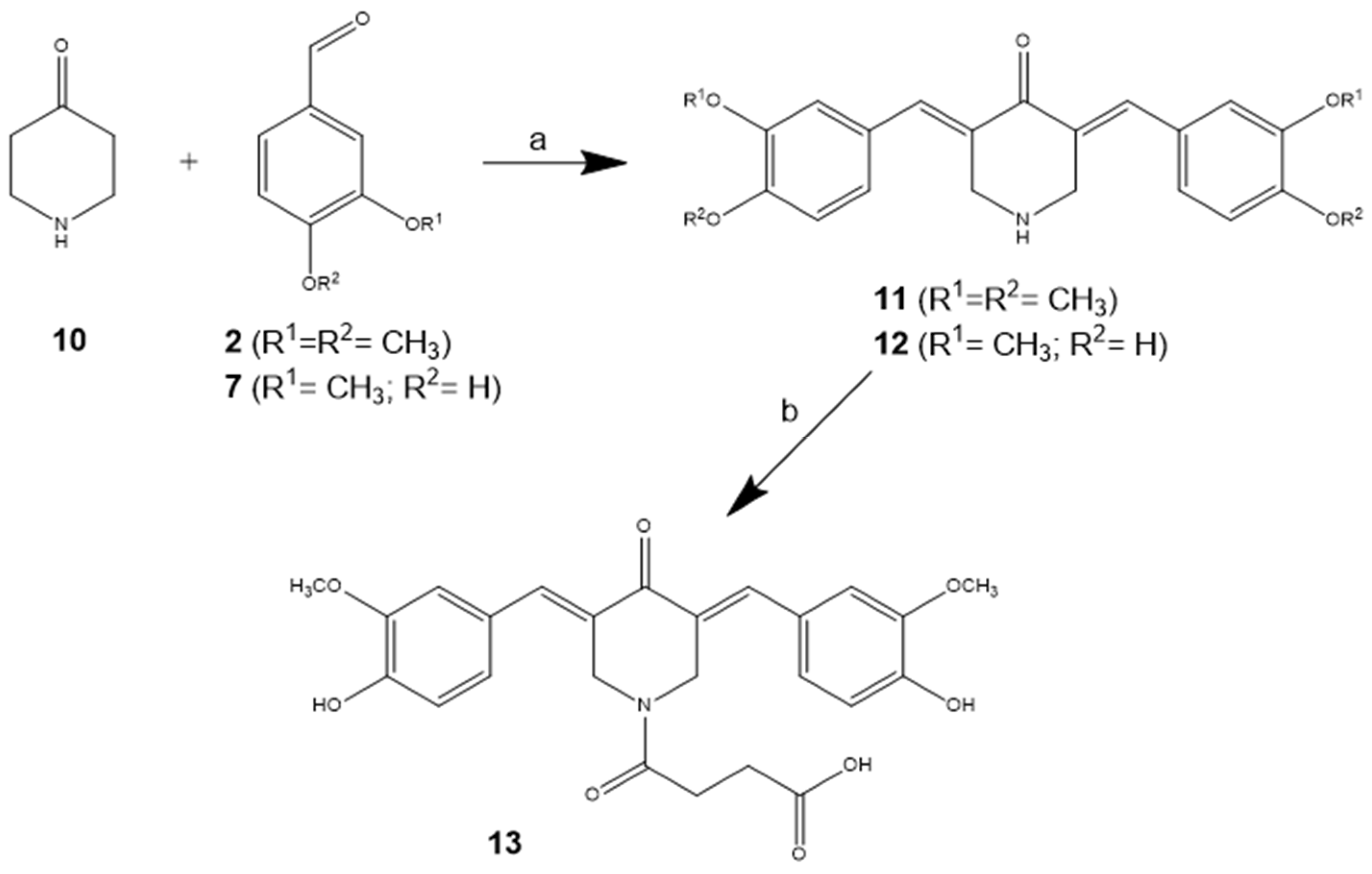
One of our major activities in our laboratory is the synthesis and pharmacological testing of new derivatives of the cyclic or heterocyclic C5-curcuminoid family, as well as analogs, such as cyclic chalcones [31,33,43]. As a continuation of this work, we decided to prepare couples of derivatives 4 and 5, 8 and 9 or 11 and 12 with phenolic and similar but nonphenolic substituents to test their differences in terms of antioxidant ability. Phenolic members 5 and 9 of the couples were formed in the acid-catalyzed Claisen-Schmidt condensation, while the others were obtained in a basic reaction milieu (Scheme 1, Scheme 2 and Scheme 3). Methanol saturated with HCl(g) was used as an acid.
4-Hydroxy-cyclohexanone (6) is a new cyclic ketone used in the design and synthesis of C5-curcuminoids. We introduced it [33] to build an additional element into molecules with hydrogen donor capabilities.
In addition to homocyclic chalcone couples 4 and 5 or homocyclic C5-curcuminoids 8 and 9, heterocyclic pairs 11 and 12 were also prepared to determine whether there was a difference in antioxidant activity. Derivative 13 contains an amide substituent on the central nitrogen, which was introduced by an acylation reaction using succinic anhydride. This substituent was a very good performer in our previous investigations [31] on astrocytoma and neuroblastoma cells. In addition, the structure of compound B (Figure 1) has mitochondria-directed neuroprotective characteristics.
2E-(3′,4′-Dimethoxybenzylidene)-1-tetralone (4) was prepared according to a previously described method for the preparation of 2-(3′,4′-methylenedioxybenzylidene)-1-tetralone in [32], starting from 1,2-dimethoxybenzaldehyde (2) and α-tetralone (1).
2-(3′,4′-Dihydroxybenzylidene)-1-tetralone (5): 5 mmol of α-tetralone (1) and 5 mmol 1,2-dihydroxybenzaldehyde (3) were dissolved in 30 mL of dry methanol saturated with dry hydrogen chloride gas. This solution was left to stand at ambient temperature overnight, diluted with 100 mL of water, and the separated crystals were collected on a glass filter. The crude product was recrystallized from methanol to produce fine pale-yellow crystals. Yield: 76%. Mp: 184–186 °C. 1H NMR (400 MHz, DMSO-d6) δ (ppm) 2.93 (t, J = 6.7 Hz, 2H), 3.09 (t, J = 6.2 Hz, 2H), 6.80–6.85 (m, 1H), 6.90 (dd, J = 1.8 Hz, 8.3 Hz, 1H), 6.99 (d, J = 1.8 Hz, 1H), 7.33–7.42 (m, 2H), 7.55 (dd, J = 1.2 Hz, 7.4 Hz, 1H), 7.58 (s, 1H), 7.93 (d, J = 7.7 Hz, 1H). 13C NMR (100 MHz, DMSO-d6) δ (ppm) 26.62, 27.74, 115.59, 117.19, 122.87, 126.44, 126.81, 127.16, 128.29, 132.03, 133.05, 133.11, 136.59, 143.03, 145.00, 146.72, 186.48. MS: m/z for C17H14O3 [M + H]+ calculated: 267.291; observed: 267.155. NMR and MS spectra are shown in Figures S1 and S2, respectively.
(2E,6E)-2,6-Bis(3′,4′-dimethoxybenzylidene)-4-hydroxycyclohexanone (8) was prepared according to a previously described method [33].
(2E,6E)-2,6-Bis(4′-hydroxy-3′-methoxybenzylidene)-4-hydroxycyclohexanone (9)was prepared according to a method described before [33].
(2E,6E)-2,6-Bis(3′,4′-dimethoxybenzylidene)-piperid-4-one (11) was prepared according to a previously described method [34].
(2E,6E)-2,6-Bis(4′-hydroxy-3′-methoxybenzylidene)-piperid-4-one (12) was prepared according to a method described before [35].
4-[(3′E,5′E)-3′,5′-bis(4′-hydroxy-3′-methoxybenzylidene)-piperid-4′-one-1′-yl]-4-oxobutane-1-carboxylic acid (13): was prepared from compound 12 and succinic anhydride according to a method described for other derivatives in [23]. Yield: 71%. Mp.: 205–206 °C, recrystallized from methanol. 1H-NMR (500 MHz, DMSO-D6) δ (ppm) 2.33 (m, 2H), 2.45 (m, 2H), 3.83 (m, 6H), 4.83 (m, 4H), 6.89 (m, 2H), 7.04 (m, 2H), 7.13 (s, 2H), 7.63 (m, 2H). 13C-NMR (125 MHz, DMSO-D6) δ (ppm) 27.0, 28.6, 42.5 *, 45.9 *, 55.6, 115.0, 115.6, 124.3, 125.6 *, 125.8 *, 129.4 *, 129.8 *, 136.3, 147.5, 148.4, 169.8, 173.4, 185.4 (*: doubling of signals). MS: m/z for C25H25NO8 [M + H]+ calculated: 468.468; observed: 468.166. NMR and MS spectra are shown in Figures S1 and S2, respectively.

3.2. IC50 Values

6-OHDA-mediated oxidative stress was observed in differentiated SH-SY5Y cells. The cells were treated with seven different cyclic C5-curcuminoids and chalcones (4, 5, 8, 9, 11, 12, and 13) for 24 h, and cell viability was determined. Considering the IC50 values, compounds 5, 9, and 13 exhibited the highest values, indicating that the cells were more tolerant to these samples. IC50 values and their concentrations used in the experiments are listed in Table 2. Rasagiline, an MAO-B inhibitor, was used as a positive control for the experiments.

3.3. Cyclic C5-Curcuminoids and Chalcones Decrease the Production of ROS by Increasing Antioxidant Defense

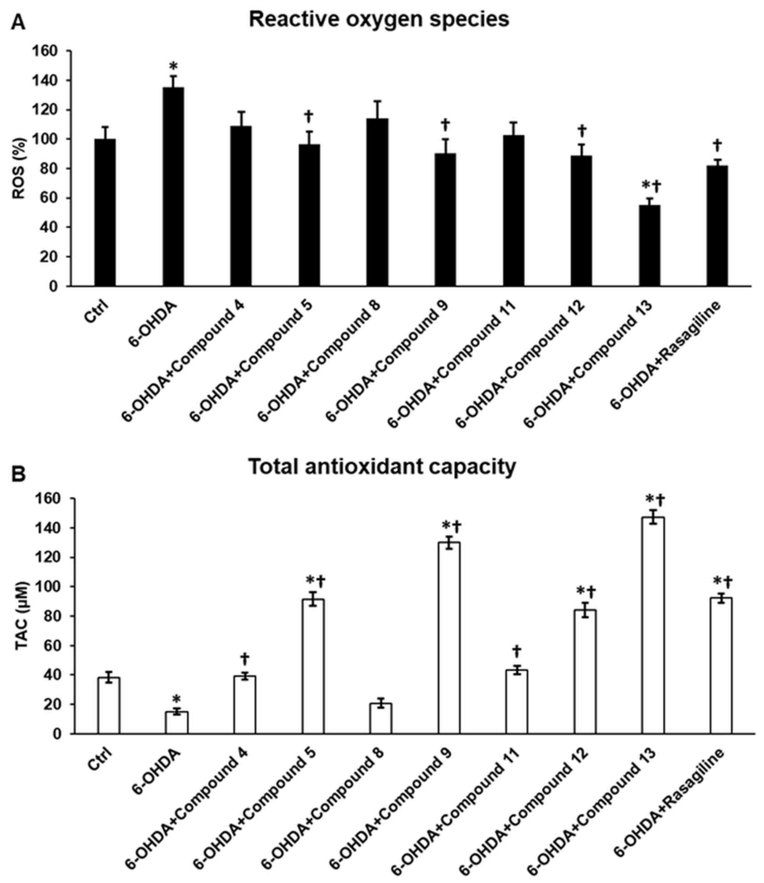
6-OHDA treatment of differentiated cells significantly increased ROS levels, indicating the induction of oxidative stress, a key feature of PD. Compounds 5, 9, 12, and 13 significantly decreased ROS production compared to 6-OHDA (135.43 ± 7.2%) and control (100 ± 8.06%). Moreover, compound 13 (55.16 ± 4.56%), which was the most potent curcuminoid, was more efficient in reducing ROS levels than rasagiline (82.25 ± 3.68%) (Figure 3A). However, the extreme ROS-reducing capacity of compound 13 may lead to a contrary biological phenomenon, known as reductive stress, a harmful process for the cells.
As ROS levels were altered in the presence of the sample compounds, we also assessed the TAC. Our findings revealed that the TAC exhibited changes that were inversely related to the alterations in ROS levels. Compounds 5, 9, 12, and 13 significantly elevated TAC levels compared to 6-OHDA (15 ± 2.11 µM) and control (38.33 ± 3.54 µM). Compound 13 (147.5 ± 4.77 µM) was the most effective and was more efficient than rasagiline (92.36 ± 3.12 µM) (Figure 3B). However, compounds 4 and 11 also increased TAC relative to 6-OHDA treatment (Figure 3B). Supplementary Figure S3 presents the results obtained from two additional treatment concentrations of the test compounds.

3.4. Cyclic C5-Curcuminoid and Chalcone Derivatives Influence the Activity of the Antioxidant Enzymes GPx and SOD

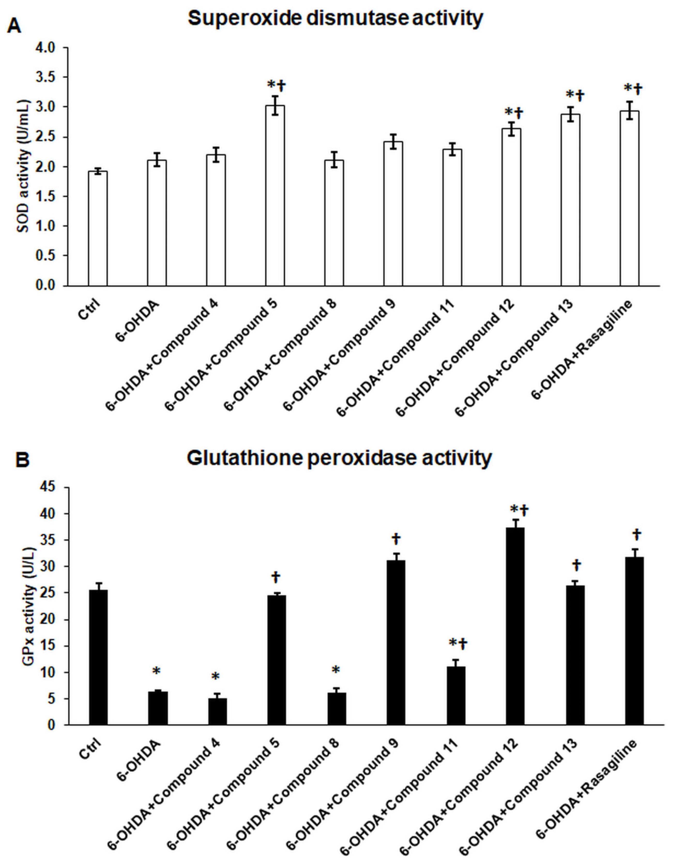
The activity of antioxidant enzymes was also determined based on TAC results. The 6-OHDA treatment of differentiated SH-SY5Y cells significantly decreased GPx activity but not SOD activity (Figure 4A,B). Compounds 5, 9, 12, and 13 demonstrated increased GPx activity, while compound 11 exhibited lower efficiency (Figure 4A). Rasagiline also effectively elevated GPx levels, with only compound 12 surpassing rasagiline in GPx levels (Figure 4A). Furthermore, compound 12 was the sole compound capable of enhancing GPx activity compared to the control.
In SOD activity measurements, we found that our sample compounds were less effective against SOD. Only compounds 5, 12, and 13 increased SOD activity with the same potency as that of rasagiline.

3.5. Cyclic C5-Curcuminoids 9, 12, 13 and Cyclic Chalcone 5 Inhibit 6-OHDA-Induced Iron Accumulation and Lipid Peroxidation

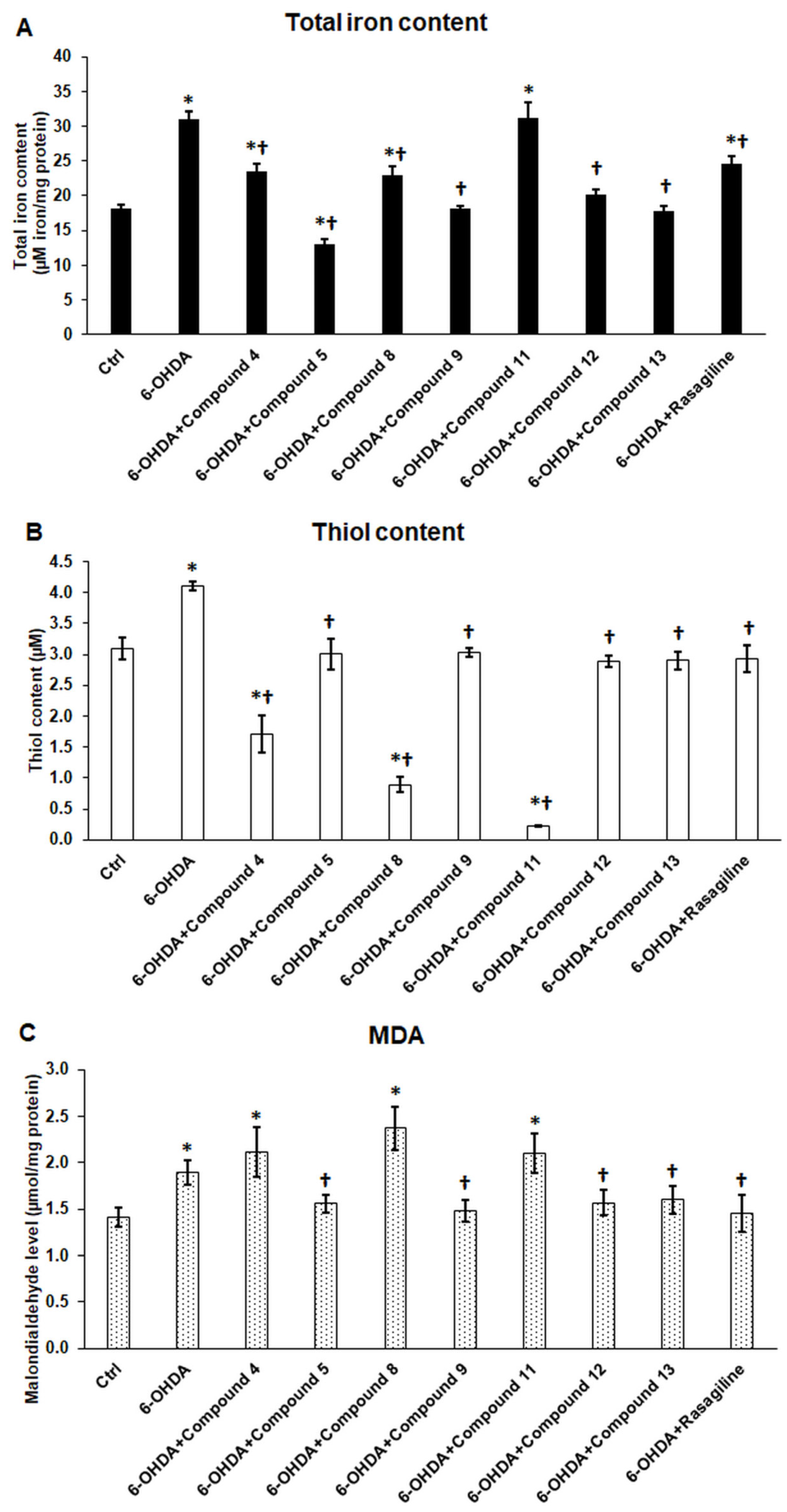
Iron accumulation in Parkinsonian neurons contributes to free radical production, thereby inducing oxidative stress and lipid peroxidation. Treatment with 6-OHDA significantly elevated the intracellular iron levels. Notably, all compounds, except compound 11, demonstrated some capacity to reduce the iron content (Figure 5A). Compound 5 demonstrated the highest efficacy, as it was the sole compound capable of reducing iron levels (13.08 ± 0.74 µM iron/mg protein) below the control level (18.15 ± 0.59 µM iron/mg protein) (Figure 5A).
Thiol levels are a suitable marker of oxidative stress. The elevated thiol levels upon 6-OHDA treatment in differentiated SH-SY5Y cells indicated the initiation of lipid peroxidation (Figure 5B). Compounds 5, 9, 12, and 13, as well as rasagiline, reduced thiol levels compared with 6-OHDA. However, they did not differ significantly from that in the control group (Figure 5B). The findings indicate that the four derivatives may offer protection against lipid peroxidation. Notably, the other three compounds, 4, 8, and 11, significantly reduced thiol levels (Figure 5B), suggesting that these compounds may interact with free thiol groups within the cells, resulting in their depletion.
MDA level, which is produced by lipid peroxidation, significantly increased upon 6-OHDA treatment of the differentiated SH-SY5Y cells. Compounds 5, 9, 12, and 13 reversed the effect of 6-OHDA, albeit without reducing the MDA levels to those observed in the control group. Notably, compounds 4, 8, and 11, which reduced intracellular total thiol levels, resulted in elevated MDA concentrations within the cells, indicating the deteriorating effects on the cells.

3.6. Cyclic C5-Curcuminoids and Cyclic Chalcones Affect Mitochondrial Function and Inhibit Apoptosis

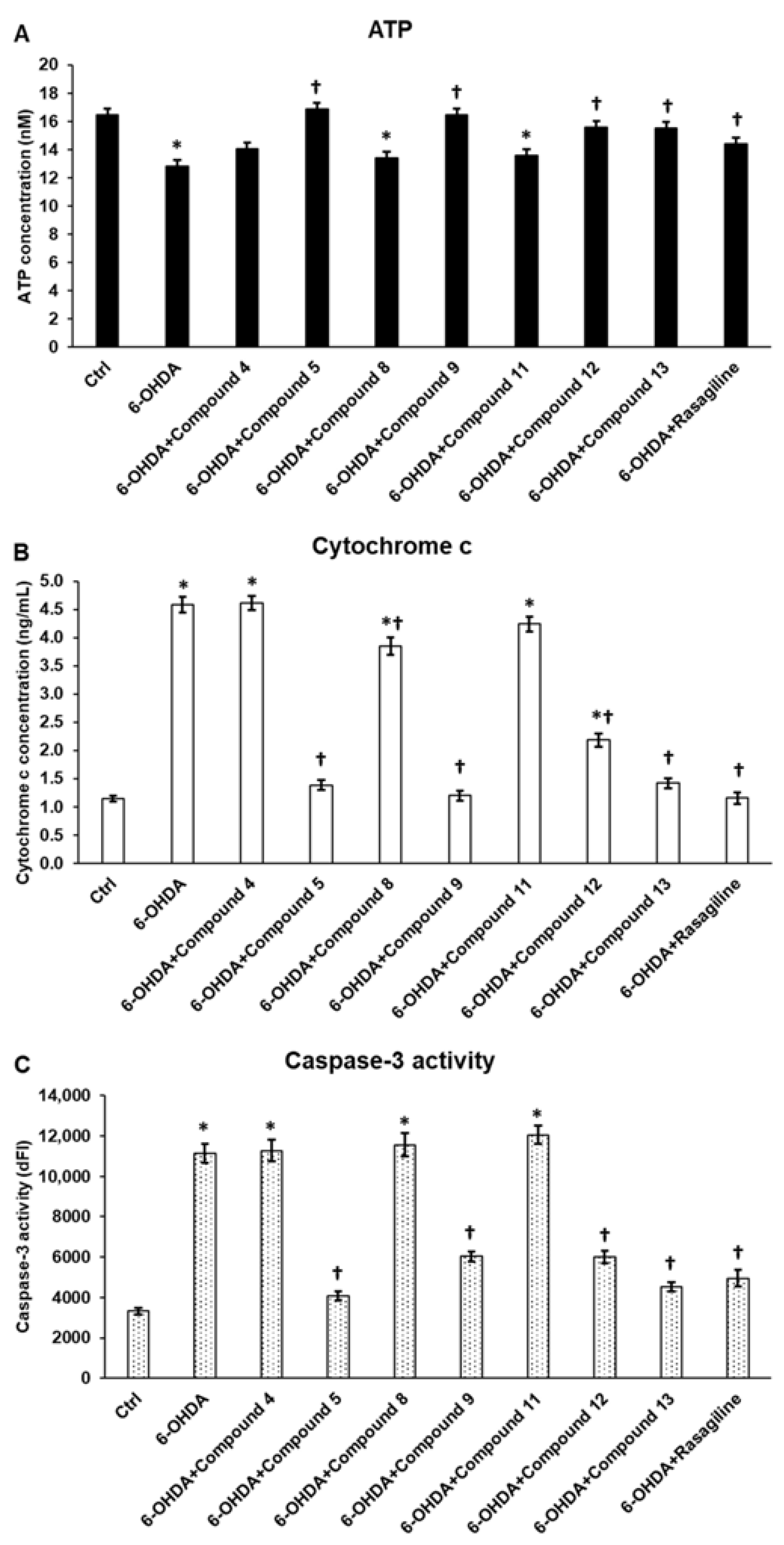
Increased ROS production may lead to mitochondrial dysfunction, reduced energy production, the release of cytochrome c, and the activation of caspase-3, an effector caspase, leading to apoptosis. The 6-OHDA treatment of differentiated SH-SY5Y cells significantly reduced ATP levels, increased cytochrome c release, and enhanced apoptosis (Figure 6A,B). Compounds 5, 9, 12, and 13 restored energy production to the control level (Figure 6A). In parallel with these findings, the aforementioned compounds significantly decreased cytochrome c concentration and caspase-3 activation (Figure 6A–C). Compound 5 was proven to act the most effectively in the in vitro 6-OHDA-induced neuronal damage model. The effect of rasagiline was similar to that of compounds 5 and 13 (Figure 6). The observed change in the case of compounds 4, 8 and 11 may not reflect the negative effects of the compounds on mitochondrial function, but rather the influence of 6-OHDA, as there was no significant difference between the 6-OHDA treatment and the 6-OHDA combined with compounds 4, 8, and 11.

3.7. Cyclic C5-Curcuminoids 9, 12, 13 and Cyclic Chalcone 5 Exhibit Minimal Influence on Anti-Inflammatory Cytokine Production of the OHDA-Induced Differentiated SH-SY5Y Cells

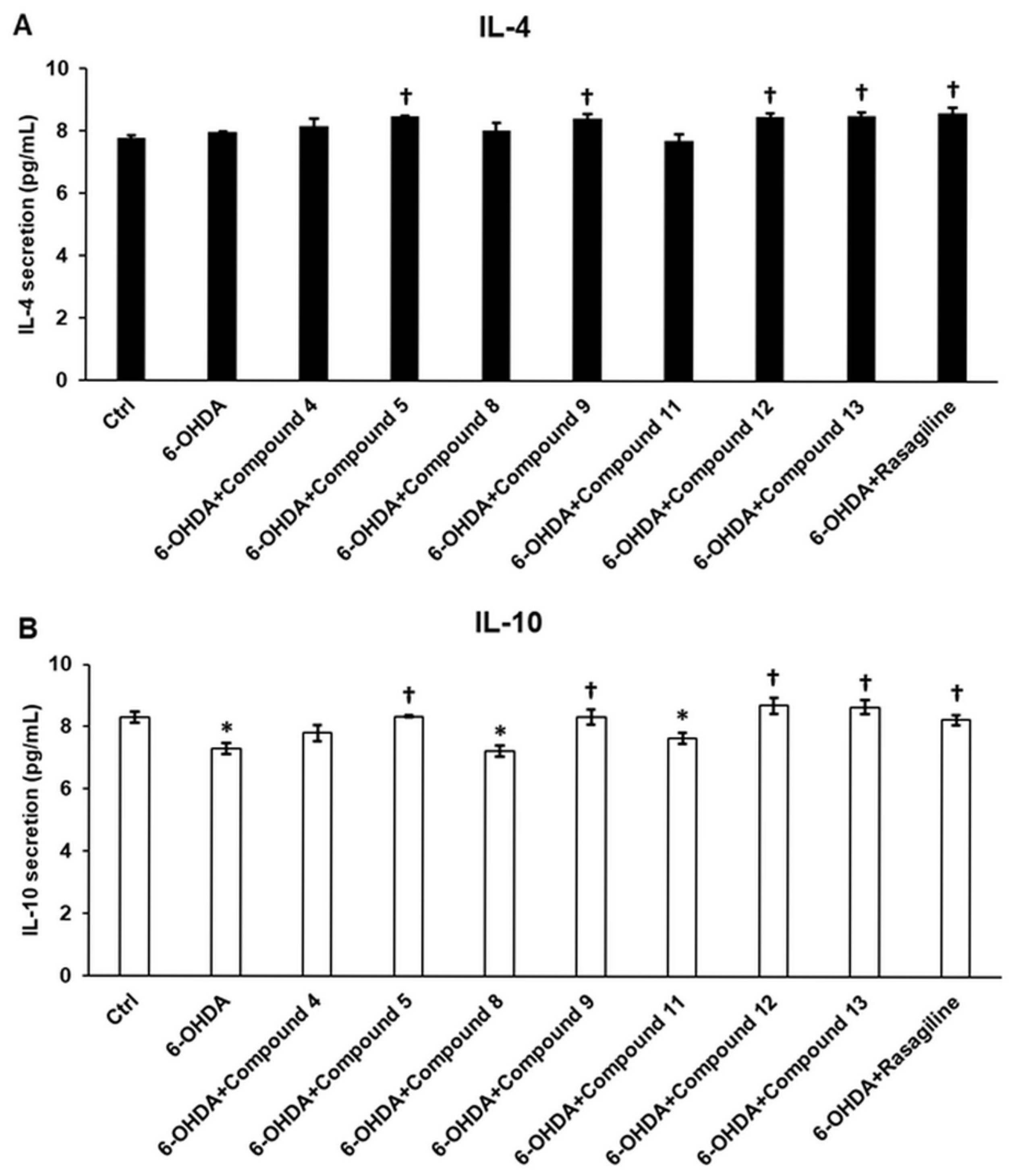
Inflammation is a crucial process in the progression of PD. The secretion of pro-inflammatory and/or anti-inflammatory cytokines influences the function of microglia, which serve as the brain’s immune cells, and can either exacerbate or mitigate neurodegeneration. 6-OHDA treatment of differentiated SH-SY5Y cells did not alter IL-4 levels but significantly decreased the release of IL-10 (Figure 7A,B). Four sample compounds, 5, 9, 12, and 13, caused minor elevations in both IL-4 and IL-10 levels, similar to the effects of rasagiline on anti-inflammatory cytokines (Figure 7A,B). Interestingly, compounds 8 and 11, akin to 6-OHDA, significantly decreased IL-10 secretion from the neurons (Figure 7B).
Based on these findings, it cannot be definitively concluded that the examined chalcones and curcuminoids possess significant anti-inflammatory properties. Their effect is limited to a modest enhancement in the production and release of the anti-inflammatory cytokines IL-4 and IL-10.

3.8. Cyclic C5-Curcuminoids and Cyclic Chalcones Reduce Pro-Inflammatory Cytokine Release of the OHDA-Induced Differentiated SH-SY5Y Cells

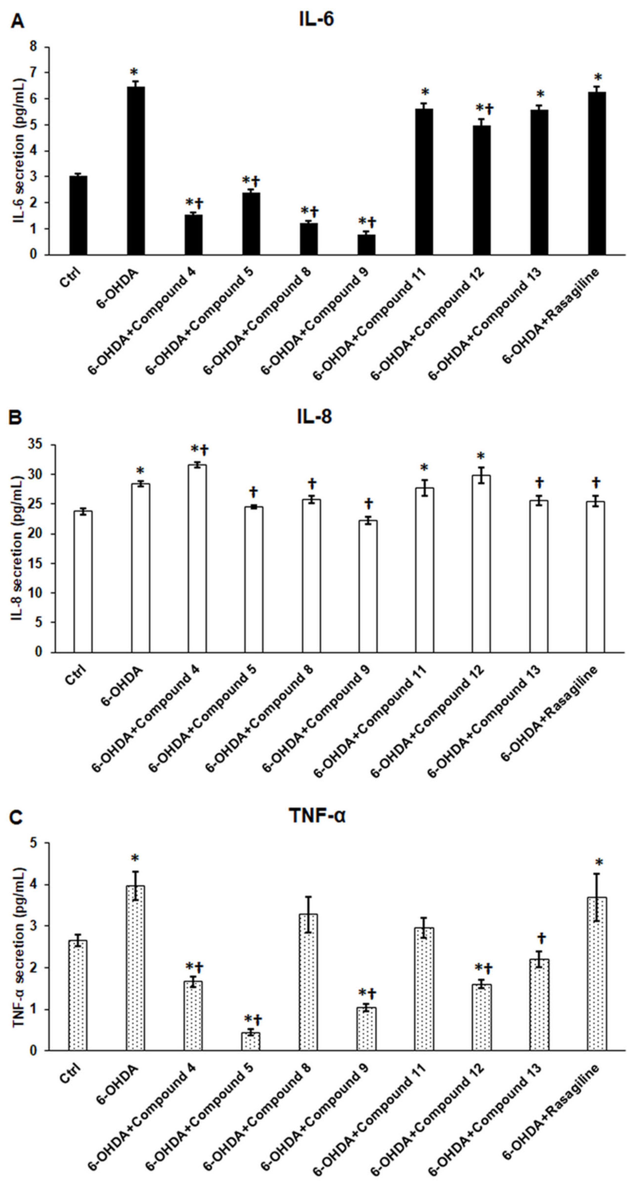
The secretion levels of the pro-inflammatory cytokines IL-6, IL-8, and TNF-α were also assessed. Treatment of differentiated SH-SY5Y cells with 6-OHDA resulted in a significant increase in the release of all three pro-inflammatory cytokines (Figure 8). We also observed that the sample compounds had few specific effects on these cytokines. Compounds 4, 5, 8, and 9 significantly decreased IL-6 secretion compared to 6-OHDA and the control; however, compounds 11, 12, and 13, as well as rasagiline, increased its secretion compared to the control (Figure 8A).
In the case of IL-8, compounds 5, 8, 9, and 13, along with rasagiline, significantly reduced IL-8 secretion compared to 6-OHDA treatment (Figure 8B). Conversely, compound 4 notably increased IL-8 release from the neurons (Figure 8B). Regarding TNF-α, compounds 4, 5, 9, 12, and 13, but not rasagiline, significantly decreased TNF-α secretion (Figure 8C).
According to these findings, only compounds 5 and 9 effectively reduced the levels of all three pro-inflammatory cytokines, while the effects of the other five compounds remain controversial.

3.9. Cyclic C5-Curcuminoids 9, 12, 13, and Cyclic Chalcone 5 Reduce the Release of Fractalkine from the OHDA-Induced Differentiated SH-SY5Y Cells

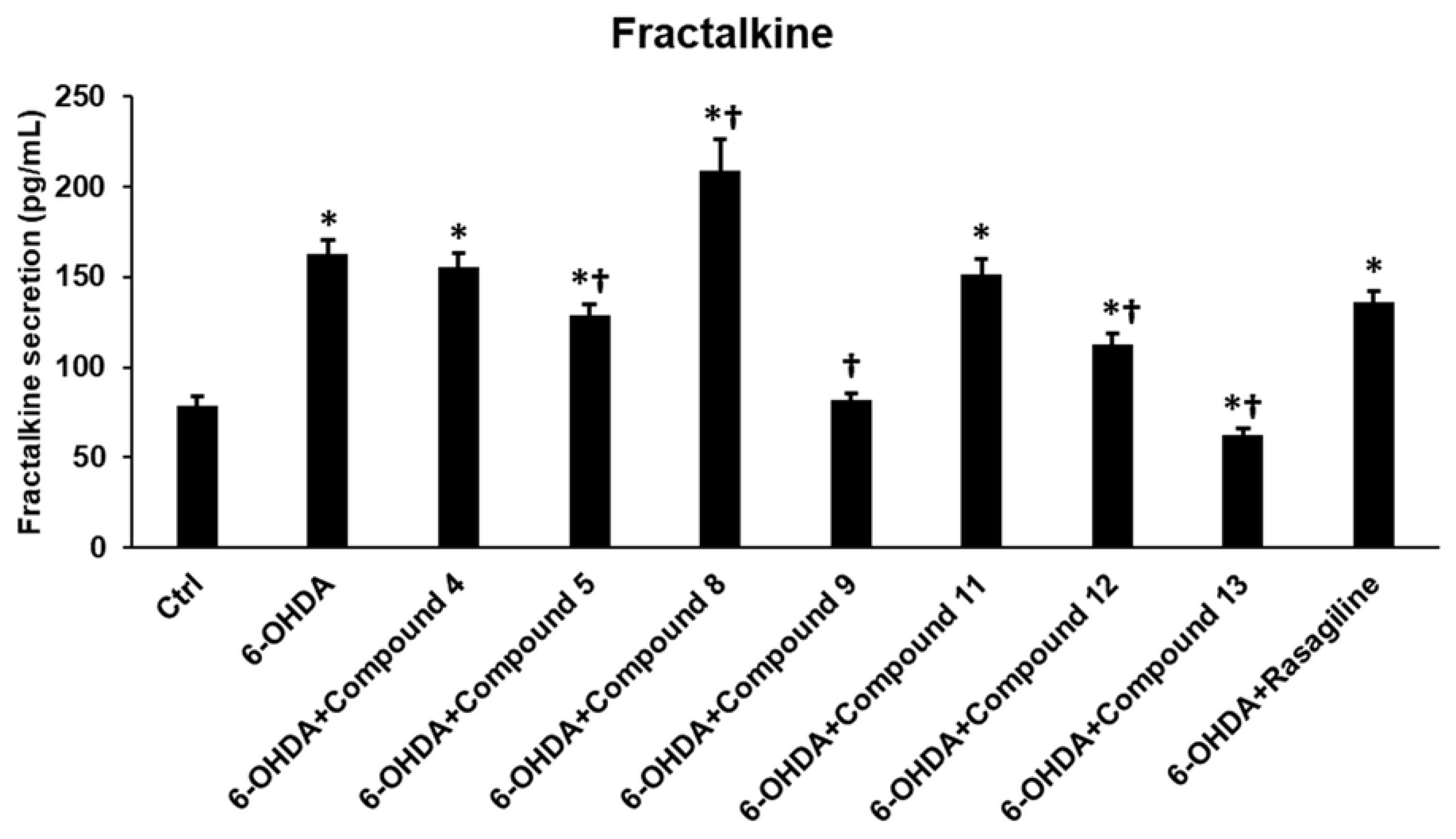
Soluble fractalkine, an inflammatory chemokine, contributes to the activation of microglia in PD. Analysis of fractalkine levels showed that compounds 5, 9, 12, and 13 significantly reduced the release of soluble fractalkine from differentiated SH-SY5Y cells compared to 6-OHDA treatment (Figure 9). Conversely, compound 8 markedly increased fractalkine secretion, while compounds 4 and 11, along with rasagiline, did not affect fractalkine production compared to 6-OHDA (Figure 9).
The results indicate that compounds 5 and 9 effectively reduced the levels of all four inflammatory proteins (IL-6, IL-8, TNF-α, and fractalkine), thereby suggesting their potential role as anti-inflammatory agents.

4. Discussion

Parkinson’s disease (PD) progression includes the overproduction of reactive oxygen species, iron accumulation, lipid peroxidation, and mitochondrial dysfunction, resulting in neuronal cell death [3]. The overactivation of microglia, which are the innate immune cells of the brain, contributes to the development and deterioration of the disease by releasing pro-inflammatory cytokines and promoting inflammation [44,45].
The utilization of plant-derived molecules and their synthetic derivatives is gaining ground in the treatment of difficult-to-treat diseases, mainly as complementary therapies [46]. The prevalence of neurodegenerative diseases (such as Parkinson’s disease, Alzheimer’s disease, Huntington’s disease, and amyotrophic lateral sclerosis) is steadily increasing worldwide, placing an ever-greater burden on society [47]. Most therapies aim only to relieve symptoms and slow the progression of the disease, mainly due to the complex background of neurodegeneration [48]. New approaches and strategies are being developed, notably stem cell treatment and gene therapy [49,50].
Turmeric (Curcuma longa L.) contains curcumin, the most abundant curcuminoid. The other three natural curcuminoids in Curcuma are demethoxycurcumin, bisdemethoxycurcumin, and cyclo-curcumin [18,51,52]. Curcumin has been shown to possess neuroprotective potential in PD models [53,54,55,56], and exhibits antibacterial, antifungal, and anticancer activities [57,58]. Recently, it was reported that the ethanol extract of Curcuma has free radical scavenging capacity [59], suggesting a beneficial effect of curcumin on reactive oxygen species. Since oxidative stress is one of the underlying causes of the progression of PD, these observations support that curcuminoids are potential antioxidant molecules and also provide neuroprotection [29,57,60]. However, no experimental data are available on the effects of synthetic cyclic C5-curcuminoids in a Parkinson’s model.
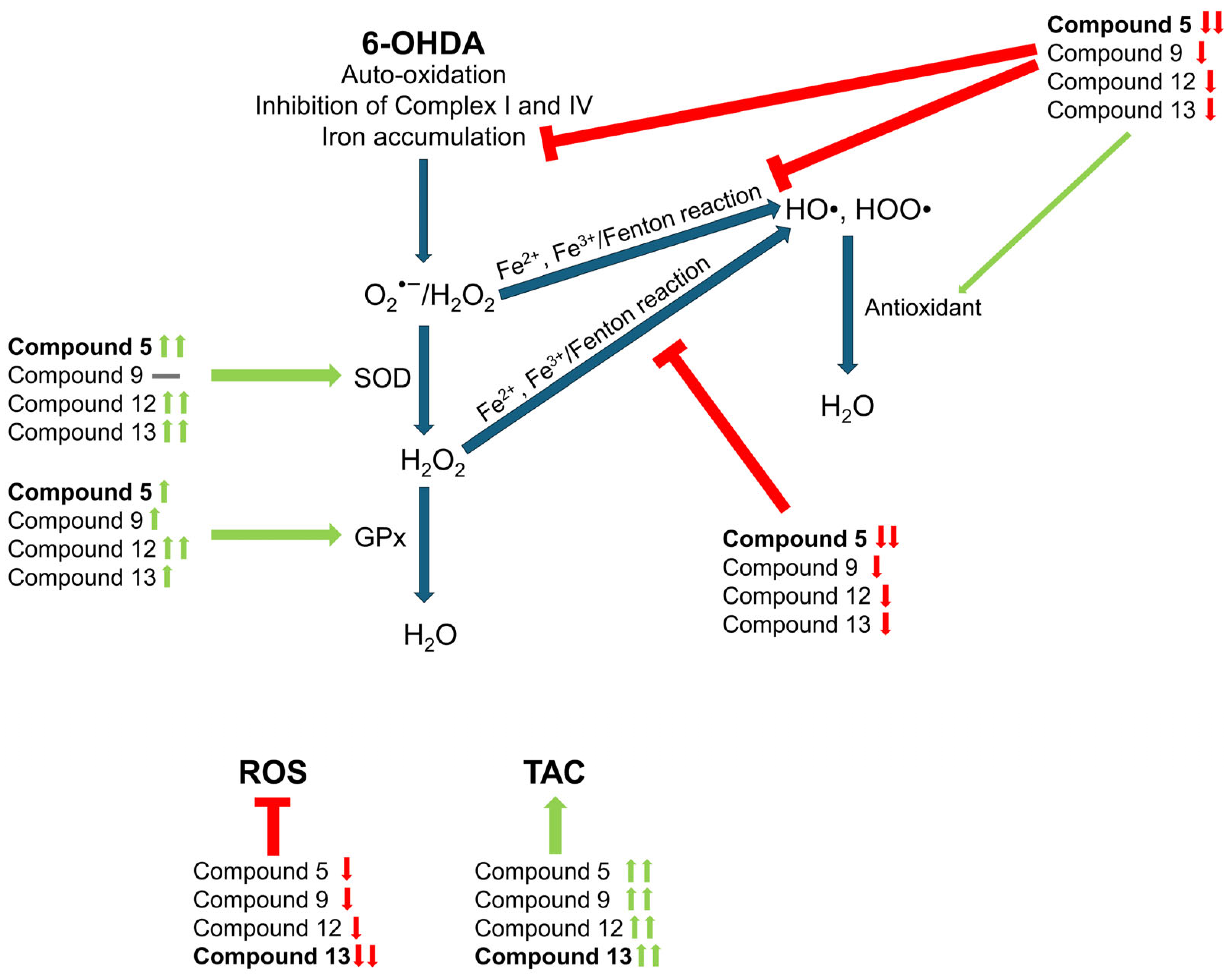
In this study, we investigated novel synthetic cyclic C5-curcuminoids for their potential antioxidant activity within an in vitro neurodegeneration model using ATRA-differentiated SH-SY5Y cells, where oxidative stress was induced by 6-OHDA treatment. Previous studies have demonstrated [37,61,62,63] that 6-OHDA treatment leads to increased iron accumulation. This occurs due to the elevated expression of iron importer transferrin receptor-1 and heme oxygenase-1, which facilitates the release of free iron into the cytosol through heme degradation. Additionally, 6-OHDA slightly decreases the expression of the ferroportin iron exporter, potentially contributing to iron accumulation [37]. Consequently, this enhances the reactive iron (Fe2+) content within the labile iron pool, potentially initiating the Fenton reaction and elevating ROS levels. Simultaneously, the auto-oxidation of 6-OHDA in cells, in the presence of oxygen, iron, or copper, leads to the generation of superoxide anion and hydrogen peroxide [64,65,66]. Furthermore, 6-OHDA inhibits Complex I and Complex IV in the mitochondrial respiratory chain, resulting in increased superoxide production [64,67,68].
Rasagiline, a MAO-B inhibitor known for its antioxidant and anti-apoptotic properties, was utilized as a positive control, providing neuroprotection [69,70]. Our investigation focused on the cellular antioxidant defense mechanisms, which involved assessing ROS production, evaluating TAC, and measuring the activity of GPx and SOD. These findings provide insights into the level of antioxidant protection in cells, attributed to small molecules and antioxidant enzymes [71].
6-OHDA treatment of differentiated SH-SY5Y cells induced ROS production but decreased TAC and GPx activity [65,72,73]. The addition of cyclic sample compounds to 6-OHDA-treated cells separated the participating derivatives into two distinct groups. Compounds 5, 9, 12, and 13 significantly reduced ROS levels while enhancing TAC, GPx, and SOD activity. Notably, compounds 9 and 12 demonstrated higher efficacy compared to rasagiline, with the exception of SOD activity. Additionally, in the case of compound 12, both SOD and GPx activities were significantly increased. The observed extreme ROS-reducing capacity of compound 13 may lead to a contrary biological phenomenon, known as reductive stress, which can result in an excess of reducing agents, mitochondrial dysfunction, metabolic disruption, and potentially increased inflammation [74,75,76]. The SOD enzymes, MnSOD and Zn/CuSOD, are essential for eliminating superoxide anions, which subsequently produce hydrogen peroxide. This hydrogen peroxide is then neutralized by GPx enzymes. Together with the thioredoxin system, these enzymes form the primary defense against oxidative damage [77]. In contrast, compounds 4, 8, and 11 in the second group did not alter ROS levels or GPx and SOD activity. However, compounds 4 and 11 did show a modest increase in TAC levels. This effect may be attributed to the direct free-radical scavenging properties of curcuminoids [59]. The ability of curcumin and curcuminoids to reduce ROS was also noted in SH-SY5Y cells infected with SARS-CoV-2 [78].
Hydroxyl radicals are produced by free iron in the cytoplasm through the Fenton reaction [77]. When excess free radicals reach macromolecules, they can inflict significant damage, leading to lipid and protein peroxidation and posing a risk to DNA integrity [79,80]. In differentiated SH-SY5Y cells, 6-OHDA increased iron content [81]. Notably, all compounds reduced iron levels, though at different rates. Compounds 5, 9, 12, and 13 lowered the iron content to the control level, while cells treated with compounds 4 and 8 exhibited significantly higher iron levels than the control, indicating their reduced effectiveness. Synthetic cyclic C5-curcuminoids effectively diminished iron accumulation, thereby contributing to a reduction in ROS production [9].
Based on the intracellular iron content, the investigated test molecules can be separated into two groups. One plausible chemical explanation for the differentiation between the two groups of tested compounds is the ability of Fe(III) to form complexes with phenolic ligands, as seen in derivatives 5, 9, 12, and 13 of the first group. A recent review article highlighted the interaction of Fe(III) with natural antioxidants, such as the Fe(III)–curcumin complex [82]. The authors concluded that curcumin has the capacity to sequester free iron, thereby preventing its detrimental accumulation within living cells. It is highly probable that, in our experiments, cells could be shielded from the accumulation of free iron through this chemical binding, resulting in neuroprotection in the case of phenolic compounds 5, 9, 12, and 13. Conversely, the complex-forming reactivity is significantly diminished in the second group (nonphenolic compounds 4, 8, and 11) due to methyl substitution blocking the phenolic ligand, thereby permitting the progression of neurodegeneration.
The thiol-containing molecules, like glutathione (GSH), protein thiols, thioredoxin, α-lipoic acid, and N-acetylcysteine, are essential for eliminating free radicals, which can result in lipid peroxidation and subsequent cellular dysfunction [83]. Thiol measurements indicated that treatment with 6-OHDA resulted in elevated thiol levels, whereas compounds 5, 9, 12, and 13 mitigated this effect. According to literature, an increase in thiol levels is indicative of the early stages of oxidative stress and lipid peroxidation [72]. During this phase, the sulfhydryl (-SH) groups of proteins, specifically cysteine and methionine amino acid residues, interact with the reactive species. This initial oxidative stress can lead to an increase in intracellular thiol levels, which subsequently decline as the concentration of ROS rises and thiol consumption, particularly of GSH, intensifies to protect the cells [84,85]. In our study, the measurement of thiols encompasses not only protein thiols and GSH but also other thiol-containing molecules such as thioredoxin. Alpha-lipoic acid, another thiol-containing compound, functions as a neuroprotective agent in PD [61] and can enhance GSH synthesis. Similarly, N-acetylcysteine is a thiol-containing antioxidant that reduces oxidative stress and promotes GSH synthesis primarily through deacetylation, which supplies cysteine for GSH synthesis [86]. It also exerts beneficial effects on dopaminergic neurons in PD [87]. Based on the total thiol measurements, we posit that the in vitro neural damage model system represents the early stage of oxidative stress, which is mitigated by compounds 5, 9, 12, and 13 but not by compounds 4, 8, and 11. In the latter case, we suggest that these cyclic C5-curcuminoids exhibit thiol-binding activity rather than ROS scavenging activity [88,89,90].
Analysis of the TAC and thiol concentrations suggests that the observed effects of the tested compounds may appear contradictory. Total antioxidant capacity measurements determine the capacity of small molecule antioxidants and antioxidant enzymes, such as SOD, GPx, and catalase. 6-OHDA treatment of differentiated SH-SY5Y cells decreased the TAC. Although the thiol level was elevated, GPx activity significantly decreased. In the case of compounds 5, 9, 12, and 13, the thiol content was similar to that of the control, but TAC was significantly increased. This can be explained by the increased GPx activity in the case of compounds 9 and 12, and the elevated SOD activity in the case of compounds 5 and 13. Moreover, in the case of compound 12, in which the highest TAC level was measured, both SOD and GPx activities were significantly increased. We cannot exclude the direct free radical scavenging capability of the test compounds, which can also increase TAC [89,90,91]. The proposed effects of compounds 5, 9, 12, and 13 are described in Scheme 4.
We evaluated the levels of MDA, a marker for lipid peroxidation. The data reveal that 6-OHDA treatment triggered lipid peroxidation, whereas compounds 5, 9, 12, and 13 alleviated the impact of 6-OHDA, although they did not bring MDA levels down to those of the control group. Remarkably, compounds 4, 8, and 11, which led to a reduction in intracellular total thiol levels, resulted in an increase in MDA concentrations within the cells. However, these increases were not statistically significant compared to 6-OHDA alone. Nonetheless, these observations suggest that compounds 4, 8, and 11 merit further investigation as potential anticancer agents.
Increased production of ROS also impacts mitochondrial function. Free radicals target the mitochondrial membrane, leading to a reduction in ATP production and the subsequent release of cytochrome c, which activates apoptosis [7,92]. In our neurodegeneration model, ATP levels were significantly reduced following 6-OHDA treatment, indicating a decrease in oxidative phosphorylation [93]. Compounds 5, 9, 12, and 13 were able to maintain ATP production at control levels, whereas treatment of 6-OHDA-induced SH-SY5Y cells with compounds 4, 8, and 11 did not mitigate the adverse effects of 6-OHDA on ATP levels.
The release of cytochrome c is contingent upon the integrity of the mitochondrial membrane. ROS can target membrane proteins, resulting in the enhanced release of cytochrome c or the opening of the permeability transition pore complex (PTPC), which increases the permeability of the outer membrane and prompts cytochrome c release [94,95]. It has been demonstrated that curcuminoids can induce apoptosis by promoting cytochrome c release and activating caspase-3 in cancer cells [96,97,98]. In our in vitro neurodegeneration model, 6-OHDA was found to elevate cytochrome c levels and increase relative caspase-3 activity in neurons [99]. Compounds 5, 9, 12, and 13, similar to rasagiline, effectively reduced cytochrome c concentrations and significantly decreased caspase-3 activity compared to 6-OHDA, highlighting the anti-apoptotic properties of these cyclic C5-curcuminoids. Conversely, compounds 4, 8, and 11 did not affect cytochrome c levels or caspase-3 activity relative to 6-OHDA, indicating a lack of anti-apoptotic effect on differentiated SHSY-5Y cells. Furthermore, compounds 5 and 9 emerged as the most potent in reducing the concentration and activity of apoptosis-related proteins.
Based on the overall results, the test compounds utilized in this study, specifically cyclic C5-curcuminoids and chalcones, can be biologically categorized into two distinct groups. The first group of compounds (5, 9, 12, and 13) demonstrated efficacy in mitigating oxidative stress, reducing oxidative damage, and enhancing mitochondrial function. Additionally, this group was effective in decreasing apoptosis. Conversely, the second group (compounds 4, 8, and 11) did not exhibit success in inhibiting oxidative stress or apoptosis. The biological differentiation between the two groups corresponded with their chemical classification. The first group comprised phenolic compounds (5, 9, 12, and 13), whereas the second group consisted of nonphenolic derivatives (4, 8, and 11). The following chemical explanations for the separation of the tested compounds can be proposed:
In our previous study [100], we demonstrated that similar compounds are capable of participating in catalytic hydrogen transfer reactions. Such hydrogen transfer reactions, involving enzymes and other peptides, are ubiquitous in biological systems [101,102]. The hydrogen-donating capabilities of our model compounds 5, 9, 12, and 13 in biochemical hydrogen-transfer reactions are attributed to their potential structural rearrangements, as illustrated in Scheme 5.
Compounds 5, 9, 12, and 13, while giving off hydrogen, were oxidized to their corresponding quinoidal products 1420. Compound 5, for example, was ortho quinone 15, compound 9 was para quinone 18, 12 to 19, and derivatives 13 to 20. The pronounced hydrogen donor property is responsible for several of the aforementioned beneficial effects, indicating that the first group of compounds (5, 9, 12, and 13) may serve as potential neuroprotective agents. The group of hydrogen-donor phenolic compounds (5, 9, 12, and 13) and their quinoidal products formed in hydrogen-transfer reactions (1420) mimic the natural quinone–hydroquinone equilibrium. One of our hypotheses posits that the potential reduction of oxidized derivatives 1420 back to compounds 5, 9, 12, and 13, utilizing physiological cellular hydrogen sources, may result in enhanced cellular protection until the tested compounds are metabolized. This hypothesis, however, requires further investigation and validation.
In contrast, the methyl substitution of phenolic OH groups with methoxy groups in compounds 4, 8, and 11 in the second group prevents such rearrangements, eliminating any potential for hydrogen donor activity. Moreover, these methoxy substituents promote thiol addition, known as the thia Michael reaction, over the benzylidene double bonds. These derivatives (4, 8, and 11) are associated with cellular toxicity rather than protection, which contributes to their harmful impact on cell viability.
Inflammation in PD is driven by neurons, microglia, brain immune cells, and astrocytes, all of which contribute to the inflammatory process [103,104,105]. Pro-inflammatory cytokines such as IL-6, TNF-α, and IL-8, along with chemokines like fractalkine and other biomolecules like glutamate, can activate microglia released by dopaminergic neurons in PD [106,107]. As PD progresses, microglial overactivation intensifies, worsening the disease [44,108]. Conversely, anti-inflammatory cytokines like IL-4 can encourage the anti-inflammatory M2 type of microglia [108]. Thus, IL-4 released by dopaminergic neurons in PD may offer neuroprotection [109]. Additionally, IL-10 modulates microglial activity, enhances phagocytic activity, and provides neuroprotection [110].
Our findings indicated that treating differentiated SH-SY5Y cells with 6-OHDA did not affect IL-4 levels but did increase the release of the anti-inflammatory cytokine IL-10. Compounds 5, 9, 12, and 13, akin to rasagiline, minimally enhanced the secretion of IL-4, a pattern also observed with IL-10; therefore, it cannot be definitively concluded that the examined chalcones and curcuminoids possess significant anti-inflammatory properties. Compounds 4, 8, and 11 did not alter IL-4 levels and, similar to 6-OHDA, reduced IL-10 release from neurons.
An analysis of the concentrations of the pro-inflammatory cytokines IL-6, IL-8, and TNF-α did not yield consistent results when compared with anti-inflammatory cytokines. Notably, compounds 4, 5, 8, and 9 significantly reduced IL-6 secretion, whereas compounds 11, 12, and 13, along with rasagiline, increased its levels. IL-8 levels were inversely correlated with those of anti-inflammatory cytokines. Compounds 5, 9, and 13 significantly reduced IL-8 release, whereas compounds 4, 8, and 11 elevated its level. TNF-α, a key mediator of inflammation, contributes to neurotoxicity in PD [111]. Its levels present a distinct perspective on the effects of the examined curcuminoids. Compounds 4, 5, 9, 12, and 13 decreased the TNF-α levels secreted by neurons, whereas compounds 8 and 11 and, interestingly, rasagiline did not.
Fractalkine, an uncommon chemokine, has been discovered in the central nervous system [112]. Membrane-bound fractalkine in the neurons maintains the resting state of microglia via the CX3CR1 fractalkine receptor expressed by the microglia [113]. On the other hand, the soluble fractalkine released from the membrane-bound form triggers microglial activation. It has also been reported that fractalkine levels increase in PD [114]. 6-OHDA treatment significantly increased fractalkine levels, whereas compounds 5, 9, 12, and 13, but not rasagiline, reduced fractalkine release from the differentiated SH-SY5Y cells. The findings indicate that cyclic C5-curcuminoids and chalcones confer neuroprotection by diminishing the secretion of soluble fractalkine, thereby contributing to the maintenance of microglial quiescence and attenuating the release of pro-inflammatory cytokines from microglia.
Interestingly, the effects of the sample compounds on inflammation could not be distinctly categorized into the previously mentioned two groups. While the anti-inflammatory properties of the first group of tested compounds are characterized by slightly elevated levels of anti-inflammatory cytokines and increased soluble fractalkine secretion, the pro-inflammatory cytokine measurements exclude compounds 12 and 13 due to their inflammation-promoting effects.

5. Conclusions

Based on the findings of this study, it can be concluded that four of the seven derivatives investigated, specifically cyclic chalcone 5 and synthetic C5-curcuminoids 9, 12, and 13, demonstrated antioxidant and anti-apoptotic effects in our in vitro model of neurodegeneration. Nevertheless, the evaluation of anti-inflammatory effects led to the exclusion of compounds 12 and 13, as compound 12 was associated with an increase in IL-6 and IL-8 pro-inflammatory cytokines, and compound 13 was linked to elevated IL-6 secretion. In addition, compound 13 markedly diminished the levels of ROS, which can induce reductive stress, a harmful condition for cells. Although these test compounds show promise as drug candidates, further in vitro investigation of cyclic chalcone 5 and synthetic C5-curcuminoid 9 is necessary to assess their impact on microglia, which play a critical role in disease progression. Subsequently, formulation and in vivo studies using an animal model of PD are required to validate their therapeutic efficacy.

Supplementary Materials

The following supporting information can be downloaded at https://www.mdpi.com/article/10.3390/antiox14091057/s1: Figure S1: MS spectra; Figure S2: NMR spectra (H and C), Figure S3: Determination of ROS production and total antioxidant capacity in 6-OHDA-induced differentiated SH-SY5Y cells using three concentrations of test compounds.

Author Contributions

Conceptualization, E.P.; methodology, E.P., I.H., G.K. and G.G.-F.; formal analysis, I.H., G.K. and G.G.-F.; investigation, E.P.; writing—original draft preparation, E.P. and I.H.; supervision, E.P.; funding acquisition, E.P. All authors have read and agreed to the published version of the manuscript.

Funding

This project was supported by the János Bolyai Research Scholarship of the Hungarian Academy of Sciences (BO/00251/24/5, Edina Pandur).

Institutional Review Board Statement

Not applicable.

Informed Consent Statement

Not applicable.

Data Availability Statement

The original contributions presented in this study are included in the article/Supplementary Materials. Further inquiries can be directed to the corresponding author.

Conflicts of Interest

The authors declare no conflicts of interest.

Abbreviations

The following abbreviations are used in this manuscript:
6-OHDA6-hydroxydopamine
ATRAall-trans retinoic acid
CX3CR1fractalkine receptor
DMSOdimethyl sulfoxide
FBSfetal bovine serum
FIfluorescence intensity
GPxglutathione peroxidase
GSHglutathione
H2O2hydrogen peroxide
HO•hydroxyl radical
HOO•hydroperoxyl
HPLChigh-performance liquid chromatography
ILinterleukin
MAO-Bmonoamine oxidase B
MDAmalondialdehyde
MDRmultiple-drug-resistance
MSmass spectrometry
NMRnuclear magnetic resonance
O2•−superoxide anion
P/Spenicillin/streptomycin
PBSphosphate-buffered saline
PDParkinson’s disease
ROSreactive oxygen species
SODsuperoxide dismutase
TACtotal antioxidant capacity
TNF-αtumor necrosis factor alpha

References

  1. Dorsey, E.R.; Bloem, B.R. The Parkinson Pandemic—A Call to Action. JAMA Neurol. 2018, 75, 9. [Google Scholar] [CrossRef] [PubMed]
  2. Adam, H.; Gopinath, S.C.B.; Md Arshad, M.K.; Adam, T.; Parmin, N.A.; Husein, I.; Hashim, U. An Update on Pathogenesis and Clinical Scenario for Parkinson’s Disease: Diagnosis and Treatment. 3 Biotech 2023, 13, 142. [Google Scholar] [CrossRef]
  3. Livingston, G.; Huntley, J.; Liu, K.Y.; Costafreda, S.G.; Selbæk, G.; Alladi, S.; Ames, D.; Banerjee, S.; Burns, A.; Brayne, C.; et al. Dementia Prevention, Intervention, and Care: 2024 Report of the Lancet Standing Commission. Lancet 2024, 404, 572–628. [Google Scholar] [CrossRef]
  4. Trevisan, L.; Gaudio, A.; Monfrini, E.; Avanzino, L.; Di Fonzo, A.; Mandich, P. Genetics in Parkinson’s Disease, State-of-the-Art and Future Perspectives. Br. Med. Bull. 2024, 149, 60–71. [Google Scholar] [CrossRef]
  5. Arya, R.; Haque, A.K.M.A.; Shakya, H.; Billah, M.M.; Parvin, A.; Rahman, M.-M.; Sakib, K.M.; Faruquee, H.M.; Kumar, V.; Kim, J.-J. Parkinson’s Disease: Biomarkers for Diagnosis and Disease Progression. Int. J. Mol. Sci. 2024, 25, 12379. [Google Scholar] [CrossRef]
  6. Henrich, M.T.; Oertel, W.H.; Surmeier, D.J.; Geibl, F.F. Mitochondrial Dysfunction in Parkinson’s Disease—A Key Disease Hallmark with Therapeutic Potential. Mol. Neurodegener. 2023, 18, 83. [Google Scholar] [CrossRef]
  7. Kawabata, T. Iron-Induced Oxidative Stress in Human Diseases. Cells 2022, 11, 2152. [Google Scholar] [CrossRef]
  8. Ding, X.; Gao, L.; Han, Z.; Eleuteri, S.; Shi, W.; Shen, Y.; Song, Z.; Su, M.; Yang, Q.; Qu, Y.; et al. Ferroptosis in Parkinson’s Disease: Molecular Mechanisms and Therapeutic Potential. Ageing Res. Rev. 2023, 91, 102077. [Google Scholar] [CrossRef] [PubMed]
  9. Chang, K.-H.; Chen, C.-M. The Role of Oxidative Stress in Parkinson’s Disease. Antioxidants 2020, 9, 597. [Google Scholar] [CrossRef]
  10. Asano, H.; Tian, Y.-S.; Hatabu, A.; Takagi, T.; Ueda, M.; Ikeda, K. Safety Comparisons among Monoamine Oxidase Inhibitors against Parkinson’s Disease Using FDA Adverse Event Reporting System. Sci. Rep. 2023, 13, 19272. [Google Scholar] [CrossRef] [PubMed]
  11. Murakami, H.; Shiraishi, T.; Umehara, T.; Omoto, S.; Iguchi, Y. Recent Advances in Drug Therapy for Parkinson’s Disease. Intern. Med. 2023, 62, 33–42. [Google Scholar] [CrossRef] [PubMed]
  12. Giladi, N.; Gurevich, T.; Djaldetti, R.; Adar, L.; Case, R.; Leibman-Barak, S.; Sasson, N.; Caraco, Y. ND0612 (Levodopa/Carbidopa for Subcutaneous Infusion) in Patients with Parkinson’s Disease and Motor Response Fluctuations: A Randomized, Placebo-Controlled Phase 2 Study. Park. Relat. Disord. 2021, 91, 139–145. [Google Scholar] [CrossRef] [PubMed]
  13. Woitalla, D.; Buhmann, C.; Hilker-Roggendorf, R.; Höglinger, G.; Koschel, J.; Müller, T.; Weise, D. Role of Dopamine Agonists in Parkinson’s Disease Therapy. J. Neural. Transm. 2023, 130, 863–873. [Google Scholar] [CrossRef]
  14. Hariz, M.; Blomstedt, P. Deep Brain Stimulation for Parkinson’s Disease. J. Intern. Med. 2022, 292, 764–778. [Google Scholar] [CrossRef]
  15. Armstrong, M.J.; Okun, M.S. Diagnosis and Treatment of Parkinson Disease. JAMA 2020, 323, 548. [Google Scholar] [CrossRef] [PubMed]
  16. Dzobo, K. The Role of Natural Products as Sources of Therapeutic Agents for Innovative Drug Discovery. In Comprehensive Pharmacology; Elsevier: Amsterdam, The Netherlands, 2022; pp. 408–422. [Google Scholar]
  17. Newman, D.J.; Cragg, G.M. Natural Products as Sources of New Drugs over the Nearly Four Decades from 01/1981 to 09/2019. J. Nat. Prod. 2020, 83, 770–803. [Google Scholar] [CrossRef]
  18. González, Y.; Mojica-Flores, R.; Moreno-Labrador, D.; Cubilla-Rios, L.; Rao, K.S.J.; Fernández, P.L.; Larionov, O.V.; Lakey-Beitia, J. Polyphenols with Anti-Inflammatory Properties: Synthesis and Biological Activity of Novel Curcumin Derivatives. Int. J. Mol. Sci. 2023, 24, 3691. [Google Scholar] [CrossRef]
  19. Bērziņa, L.; Mieriņa, I. Antiradical and Antioxidant Activity of Compounds Containing 1,3-Dicarbonyl Moiety: An Overview. Molecules 2023, 28, 6203. [Google Scholar] [CrossRef]
  20. Cozmin, M.; Lungu, I.I.; Gutu, C.; Stefanache, A.; Duceac, L.D.; Șoltuzu, B.D.; Damir, D.; Calin, G.; Bogdan Goroftei, E.R.; Grierosu, C.; et al. Turmeric: From Spice to Cure. A Review of the Anti-Cancer, Radioprotective and Anti-Inflammatory Effects of Turmeric Sourced Compounds. Front. Nutr. 2024, 11, 1399888. [Google Scholar] [CrossRef]
  21. Pál, P. Chalcones and Their Synthetic Analogs; Nova Science Publishers: Hauppauge, NY, USA, 2020. [Google Scholar]
  22. Kuzminska, J.; Szyk, P.; Mlynarczyk, D.T.; Bakun, P.; Muszalska-Kolos, I.; Dettlaff, K.; Sobczak, A.; Goslinski, T.; Jelinska, A. Curcumin Derivatives in Medicinal Chemistry: Potential Applications in Cancer Treatment. Molecules 2024, 29, 5321. [Google Scholar] [CrossRef]
  23. Vieira, T.M.; Tanajura, L.S.; Heleno, V.C.G.; Magalhães, L.G.; Crotti, A.E.M. Monoketone Curcuminoids: An Updated Review of Their Synthesis and Biological Activities. Future Pharmacol. 2024, 4, 54–77. [Google Scholar] [CrossRef]
  24. Roayapalley, P.K.; Sakagami, H.; Satoh, K.; Amano, S.; Bandow, K.; Aguilera, R.J.; Hernandez, K.G.C.; Schiaffino Bustamante, A.Y.; Dimmock, S.G.; Sharma, R.K.; et al. Cytotoxic Tumour-Selective 1,5-Diaryl-3-Oxo-1,4-Pentadienes Mounted on a Piperidine Ring. Medicines 2021, 8, 78. [Google Scholar] [CrossRef]
  25. ElNaggar, A.C.; Saini, U.; Naidu, S.; Wanner, R.; Sudhakar, M.; Fowler, J.; Nagane, M.; Kuppusamy, P.; Cohn, D.E.; Selvendiran, K. Anticancer Potential of Diarylidenyl Piperidone Derivatives, HO-4200 and H-4318, in Cisplatin Resistant Primary Ovarian Cancer. Cancer Biol. Ther. 2016, 17, 1107–1115. [Google Scholar] [CrossRef]
  26. Shi, L.; Gao, L.; Cai, S.; Xiong, Q.; Ma, Z. A Novel Selective Mitochondrial-Targeted Curcumin Analog with Remarkable Cytotoxicity in Glioma Cells. Eur. J. Med. Chem. 2021, 221, 113528. [Google Scholar] [CrossRef]
  27. Nagargoje, A.A.; Akolkar, S.V.; Siddiqui, M.M.; Subhedar, D.D.; Sangshetti, J.N.; Khedkar, V.M.; Shingate, B.B. Quinoline Based Monocarbonyl Curcumin Analogs as Potential Antifungal and Antioxidant Agents: Synthesis, Bioevaluation and Molecular Docking Study. Chem. Biodivers. 2020, 17, e1900624. [Google Scholar] [CrossRef]
  28. Nivedha, J.; Kanimozhi, K.; Olikkavi, S.; Vidhyasagar, T.; Vijayakumar, N.; Uma, C.; Rajeswari, K.; Vennila, L. In Vitro Screening for Antioxidant and Antimicrobial Properties of 3,5-Bis(E-Thienylmethylene) Piperidin-4-One, a Curcumin Analogue. Pharmacogn. Res. 2022, 14, 276–283. [Google Scholar] [CrossRef]
  29. Oglah, M.K.; Mustafa, Y.F.; Bashir, M.K.; Jasim, M.H.; Mustafa, Y.F. Curcumin and Its Derivatives: A Review of Their Biological Activities. Syst. Rev. Pharm. 2020, 11, 472–481. [Google Scholar] [CrossRef]
  30. Chang, Y.-H. Curcumin as a Potential Therapeutic Agent for Parkinson’s Disease: A Systematic Review. Front. Pharmacol. 2025, 16, 1593191. [Google Scholar] [CrossRef]
  31. Huber, I.; Pandur, E.; Sipos, K.; Barna, L.; Harazin, A.; Deli, M.A.; Tyukodi, L.; Gulyás-Fekete, G.; Kulcsár, G.; Rozmer, Z. Novel Cyclic C5-Curcuminoids Penetrating the Blood-Brain Barrier: Design, Synthesis and Antiproliferative Activity against Astrocytoma and Neuroblastoma Cells. Eur. J. Pharm. Sci. 2022, 173, 106184. [Google Scholar] [CrossRef]
  32. Al Nakib, T.; Bezjak, V.; Meegan, M.; Chandy, R. Synthesis and Antifungal Activity of Some 3-Benzylidenechroman-4-Ones, 3-Benzylidenethiochroman-4-Ones and 2-Benzylidene-1-Tetralones. Eur. J. Med. Chem. 1990, 25, 455–462. [Google Scholar] [CrossRef]
  33. Huber, I.; Zupkó, I.; Gyovai, A.; Horváth, P.; Kiss, E.; Gulyás-Fekete, G.; Schmidt, J.; Perjési, P. A Novel Cluster of C5-Curcuminoids: Design, Synthesis, In Vitro Antiproliferative Activity and DNA Binding of Bis(Arylidene)-4-Cyclanone Derivatives Based on 4-Hydroxycyclohexanone Scaffold. Res. Chem. Intermed. 2019, 45, 4711–4735. [Google Scholar] [CrossRef]
  34. Das, S.; Das, U.; Selvakumar, P.; Sharma, R.K.; Balzarini, J.; De Clercq, E.; Molnár, J.; Serly, J.; Baráth, Z.; Schatte, G.; et al. 3,5-Bis(Benzylidene)-4-oxo-1-phosphonopiperidines and Related Diethyl Esters: Potent Cytotoxins with Multi-Drug-Resistance Reverting Properties. ChemMedChem 2009, 4, 1831–1840. [Google Scholar] [CrossRef]
  35. Fawzy, I.M.; Youssef, K.M.; Ismail, N.S.M.; Gullbo, J.; Abouzid, K.A.M. Newly Designed and Synthesized Curcumin Analogs with in Vitro Cytotoxicity and Tubulin Polymerization Activity. Chem. Biol. Drug Des. 2015, 86, 80–90. [Google Scholar] [CrossRef]
  36. Pandur, E.; Varga, E.; Tamási, K.; Pap, R.; Nagy, J.; Sipos, K. Effect of Inflammatory Mediators Lipopolysaccharide and Lipoteichoic Acid on Iron Metabolism of Differentiated SH-SY5Y Cells Alters in the Presence of BV-2 Microglia. Int. J. Mol. Sci. 2018, 20, 17. [Google Scholar] [CrossRef]
  37. Pandur, E.; Major, B.; Rák, T.; Sipos, K.; Csutak, A.; Horváth, G. Linalool and Geraniol Defend Neurons from Oxidative Stress, Inflammation, and Iron Accumulation in In Vitro Parkinson’s Models. Antioxidants 2024, 13, 917. [Google Scholar] [CrossRef]
  38. Pap, R.; Pandur, E.; Jánosa, G.; Sipos, K.; Nagy, T.; Agócs, A.; Deli, J. Lutein Decreases Inflammation and Oxidative Stress and Prevents Iron Accumulation and Lipid Peroxidation at Glutamate-Induced Neurotoxicity. Antioxidants 2022, 11, 2269. [Google Scholar] [CrossRef]
  39. Pap, R.; Pandur, E.; Jánosa, G.; Sipos, K.; Fritz, F.R.; Nagy, T.; Agócs, A.; Deli, J. Protective Effects of 3′-Epilutein and 3′-Oxolutein against Glutamate-Induced Neuronal Damage. Int. J. Mol. Sci. 2023, 24, 12008. [Google Scholar] [CrossRef] [PubMed]
  40. Pandur, E.; Pap, R.; Sipos, K. Activated THP-1 Macrophage-Derived Factors Increase the Cytokine, Fractalkine, and EGF Secretions, the Invasion-Related MMP Production, and Antioxidant Activity of HEC-1A Endometrium Cells. Int. J. Mol. Sci. 2024, 25, 9624. [Google Scholar] [CrossRef] [PubMed]
  41. Riemer, J.; Hoepken, H.H.; Czerwinska, H.; Robinson, S.R.; Dringen, R. Colorimetric Ferrozine-Based Assay for the Quantitation of Iron in Cultured Cells. Anal. Biochem. 2004, 331, 370–375. [Google Scholar] [CrossRef]
  42. Chiu, C.-C.; Yeh, T.-H.; Lu, C.-S.; Huang, Y.-C.; Cheng, Y.-C.; Huang, Y.-Z.; Weng, Y.-H.; Liu, Y.-C.; Lai, S.-C.; Chen, Y.-L.; et al. PARK14 PLA2G6 Mutants Are Defective in Preventing Rotenone-Induced Mitochondrial Dysfunction, ROS Generation and Activation of Mitochondrial Apoptotic Pathway. Oncotarget 2017, 8, 79046–79060. [Google Scholar] [CrossRef]
  43. Huber, I.; Rozmer, Z.; Gyöngyi, Z.; Budán, F.; Horváth, P.; Kiss, E.; Perjési, P. Structure Activity Relationship Analysis of Antiproliferative Cyclic C5-Curcuminoids without DNA Binding: Design, Synthesis, Lipophilicity and Biological Activity. J. Mol. Struct. 2020, 1206, 127661. [Google Scholar] [CrossRef]
  44. Trainor, A.R.; MacDonald, D.S.; Penney, J. Microglia: Roles and Genetic Risk in Parkinson’s Disease. Front. Neurosci. 2024, 18, 1506358. [Google Scholar] [CrossRef] [PubMed]
  45. Gao, C.; Jiang, J.; Tan, Y.; Chen, S. Microglia in Neurodegenerative Diseases: Mechanism and Potential Therapeutic Targets. Signal Transduct. Target. Ther. 2023, 8, 359. [Google Scholar] [CrossRef] [PubMed]
  46. Chaachouay, N.; Zidane, L. Plant-Derived Natural Products: A Source for Drug Discovery and Development. Drugs Drug Candidates 2024, 3, 184–207. [Google Scholar] [CrossRef]
  47. Huang, Y.; Li, Y.; Pan, H.; Han, L. Global, Regional, and National Burden of Neurological Disorders in 204 Countries and Territories Worldwide. J. Glob. Health 2023, 13, 04160. [Google Scholar] [CrossRef]
  48. Wareham, L.K.; Liddelow, S.A.; Temple, S.; Benowitz, L.I.; Di Polo, A.; Wellington, C.; Goldberg, J.L.; He, Z.; Duan, X.; Bu, G.; et al. Solving Neurodegeneration: Common Mechanisms and Strategies for New Treatments. Mol. Neurodegener. 2022, 17, 23. [Google Scholar] [CrossRef]
  49. Palanisamy, C.P.; Pei, J.; Alugoju, P.; Anthikapalli, N.V.A.; Jayaraman, S.; Veeraraghavan, V.P.; Gopathy, S.; Roy, J.R.; Janaki, C.S.; Thalamati, D.; et al. New Strategies of Neurodegenerative Disease Treatment with Extracellular Vesicles (EVs) Derived from Mesenchymal Stem Cells (MSCs). Theranostics 2023, 13, 4138–4165. [Google Scholar] [CrossRef]
  50. de la Fuente, A.G.; Pelucchi, S.; Mertens, J.; Di Luca, M.; Mauceri, D.; Marcello, E. Novel Therapeutic Approaches to Target Neurodegeneration. Br. J. Pharmacol. 2023, 180, 1651–1673. [Google Scholar] [CrossRef]
  51. Stohs, S.J.; Chen, O.; Ray, S.D.; Ji, J.; Bucci, L.R.; Preuss, H.G. Highly Bioavailable Forms of Curcumin and Promising Avenues for Curcumin-Based Research and Application: A Review. Molecules 2020, 25, 1397. [Google Scholar] [CrossRef]
  52. Shahrajabian, M.H.; Sun, W. The Golden Spice for Life: Turmeric with the Pharmacological Benefits of Curcuminoids Components, Including Curcumin, Bisdemethoxycurcumin, and Demethoxycurcumins. Curr. Org. Synth. 2024, 21, 665–683. [Google Scholar] [CrossRef]
  53. Di Martino, R.M.C.; Pruccoli, L.; Bisi, A.; Gobbi, S.; Rampa, A.; Martinez, A.; Pérez, C.; Martinez-Gonzalez, L.; Paglione, M.; Di Schiavi, E.; et al. Novel Curcumin-Diethyl Fumarate Hybrid as a Dualistic GSK-3β Inhibitor/Nrf2 Inducer for the Treatment of Parkinson’s Disease. ACS Chem. Neurosci. 2020, 11, 2728–2740. [Google Scholar] [CrossRef] [PubMed]
  54. Ramires, O.V., Jr.; Alves, B.d.S.; Barros, P.A.B.; Rodrigues, J.L.; Ferreira, S.P.; Monteiro, L.K.S.; Araújo, G.d.M.S.; Fernandes, S.S.; Vaz, G.R.; Dora, C.L.; et al. Nanoemulsion Improves the Neuroprotective Effects of Curcumin in an Experimental Model of Parkinson’s Disease. Neurotox. Res. 2021, 39, 787–799. [Google Scholar] [CrossRef] [PubMed]
  55. El Nebrisi, E.; Javed, H.; Ojha, S.K.; Oz, M.; Shehab, S. Neuroprotective Effect of Curcumin on the Nigrostriatal Pathway in a 6-Hydroxydopmine-Induced Rat Model of Parkinson’s Disease Is Mediated by A7-Nicotinic Receptors. Int. J. Mol. Sci. 2020, 21, 7329. [Google Scholar] [CrossRef]
  56. Xu, L.; Hao, L.-P.; Yu, J.; Cheng, S.-Y.; Li, F.; Ding, S.-M.; Zhang, R. Curcumin Protects against Rotenone-Induced Parkinson’s Disease in Mice by Inhibiting Microglial NLRP3 Inflammasome Activation and Alleviating Mitochondrial Dysfunction. Heliyon 2023, 9, e16195. [Google Scholar] [CrossRef]
  57. Nebrisi, E. El Neuroprotective Activities of Curcumin in Parkinson’s Disease: A Review of the Literature. Int. J. Mol. Sci. 2021, 22, 11248. [Google Scholar] [CrossRef]
  58. Dai, C.; Lin, J.; Li, H.; Shen, Z.; Wang, Y.; Velkov, T.; Shen, J. The Natural Product Curcumin as an Antibacterial Agent: Current Achievements and Problems. Antioxidants 2022, 11, 459. [Google Scholar] [CrossRef]
  59. Wu, H.; Liu, Z.; Zhang, Y.; Gao, B.; Li, Y.; He, X.; Sun, J.; Choe, U.; Chen, P.; Blaustein, R.A.; et al. Chemical Composition of Turmeric (Curcuma longa L.) Ethanol Extract and Its Antimicrobial Activities and Free Radical Scavenging Capacities. Foods 2024, 13, 1550. [Google Scholar] [CrossRef]
  60. Sathyabhama, M.; Priya Dharshini, L.C.; Karthikeyan, A.; Kalaiselvi, S.; Min, T. The Credible Role of Curcumin in Oxidative Stress-Mediated Mitochondrial Dysfunction in Mammals. Biomolecules 2022, 12, 1405. [Google Scholar] [CrossRef]
  61. Tai, S.; Zheng, Q.; Zhai, S.; Cai, T.; Xu, L.; Yang, L.; Jiao, L.; Zhang, C. Alpha-Lipoic Acid Mediates Clearance of Iron Accumulation by Regulating Iron Metabolism in a Parkinson’s Disease Model Induced by 6-OHDA. Front. Neurosci. 2020, 14, 612. [Google Scholar] [CrossRef] [PubMed]
  62. Jia, R.; Liu, Y.; Shuai, K.; Zhou, C.; Chen, L.; Zhu, L.; Wu, X.-M. The Relationship between Iron and LRRK2 in a 6-OHDA-Induced Parkinson’s Disease Model. Int. J. Mol. Sci. 2023, 24, 3709. [Google Scholar] [CrossRef] [PubMed]
  63. Xu, Q.; Kanthasamy, A.G.; Jin, H.; Reddy, M.B. Hepcidin Plays a Key Role in 6-OHDA Induced Iron Overload and Apoptotic Cell Death in a Cell Culture Model of Parkinson’s Disease. Park. Dis. 2016, 2016, 8684130. [Google Scholar] [CrossRef]
  64. Drechsel, D.A.; Patel, M. Role of Reactive Oxygen Species in the Neurotoxicity of Environmental Agents Implicated in Parkinson’s Disease. Free. Radic. Biol. Med. 2008, 44, 1873–1886. [Google Scholar] [CrossRef]
  65. Pantic, I.; Cumic, J.; Skodric, S.R.; Dugalic, S.; Brodski, C. Oxidopamine and Oxidative Stress: Recent Advances in Experimental Physiology and Pharmacology. Chem. Biol. Interact. 2021, 336, 109380. [Google Scholar] [CrossRef] [PubMed]
  66. Saito, Y.; Nishio, K.; Ogawa, Y.; Kinumi, T.; Yoshida, Y.; Masuo, Y.; Niki, E. Molecular Mechanisms of 6-Hydroxydopamine-Induced Cytotoxicity in PC12 Cells: Involvement of Hydrogen Peroxide-Dependent and -Independent Action. Free. Radic. Biol. Med. 2007, 42, 675–685. [Google Scholar] [CrossRef]
  67. Glinka, Y.; Tipton, K.F.; Youdim, M.B.H. Nature of Inhibition of Mitochondrial Respiratory Complex I by 6-Hydroxydopamine. J. Neurochem. 1996, 66, 2004–2010. [Google Scholar] [CrossRef]
  68. Tirichen, H.; Yaigoub, H.; Xu, W.; Wu, C.; Li, R.; Li, Y. Mitochondrial Reactive Oxygen Species and Their Contribution in Chronic Kidney Disease Progression Through Oxidative Stress. Front. Physiol. 2021, 12, 627837. [Google Scholar] [CrossRef]
  69. Lecht, S.; Lahiani, A.; Klazas, M.; Naamneh, M.S.; Rubin, L.; Dong, J.; Zheng, W.; Lazarovici, P. Rasagiline Exerts Neuroprotection towards Oxygen–Glucose-Deprivation/Reoxygenation-Induced GAPDH-Mediated Cell Death by Activating Akt/Nrf2 Signaling. Biomedicines 2024, 12, 1592. [Google Scholar] [CrossRef]
  70. Naoi, M.; Maruyama, W.; Shamoto-Nagai, M. Neuroprotective Function of Rasagiline and Selegiline, Inhibitors of Type B Monoamine Oxidase, and Role of Monoamine Oxidases in Synucleinopathies. Int. J. Mol. Sci. 2022, 23, 11059. [Google Scholar] [CrossRef]
  71. Kozlov, A.V.; Javadov, S.; Sommer, N. Cellular ROS and Antioxidants: Physiological and Pathological Role. Antioxidants 2024, 13, 602. [Google Scholar] [CrossRef] [PubMed]
  72. Pieńkowska, N.; Bartosz, G.; Sadowska-Bartosz, I. Effect of 6-Hydroxydopamine Increase the Glutathione Level in SH-SY5Y Human Neuroblastoma Cells. Acta Biochim. Pol. 2023, 70, 457–464. [Google Scholar] [CrossRef]
  73. Li, J.; Li, R.; Wu, X.; Hoo, R.L.-C.; Lee, S.M.-Y.; Cheung, T.M.-Y.; Ho, B.S.-Y.; Leung, G.P.-H. Amauroderma rugosum Protects PC12 Cells against 6-OHDA-Induced Neurotoxicity through Antioxidant and Antiapoptotic Effects. Oxid. Med. Cell. Longev. 2021, 2021, 6683270. [Google Scholar] [CrossRef]
  74. Mercola, J. Reductive Stress and Mitochondrial Dysfunction: The Hidden Link in Chronic Disease. Free. Radic. Biol. Med. 2025, 233, 118–131. [Google Scholar] [CrossRef]
  75. Mladenov, M.; Sazdova, I.; Hadzi-Petrushev, N.; Konakchieva, R.; Gagov, H. The Role of Reductive Stress in the Pathogenesis of Endocrine-Related Metabolic Diseases and Cancer. Int. J. Mol. Sci. 2025, 26, 1910. [Google Scholar] [CrossRef]
  76. Pérez-Torres, I.; Guarner-Lans, V.; Rubio-Ruiz, M.E. Reductive Stress in Inflammation-Associated Diseases and the Pro-Oxidant Effect of Antioxidant Agents. Int. J. Mol. Sci. 2017, 18, 2098. [Google Scholar] [CrossRef]
  77. Jomova, K.; Alomar, S.Y.; Alwasel, S.H.; Nepovimova, E.; Kuca, K.; Valko, M. Several Lines of Antioxidant Defense against Oxidative Stress: Antioxidant Enzymes, Nanomaterials with Multiple Enzyme-Mimicking Activities, and Low-Molecular-Weight Antioxidants. Arch. Toxicol. 2024, 98, 1323–1367. [Google Scholar] [CrossRef]
  78. Nicoliche, T.; Bartolomeo, C.S.; Lemes, R.M.R.; Pereira, G.C.; Nunes, T.A.; Oliveira, R.B.; Nicastro, A.L.M.; Soares, É.N.; da Cunha Lima, B.F.; Rodrigues, B.M.; et al. Antiviral, Anti-Inflammatory and Antioxidant Effects of Curcumin and Curcuminoids in SH-SY5Y Cells Infected by SARS-CoV-2. Sci. Rep. 2024, 14, 10696. [Google Scholar] [CrossRef]
  79. Viedma-Poyatos, Á.; González-Jiménez, P.; Langlois, O.; Company-Marín, I.; Spickett, C.M.; Pérez-Sala, D. Protein Lipoxidation: Basic Concepts and Emerging Roles. Antioxidants 2021, 10, 295. [Google Scholar] [CrossRef] [PubMed]
  80. Zheng, Y.; Sun, J.; Luo, Z.; Li, Y.; Huang, Y. Emerging Mechanisms of Lipid Peroxidation in Regulated Cell Death and Its Physiological Implications. Cell Death Dis. 2024, 15, 859. [Google Scholar] [CrossRef] [PubMed]
  81. Lee, W.J.; Lee, H.G.; Hur, J.; Lee, G.H.; Won, J.P.; Kim, E.; Hwang, J.S.; Seo, H.G. PPARδ Activation Mitigates 6-OHDA-Induced Neuronal Damage by Regulating Intracellular Iron Levels. Antioxidants 2022, 11, 810. [Google Scholar] [CrossRef]
  82. Malacaria, L.; Corrente, G.A.; Beneduci, A.; Furia, E.; Marino, T.; Mazzone, G. A Review on Coordination Properties of Al(III) and Fe(III) toward Natural Antioxidant Molecules: Experimental and Theoretical Insights. Molecules 2021, 26, 2603. [Google Scholar] [CrossRef]
  83. Kurutas, E.B. The Importance of Antioxidants Which Play the Role in Cellular Response against Oxidative/Nitrosative Stress: Current State. Nutr. J. 2015, 15, 71. [Google Scholar] [CrossRef]
  84. Kükürt, A.; Gelen, V.; Faruk Başer, Ö.; Ahmet Deveci, H.; Karapehlivan, M. Thiols: Role in Oxidative Stress-Related Disorders. In Accenting Lipid Peroxidation; IntechOpen: London, UK, 2021. [Google Scholar]
  85. Ates, I.; Kaplan, M.; Inan, B.; Alısık, M.; Erel, O.; Yilmaz, N.; Guler, S. How Does Thiol/Disulfide Homeostasis Change in Prediabetic Patients? Diabetes Res. Clin. Pract. 2015, 110, 166–171. [Google Scholar] [CrossRef]
  86. Cetinkaya, A.; Bulbuloglu, E.; Kurutas, E.B.; Ciralik, H.; Kantarceken, B.; Buyukbese, M.A. Beneficial Effects of N-Acetylcysteine on Acetic Acid-Induced Colitis in Rats. Tohoku J. Exp. Med. 2005, 206, 131–139. [Google Scholar] [CrossRef]
  87. Monti, D.A.; Zabrecky, G.; Kremens, D.; Liang, T.; Wintering, N.A.; Bazzan, A.J.; Zhong, L.; Bowens, B.K.; Chervoneva, I.; Intenzo, C.; et al. N-Acetyl Cysteine Is Associated with Dopaminergic Improvement in Parkinson’s Disease. Clin. Pharmacol. Ther. 2019, 106, 884–890. [Google Scholar] [CrossRef]
  88. Luis, P.B.; Boeglin, W.E.; Schneider, C. Thiol Reactivity of Curcumin and Its Oxidation Products. Chem. Res. Toxicol. 2018, 31, 269–276. [Google Scholar] [CrossRef]
  89. Li, Y.; Toscano, M.; Mazzone, G.; Russo, N. Antioxidant Properties and Free Radical Scavenging Mechanisms of Cyclocurcumin. New J. Chem. 2018, 42, 12698–12705. [Google Scholar] [CrossRef]
  90. Rahul, R.; Arrivukkarasan, S.; Anhuradha, S. In Vitro Antioxidant and Free Radical Scavenging Activity of Curcuma Longa, Acorus Calamus and Camellia Sinensis. Food Nutr. Sci. 2022, 13, 750–760. [Google Scholar] [CrossRef]
  91. Sreejayan; Rao, M.N.A. Nitric Oxide Scavenging by Curcuminoids. J. Pharm. Pharmacol. 1997, 49, 105–107. [Google Scholar] [CrossRef]
  92. Lee, H.; Zhuang, L.; Gan, B. Energy Stress Inhibits Ferroptosis via AMPK. Mol. Cell. Oncol. 2020, 7, 1761242. [Google Scholar] [CrossRef]
  93. Gonçalves, D.F.; Courtes, A.A.; Hartmann, D.D.; da Rosa, P.C.; Oliveira, D.M.; Soares, F.A.A.; Dalla Corte, C.L. 6-Hydroxydopamine Induces Different Mitochondrial Bioenergetics Response in Brain Regions of Rat. Neurotoxicology 2019, 70, 1–11. [Google Scholar] [CrossRef]
  94. Bock, F.J.; Tait, S.W.G. Mitochondria as Multifaceted Regulators of Cell Death. Nat. Rev. Mol. Cell Biol. 2020, 21, 85–100. [Google Scholar] [CrossRef] [PubMed]
  95. Bernardi, P.; Gerle, C.; Halestrap, A.P.; Jonas, E.A.; Karch, J.; Mnatsakanyan, N.; Pavlov, E.; Sheu, S.-S.; Soukas, A.A. Identity, Structure, and Function of the Mitochondrial Permeability Transition Pore: Controversies, Consensus, Recent Advances, and Future Directions. Cell Death Differ. 2023, 30, 1869–1885. [Google Scholar] [CrossRef]
  96. Zoi, V.; Galani, V.; Lianos, G.D.; Voulgaris, S.; Kyritsis, A.P.; Alexiou, G.A. The Role of Curcumin in Cancer Treatment. Biomedicines 2021, 9, 1086. [Google Scholar] [CrossRef]
  97. Manica, D.; Silva, G.B.d.; Silva, A.P.d.; Marafon, F.; Maciel, S.F.V.d.O.; Bagatini, M.D.; Moreno, M. Curcumin Promotes Apoptosis of Human Melanoma Cells by Caspase 3. Cell Biochem. Funct. 2023, 41, 1295–1304. [Google Scholar] [CrossRef]
  98. Razali, N.S.C.; Lam, K.W.; Rajab, N.F.; Jamal, A.R.A.; Kamaludin, N.F.; Chan, K.M. Curcumin Piperidone Derivatives Induce Caspase-Dependent Apoptosis and Suppress MiRNA-21 Expression in LN-18 Human Glioblastoma Cells. Genes Environ. 2024, 46, 4. [Google Scholar] [CrossRef]
  99. Pakrashi, S.; Chakraborty, J.; Bandyopadhyay, J. Quercetin Alleviates 6-OHDA-Caused Apoptosis in SH-SY5Y Cells. Toxicol. Res. 2024, 13, tfae117. [Google Scholar] [CrossRef]
  100. Huber, I.; Zupkó, I.; Kovács, I.J.; Minorics, R.; Gulyás-Fekete, G.; Maász, G.; Perjési, P. Synthesis and Antiproliferative Activity of Cyclic Arylidene Ketones: A Direct Comparison of Monobenzylidene and Dibenzylidene Derivatives. Monatshefte Chem. Chem. Mon. 2015, 146, 973–981. [Google Scholar] [CrossRef]
  101. Banerjee, S.; Sadler, P.J. Transfer Hydrogenation Catalysis in Cells. RSC Chem. Biol. 2021, 2, 12–29. [Google Scholar] [CrossRef]
  102. Ludwig, R. Hydrogen-Transfer Reactions.Edited by J. T. Hynes, J.P. Klinman, H.-H. Limbach and R. L. Schowen. ChemPhysChem 2007, 8, 2539. [Google Scholar] [CrossRef]
  103. Linnerbauer, M.; Wheeler, M.A.; Quintana, F.J. Astrocyte Crosstalk in CNS Inflammation. Neuron 2020, 108, 608–622. [Google Scholar] [CrossRef] [PubMed]
  104. Tansey, M.G.; Wallings, R.L.; Houser, M.C.; Herrick, M.K.; Keating, C.E.; Joers, V. Inflammation and Immune Dysfunction in Parkinson Disease. Nat. Rev. Immunol. 2022, 22, 657–673. [Google Scholar] [CrossRef] [PubMed]
  105. Kam, T.-I.; Hinkle, J.T.; Dawson, T.M.; Dawson, V.L. Microglia and Astrocyte Dysfunction in Parkinson’s Disease. Neurobiol. Dis. 2020, 144, 105028. [Google Scholar] [CrossRef] [PubMed]
  106. Yang, L.; Huh, J.R.; Choi, G.B. One Messenger Shared by Two Systems: How Cytokines Directly Modulate Neurons. Curr. Opin. Neurobiol. 2023, 80, 102708. [Google Scholar] [CrossRef] [PubMed]
  107. Chu, Y.; Harms, A.S.; Boehringer, A.; Kordower, J.H. Decreased Neuronal and Increased Endothelial Fractalkine Expression Are Associated with Neuroinflammation in Parkinson’s Disease and Related Disorders. Front. Cell. Neurosci. 2025, 19, 1557645. [Google Scholar] [CrossRef]
  108. Dzamko, N. Cytokine Activity in Parkinson’s Disease. Neuronal Signal. 2023, 7, NS20220063. [Google Scholar] [CrossRef]
  109. Liu, J.; Liang, Y.; Meng, Q.; Chen, J.; Ma, J.; Zhu, H.; Cai, L.; Song, N.; Ding, J.; Fan, Y.; et al. Antagonism of β-Arrestins in IL-4–Driven Microglia Reactivity via the Samd4/MTOR/OXPHOS Axis in Parkinson’s Disease. Sci. Adv. 2024, 10, eadn4845. [Google Scholar] [CrossRef]
  110. Porro, C.; Cianciulli, A.; Panaro, M.A. The Regulatory Role of IL-10 in Neurodegenerative Diseases. Biomolecules 2020, 10, 1017. [Google Scholar] [CrossRef]
  111. Amin, R.; Quispe, C.; Docea, A.O.; Ydyrys, A.; Kulbayeva, M.; Durna Daştan, S.; Calina, D.; Sharifi-Rad, J. The Role of Tumour Necrosis Factor in Neuroinflammation Associated with Parkinson’s Disease and Targeted Therapies. Neurochem. Int. 2022, 158, 105376. [Google Scholar] [CrossRef]
  112. Zhan, L.; Qiu, M.; Zheng, J.; Lai, M.; Lin, K.; Dai, J.; Sun, W.; Xu, E. Fractalkine/CX3CR1 Axis Is Critical for Neuroprotection Induced by Hypoxic Postconditioning against Cerebral Ischemic Injury. Cell Commun. Signal. 2024, 22, 457. [Google Scholar] [CrossRef]
  113. Zhao, J.; Li, Q.; Ouyang, X.; Wang, F.; Li, Q.; Xu, Z.; Ji, D.; Wu, Q.; Zhang, J.; Lu, C.; et al. The Effect of CX3CL1/ CX3CR1 Signal Axis on Microglia in Central Nervous System Diseases. J. Neurorestoratology 2023, 11, 100042. [Google Scholar] [CrossRef]
  114. Angelopoulou, E.; Paudel, Y.N.; Shaikh, M.F.; Piperi, C. Fractalkine (CX3CL1) Signaling and Neuroinflammation in Parkinson’s Disease: Potential Clinical and Therapeutic Implications. Pharmacol. Res. 2020, 158, 104930. [Google Scholar] [CrossRef] [PubMed]
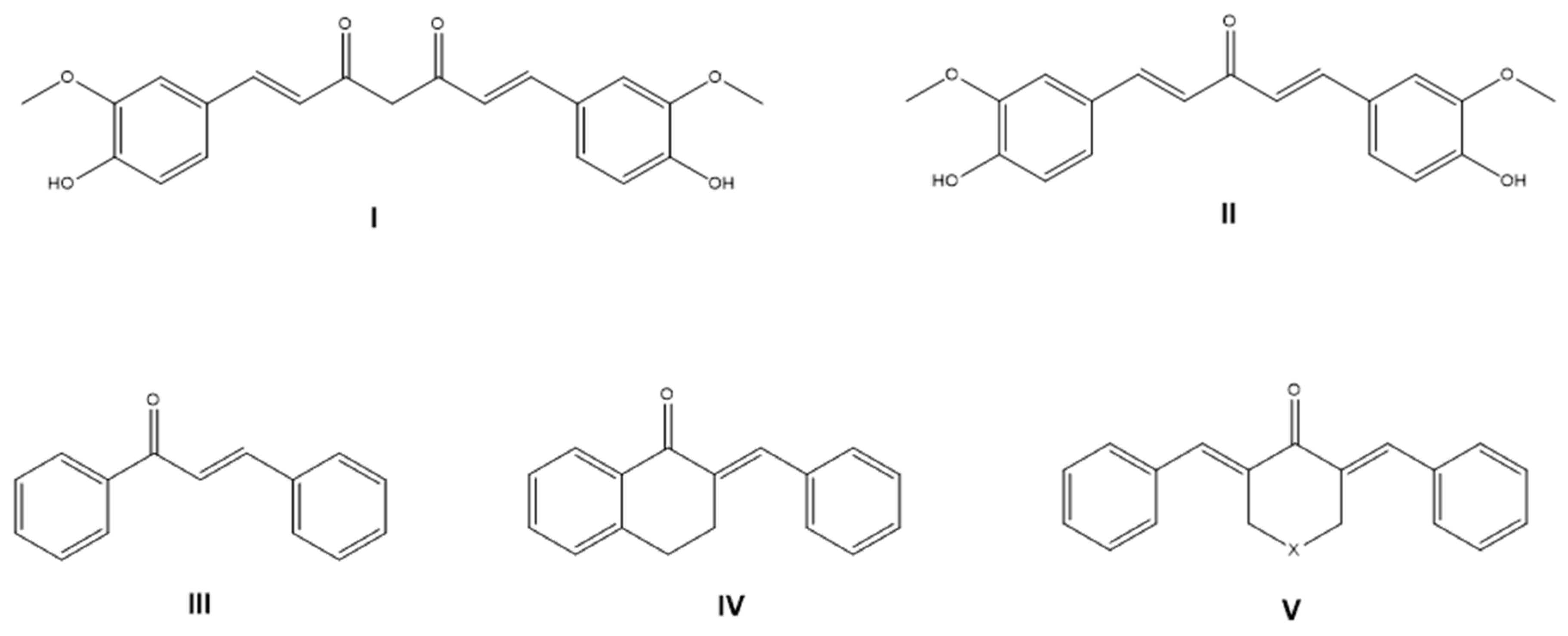
Figure 1. Structurally related aromatic cross-conjugated compounds, such as curcumin (I) as β-diendione, C5-curcumin (II) as β-dienone, and chalcones (III) as β-enones, are natural models of synthetic analogs of cyclic chalcones (IV) or cyclic C5-curcuminoids ((V), X = CH2, N, O, or S).
Antioxidants 14 01057 g001
Figure 2. Structures of the synthetic 4-piperidone-derived cyclic C5-curcuminoids (AD) with proven antioxidant activity or neuroprotective ability. The presence of phenolic OH groups in the molecule is not obligatory for free-radical neutralizing/antioxidant activity.
Antioxidants 14 01057 g002
Scheme 1. Synthetic pathway for cyclic chalcone analogs 4 and 5. Claisen-Schmidt condensation was performed starting from tetralone (1) in the presence of aromatic aldehyde 2 for dimethoxy compound 4 in a basic medium, and with aldehyde 3 for diphenol derivative 5 in an acidic medium.
Antioxidants 14 01057 sch001
Scheme 2. Synthesis of cyclic C5-curcuminoids 8 and 9. 4-hydroxy-cyclohexanone (6) was used in the base-catalyzed Claisen-Schmidt condensation of 8 and in the acid-catalyzed Claisen-Schmidt condensation of 9.
Antioxidants 14 01057 sch002
Scheme 3. Cyclic C5-curcumin derivatives 11 and 12 were obtained from the acid-catalyzed Claisen-Schmidt condensation (a). Compound 12 was transformed into 13 via acylation with succinic anhydride in the presence of triethylamine (b).
Antioxidants 14 01057 sch003
Figure 3. Determination of ROS production and total antioxidant capacity in 6-OHDA-induced differentiated SH-SY5Y cells. ROS determination was carried out using a Fluorometric Intracellular ROS Kit. ROS levels were compared to the control cells and expressed as a percentage. The TAC levels were determined using an Antioxidant Capacity Assay Kit. (A) Intracellular ROS levels in 6-OHDA-induced differentiated SH-SY5Y cells after treatment with sample compounds and rasagiline. (B) Total antioxidant capacity of 6-OHDA-induced differentiated SH-SY5Y cells after treatment with sample compounds and rasagiline. Each column shows the mean ± SD derived from three independent experiments (n = 3). The * signifies a p-value of less than 0.05 in comparison to the control. The † indicates p < 0.05, compared with the 6-OHDA treatment. Abbreviations: 6-OHDA, 6-hydroxydopamine; ROS, reactive oxygen species.
Antioxidants 14 01057 g003
Figure 4. Superoxide dismutase and glutathione peroxidase activity measurements of the 6-OHDA-induced differentiated SH-SY5Y cells. SOD activity was measured using the Superoxide Dismutase Activity Assay Kit. GPx activity was determined using the Glutathione Peroxidase Assay Kit. (A) SOD activity in 6-OHDA-induced differentiated SH-SY5Y cells after treatment with the sample compounds and rasagiline. (B) GPx activity in 6-OHDA-induced differentiated SH-SY5Y cells after treatment with the sample compounds and rasagiline. Each column shows the mean ± SD derived from three independent experiments (n = 3). The * signifies a p-value of less than 0.05 in comparison to the control. The † indicates p < 0.05, compared with the 6-OHDA treatment. Abbreviations: 6-OHDA, 6-hydroxydopamine; GPx, glutathione peroxidase; SOD, superoxide dismutase.
Antioxidants 14 01057 g004
Figure 5. Total intracellular iron content and thiol concentrations and MDA levels in 6-OHDA-induced differentiated SH-SY5Y cells. The total intracellular iron content was determined using a colorimetric ferrozine-based assay. The total intracellular thiol levels were measured using a Thiol Assay. (A) Total iron content of 6-OHDA-induced differentiated SH-SY5Y cells after treatment with the sample compounds and rasagiline. (B) Thiol levels in 6-OHDA-induced differentiated SH-SY5Y cells after treatment with sample compounds and rasagiline. (C) MDA levels in 6-OHDA-induced differentiated SH-SY5Y cells after treatment with sample compounds and rasagiline. Each column shows the mean ± SD derived from three independent experiments (n = 3). The * signifies a p-value of less than 0.05 in comparison to the control. The † indicates p < 0.05, compared with the 6-OHDA treatment. Abbreviations: 6-OHDA, 6-hydroxydopamine.
Antioxidants 14 01057 g005
Figure 6. ATP and cytochrome c levels and caspase-3 activity in 6-OHDA-induced differentiated SH-SY5Y cells. ATP levels were determined using a luminescent ATP Detection Kit. The intracellular cytochrome c concentration was measured using a Cytochrome c ELISA Kit. Caspase-3 activity was calculated using a fluorescent Caspase-3 Assay. (A) ATP levels in 6-OHDA-induced differentiated SH-SY5Y cells after treatment with the sample compounds and rasagiline. (B) Cytochrome c levels in 6-OHDA-induced differentiated SH-SY5Y cells after treatment with sample compounds and rasagiline. (C) Caspase-3 activity in 6-OHDA-induced differentiated SH-SY5Y cells after treatment with the sample compounds and rasagiline. Each column shows the mean ± SD derived from three independent experiments (n = 3). The * signifies a p-value of less than 0.05 in comparison to the control. The † indicates p < 0.05, compared with the 6-OHDA treatment. Abbreviations: 6-OHDA, 6-hydroxydopamine.
Antioxidants 14 01057 g006
Figure 7. IL-4 and IL-10 release from 6-OHDA-induced differentiated SH-SY5Y cells. The concentrations of secreted IL-4 and IL-10 from the cell culture supernatants were determined using human IL-4 and IL-10 ELISA Kits. (A) IL-4 levels in 6-OHDA-induced differentiated SH-SY5Y cells after treatment with the test compounds and rasagiline. (B) IL-10 release in 6-OHDA-induced differentiated SH-SY5Y cells after treatment with sample compounds and rasagiline. Each column shows the mean ± SD derived from three independent experiments (n = 3). The * signifies a p-value of less than 0.05 in comparison to the control. The † indicates p < 0.05, compared with the 6-OHDA treatment. Abbreviations: 6-OHDA, 6-hydroxydopamine.
Antioxidants 14 01057 g007
Figure 8. IL-6, IL-8, and TNF-α secretion by 6-OHDA-induced differentiated SH-SY5Y cells. The concentrations of secreted IL-6, IL-8, and TNF-α were determined using human IL-6, IL-8, and TNF-α ELISA Kits from cell culture supernatants. (A) IL-6 levels in 6-OHDA-induced differentiated SH-SY5Y cells after treatment with the test compounds and rasagiline. (B) IL-8 release in 6-OHDA-induced differentiated SH-SY5Y cells after treatment with sample compounds and rasagiline. (C) TNF-α secretion in 6-OHDA-induced differentiated SH-SY5Y cells after treatment with sample compounds and rasagiline. Each column shows the mean ± SD derived from three independent experiments (n = 3). The * signifies a p-value of less than 0.05 in comparison to the control. The † indicates p < 0.05, compared with the 6-OHDA treatment. Abbreviations: 6-OHDA, 6-hydroxydopamine.
Antioxidants 14 01057 g008
Figure 9. Fractalkine secretion by 6-OHDA-induced differentiated SH-SY5Y cells. The concentrations of secreted fractalkine were determined using the human Fractalkine ELISA Kit from the cell culture supernatants. Each column shows the mean ± SD derived from three independent experiments (n = 3). The * signifies a p-value of less than 0.05 in comparison to the control. The † indicates p < 0.05, compared with the 6-OHDA treatment. Abbreviations: 6-OHDA, 6-hydroxydopamine.
Antioxidants 14 01057 g009
Scheme 4. Antioxidant effects of compounds 5, 9, 12, and 13 in an in vitro neuronal damage model. 6-OHDA triggers ROS production through various mechanisms. Compounds 5, 9, 12, and 13 reduced iron accumulation and the subsequent Fenton reaction, which generates hydroxyl radicals (HO). These test compounds also increased SOD activity, thereby enhancing the conversion of superoxide anions (O2•−) to hydrogen peroxide (H2O2). GPx, whose activity is enhanced by the test compounds, then reduces hydrogen peroxide to water. Overall, these compounds elevated the TAC of the cells and lowered ROS levels. The most effective compounds are highlighted in bold.
Antioxidants 14 01057 sch004
Scheme 5. The proposed products (1420) of the possible structural rearrangements allow compounds 5, 9, 12, and 13 to act as hydrogen donors and promote neuroprotection. Compounds 5, 12, and 13 provide four hydrogen atoms, whereas derivative 9 contains six hydrogen atoms for biological hydrogen-transfer reactions.
Antioxidants 14 01057 sch005
Table 1. Concentrations of the test compounds used for reactive oxygen species and total antioxidant capacity determinations.
Test CompoundConcentrations (µM)
Compound 43; 5; 8
Compound 55; 10; 15
Compound 83; 4; 5
Compound 95; 10; 14
Compound 113; 5; 6
Compound 123; 5; 7
Compound 135; 10; 12
Table 2. IC50 values of the synthetic cyclic C5-curcuminoids and chalcones, and their concentrations used in the experiments.
IC50 (µM)SH-SY5YConcentration Used in the Experiments (µM)
Compound 49.14 ± 1.135
Compound 518.11 ± 2.1110
Compound 86.23 ± 1.095
Compound 915.11 ± 1.0310
Compound 117.46 ± 1.145
Compound 128.94 ± 1.235
Compound 1313.68 ± 1.2210
Disclaimer/Publisher’s Note: The statements, opinions and data contained in all publications are solely those of the individual author(s) and contributor(s) and not of MDPI and/or the editor(s). MDPI and/or the editor(s) disclaim responsibility for any injury to people or property resulting from any ideas, methods, instructions or products referred to in the content.